HAL Id: inserm-00315944
https://www.hal.inserm.fr/inserm-00315944
Submitted on 13 Nov 2009
HAL is a multi-disciplinary open access
archive for the deposit and dissemination of sci-entific research documents, whether they are pub-lished or not. The documents may come from teaching and research institutions in France or abroad, or from public or private research centers.
L’archive ouverte pluridisciplinaire HAL, est destinée au dépôt et à la diffusion de documents scientifiques de niveau recherche, publiés ou non, émanant des établissements d’enseignement et de recherche français ou étrangers, des laboratoires publics ou privés.
Lipid-bloated subretinal microglial cells are at the origin
of drusen appearance in CX3CR1-deficient mice.
William Raoul, Charles Feumi, Nicole Keller, Sophie Lavalette, Marianne
Houssier, Francine Behar-Cohen, Christophe Combadière, Florian Sennlaub
To cite this version:
William Raoul, Charles Feumi, Nicole Keller, Sophie Lavalette, Marianne Houssier, et al.. Lipid-bloated subretinal microglial cells are at the origin of drusen appearance in CX3CR1-deficient mice.. Ophthalmic Research, Karger, 2008, 40 (3-4), pp.115-9. �10.1159/000119860�. �inserm-00315944�
Lipid bloated subretinal microglial cells are at the origin of Drusen
appearance in CX3CR1 deficient mice
Raoul W. 1,2,3, Feumi C. 1,2,3, Keller N. 1,2,3, Lavalette S. 1,2,3, Houssier M. 1,2,3,Behar-Cohen F.
1,2,3,6
, * Combadière C. 2,4,6 * Sennlaub F. 1,2,3
*these authors contributed equally to the work
1
Inserm, UMR S 872, Paris, F-75006 France
2
Université Pierre et Marie Curie-Paris6,Paris, 75634, France
3
Centre de Recherche des Cordeliers, Université Pierre et Marie Curie – Paris 6, UMR S 872 Paris, F-75006 France
4
Inserm U543, Laboratoire d’Immunologie Cellulaire, Paris, 75013, France
5
AP-HP, Groupe Hospitalier Pitié-Salpétrière, Service d’Immunologie, Paris, 75651, France
6Hôtel Dieu, Service d’Ophtalmologie, Centre de Recherche ophtalmologique, 75006 Paris
To whom correspondance should be addressed Dr Florian Sennlaub, Inserm, UMRS 872, Centre de recherche des Cordeliers, 15 rue de l’Ecole de Médecine, 75006 Paris, France. Tel: (33) 1 40 46 78 63, Fax: (33) 1 40 46 78 65, Email : sennlaub@idf.inserm.fr.
Abstract
Drusen, the white yellowish deposits that can be seen in funduscopy, are a hallmark of age related macular disease (AMD). Histologically, drusen are believed to be dome-shaped or more confluent lipid accumulations between the retinal pigment epithelium and the choroicapillaries. Recent advances in mouse fundoscopy have revealed the presence of drusen-like structures in chemokine knockout animals in the absence of sizeable dome-shaped material below the RPE. We show that aged CX3CR1-/- mice present with drusen-like
appearance in funduscopy that is associated with a progressive age-related microglial cell (MC) accumulation in the subretinal space. We demonstrate that the anatomical equivalent of the drusen-like appearance in these mice are lipid bloated subretinal MCs rather than sub-RPE deposits (1).
Introduction
Age-related macular degeneration (AMD) is the leading causeof vision loss in elderly people in the industrialized countries (2). Its most prominent pathologic features are lesions involving
the retinal pigment epithelium (RPE) and Bruch’s membrane (BM), photoreceptor
degeneration and, in the most aggressive cases, choroidal neovascularization (CNV) (3). Early AMD is characterized by the excessive presence of yellowish white subretinal deposits called drusen, located on BM, partially covered by the RPE and clinically visible with funduscopy (3, 4). Drusen are composed of lipids and glycoproteins contain numerous degenerating organelles (5), and are believed to be formed by extracellular deposit of materials or by transformed degenerating RPE cells (6). Controversy continues to surround
their pathogenesis, as well as the causes of AMD. Some of the features of humanAMD have
been observed in transgenic mouse models. Notably CCL2 and CCR2 deficiency led to an age dependent appearance of yellowish drusen-like spots in funduscopy in the absence of
dome-shaped sub-RPE debris of an equivalent size (7). We recently demonstrated that CX3CR1 deficiency leads to subretinal MC accumulation that is associated with cardinal features of AMD (1). In these mice, drusen-like funduscopical appearance was caused by intracellular lipid accumulation in bloated subretinal microglial cells.
Results
Subretinal MC (SrMC) accumulate in CX3CR1-/- mice with age
We have previously shown that green fluorescent (GFP) subretinal MCs progressively
accumulate in CX3CR1GFP/GFP mice (1) compared to CX3CR1+/GFP mice. On RPE flatmounts
of 18-month-old CX3CR1+/+ mice (Fig. 1A), CD11b-positive SrMC were seen occasionally,
as previously reported (8). In contrast in CX3CR1-/- mice, subretinal MC juxtaposed to RPE
(Fig. 1B) accumulated significantly (Fig. 1C) similar to CX3CR1GFP/GFP mice.
Drusen observed in CX3CR1 knockout mice are subretinal bloated MC.
Subretinal deposits are characteristic in AMD. Funduscopy of senescent CX3CR1+/+
(Fig. 2A) and CX3CR1-/- (Fig. 2B) mice showed that all the CX3CR1-/- mice had numerous
drusen-like yellow nodular deposits in the deep layer of the retina. To characterize in more detail the anatomical structure that produced this drusen-like appearance in funduscopy in CX3CR1-/- mice, we “flatmounted” the eyes, thus exposing the RPE monolayer. The RPE
monolayer of CX3CR1+/+ animals, observed in tangential white light, appeared regular and
smooth (1). In contrast, in CX3CR1 knockout mice, examination of the monolayer showed elevated white spots (Fig. 2C). Higher magnification revealed the intracellular location of this white material in bloated CD11b positive cells (Fig 2D green fluorecence) adjacent to the RPE cell monolayer (phalloidin stain in red). These intracellular yellowish white deposits were present only in CD11b positive cells and histological serial sections of drusen-bearing eyes revealed no significant dome-shaped sub RPE deposits (data not shown). These
subretinal ramified cells were also positive for the specific MC marker 5D4 (data not shown) and very similar to the GFP-positive cells previously described in CX3CR1GFP/GFP mice (1).
Not all subretinal MC were bloated, but only the bloated cells appeared drusen-like on RPE flatmounts.
Subretinal bloated MCs contain multiple lipid droplets and OS derived debris.
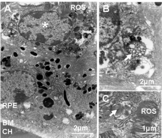
Ultrastructural analysis of senescent CX3CR1-/- eyes by transmission electron
microscopy confirmed the presence of subretinal cells between the outer segment (OS) and the RPE cells (Fig. 3A), which contained intracellular lipid deposits and remnants of OS (Fig. 3B), typically observed in RPE cells (9) as previously described (1).
Discussion
In CX3CR1-deficient mice, MC accumulated subretinally significantly in CX3CR1 -/-mice compared to CX3CR1+/+ mice. A subset of these subretinal MCs appeared bloated with
white intracellular material. Electron microscopy revealed the presence of intracellular lipid droplets that might explain the white appearance of the intra-cellular accumulation. Furthermore partially digested OS in phagosomes were found in the subretinal MCs that can usually only be seen in RPE cells. It is intriguing to speculate that the intracellular lipid accumulation in subretinal MCs occurs secondary to excessive ingestion of OS by the subretinal MCs. As bloated subretinal MCs can also be found in the human disease (10) (1), one can not exclude that bloated subretinal MC contribute to some extent to the drusen appearance in human AMD. Furthermore, one might speculate that the rounded contours and steeply sloping sides characteristic of drusen (6) may derive in part from degenerating bloated MC that are subsequently covered by RPE cells. Supporting this theory is the fact that drusen contain numerous degenerating organelles (5), the origin of which may be degenerating retinal MCs. Supporting the hypothesis that degenerating MCs contribute to drusen formation
is the fact that CX3CR1 deposits can be found in drusen in AMD patients (1). Similarly drusen deposits have been reported to contain apolipoprotein E, complement factors, major histocompatibility complex (MHC) and amyloid oligomers (6, 11-14). Activated MCs express ApoE (15), complement factors (16), MHC (17) and the beta-amyloid precursor protein (18) and might be the source of these proteins found in Drusen. MC can also be a major source of oxidative stress through respiratory bursts (19) that can cause the oxidative protein and lipid modifications which are typical for Drusen (20).
In summary, our results suggest that drusen-like appearance in CX3CR1 knockout animals and possibly in other mice are by in large caused by subretinal lipid-bloated MCs. Our study challenges the longstanding paradigm of drusen genesis as the gradual accumulation of extracellular debris from the RPE and choroid. We propose that subretinal MC accumulation might play a key role in drusen formation in AMD, where lipid bloated subretinal MCs can be found (1, 10).
Materials and methods
Animals
CX3CR1-/- mouse strain on C57BL/6 background and their controls were kept in
specific pathogen-free conditions with food and water available ad libitum and housed in a 12/12 hr light/dark (100-500 lux) cycle. Animal experiments were approved by the Institutional Animal Care and Use Committee.
Fundus photography
Mice were anesthetized by intramuscular injection of ketamine (50 mg/kg) and xylazine (10 mg/kg). Pupils were fully dilated with 1% tropicamide or 1% atropin. Coverslips positioned on the mouse cornea were used as a contact glass. Fundus photographs were taken with a digital camera mounted on an ophthalmic operating microscope (Zeiss).
Immunohistochemistry.
Antibodies used were rat monoclonal anti-CD11b (Serotec) and Rhodamine Phalloïdin (Molecular Probes). The corresponding Alexa Fluor 488-conjugated secondary antibody (Molecular Probes) was used to reveal the primary antibody and sections were counterstained with 4-6-diamino-2-phenylindole (DAPI). Flatmounts were viewed with a fluorescence microscope (BX51; Olympus).
Choroidal flatmounts and SrMC quantifications
Eyes were enucleated, fixed in 4% PFA for 15 minutes at room temperature and sectionedat the limbus; the cornea and lens were discarded (for CNV, mice were perfused with Fluorescein Dextran 106 before enucleation). The retinas were carefully peeled fromthe
RPE/choroid/sclera. Retinas and choroids were fixed for an additional 15 minutes in methanol at –20°C and incubated with the indicated primary and secondary antibodies. The choroids and retinas were radially incised, flatmounted and viewed with a fluorescence microscope (BX51; Olympus). For RPE flatmount photographs, tangential white light was applied by placing the light-conducting glass fibers of a KL2500LCD Schott lamp directly over the coverslip, next to the objective. SrMC were counted on CD11b stained whole RPE/choroidal flatmounts up to the ciliary body and on the outer segment side of the retina. The surface of the RPE was measured and SrMC density calculated.
Electron microscopy
For electron microscopy, eyes were fixed in 2.5% glutaraldehyde of cacodylate buffer (0.1 M, pH 7.4). After 1 hour, eyeballs were dissected, fixed for another 3 hours, post-fixed in 1% osmium tetroxide in cacodylate buffer, and dehydrated in graduated ethanol solution. The samples were included in epoxy resin and oriented. Ultrathin sections (80 nm) were stained for contrast with uranyl acetate and lead citrate and were observed in a JEOL 100 CX II electron microscope (JEOL, Tokyo, Japan) with 80 kV.
Statistical Analysis
Graph Pad Prism 4 (GraphPad Software) was used for data analysis and graphic representation. All values are reported as mean ± SEM. Statistical comparisons used unpaired two-sample t-tests for means and Mann-Whitney U tests. Significance was set at p < 0.05.
Acknowledgments
We thank Dr. D. Littman and Dr. S Jung for sharing the CX3CR1Kin GFP mice and Dr. R.S. Molday for the anti-rhodopsin antibody. This work was supported by grants from Inserm, ANR “Cardiovasculaire, obésité et diabète” (AO5088DS), ANR “blanc” (AO5120DD), European Grant “Innochem” 518167, PNR vision (AO6004SP). C.C. is the recipient of a contract «Interface» from Assistance Publiques-Hopitaux de Paris.
References
1. Combadière, C., Feumi, C., Raoul, W., Keller, N., Rodéro, M., Pézard, A., Lavalette,
S., Houssier, M., Jonet, L., Picard, E., et al. 2007. CX3CR1-dependent subretinal microglia cell accumulation is associated with cardinal features of age-related macular degeneration. Journal of Clinical Investigation in press.
2. Friedman, D.S., O'Colmain, B.J., Munoz, B., Tomany, S.C., McCarty, C., de Jong, P.T., Nemesure, B., Mitchell, P., and Kempen, J. 2004. Prevalence of age-related macular degeneration in the United States. Arch Ophthalmol 122:564-572.
3. Sarks, S.H. 1976. Ageing and degeneration in the macular region: a clinico-pathological study. Br J Ophthalmol 60:324-341.
4. Bird, A.C., Bressler, N.M., Bressler, S.B., Chisholm, I.H., Coscas, G., Davis, M.D., de Jong, P.T., Klaver, C.C., Klein, B.E., Klein, R., et al. 1995. An international classification and grading system for age-related maculopathy and age-related macular degeneration. The International ARM Epidemiological Study Group. Surv Ophthalmol 39:367-374.
5. Farkas, T.G., Sylvester, V., and Archer, D. 1971. The ultrastructure of drusen. Am J Ophthalmol 71:1196-1205.
6. Hageman, G.S., Luthert, P.J., Victor Chong, N.H., Johnson, L.V., Anderson, D.H., and Mullins, R.F. 2001. An integrated hypothesis that considers drusen as biomarkers of immune-mediated processes at the RPE-Bruch's membrane interface in aging and age-related macular degeneration. Prog Retin Eye Res 20:705-732.
7. Ambati, J., Anand, A., Fernandez, S., Sakurai, E., Lynn, B.C., Kuziel, W.A., Rollins, B.J., and Ambati, B.K. 2003. An animal model of age-related macular degeneration in senescent Ccl-2- or Ccr-2-deficient mice. Nat Med 9:1390-1397.
8. Ng, T.F., and Streilein, J.W. 2001. Light-induced migration of retinal microglia into the subretinal space. Invest Ophthalmol Vis Sci 42:3301-3310.
9. Tamai, M., and O'Brien, P.J. 1979. Retinal dystrophy in the RCS rat: in vivo and in vitro studies of phagocytic action of the pigment epithelium on the shed rod outer segments. Exp Eye Res 28:399-411.
10. Gupta, N., Brown, K.E., and Milam, A.H. 2003. Activated microglia in human retinitis pigmentosa, late-onset retinal degeneration, and age-related macular degeneration. Exp Eye Res 76:463-471.
11. van der Schaft, T.L., Mooy, C.M., de Bruijn, W.C., and de Jong, P.T. 1993. Early stages of age-related macular degeneration: an immunofluorescence and electron microscopy study. Br J Ophthalmol 77:657-661.
12. Mullins, R.F., Russell, S.R., Anderson, D.H., and Hageman, G.S. 2000. Drusen associated with aging and age-related macular degeneration contain proteins common to extracellular deposits associated with atherosclerosis, elastosis, amyloidosis, and dense deposit disease. Faseb J 14:835-846.
13. Luibl, V., Isas, J.M., Kayed, R., Glabe, C.G., Langen, R., and Chen, J. 2006. Drusen deposits associated with aging and age-related macular degeneration contain nonfibrillar amyloid oligomers. J Clin Invest 116:378-385.
14. Johnson, L.V., Leitner, W.P., Rivest, A.J., Staples, M.K., Radeke, M.J., and Anderson, D.H. 2002. The Alzheimer's A beta -peptide is deposited at sites of complement activation in pathologic deposits associated with aging and age-related macular degeneration. Proc Natl Acad Sci U S A 99:11830-11835.
15. Xu, Q., Bernardo, A., Walker, D., Kanegawa, T., Mahley, R.W., and Huang, Y. 2006.
Profile and regulation of apolipoprotein E (ApoE) expression in the CNS in mice with targeting of green fluorescent protein gene to the ApoE locus. J Neurosci 26:4985-4994.
16. Bellander, B.M., Bendel, O., Von Euler, G., Ohlsson, M., and Svensson, M. 2004. Activation of microglial cells and complement following traumatic injury in rat entorhinal-hippocampal slice cultures. J Neurotrauma 21:605-615.
17. Matsubara, T., Pararajasegaram, G., Wu, G.S., and Rao, N.A. 1999. Retinal microglia
differentially express phenotypic markers of antigen-presenting cells in vitro. Invest Ophthalmol Vis Sci 40:3186-3193.
18. Haass, C., Hung, A.Y., and Selkoe, D.J. 1991. Processing of beta-amyloid precursor protein in microglia and astrocytes favors an internal localization over constitutive secretion. J Neurosci 11:3783-3793.
19. Klegeris, A., and McGeer, P.L. 1994. Rat brain microglia and peritoneal macrophages
show similar responses to respiratory burst stimulants. J Neuroimmunol 53:83-90. 20. Crabb, J.W., Miyagi, M., Gu, X., Shadrach, K., West, K.A., Sakaguchi, H., Kamei,
M., Hasan, A., Yan, L., Rayborn, M.E., et al. 2002. Drusen proteome analysis: an approach to the etiology of age-related macular degeneration. Proc Natl Acad Sci U S A 99:14682-14687.
Figure legends:
Figure 1: Subretinal MC accumulate in CX3CR1-/- C57BL/6 mice with age
RPE flatmounts of aged mice show a strong accumulation of subretinal MC in CX3CR1+/+ (A) and CX3CR1-/- mice (B). Quantification of subretinal CD11b-positive cells
on RPE flatmounts reveals that subretinal MC accumulate progressively in CX3CR1-/- mice
and are significantly more numerous than in CX3CR1+/+ mice (C). Experiments were
performed on 5 eyes from different mice per group. *, P < 0.05. CX3CR1+/+ (+/+) in white
Figure 2: Drusen observed in CX3CR1 knockout animals are bloated subretinal MC Comparison of fundus photos and micrographs of a retinal pigment epithelium (RPE) flatmount in tangential light of 1-year-old CX3CR1+/+ (A) and CX3CR1-/- mice (B) reveals
multiple drusen in CX3CR1-/- mice. Close-up of a drusen (arrow) in a CX3CR1-/- mouse in
tangential light (C) superposed with CD11b-positive (green, phalloidin in red, and DAPI blue) subretinal ramified cell (D, E tangential light and DAPI merged) on RPE flatmounts.
Figure 3: Subretinal bloated MCs contain multiple lipid droplets and OS derived debris. Electron microscopy of the RPE/outer segment interface (A) in CX3CR1-/- mice
reveals subretinal cell (star) with multiple intracellular debris accumulation, juxtaposed to the RPE cell layer. Close ups revealed typical lipid droplet appearance (B) and intra-phagosomal OS remnants (C). Scale bars: 50 µm and as indicated (F). OS: outer segments; RPE: retinal pigment epithelium; BM: Bruch’s membrane; CH: choroid.