HAL Id: hal-01359398
https://hal-univ-rennes1.archives-ouvertes.fr/hal-01359398
Submitted on 10 Oct 2016HAL is a multi-disciplinary open access archive for the deposit and dissemination of sci-entific research documents, whether they are pub-lished or not. The documents may come from teaching and research institutions in France or abroad, or from public or private research centers.
L’archive ouverte pluridisciplinaire HAL, est destinée au dépôt et à la diffusion de documents scientifiques de niveau recherche, publiés ou non, émanant des établissements d’enseignement et de recherche français ou étrangers, des laboratoires publics ou privés.
Role of the tumor microenvironment in regulating
apoptosis and cancer progression
Katherine Yaacoub, Remy Pedeux, Karin Tarte, Thierry Guillaudeux
To cite this version:
Katherine Yaacoub, Remy Pedeux, Karin Tarte, Thierry Guillaudeux. Role of the tumor microen-vironment in regulating apoptosis and cancer progression. Cancer Letters, Elsevier, 2016, 378 (2), pp.150-159. �10.1016/j.canlet.2016.05.012�. �hal-01359398�
Role of the Tumor microenvironment in regulating
apoptosis and cancer progression
Katherine Yaacoub
1, 2,3, Remy Pedeux
1,3, Karin Tarte
1,2and Thierry
Guillaudeux
1,2,3,41-Université Rennes 1, 2 Rue du Thabor 35000 Rennes, France
2-UMR INSERM, 917, 2 Av. du Pr Léon Bernard, 35043 Rennes cedex France
3- INSERM ER440-OSS, CLCC Eugène Marquis, Rue Bataille Flandres Dunkerque, 35042 Rennes
4-UMS CNRS3480/US 018 INSERM BIOSIT, 2 Av. du Pr Léon Bernard, 35043 Rennes cedex, France
Corresponding Author: Thierry Guillaudeux, 2 avenue du Pr L Bernard, Rennes 35043, France, E-mail: thierry.guillaudeux@univ-rennes1.fr
Keywords: Microenvironment, apoptosis, resistance, immune escape, inflammation.
Highlights
x The impairment of cell death represents the main factor of cancer outbreak, as well as the resistance to anti-cancer agents.
x Tumor microenvironment has a critical importance in immune escape, therefore promoting tumor progression and metastasis.
x Inflammatory responses modulate the tumor microenvironment and play a decisive role in tumor development.
x Microenvironment-targeted thearpies might be a real gain to fight cancer.
Abstract
Apoptosis is a gene-directed program that is engaged to efficiently eliminate dysfunctional cells. Evasion of apoptosis may be an important gate to tumor initiation and therapy resistance. Like any other developmental program, apoptosis can be disrupted by several genetic aberrations driving malignant cells into an uncontrolled progression and survival. For its sustained growth, cancer develops in a complex environment, which provides survival signals and rescues malignant cells from apoptosis. Recent studies have clearly shown a wide interaction between tumor cells and their microenvironment, confirming the influence of the surrounding cells on tumor expansion and invasion. These non-malignant cells, not only intensify tumor cells growth, but also upgrade the process of metastasis. The strong
crosstalk between malignant cells and a reactive microenvironment is mediated by soluble chemokines and cytokines, which act on tumor cells through surface receptors. Disturbing the microenvironment signaling might be an encouraging approach for patient’s treatment. Therefore, the ultimate knowledge of “tumor-microenvironment” interactions facilitates the identification of novel therapeutic procedures that mobilize cancer cells from their supportive cells. This review focuses on cancer progression mediated by the dysfunction of apoptosis and by the fundamental relationship between tumor and reactive cells. New insights and valuable targets for cancer prevention and therapy are also presented.
Introduction
In 1842, the notion of cell death, known now as apoptosis, was introduced by Carl Vogt after his work on developmental cell death in toads. Later, in 1885, Walther Flemming was the first to propose a morphological description of apoptosis showing the deformation of the cell, DNA degradation and apoptotic body formation1. In 2002, Sydney Brenner, Robert Horvitz and John E. Suston deciphered the genetic regulation of programmed cell death, and provided “Caenorhabditis elegans” as a biological model to study apoptosis 2.
The process of apoptosis, a tightly regulated programmed cell death, occurs normally to maintain the development and homeostasis in normal cell populations. Inappropriate apoptosis is a major factor in many human diseases. Defects in apoptosis cause autoimmune diseases or cancers, while enhanced cell death may cause degenerative diseases and immunodeficiency3.
The apoptotic mechanism is triggered by two distinguished pathways: i) the “extrinsic pathway” which is initiated by a variety of death receptors -members of TNF Receptor superfamily-, such as TNFα receptor, Fas-L receptor, TRAIL receptors... and ii) the “intrinsic pathway” mediated by the mitochondria which releases apoptogenic factors from its inter-membrane space. In both pathways, most of changes depends on a group of cysteine proteases described as “caspases”, which were revealed as the central executioners of the apoptotic pathway because of their role in the cleavage of major cellular substrates such as nuclear lamins, cytoskeletal proteins (Fodrin, gelsolin) and the caspases themselves 4,5. Additional studies have demonstrated a novel apoptotic pathway mediated by the activation of caspase-12 in response to the endoplasmic reticulum stress (ER-stress). Caspase-12 is responsible for the induction of ER-stress specific caspases cascade, such as caspase-9 in a cytochrome-C independent manner, confirming the central role of caspase-12 in ER stress-mediated apoptosis 6.
Each disruption or defect in apoptosis can allow pre-neoplastic and neoplastic cells to survive and enhance tumor pathogenesis via activation of proto-oncogenes. One of the mechanisms that provides apoptotic resistance to tumor cells is the overexpression of anti-apoptotic proteins (ex: Bcl2), or the downregulation of pro-anti-apoptotic proteins (ex: BAX) 3; and the overexpression of anti-apoptotic proteins such as Bcl-2 will contribute with other proteins like c-myc to tumorigenesis 4. Once the tumor is formed, it initiates an inflammatory response and modifies the texture of the surrounding environment to convert it into a pathological entity. In contrast, the tumor microenvironment provides inappropriate signals
that lead to the maintenance of tumorigenesis and cancer therapies resistance 7. In numerous situations, tumor cells can become fully resistant to apoptosis, so they can escape from the immune system, and resist subsequently to any therapeutic strategy targeting the apoptotic pathways.
The aim of this review is i) to present the correlations between defects in apoptosis and cancers, and ii) to clarify the crosstalk between tumor cells and their microenvironment interfering with these mechanisms of apoptosis. We finally highlight the novel therapeutic issues that target common pathways of stroma and tumor cells.
1)Apoptosis
1.1) Apoptosis : vital component of cell turnover
The mechanism of apoptosis, or programmed cell death (PCD), is a vital factor of different processes including cell renewal, embryonic development, and immune system activity. Many morphological changes occur during apoptosis: cell shrinkage, formation of cytoplasmic blebs, mitochondrial break-down, chromatin condensation, and eventually disturbance of cytoplasmic membranes and release of apoptotic bodies. Two main apoptotic pathways have been extensively described, which include the extrinsic pathway and the intrinsic pathway. Each of them requires the implication and activation of caspases -cysteinyl
aspartate proteases- to allow a proteolytic cascade which promotes the apoptotic signaling
pathways.
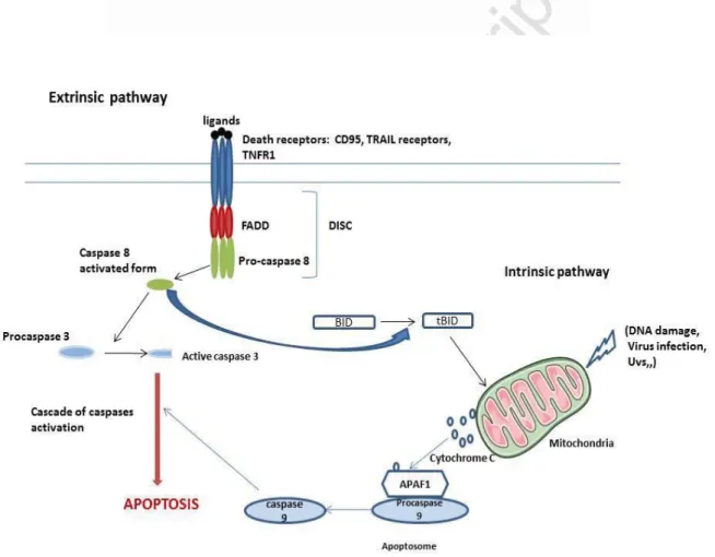
a) The extrinsic pathway, or death receptor pathway, is initiated by the activation of transmembrane death receptors (DRs) such as Fas/CD95, TRAIL receptors and all members of the TNF (Tumor necrosis factor) receptor superfamily, following the binding of their suitable ligands. Then, the activated receptors are able to recruit the adaptor protein FADD which associates with procaspase-8 and form the death-inducing signaling complex (DISC). This complex is responsible for caspase-8 activation and the induction of a downstream cascade of effectors such as caspase-3,-6, and -7, leading finally to an irreversible cell death.
b) The intrinsic pathway, also called mitochondrial pathway, is triggered by intracellular signals, such as DNA damage, oxidative stress and irradiations. These multiple forms of stresses induce the release of pro-apoptotic proteins from the mitochondria: Cytochrome c, Smac/Diablo, AIF, and EndoG. Then, Cytochrome c will bind to APAF1 (Apoptotic Protease Activating Factor-1) to form a large complex -the apoptosome- which in turn recruits pro-caspase-9 leading to caspase-9 activation. Activated caspase-9will induce additional caspases such as caspase-3. While Smac/DIABLO plays an essential role in blocking the activity of IAPs (inhibitors of apoptosis proteins) allowing apoptosis to occur. These two major apoptotic pathways end by the execution phase which is characterized by the degradation of the nuclear material and cytoskeletal proteins, contributing to cell death. In most cells, a “cross-talk” between the extrinsic and intrinsic pathways occurs through the cleavage of BID in t-BID by
activated caspase-8. Truncated BID (t-BID) permeabilizes the mitochondria and promotes the activation of additional caspases molecules (caspase-9,-3,-6 and -7) amplifying the apoptotic signal 8–10(Fig.1).
c) A third apoptotic pathway has been more recently described which highly underscores the role of the ER stress. This pathway initiates cell death through caspases activation, primarily caspase-4 in humans and caspase-12 in mice. Once triggered and activated by the ER stress, caspase-12 induces the activation of downstream caspases like caspase-9 and -3, responsible for ER stress–induced apoptosis6. Several models of caspase-4 activation have been proposed, one of them brings out the role of IRE1α, an ER-transmembrane protein which transduces the stress signals initiated by the accumulation of misfolded proteins, from the ER to the cytoplasm and nucleus. In response to ER stress, procaspase-4 localized into the ER and interacts with IRE1α through TRAF2 (Tumor Necrosis Factor receptor-Associated Factor 2), leading to the auto-processing and activation of caspase-4. The mechanisms of caspase-4 activation by ER apoptotic signals or how TRAF2 transmits ER signals from IRE1α to its downstream effector caspase-4 are still unknown11. In contrast, the overexpression of Calreticulin, an ER luminal protein, resulted in an increased release of cytochrome c from the mitochondria and an enhancement of caspase activity during apoptosis. These findings suggest that ER and mitochondria pathways are tightly linked, and this correlation involves Ca2+ which is released from the ER and accumulates into the mitochondria12.
1.2) Defects of apoptosis and tumorigenesis
Several forms of mutations contribute to apoptosis resistance, facilitating the uncontrolled cell growth of tumor cells, invasiveness and metastatic ability. The anti-apoptotic gene “Bcl2” is activated by a chromosomal translocation, contributing to lymphomagenesis. The p53, tumor suppressor gene, is considered as a central regulator of apoptosis. It recognizes DNA damage and thereafter arrests the cell cycle and triggers the DNA repair mechanisms. Due to its rescuer role, the downregulation of p53 enhances tumorigenesis in many cancer types. In addition, any loss or defect in the function of one factor of the intrinsic or extrinsic pathways can enhance tumor progression. For example, deficient function of APAF1 contributes to oncogenesis in ovarian and melanoma cancer cell lines, when perturbation of the FAS pathway, such as Fas gene mutation, contributes to non-Hodgkin’s lymphomas and other cancer types13. Moreover, the amplified resistance to apoptotic cell death is a hallmark of quickly proliferating cancer cells. Then, the inhibition of this anti-apoptotic defense could improve cancer therapies14. Indeed, the overexpression of anti-apoptotic proteins such as Bcl-2, survivin and Mcl-1, which act as negative regulators of the mitochondrial pathway, leads to cell death resistance in follicular lymphomas. Quercetin, a natural flavonoid, restores TRAIL-mediated cell death by induction of the proteasomal degradation of Mcl-1 and by suppressing survivin mRNA expression15. The absence of caspases also promotes cell
survival and clonogenic growth. Downregulation of caspase-2 or -8 in murine embryonic fibroblasts (MEFs) enhances cell proliferation and tumorigenesis. Besides, there is a correlation between the absence of autophagy and carcinogenesis. Mutations of autophagic genes contribute deeply to tumorigenesis. For example, Atg5 mutations has been observed in gastric cancer16. In addition, reduced expression of Beclin 1, an autophagy inducer, is observed in breast and ovarian carcinomas due to a significant deletion of the corresponding gene. Hence, resistance to autophagy and apoptosis greatly contributes to cancer drugs resistance. Then, proteins involved in the autophagic and the apoptotic pathways might be important targets of cancer therapies17.
1.3) Resistant tumor cells
Drug resistance is considered as an essential mechanism for inhibiting chemotherapy efficiency. Resistance to chemical drugs could be either intrinsic, indicating that tumor cells pre-contain resistance factors that weaken drugs efficiency, or acquired resistance caused by either several mutations, or an increased expression of the therapeutic target, or an induction of an alternative pathway. In addition, multidrug resistance has been established as a result of the presence of specific “transporters” in tumor-cell membranes. Two major pumps, members of the ATP-binding cassette transmembrane transporters family, have been well described: The P-Glycoprotein and the multidrug resistance-associated protein (MRP) that rejects therapeutic drugs out of the tumor cells. The overexpression of the P-glycoprotein, encoded by the MDR-1 gene, in many types of tumors including leukemia, lymphoma, kidney, liver and colon cancers, provides multidrug resistance to tumor cell lines18. At first, the overexpression of the P-Glycoprotein was considered as the main and only factor of cancer drug resistance. However, Zosuquidar and tariguidar showed that highly efficient MDR-1 inhibitors, failed to remove drug resistance in many clinical trials. Moreover, the chemotherapeutic resistance of cancer cells with no P-GP overexpression proves the intervention of other factors that prevent the complete response to drugs 18,19. Anti-apoptotic proteins of the Bcl-2 family, localized on the mitochondria, play a central role in cell resistance to death, preventing effective therapies. The upregulation of Bcl2 in small cell lung cancers (SCLC) decreased the apoptotic rate by inhibiting the loss of the mitochondrial transmembrane potential and the release of cytochrome c , conferring resistance to chemotherapy20. Mcl-1 is an anti-apoptotic Bcl-2 family protein which plays an essential role in cell survival by inhibiting Bim-induced activation of the proapoptotic protein Bax. McL-1 is amplified in a variety of cancers and it is implicated in apoptosis resistance and cell survival in tumor cells. Zhao et al. have demonstrated that glucose catabolism inhibited apoptotic cell death by the attenuation of Mcl-1 degradation. The amplified glucose metabolism in cancer cells induces the activity of PKC (protein kinase C) which inhibits GSK-3 alpha/beta (glycogen synthase kinase), known as a promoter of Mcl-1 degradation21.
X-linked inhibitor of apoptosis protein (XIAP) is an anti-apoptotic protein implicated in apoptosis suppression via potent inhibition through direct binding of caspases-3, -7 and -9. Meanwhile, Smac (Second mitochondria -derived activator of caspases) is a potent inhibitor of XIAP, preventing its inhibition on caspases and thus allowing apoptosis to proceed. The high expression of the XIAP protein correlates with the progression of different types of
cancers as well as chemotherapy resistance22. In rectal cancer, an increased expression of XIAP was observed in tumor tissue compared to matched-normal tissue, accordingly with the degree of radio chemotherapy resistance. However, Smac levels did not increase with the radio-chemotherapy resistance, and it was similarly expressed in tumor and normal tissues. This disturbance of XIAP/Smac balance may strongly contribute to tumor cells survival and therapies resistance23.
One of the mechanisms by which tumors resist to apoptosis is the upregulation of the anti-apoptotic protein “c-FLIP” (cellular FLICE inhibitory protein) which blocks apoptosis mediated by death receptors, such as Fas, DR4 (TRAIL-R1) and DR5 (TRAIL-R2). C-FLIP binds to FADD within the DISC to prevent caspase-8 homodimerization, self-processing and then activation. The overexpression of c-FLIP is identified in various cancers, such as NSCLC (Non-small cell lung carcinoma) and it is correlated with shorter overall survival24.
Resistance to apoptosis is one of the characteristics of human cancers. Evasion of apoptosis contributes to tumor progression and resistance to therapies. As discussed above, this chemoresistance phenotype is the result of several genetic alterations, but recent studies have suggested that apoptosis resistance could also be associated with tumor microenvironment which is recognized currently as a major factor impacting cancer growth and therapies sensitivity.
1.4) Role of tumor microenvironment in modulating resistance to apoptosis
A large number of cancer drugs which display a potent efficiency against tumor cells in vitro, lose their cytotoxicity when used in patients. This tumor resistance to apoptosis and chemical drugs was widely investigated in many laboratories over the years to finally identify several factors that reduce the efficacy of cancer drugs. The role of tumor microenvironment has drawn the attention of researchers as a pivotal factor influencing tumor resistance. Many components of the tumor microenvironment have been identified, having the ability to affect tumor sensitivity or resistance to chemotherapy-mediated apoptosis and playing a major role in tumor survival and progression.
Tumors are located within a heterogeneous area of stromal cells ( endothelial cells, fibroblasts, immune cells, adipocytes, mesenchymal stem cells, etc.) and each member of this population can be implicated in conferring cell death resistance to tumor cells and thereby chemoresistance.
Endothelial cells are responsible for angiogenesis, which is highly correlated with apoptosis resistance. Tumor cells produce VEGF (vascular endothelial growth factor), an angiogenesis stimulator, which stimulates the proliferation and differentiation of endothelial cells. Thus, induced endothelial cells initiate tumor vascularization to transport oxygen and nutrients to cancer cells, promoting their survival79.
Other microenvironmental cells, as fibroblasts, are also involved in affecting the sensitivity of tumor cells to drugs by IL-6 secretion, a pleiotropic cytokine which upgrade the resistance of multiple myeloma cells to cell death by the activation of the anti-apoptotic protein BcL-xL expression and JAK/STAT pathway81.
As well, the tumor surrounded carcinoma-associated mesenchymal stem cells (CA-MSC) are able to form hypoxic niches with high production of HIF-α. It has been demonstrated that
this hypoxic area is correlated with leukemia cell survival and chemoresistance to anti-leukemia drugs82.
Adipocytes represent also one of the abundant proportion of microenvironment cells, which secrete growth factors and enhance tumor development. Adipocytes can protect leukemia cells from chemotherapy treatments by stimulating the expression of anti-apoptotic proteins Pim-2 and Bcl-2. The secretion of IL-6 by cancer-associated adipocytes leads to ChK1 phosphorylation and resulted in cancer cell survival and radiotherapy resistance80.
Besides, to evaluate the role of cancer cell external environment in the resistance to treatment, researchers have treated mesothelioma cell lines with TRAIL (TNF-related apoptosis inducing ligand) plus cycloheximide. Results showed that cells grown in vitro as a monolayer went into a total apoptosis. However, mesothelioma fragments grown as spheroids which maintained multiple characteristics of the original tumors (mesothelioma cell viability, three-dimensional form of the tumor, the presence of non-malignant cells such as macrophages, and a collagen-rich extracellular matrix) exhibit an apoptotic resistance to TRAIL. These findings strongly support the hypothesis that microenvironmental factors contribute to modulate sensitivity to cancer drugs25.
2. Composition and functions of cancer cell
microenvironment
The evolving interaction between tumor cells and its surrounding microenvironmental cells plays an essential role in tumor progression. The microenvironment contributes to the induction, selection and expansion of tumor cells. Neoplastic tissue attracts stromal cells by cytokines/chemokines and growth factors secretions, in turn, cellular and extracellular elements of the microenvironment influence the invasion and proliferation of cancer cells26. The use of novel model systems obviously revealed the implication of the microenvironment in tumor progression. Injected murine tumoral mammary epithelial cells into mammary glands of hosts with irradiated mammary stroma gave rise to more tumors that appear quickly and larger than in unirradiated animals. These findings suggest that radiations effects on stroma can contribute to neoplastic progression in-vivo27.
2.1 Hodgkin’s Lymphoma
Hodgkin’s Lymphoma (HL) is characterized by Hodgkin Reed-Sternberg (HRS) cells which express a wide diversity of chemokines and cytokines (ex: IL5, CCL5, TNFα,..) affecting its microenvironment composition, and resulting in an abnormal immune response. Besides, cells in the microenvironment also release a set of cytokines and chemokines (ex: CXCL12, IL12, TGFβ,..) that contribute to an inflammatory response28. In B cell lymphomas, the microenvironment is composed of blood vessels, stromal cells, immune cells and extracellular matrix, on which malignant cells are absolutely depending to acquire survival and proliferation signals. Various studies demonstrated that CD4+ T cells are the most abundant cells in Hodgkin’s Lymphoma microenvironment, predominantly Th2 and Treg
cells. HRS cells produce several chemokines such as CCL5, CCL17 and CCL22, which attract Treg and Th2 cells expressing appropriate CCR4 receptors. Furthermore, IL5 and CCL28 (also called MEC mucosae-associated epithelial chemokine) derived from HRS, attract eosinophils, while the secretion of colony stimulating factor (CSF1) and CX3CL1 by HRS results in macrophages infiltration that enhances tumor progression and suppresses CD8 (+) T lymphocytes-mediated antitumor immunity. In addition, HRS cells expressing CD74 receptor, bind the macrophage inhibitory factor (MIF) which promotes their proliferation29–33. Recently, IL13 has been reported to play an important role in stimulating the proliferation of HRS cells. IL13 is secreted by HRS cells. Eighty nine percent of patients with Hodgkin’s Lymphoma showed an expression of IL13-Rα1 receptor, confirming that IL13 plays a crucial role in the autocrine growth of HL cells34,35. Besides, the neoplastic HRS cells effectively induce Eotaxin expression by fibroblasts. Production of TNFα by HRS seems to be responsible for this induction, because blocking TNFα inhibits Eotaxin expression. Eotaxin, a chemoattractant that binds on its CCR3 receptor on Th2 cells and eosinophils, contributes to their recruitment in Hodgkin’s disease and leads to the development and proliferation of HRS cells36(Fig.2).
2.2 Non Hodgkin’s Lymphoma
Recent studies showed that follicular lymphoma (FL) and MALT lymphoma cells require T cell-derived signals for their growth. The FL microenvironment is rich in Tfh cells (follicular helper T cell) which interact with CD40 receptor expressed on B cells via CD40-L, to produce high quantity of IL-4, an important cytokine for B cells stimulation and proliferation. IL-4 induces STAT6, an activator of transcription, in FL cells in a paracrine manner. These results suggest that Tfh that infiltrate follicular lymphomas are certainly implicated in tumor growth, and therapy directed against this specific population could be a promising strategy37. B-cell activating factor (BAFF) and APRIL (a proliferation inducing-ligand), two members of TNF superfamily, are also produced by immune cells (macrophages, monocytes and dendritic cells) and contribute to normal B cell development. BAFF-deficiency resulted in a significant loss of marginal and follicular B cells38. In contrast, an overexpression of BAFF and APRIL was observed in various B-cell malignancies such as B-cell Non-Hodgkin’s lymphomas and chronic lymphocytic leukemia. A significant upregulation of TACI (a common receptor of BAFF and APRIL) was also found in multiple myelomas and thyroid carcinomas,
with an association between lymphoma prognosis and TACI expression39.
In addition, stromal cells are highly implicated in supporting cancer growth and abnormal proliferation. In follicular lymphoma, the interaction with the surrounding stromal cells prevents apoptosis. Bone marrow mesenchymal stem cells (BM-MSCs), a subset of stromal cells, provide a highly supportive microenvironment for malignant B cells survival. A bidirectional interaction between BM-MSCs and FL cells has been described. BM-MSCs produce several chemokines, CXCL12 is considered as the most efficient chemoattractant which induces chemotaxis of FL cells that strongly express the CXCR4 chemokine receptor. The migrating malignant B cells toward CXCL12 produce high levels of TNFα (Tumor necrosis factor) and lymphotoxin α1/β2, responsible for BM-MSCs differentiation into fibroblastic reticular cells (FRCs) that support tumor development40. Furthermore, stromal cells produce
different types of Hedgehog proteins (Hh) that play an important role in lymphoid cells proliferation in NHL and multiple myeloma. Recently, it has been shown that in Eμ-Myc mice, Hedgehog proteins produced by the stroma contribute to the establishment of a supportive environment for the survival of lymphoma B cells through the activation of the anti-apoptotic protein Bcl2, preventing Myc-overexpressing cells from apoptosis. Inhibition of the Hh pathway by cyclopamine downregulates Bcl2 expression and reduces tumor mass in mice41. Besides, the physical interaction of NHL cells with Bone marrow-stromal cells (BM-SCs) induce the production of BAFF, a functional factor for B cells survival. The depletion of BAFF in BM-SCs by small hairpin RNA could remove stroma-mediated drug resistance and improve B cell response to chemotherapy42 (Fig.2).
2.3 Solid tumors
Fibroblasts, considered as the main component of tumor stroma, have been found to be highly activated in cancer. These cancer-associated fibroblasts (CAFs) contribute to the progression of many solid tumor types such as endometrial, breast, prostate and pancreatic cancers, and they are able to enhance cancer cell initiation, invasion and chemoresistance. Media collected from CAFs’ cultures induce a significant proliferation of both primary and endometrial cancer cell lines in comparison with non-treated cells. In contrast, fibroblasts derived from normal endometrial tissue did not affect endometrial cancer cells growth, thus demonstrating the specificity of CAFs in tumor cell growth. An upregulation of AKT and ERK phosphorylation was significantly observed in endometrial cells treated with CAFs media, indicating the role of PI3K/AKT and MAPK/ERK pathways in fibroblasts mediated endometrial cancer progression. These two pathways are activated by many cytokines such as IL-6, IL-8, macrophage chemoattractant protein (MCP-1), RANTES and vascular endothelial growth factor (VEGF), secreted by CAFs to induce cell proliferation. These data demonstrate the pro-tumorigenic role of the surrounding microenvironment, and a profound understanding of these interactions may serve as a novel way to cancer therapies43,44.
3. Microenvironment-mediated immune escape
The interaction between the neoplastic cells and their microenvironment provides tolymphoma cells the ability to escape from antitumor immune response. Tumor cells modify their microenvironment composition in several ways to avoid immune attack. These
procedures include the alteration of surface molecules expression and the recruitment of immunosuppressive cells.
3.1) The loss of surface major histocompatibility complex I and II
B cell lymphomas express on their surface the major histocompatibility complex class I and II (MHC I and MCH II), two molecules involved in antigen presentation to T cells. The loss of MHC I/II molecules contributes widely to immune escape as it could harm the activation of CD4+ T cells and CD8+ cytotoxic T cells (CTLs) which recognize tumor antigens presented by MHC II and MHC I respectively. About half of the testicular and central nervous system lymphomas showed a total loss of MHC II expression, resulting from the presence of homozygous deletions of the MHC II region on chromosome 6p21.3. The absence of MHC II expression abrogates antigen recognition by CD4+ T cells, leading to an impaired activation of CTLs and a promotion of tumor growth45,46.
Furthermore, in diffuse large B-cell lymphoma (DLBCL), 75% of cases display abnormal β2-microglobulin (the β2-M is a component of the MHC I molecules on the cell surface) protein expression, with 29 % of them harboring mutations and deletions in the β2-M gene. The loss or defect of β2-M expression is accompanied with the lack of MHC I expression, thereby it provides immune escape from CTLs cells, suggesting a crucial role of this gene in DLBCL pathogenesis.
3.2) The loss of cell adhesion molecule
The majority of DLBCL tumors also showed a loss of CD58 protein expression, a cell adhesion molecule and a ligand for the CD2 receptor on T cells and Natural Killer (NK) cells. The loss of CD58 expression disrupts DLBCL cells recognition by NK cells and affects NK-mediated cytolysis, a mechanism that enhances the escape from immune monitoring. The simultaneous inactivation of β2-M and CD58 in DLBCL indicates that they are co-selected, having a combined role during the immune escape47(Fig.3a).
3.3) Replacement of MHC I by HLA-G protein
Additional mechanisms of immune evasion include the expression of HLA-G protein, a non-classical MHC I gene and a ligand for an inhibitory receptor immunoglobulin-like transcript 2 (ILT2) and immunoglobulin-like transcript 4 (ILT4) on NK cells, a subset of T cells, macrophages, monocytes and dendritic cells. Fifty four percent of classical Hodgkin Lymphomas (cHL) display an expression of HLA-G protein on their HRS cells associated with the absence of MHC I expression. The expression of HLA-G protein inhibits NK cells function, and it is considered as a potential factor for immune escape from NK/ CTLs mediated response48(Fig.3b).
3.4) Overexpression of death ligands: CD95-L and PD-L1/2
Among the surface molecules overexpressed on HRS cells in classic HL, Fas-L (CD95-L) protein is upregulated in all the neoplastic cells of nodular sclerosis (NS) and mixed cellularity (MC), inducing apoptosis in tumor-specific Fas positive CTLs, thereby allowing tumor escape from host defenses. Knocking down Fas using anti-Fas siRNA could increase CTLs survival and expansion, and this may be an efficient therapy for HL disease49,50.
overexpressed on HRS cells of cHL and MLBCL (mediastinal large B-cell lymphomas) respectively, which are two molecules that bind to PD1, a co-inhibitory receptor (programmed cell death 1) expressed on CTLs and CD4+ cells, providing inhibitory signals and inducing T cell exhaustion. PD-L1/L2 genes are localized on chromosome 9p24. Interestingly, an amplification of 9p24.1 associated with an increased PDL1/PDL2 protein expression has been observed in cHL and MLBCL. In addition, JAK2 is localized on 9p24.1 and can be co-amplified with PDL1/2 loci, resulting in an increased activity of JAK2 protein which enhances PD1 ligands transcription and expression51,52 (Fig.3c). Furthermore, PD1/PD Ligand
1 pathway plays a fundamental role in converting naïve T4 cells into T-reg cells, indicating its implication in T-reg induction, development and functional maintenance. Induced T-reg (iT reg) could repress the expansion and function of effector T cells (T eff), and inhibit T cell responses and immune attack53.
3.5) Recruitment of immunosuppressive cells
In accordance with the upper findings , many studies focused on the pivotal role of T-reg in B cell lymphomas survival. Lately, it has been demonstrated that there was a strong imbalance between the number of CD8+ T cells and Treg cells in the tumor microenvironment. The large accumulation of intratumoral T-reg is able to inhibit the development and granule production of CD8+ cells, and to suppress strongly their cytotoxic activity, thus removing the capacity of infiltrating CD8+ cells to kill and destroy lymphoma B cells in NHL54. Furthermore,
malignant B cells of FL are surrounded by CD4+ T cells, a subset of cells highly expressing CXCR5 (CXCL13 chemokine receptor) and ICOS (inducible costimulator). Two populations of CD4+ have been detected, CD25pos follicular regulatory T cells (T FR) and CD25neg follicular helper T cells (T FH), indicating the high heterogeneity of tumor CD4+ T cells. T FH provide many supportive signals to B cells, such as IL4 that exhibits an anti-apoptotic activity on normal B cells, and CD40-L implicated in B cell survival and differentiation. Also, T FH are deeply correlated with T FR cells by secreting CXCL13 that recruits CXCR5 positive T FR cells into tumor follicles, allowing regulatory cells to exert their inhibitory functions towards CD4+ effector T cells and CD8+ cytotoxic T cells and to remove anti-tumor immune response55(Fig.4). In addition, malignant Hodgkin’s Lymphoma cells produce many types of cytokines which play a fundamental role in immunosuppressive cells accumulation. HRS liberate IL-7, a co-stimulator of T-reg proliferation and expansion56, and IL-10 which inhibits
the immune response of CTLs, contributing to cytotoxicity evasion57.
4. Role of inflammation in tumor promotion
Inflammation induced by several infections or external carcinogen agents plays an important role in tumor development. Chronic inflammation results in an upregulation of many factors which provide a potential microenvironment contributing strongly to tumorigenesis.
Recently, inflammation has been well established as a crucial element that promotes lymphomagenesis, and a deep correlation has been demonstrated between the use of anti-inflammatory drugs and lymphoma risk58. Cyclooxygenases (COX) 1 and 2 are the essential
enzymes implicated in the regulation of inflammation. COX1/2 are responsible for pro-inflammatory prostanoids production, such as prostaglandins (PGs) and thromboxanes (TXs). An upregulated COX-2 expression is often found in hematological malignancies59. In the case
of B cell lymphomas, a significant increase of COX-2 protein expression has been detected in all lymphoma cell line compared to primary B cells, and the treatment with Celecoxib, a selective inhibitor of COX-2 activity, blocks the proliferation and induces apoptosis in 85% of lymphoma cells60. Celecoxib treatment, not only reduces PGE2 production by follicular
lymphoma stromal cells, but it also sensitizes NHL cells to apoptosis through COX2-independent pathway by decreasing the expression of the anti-apoptotic proteins McL-1 and survivin, and by increasing the expression of Bax, a proapoptotic protein. At the same time, celecoxib induced an ER stress and ROS production resulting in lymphoma B cells apoptosis. In addition, celecoxib promotes the apoptotic activity of TRAIL contributing to TRAIL-mediated cell death61. Similarly, 71.6% of DLBCL cases showed a remarkable expression of COX2, and there was a correlation between COX2 and VEGF-A expression (a main factor of angiogenesis) confirming the potential role of angiogenesis in the DLBCL malignancy. These findings suggest the use of anti-angiogenic and anti-inflammatory drugs with cancer treatments to increase the therapeutic efficiency62. Eicosanoids, including COX-derived prostaglandins and LOX-derived leukotrienes, are pro-inflammatory molecules responsible for inflammatory responses and they play an important role in promoting tumor development63. In acute myeloid leukemia (AML), COX2/PGE2 pathway is implicated in
tumor progression. PGE2 is able to activate anti-apoptotic proteins such as Bcl-2 and BcLXL, and decreases the expression of the pro-apoptotic protein AIF (apoptosis inducing factor), thus promoting tumor growth. In addition, PGE2 enhances COX2 upregulation in leukemia cells via an autocrine loop. MAPK/PKA pathway has been demonstrated to mediate PGE2 autocrine signaling64.
The benign inflammatory Mast cells indirectly contribute to Hodgkin’s lymphoma progression by modulating its microenvironment. Mast cells release CCL2 chemokine and many proangiogenic factors such as VEGF-A, angiopoietin-1, endoglin and Fibroblast Growth Factor (FGF), thereby inducing neovascularization and fibrosis which contribute to tumor cells progression and invasion65. Moreover, in classical Hodgkin lymphoma (cHL), HRS are surrounded by a large number of inflammatory cells including mast cells, eosinophils and macrophages which produce a high amount of Cysteinyl_leukotrienes (CysLTs) such as LTD4. Upon LTD4 stimulation, an increase of IL-6, IL-8, TNFα, CCL3 and CCL4 secretion has been detected in HRS cells. Several transcription factors are activated by LTD4, such as EGR1 (Early growth response protein 1) which plays a critical role in LTD4-induced transcription of these cytokines66. Leukotriene B4 (LTB4) also plays a crucial role in tumor growth and cell
proliferation enhancement in many type of cancers including lymphoma. LY293111 is a leukotriene B4 receptor antagonist. The use of this molecule in anaplastic large-cell lymphoma (ALCL) inhibited the proliferation of malignant B cells and induced cell cycle arrest. Pretreatment with LY293111 inhibits the phosphorylation of ERK1/2 and AKT, also increases the phosphorylation of JNK, thus playing a crucial role in triggering apoptosis67.
pro-inflammatory cytokine implicated in various processes including tumor growth/evasion and vascular reconstitution. Tumor cells-derived TNFα, through the binding on its own receptor TNFR1, induces TAMs (tumor–associated macrophages) to produce VEGF-C (vascular endothelial growth factor) that activates VEGFR3 receptor on lymphatic endothelial cells. VEGF-C/VEGFR3 signaling pathway is essential for lymphatic network formation and promotes lymphangiogenesis and lymphatic metastasis68.
5) Tumor microenvironment: A novel target for
cancer therapies
The supportive and protective interactions between malignant cells and their microenvironment contribute to cancer emergence and progression. Therefore, novel anti-cancer therapies are designed to target not only the tumor cells, but also to modify the surrounding microenvironment and alter these interactions to accomplish significant outcomes. These approaches aim to weaken the supportive microenvironment by enhancing the immune response, disturbing the angiogenesis and preventing the activity of surface receptors on malignant cells.
Given the importance of the immune system in impairing cancer development, focusing on modulating immune cell phenotypes is a worthwhile therapeutic strategy. Lenalidomide is an immunomodulatory drug. It showed recently a clinical effect in chronic lymphocytic leukemia (CLL). Lenalidomide does not affect directly the development of leukemia cells, but it increases the proliferation of immune cells such as Natural killer NK and CD4 T cells, responsible for apoptosis induction. Lenalidomide treatment activates the IL-2 production by CD4 T cells, an interleukin that induces the cytotoxic effect of NK cells against malignant cells69. Another potent anticancer therapy altering the tumor microenvironment is the
application of the nitrogen mustard cyclophosphamide on humanized mouse model of B cell leukemia. Interestingly, cyclophosphamide is able to induce the production of stress-related cytokines such as ASAP (acute secretory activating phenotype), CCL4, TNFα, IL-8 and VEGF by the tumor cells. Secreted molecules increase the accumulation and infiltration of macrophages in the bone marrow, and induce their phagocytic activity70. Besides, CD80 is a
cell surface receptor which is implicated in the co-stimulation of T cell proliferation by binding to CD28, and the inhibition of T cell activation by binding to CTLA4 (cytotoxic T-lymphocyte antigen 4). CD80 is constitutively expressed on malignant B cells, thus it has been reported as a target of the clinical treatment Galiximab (anti-CD80) in follicular lymphoma. Galiximab exhibits a significant inhibition of tumor growth. The anti-tumor activity of Galiximab is reinforced by the combination of Galiximab with Fludarabine, a cytotoxic chemotherapy drug71. Furthermore, CD20 -a surface antigen expressed on normal and malignant B cells- has been identified as a target for B-cell lymphoma treatment using Rituximab, an anti-CD20 monoclonal antibody. The infiltrating B lymphocytes provide growth factors (IL-10) which contribute to HRS survival. Rituximab, by killing malignant and normal B lymphocytes, prevents HRS from survival signals and leads to lesions regression72. In refractory Hodgkin Lymphoma, the combination of Rituximab with Gemcitabine, a cytotoxic
drug which targets the surrounding Treg cells, has shown promising results in 48% of patients, and it might be an effective alternative antitumor strategy73.
On the other side, suppressing the angiogenesis and vascular integrity might be deeply useful in cancer therapies. Treatment with Imatinib, inhibitor of platelet-derived growth factor receptor β (PDGFR-β), induces the apoptosis of tumor-associated PDGFR positives pericytes, thus decreasing the microvascular density and stability. The inhibition of vascular cell proliferation by Imatinib abrogates lymphoma tumor growth and prevents distant metastasis74.
Besides, malignant cells are broadly depending on the downstream signaling pathways linked to surface receptors. Thus, targeting the expression of TNF receptor family on malignant cells remains a powerful strategy in antitumor approaches. Brentuximab vedotin (SGN-35), a monoclonal antibody is used to target CD30, a highly expressed member of TNFR in Primary effusion lymphoma (PEL). SGN-35 is able to induce cell cycle arrest and apoptosis of PEL cells in vitro, and it extends the survival of mice in vivo, thereby it is considered as an effective therapy for PEL patients75. Furthermore, leukemia cells express chemokine
receptors such as CXCR4 that allows their migration and homing into tumor supportive niches (for example, the bone marrow) where normal stromal cells produce CXCL12, which binds to CXCR4 and plays an important role in tumor survival and angiogenesis, conferring drug resistance76. It has been demonstrated that the administration of AMD3100 -a CXCR4
antagonist- interrupts the interaction between ALL cells (acute lymphoblastic leukemia) and stromal cells providing the potent supportive CXCL12 chemokine. AMD3100 is able to mobilize precursor B ALL cells into the peripheral blood where they are more accessible to cytotoxic drugs. Thus, targeting adhesion to a supportive stroma and mobilizing tumor cells into the blood stream might be a successful strategy to improve antitumor cures77. In addition, B-cell receptor (BCR) signaling pathway is essential to integrin (VLA-4)-mediated adhesion of B cells to stromal cells, so it is involved in the survival and pathogenesis of B cell malignancies. Bruton’s tyrosine kinase (BTK) plays a key role in BCR pathway, thus it is considered as an essential factor for the homing of MCL (mantle cell lymphoma) to the supportive microenvironment. Treatment with Ibrutinib, an inhibitor of BTK, prevents BCR- and CXCL12/CXCL13- mediated adhesion and chemotaxis of MCL cell lines and MCL primary cells to the stromal cells, and leads to a mobilizing of malignant cells into the blood stream where they are likely cleared because of the lack of cytokines and growth factors 78.
Conclusion
Cell death is one of the most studied topics in biology, and understanding its underlying mechanism opens the door to treatment strategies in many diseases. The imbalance between cell division and cell death, due to defects and troubles in apoptotic pathways, leads to malignant transformation and cancer development. Hence, the inactivation of apoptotic responses may contribute to treatment resistance and cancer cell survival. Drugs that target the apoptotic abnormalities can restore cell death pathways and promote tumor cell elimination. In the past decade, advanced studies showed that tumor microenvironment is able to reinforce the neoplastic features of cancer cells, shedding the light on the complex tumor cell/host cell interplay to improve anticancer therapies. Released cytokines by tumor
surrounding cells promote immune suppressor cells recruitment, cancer progression and drug resistance. Thereby, it is necessary to take into account all signal molecules mediating this crosstalk to better design future therapies. As well, chronic inflammation contributes to cancer development. Several clinical trials support the use of anti-inflammatory drugs in cancer therapies, such as cyclooxygenase-2 inhibitors, which slow down tumor progression and reduce mortality. A combination of chemotherapy with anti-inflammatory drugs could be necessary to reach high levels of effectiveness.
In conclusion, to maximize the effect of anti-cancer treatment, it is important to focus on tumor cells as well on the potential non-malignant surrounding cells. A better understanding of microenvironment cellular components and their behavior may help to develop new promising strategies for cancer eradication.
REFERENCES
1. Cotter, T. G. Apoptosis and cancer: the genesis of a research field. Nat. Rev. Cancer 9, 501–507 (2009). 2. Putcha, G. V & Johnson Jr., E. M. Men are but worms: neuronal cell death in C elegans and vertebrates.
Cell Death Differ 11, 38–48 (2004).
3. Hassan, M., Watari, H., AbuAlmaaty, A., Ohba, Y. & Sakuragi, N. Apoptosis and molecular targeting therapy in cancer. Biomed Res. Int. 2014, 150845 (2014).
4. Igney, F. H. & Krammer, P. H. Death and Anti-Death: Tumour Resistance To Apoptosis. Nat. Rev. Cancer
2, 277–288 (2002).
5. Hengartner, M. O. The biochemistry of apoptosis. Nature 407, 770–6 (2000).
6. Morishima, N., Nakanishi, K., Yasuhiko, Y. & Chem, J. B. An Endoplasmic Reticulum Stress-specific CYTOCHROME c-INDEPENDENT An Endoplasmic Reticulum Stress-specific Caspase Cascade in. J. Biol.
Chem. 277, 34287–34294 (2002).
7. Bissell, M. J. & Hines, W. C. Why don’t we get more cancer? A proposed role of the microenvironment in restraining cancer progression. Nat. Med. 17, 320–329 (2011).
8. Elmore, S. Apoptosis: A Review of Programmed Cell Death. Toxicol. Pathol. 35, 495–516 (2007).
9. Youle, R. J. & Strasser, A. The BCL-2 protein family: opposing activities that mediate cell death. Nat. Rev.
Mol. Cell Biol. 9, 47–59 (2008).
11. Papers, J. B. C. JBC Papers in Press. Published on January 29, 2001 as Manuscript M010677200 1. 2, (2001).
12. Nakamura, K. et al. Changes in endoplasmic reticulum luminal environment affect cell sensitivity to
apoptosis. J. Cell Biol. 150, 731–40 (2000).
13. Okada, H. & Mak, T. W. Pathways of apoptotic and non-apoptotic death in tumour cells. Nat. Rev.
Cancer 4, 592–603 (2004).
14. Salminen, A., Ojala, J. & Kaarniranta, K. Apoptosis and aging: Increased resistance to apoptosis enhances the aging process. Cell. Mol. Life Sci. 68, 1021–1031 (2011).
15. Jacquemin, G. et al. Quercetin-mediated Mcl-1 and survivin downregulation restores TRAIL-induced
apoptosis in non-Hodgkin’s lymphoma B cells. Haematologica 97, 38–46 (2012).
16. Zhivotovsky, B. & Orrenius, S. Cell death mechanisms: Cross-talk and role in disease. Exp. Cell Res. 316, 1374–1383 (2010).
17. Kim, R. Recent advances in understanding the cell death pathways activated by anticancer therapy.
Cancer 103, 1551–1560 (2005).
18. Holohan, C., Van Schaeybroeck, S., Longley, D. B. & Johnston, P. G. Cancer drug resistance: an evolving paradigm. Nat. Rev. Cancer 13, 714–726 (2013).
19. Gottesman, M. M., Fojo, T. & Bates, S. E. Multidrug Resistance in Cancer: Role of Atp-Dependent Transporters. Nat. Rev. Cancer 2, 48–58 (2002).
20. Sartorius, U. a & Krammer, P. H. Upregulation of Bcl-2 is involved in the mediation of chemotherapy resistance in human small cell lung cancer cell lines. Int. J. Cancer 97, 584–592 (2002).
21. Zhao, Y. et al. Glycogen synthase kinase 3α and 3β mediate a glucose-sensitive antiapoptotic signaling
pathway to stabilize Mcl-1. Mol. Cell. Biol. 27, 4328–4339 (2007).
22. Flanagan, L. et al. XIAP impairs Smac release from the mitochondria during apoptosis. Cell Death Dis. 1,
e49 (2010).
23. Flanagan, L. et al. High levels of X-linked Inhibitor-of-Apoptosis Protein (XIAP) are indicative of radio
chemotherapy resistance in rectal cancer. Radiat. Oncol. 10, 131 (2015).
24. Riley, J. S. et al. Prognostic and therapeutic relevance of FLIP and procaspase-8 overexpression in
non-small cell lung cancer. Cell Death Dis. 4, e951 (2013).
apoptotic resistance. Am. J. Respir. Cell Mol. Biol. 33, 541–8 (2005).
26. Liotta, L. A. & Kohn, E. C. The microenvironment of the tumour-host interface. Nature 411, 375–379 (2001).
27. Barcellos-Hoff, M. H. & Ravani, S. a. Irradiated mammary gland stroma promotes the expression of tumorigenic potential by unirradiated epithelial cells. Cancer Res. 60, 1254–1260 (2000).
28. Khan, G. Epstein-Barr virus, cytokines, and inflammation: A cocktail for the pathogenesis of Hodgkin’s lymphoma? Exp. Hematol. 34, 399–406 (2006).
29. Maggio, E. M. et al. Common and differential chemokine expression patterns in rs cells of NLP, EBV
positive and negative classical Hodgkin lymphomas. Int J Cancer 99, 665–672 (2002).
30. Fischer, M. et al. Expression of CCL5/RANTES by Hodgkin and Reed-Sternberg cells and its possible role
in the recruitment of mast cells into lymphomatous tissue. Int. J. Cancer 107, 197–201 (2003). 31. Hedvat, C. V et al. Macrophage-derived chemokine expression in classical Hodgkin’s lymphoma:
application of tissue microarrays. Mod Pathol 14, 1270–1276 (2001).
32. Stein, R. et al. Antiproliferative activity of a humanized anti-CD74 monoclonal antibody, hLL1, on B-cell
malignancies. Blood 104, 3705–3711 (2004).
33. Hanamoto, H. et al. Expression of CCL28 by Reed-Sternberg cells defines a major subtype of classical
Hodgkin’s disease with frequent infiltration of eosinophils and/or plasma cells. Am. J. Pathol. 164, 997– 1006 (2004).
34. Kapp, U. et al. Interleukin 13 is secreted by and stimulates the growth of Hodgkin and Reed-Sternberg
cells. J. Exp. Med. 189, 1939–46 (1999).
35. Skinnider, B. F. et al. Interleukin 13 and interleukin 13 receptor are frequently expressed by Hodgkin
and Reed-Sternberg cells of Hodgkin lymphoma. Blood 97, 250–255 (2001).
36. Jundt, F. et al. Hodgkin/Reed-Sternberg cells induce fibroblasts to secrete eotaxin, a potent
chemoattractant for T cells and eosinophils. Blood 94, 2065–71 (1999).
37. Umetsu, D. T., Esserman, L., Donlon, T. A., DeKruyff, R. H. & Levy, R. Induction of proliferation of human follicular (B type) lymphoma cells by cognate interaction with CD4+ T cell clones. J. Immunol. 144, 2550–2557 (1990).
38. Schiemann, B. An Essential Role for BAFF in the Normal Development of B Cells Through a BCMA-Independent Pathway. Science (80-. ). 293, 2111–2114 (2001).
39. Moreaux, J., Veyrune, J.-L., De Vos, J. & Klein, B. APRIL is overexpressed in cancer: link with tumor progression. BMC Cancer 9, 83 (2009).
40. Amé-Thomas, P. et al. Human mesenchymal stem cells isolated from bone marrow and lymphoid
organs support tumor B-cell growth: Role of stromal cells in follicular lymphoma pathogenesis. Blood
109, 693–702 (2007).
41. Dierks, C. et al. Essential role of stromally induced hedgehog signaling in B-cell malignancies. Nat. Med.
13, 944–951 (2007).
42. Lwin, T. et al. Lymphoma cell adhesion-induced expression of B cell-activating factor of the TNF family
in bone marrow stromal cells protects non-Hodgkin’s B lymphoma cells from apoptosis. Leuk. Off. J.
Leuk. Soc. Am. Leuk. Res. Fund, U.K 23, 170–177 (2009).
43. Subramaniam, K. S. et al. Cancer-associated fibroblasts promote proliferation of endometrial cancer cells. PLoS One 8, e68923 (2013).
44. Yan Mao, Evan T. Keller, David H. Garfield, Kunwei Shen, and J. W. Stroma Cells in Tumor Microenvironment and Breast Cancer. Cancer Metastasis Rev 32, 303–315 (2013).
45. Huang, X. et al. Expression of HLA class I and HLA class II by tumor cells in Chinese classical Hodgkin
lymphoma patients. PLoS One 5, e10865 (2010).
46. Schuuring, E. et al. Extensive genetic alterations of the HLA region , including homozygous deletions of
HLA class II genes in B-cell lymphomas arising in Extensive genetic alterations of the HLA region , including homozygous deletions of HLA class II genes in B-cell lymphoma. 3569–3577 (2014).
47. Challa-Malladi, M. et al. Combined genetic inactivation of β2-Microglobulin and CD58 reveals frequent
escape from immune recognition in diffuse large B cell lymphoma. Cancer Cell 20, 728–40 (2011). 48. Diepstra, A. et al. HLA-G protein expression as a potential immune escape mechanism in classical
Hodgkin’s lymphoma. Tissue Antigens 71, 219–226 (2008).
49. Verbeke, C. S., Wenthe, U., Grobholz, R. & Zentgraf, H. Fas ligand expression in Hodgkin lymphoma. Am.
J. Surg. Pathol. 25, 388–94 (2001).
50. Dotti, G. et al. Human cytotoxic T lymphocytes with reduced sensitivity to Fas-induced apoptosis. Blood
105, 4677–84 (2005).
51. Green, M. R. et al. Integrative analysis reveals selective 9p24.1 amplification, increased PD-1 ligand expression, and further induction via JAK2 in nodular sclerosing Hodgkin lymphoma and primary mediastinal large B-cell lymphoma. Blood 116, 3268–3277 (2010).
of Hodgkin lymphoma. Blood 111, 3220–3224 (2008).
53. Francisco, L. M. et al. PD-L1 regulates the development, maintenance, and function of induced regulatory T cells. J. Exp. Med. 206, 3015–3029 (2009).
54. Yang, Z.-Z., Novak, A. J., Ziesmer, S. C., Witzig, T. E. & Ansell, S. M. Attenuation of CD8(+) T-cell function by CD4(+)CD25(+) regulatory T cells in B-cell non-Hodgkin’s lymphoma. Cancer Res. 66, 10145–10152 (2006).
55. Ame-Thomas, P. et al. Characterization of intratumoral follicular helper T cells in follicular lymphoma:
role in the survival of malignant B cells. Leukemia 26, 1053–1063 (2012).
56. Cattaruzza, L. et al. Functional coexpression of Interleukin (IL)-7 and its receptor (IL-7R) on Hodgkin and
Reed-Sternberg cells: Involvement of IL-7 in tumor cell growth and microenvironmental interactions of Hodgkin’s lymphoma. Int J Cancer 125, 1092–1101 (2009).
57. Herbst, H. et al. Frequent expression of interleukin-10 by Epstein-Barr virus-harboring tumor cells of
Hodgkin’s disease. Blood 87, 2918–29 (1996).
58. Flick, E. D., Chan, K. A., Bracci, P. M. & Holly, E. A. Original Contribution Use of Nonsteroidal
Antiinflammatory Drugs and Non-Hodgkin Lymphoma : A Population-based Case-Control Study. 164, 497–504 (2006).
59. FitzGerald, G. A. COX-2 and beyond: approaches to prostaglandin inhibition in human disease. Nat. Rev.
Drug Discov. 2, 879–890 (2003).
60. Wun, T., McKnight, H. & Tuscano, J. M. Increased cyclooxygenase-2 (COX-2): a potential role in the pathogenesis of lymphoma. Leuk. Res. 28, 179–190 (2004).
61. Gallouet, A.-S. et al. COX-2-Independent Effects of Celecoxib Sensitize Lymphoma B Cells to
TRAIL-Mediated Apoptosis. Clin. Cancer Res. 20, 2663–2673 (2014).
62. Paydas, S., Ergin, M., Seydaoglu, G., Erdogan, S. & Yavuz, S. Prognostic [corrected] significance of angiogenic/lymphangiogenic, anti-apoptotic, inflammatory and viral factors in 88 cases with diffuse large B cell lymphoma and review of the literature. Leuk. Res. 33, 1627–35 (2009).
63. Wang, D. & DuBois, R. N. Eicosanoids and cancer. Nat. Rev. Cancer 10, 181–193 (2010).
64. Shehzad, A., Lee, J. & Lee, Y. S. Autocrine prostaglandin E 2 signaling promotes promonocytic leukemia cell survival via COX-2 expression and MAPK pathway. 48, 109–114 (2015).
65. Mizuno, H. et al. Mast cells promote the growth of Hodgkin’s lymphoma cell tumor by modifying the
66. Han, H. et al. Early growth response gene (EGR)-1 regulates leukotriene D4-induced cytokine
transcription in Hodgkin lymphoma cells. Prostaglandins Other Lipid Mediat. 1–9 (2015). doi:10.1016/j.prostaglandins.2015.06.004
67. Zhang, W. et al. Leukotriene B4 receptor inhibitor LY293111 induces cell cycle arrest and apoptosis in
human anaplastic large-cell lymphoma cells via JNK phosphorylation. Leukemia 19, 1977–84 (2005). 68. Ji, H. et al. TNFR1 mediates TNF-α-induced tumour lymphangiogenesis and metastasis by modulating
VEGF-C-VEGFR3 signalling. Nat. Commun. 5, 4944 (2014).
69. Acebes-Huerta, A. et al. Lenalidomide induces immunomodulation in chronic lymphocytic leukemia and
enhances antitumor immune responses mediated by NK and CD4 T cells. Biomed Res. Int. 2014, 265840 (2014).
70. Pallasch, C. P. et al. Sensitizing protective tumor microenvironments to antibody-mediated therapy. Cell
156, 590–602 (2014).
71. Hariharan, K. et al. Galiximab (anti-CD80)-induced growth inhibition and prolongation of survival in vivo
of B-NHL tumor xenografts and potentiation by the combination with fludarabine. Int. J. Oncol. 43, 670–6 (2013).
72. Younes, A. et al. A pilot study of rituximab in patients with recurrent, classic Hodgkin disease. Cancer
98, 310–4 (2003).
73. Y., O. et al. Phase 2 study of gemcitabine in combination with rituximab in patients with recurrent or
refractory Hodgkin lymphoma. Cancer 112, 831–836 (2008).
74. Ruan, J. et al. Imatinib disrupts lymphoma angiogenesis by targeting vascular pericytes. Blood 121,
5192–5202 (2013).
75. Bhatt, S. et al. CD30 targeting with brentuximab vedotin : a novel therapeutic approach to primary
effusion lymphoma. 122, 1233–1242 (2013).
76. Orimo, A. & Weinberg, R. A. Stromal Fibroblasts in Cancer: A Novel Tumor-Promoting Cell Type. Cell
Cycle 5, 1597–1601 (2006).
77. Juarez, J. et al. CXCR4 antagonists mobilize childhood acute lymphoblastic leukemia cells into the
peripheral blood and inhibit engraftment. Leukemia 21, 1249–1257 (2007).
78. Chang, B. Y. et al. Egress of CD19+CD5+ cells into peripheral blood following treatment with the Bruton
tyrosine kinase inhibitor ibrutinib in mantle cell lymphoma patients. Blood 122, 2412–2424 (2013).
stromal cells protect tumor cells from cell death. Int. J. Mol.13, 9545-9571 (2012).
80. Bochel, L. et al. Cancer-associated adipocytes promotes breast tumor radioresistance. Biochem. Biophys.
Res. Commun 22, 102-106 (2011).
81. Shain, KH. et al. The tumor microenvironment as a determinant of cancer cell survival: a possible mechanism for de novo drug resistance. Curr. Opin. Oncol 12, 557-563 (2000).
82. Benito, J. et al. Pronounced hypoxia in models of murine and human leukemia: High efficacy of hypoxia- activated prodrug PR-104. PLoS One 8, e23108 (2011).
Figure 1. Schematic representation of the apoptotic pathways. During the extrinsic pathway, binding of the TNF family ligands on their specific death receptors leads to DISC formation and caspase 8 activation. This is responsible for the activation of additional caspases such as caspase-3. In the intrinsic pathway, many stressing conditions contribute to the permeabilization of the
mitochondria and the release of Cytochrome c which induces the formation of a large complex «the apoptosome» and the activation of caspase-9. Caspase-9 activates down-stream caspases such as caspase-3 resulting in cell apoptosis (This figure was prepared using tools from Servier Medical Art (http://www.servier.fr/servier-medical-art).
Figure 2. The interaction between malignant B cell HRS and its supportive microenvironment.HRS is surrounded by different types of non-malignant cells attracted by molecules produced by HRS cell. As well, the cells in the microenvironment provide a variety of cytokines and chemokines responsible for HRS survival. IL, interleukin; CCR3, CC-chemokine receptor 3; Th2, T helper cell; Treg, T regulatory cell; CXCL12, chemokine ligand ; BAFF, B-cell activating factor; LTα1/β2, lymphotoxin; TNFα, tumor necrosis factor; MIF, macrophage inhibitory factor; CSF1, colony stimulating factor; CD40-L, CD40 ligand; APRIL, a proliferation-inducing ligand; HRS, Hodgkin Reed-Sternberg cells. Figure prepared using tools from Servier Medical Art (http://www.servier.fr/servier-medical-art).
Figure 3. Mechanisms responsible for immune surveillance escape. The figure shows a variety of interactions disturbing the efficacy of immune system attacks. a. the loss of MHC I expression prevents the presentation of tumor antigen to immune cells. The absence of MHC I is correlated with the loss of CD58 which induces the activation of NK cells. b. the expression of HLA-G decreases antitumor immune function by providing inhibitory signals to immune cells such as NK cells via the induction of inhibitory receptors. c. Malignant cells highly express the Fas-ligand which is able to induce apoptosis of CTLs cells by binding to their death receptor Fas. Also, lymphoma cells express programmed cell death ligand 1 and 2 (PDL1/PDL2) that bind on PD1 receptor leading to T cells depletion. NK, natural killer; MHC I, major histocompatibility complex class I; TCR, T cell receptor; CTLs, cytotoxic T cells; DLBCL, diffuse large B cell lymphoma; HLA-G , histocompatibility antigen class I α-chain G; cHL, classical Hodgkin lymphoma; PD1, programmed cell death protein 1; PDL1/L2, programmed cell death ligand 1/2. Figure prepared using tools from Servier Medical Art (http://www.servier.fr/servier-medical-art).
Figure 4. The role play by the T-reg cells in cytotoxicity evasion. Lymphoma cells are accompanied by an increase in T cell subset, in particular T-reg cells. HRS cells recruit T-reg cells by producing several factors such as CCL22 which attract T-reg into the malignant zone. In turn, T-reg cells inhibit CTL and CD4+ cell functions by providing the IL10 and IL35, inhibitory cytokines. The tumor microenvironment is enriched of Tfh cells that interact with HRS cells via CD40-Ligand and provides IL4, a paracrine enhancer of proliferation. As well, Tfh produce a large number of CXCL13, a chemokine that attracts T-reg cells. CCL22, chemokine ligand 22; CXCR5, chemokine receptor 5. Figure prepared using tools from Servier Medical Art (http://www.servier.fr/servier-medical-art).


![Environnements, cultures et développements [Éditorial]](data:image/gif;base64,R0lGODlhAQABAIAAAP///wAAACH5BAEAAAAALAAAAAABAAEAAAICRAEAOw==)