HAL Id: hal-01120455
https://hal-univ-rennes1.archives-ouvertes.fr/hal-01120455
Submitted on 25 Feb 2015
HAL is a multi-disciplinary open access archive for the deposit and dissemination of sci-entific research documents, whether they are pub-lished or not. The documents may come from teaching and research institutions in France or abroad, or from public or private research centers.
L’archive ouverte pluridisciplinaire HAL, est destinée au dépôt et à la diffusion de documents scientifiques de niveau recherche, publiés ou non, émanant des établissements d’enseignement et de recherche français ou étrangers, des laboratoires publics ou privés.
HAMP mutations, to iron overload
Marie-Pascale Beaumont-Epinette, Jean-Bernard Delobel, Martine Ropert,
Yves Deugnier, Olivier Loréal, Anne-Marie Jouanolle, Pierre Brissot, Edouard
Bardou-Jacquet
To cite this version:
Marie-Pascale Beaumont-Epinette, Jean-Bernard Delobel, Martine Ropert, Yves Deugnier, Olivier Loréal, et al.. Hereditary hypotransferrinemia can lead to elevated transferrin saturation and, when associated to HFE or HAMP mutations, to iron overload. Blood Cells, Molecules and Diseases, Elsevier, 2015, 54 (2), pp.151-154. �10.1016/j.bcmd.2014.11.020�. �hal-01120455�
A
C
C
EPT
ED
M
A
N
U
SC
R
IPT
Hereditary Hypotransferrinemia can lead to elevated transferrin saturation and, WHEN associated to HFE or
HAMP mutations,to iron overload
Running Title: Hypotransferrinemia
Beaumont-Epinette Marie-Pascale* 1,2 , Delobel Jean-Bernard*3,Ropert Martine 1,4, Deugnier Yves1,3,6, Loréal Olivier 1,3,5, Jouanolle Anne-Marie1,2, Brissot Pierre1,3,5,6, Bardou-Jacquet Edouard1,3,5,6
*Both authors contributed equally to this work
1) CHU Rennes, French reference center for rare iron overload diseases of genetic origin Rennes, F-35033 Rennes, France
2) CHU Rennes, Laboratory of Molecular Genetics, F-35033 Rennes, France 3) CHU Rennes, Liver disease department, F-35033 Rennes, France
4) CHU Rennes Laboratory of Biochemistry, F-35033 Rennes, France 5) INSERM, UMR 991, F-35033 Rennes, France
6) University of Rennes1, F-35033 Rennes, France
Corresponding Author:
- Edouard Bardou-Jacquet.
- Université Rennes1, Inserm UMR 991, 2 rue Henri Le Guilloux, Rennes, FR 35000
- Phone +33 299 284 297 Fax : +33 299 284 112 - Mail : edouard.bardou-jacquet@chu-rennes.fr
Conflict of interest:
The authors state that there is no conflict of interest relevant to this manuscript.
Author’s contribution:
BEMP and DJB gathered and analyzed the data and critically reviewed the manuscript. RM
performed the biochemical analysis. YD and PB recruited the patient, analyzed the data and
reviewed the manuscript. JAM and BEMP performed the genetic testing. LO analyzed the
data and reviewed the manuscript. BJE gathered and analyzed the data and wrote the
manuscript.
Abbreviations:
A
C
C
EPT
ED
M
A
N
U
SC
R
IPT
LPI: labile plasma iron LIC: liver iron content
MRI: magnetic resonance imaging SNP: single nucleotide polymorphism TFsat: serum transferrin saturation
A
C
C
EPT
ED
M
A
N
U
SC
R
IPT
AbstractAs our understanding of iron metabolism improves through the more accurate description of
iron metabolism actors, new causes of iron overload are identified. We, here, report 16 cases
of hereditary hypotransferrinemia related to 4 previously undescribed TF (transferrin)
mutations (p.Val221Gly, p.Arg609Trp, p.Glu370Lys, p.Tyr533X and p.Cys421Arg). We
show that, besides increasing serum transferrin saturation without iron overload,
hypotransferrinemia, when associated to mutations in HFE or HAMP or to acquired factors,
can lead to clinically relevant iron burden. These cases emphasize the usefulness of serum
transferrin determination in the diagnostic evaluation of iron overload and the importance for
A
C
C
EPT
ED
M
A
N
U
SC
R
IPT
IntroductionUnderstanding of iron metabolism has made important progress as new genes and their roles
have been described in iron metabolism [1]. Mutations of those genes are searched for as part
of the diagnosis of primary iron overload [2].
The TF gene encodes transferrin which is the iron carrier protein in bloodstream [3]. In the
clinical setting, serum transferrin saturation (TFsat) is a pivotal parameter as it represents the
biologically available iron, and elevated transferrin saturation is highly suggestive of iron
overload.
Atransferrinemia is characterized by undetectable serum transferrin, liver iron overload,
severe anaemia, and juvenile onset [4-8]. In accordance with its autosomal recessive
transmission, heterozygous relatives showed halved transferrin level but normal
haematological parameters. However, data regarding their iron metabolism parameters
remains poorly documented.
Genome-wide association studies showed that TF variants partly explained variations in
serum transferrin levels of asymptomatic subjects, and may affect TFsat and serum ferritin
[9]. Moreover, in vivo study showed that serum transferrin may have a direct role in hepcidin
expression [10]. This suggests that serum transferrin level related to TF mutations could
impact serum transferrin saturation levels and iron metabolism.
Here, we report, in seven families, 16 cases of hypotransferrinemia related to 4 previously
undescribed TF mutations. We show that, beside increasing TFsat without iron overload,
hypotransferrinemia in association to mutations in HFE or HAMP can lead to clinically
relevant iron overload.
A
C
C
EPT
ED
M
A
N
U
SC
R
IPT
TF mutations were screened following the identification of hypotransferrinemia (serum
transferrin <1.5g/L) in 7 patients referred for suspected iron overload.
In all patients, conditions able to decrease transferrin concentration such as malnutrition,
protein malabsorption, hepatocellular failure, and urinary protein losses were ruled out.
The coding region and intronic flanking sequences of the TF gene (RefSeq NM_001063.3)
were sequenced using the BigDye terminator cycle sequencing kit on a 3130 Genetic
Analyser (Applied Biosystems).
The coding sequences of HFE, HJV, HAMP, TFR2, SLC40A1, CP, FTL, FH1, were analysed
by Next Generation Sequencing (NGS) with the Ion Torrent Ampliseq technology on a
Personal Genome Machine.
The potential consequences of TF mutations were assessed using the algorithm SIFT
(http://sift.jcvi.org/) and Polyphen-2 (http://genetics.bwh.harvard.edu/pph2/bgi.shtml).
Serum hepcidin levels were determined using enzyme immunoassay (Peninsula Laboratories,
Bachem, San Carlos USA). Serum hepcidin/ferritin ratios were calculated as follows: (serum
hepcidin/serum ferritin) X100. Non-transferrin bound iron (NTBI) and Labile Plasma Iron
(LPI) were determined as previously described [11].
Family screening was advised for probands carrying mutation(s). Written informed consent
was obtained from each patient and the study was performed in accordance with the
Declaration of Helsinki and with the French regulation on medical genetic diagnosis.
Results
Table I describes the clinical characteristics of the patients.
Patient 1 (proband of family 1, Figure 1) had the p.Val221Gly (c.662T>G) TF mutation and
A
C
C
EPT
ED
M
A
N
U
SC
R
IPT
Interestingly, one brother (patient 4) was diagnosed with unexplained iron overload 10 years
earlier (serum ferritin was 2500 µg/L (N<300µg/L) at diagnosis; he had weekly phlebotomies
for several months and maintenance phlebotomy since then). Patient 4 harboured both
mutations (p.Val221Gly in TF, p.Gly71Asp in HAMP). Moreover, he was a compound
heterozygote for the p.Cys282Tyr and p.His63Asp mutations in HFE. Family 1 pedigree
suggests a good segregation between mutation in both TF and HAMP and the development of
iron overload. This phenotype could even be more severe when associated with HFE
mutation. Relatives with TF mutation and HFE compound heterozygosity only, did not
develop iron overload (Patients 3 and 5), neither did relatives with the sole HAMP mutation
(Figure 1: patients III.1 and III.3).
Patient 6, and patients 7, 8, 9 belonged to two unrelated families but had the same p.Tyr533X
(c.1599_1600del) mutation. Patient 6 had increased TFsat and iron overload. He underwent
venesections and 6.2g of iron were removed. Despite having the same serum transferrin levels
as patient 6, patients 7, 8 and 9 showed no signs of iron overload. Of note, patient 6 had also
the previously described SNP rs1799852 (c.739C>T, p.Leu247Leu)[9].
Patients 10 and 11 had the undescribed p.Cys421Arg (c.1261T>C) mutation. Both had slight
iron overload.
Patient 12 had the undescribed splicing mutation c.1623-1 G>T which could induce a loss of
exon 14, and the polymorphism p.Leu247Leu. This mutation was absent in his sister who had
normal serum transferrin and normal iron parameters.
Patient 13 had the p.Tyr445Cys (c.1334A>G) and the previously described p.Arg609Trp
(c.1825C>T) mutation [8]. He had significant iron overload. However, he also had overt
alcoholic liver disease and metabolic syndrome. His sister presented the p.Tyr445Cys
A
C
C
EPT
ED
M
A
N
U
SC
R
IPT
(patient 14) had the p.Arg609Trp mutation only and presented slightly decreased serum
transferrin.
Patients 15 and 16 had the p.Glu370Lys (c.1108G>A) mutation. Patient 15 was heterozygous
for the p.Cys282Tyr HFE mutation and presented slight iron overload, while patient 16 had
increased transferrin saturation but barely increased serum ferritin.
According to Polyphen-2, TF mutations p.Cys421Arg, p.Tyr445Cys, p.Arg609Trp,
p.Glu370Lys and p.Val221Gly were probably damaging. According to SIFT, TF mutations
p.Tyr445Cys, p.Arg609Trp, and p.Val221Gly were deleterious, while TF mutations
p.Cys421Arg and p.Glu370Lys were tolerated. The HAMP p.Gly71Asp mutation was
predicted tolerated or benign according to SIFT and Polypehn-2, respectively.
No additional mutations were found in the other sequenced genes. Of note, only patient 2
presented with anaemia which was likely related to her haematological malignancy.
Discussion
To our knowledge, this is the first report describing the consequences of genetic
hypotransferrinemia on iron metabolism. We show that heterozygous mutations in TF can
lead to low serum transferrin levels and increased transferrin saturation without affecting
erythropoiesis. Moreover, we show that low transferrin levels when associated to genetic or
acquired modifiers can lead to iron overload. Overall, our data emphasize the usefulness of
serum transferrin level assessment in the diagnosis evaluation of iron overload.
Interestingly, levels of serum transferrin may be different between patients despite carrying
identical mutations, suggesting additional, yet unidentified, regulatory factors. However,
despite the low levels of functional protein, haematopoiesis was normal confirming
A
C
C
EPT
ED
M
A
N
U
SC
R
IPT
Contrary to haematopoiesis, our results shows that iron metabolism regulation was impaired
in some patients, but serum transferrin was not predictive of iron overload since some patients
had both normal serum ferritin values and TFsat. It is noteworthy that TFsat was not strictly
correlated to the protein level, suggesting an adaptive mechanism which accordingly would
lower serum iron levels.
NTBI and LPI, which could have been raised in this situation of high TFsat, were normal.
This may be partly related to the relatively moderate increase of TFsat in most cases (only 3
had Tfsat over 75%) but suggests also that in case of low serum transferrin levels, an adaptive
mechanism regulates iron release into the bloodstream to maintain a close to normal
transferrin saturation. However, in the presence of genetic or acquired factors, this mechanism
could be overwhelmed. The normal serum hepcidin and serum hepcidin/ferritin ratio suggest
that this mechanism is not related to hepcidin secretion.
Most of the patients were free of iron overload. Whenever significant iron overload was found
it was not clearly correlated with serum transferrin levels or transferrin saturation levels. This
may be related to the absence of NTBI and suggests that hypotransferrinemia is only a factor
of susceptibility to iron overload, which would be only expressed in the presence of acquired
or genetic factors.
Family 1 was especially informative regarding the impact of associated genetic factor. The
pedigree clearly showed that simultaneous mutations in TF and HAMP lead to iron overload
in patients without acquired factors. The p.Gly71Asp mutation in HAMP has already been
described although its relevance remains controversial [12, 13]. However, in our subject, we
excluded mutations in other genes involved in iron metabolism, and no acquired factors were
associated, which strongly suggests the pathological role of the p.Gly71Asp mutation. The
A
C
C
EPT
ED
M
A
N
U
SC
R
IPT
sole TF mutation did not result in iron overload while, when associated with both TF and
HAMP mutations, a more severe disease may develop.
Otherwise, gender may have been another factor leading to the expression of iron overload in
our subjects. It is noteworthy that all men had high serum ferritin levels, which is in line with
the well-known overexpression of HFE haemochromatosis in males and with the recent
finding of hepcidin regulation by testosterone [14].
In conclusion, we suggest that heterozygous TF mutation leads to haplosufficiency regarding
haematopoiesis but induces susceptibility to iron overload due to haploinsufficiency regarding
a still unidentified impact on iron metabolism regulation. On a practical point of view,
considering not only transferrin saturation but also serum transferrin level must be the rule
when facing with putative iron overload.
Acknowledgment: This study was supported by grants from the Programme National
A
C
C
EPT
ED
M
A
N
U
SC
R
IPT
References[1] P. Brissot, E. Bardou-Jacquet, A.M. Jouanolle, O. Loreal, Iron disorders of genetic origin: a changing world, Trends in molecular medicine, 17 (2011) 707-713.
[2] E. Bardou-Jacquet, Z. Ben Ali, M.P. Beaumont-Epinette, O. Loreal, A.M. Jouanolle, P. Brissot, Non-HFE hemochromatosis: Pathophysiological and diagnostic aspects, Clinics and research in hepatology and gastroenterology, 38 (2014) 143-154.
[3] K. Gkouvatsos, G. Papanikolaou, K. Pantopoulos, Regulation of iron transport and the role of transferrin, Biochimica et biophysica acta, 1820 (2012) 188-202.
[4] E. Beutler, T. Gelbart, P. Lee, R. Trevino, M.A. Fernandez, V.F. Fairbanks, Molecular characterization of a case of atransferrinemia, Blood, 96 (2000) 4071-4074.
[5] A. Hayashi, Y. Wada, T. Suzuki, A. Shimizu, Studies on familial hypotransferrinemia: unique clinical course and molecular pathology, American journal of human genetics, 53 (1993) 201-213.
[6] R.L. Hamill, J.C. Woods, B.A. Cook, Congenital atransferrinemia. A case report and review of the literature, American journal of clinical pathology, 96 (1991) 215-218.
[7] N. Goya, S. Miyazaki, S. Kodate, B. Ushio, A family of congenital atransferrinemia, Blood, 40 (1972) 239-245. [8] R. Athiyarath, N. Arora, F. Fuster, R. Schwarzenbacher, R. Ahmed, B. George, M. Chandy, A. Srivastava, A.M. Rojas, M. Sanchez, E.S. Edison, Two novel missense mutations in iron transport protein transferrin causing hypochromic microcytic anaemia and haemosiderosis: molecular characterization and structural implications, British journal of haematology, 163 (2013) 404-407.
[9] B. Benyamin, A.F. McRae, G. Zhu, S. Gordon, A.K. Henders, A. Palotie, L. Peltonen, N.G. Martin, G.W. Montgomery, J.B. Whitfield, P.M. Visscher, Variants in TF and HFE explain approximately 40% of genetic variation in serum-transferrin levels, American journal of human genetics, 84 (2009) 60-65.
[10] T.B. Bartnikas, N.C. Andrews, M.D. Fleming, Transferrin is a major determinant of hepcidin expression in hypotransferrinemic mice, Blood, 117 (2011) 630-637.
[11] C. Le Lan, O. Loreal, T. Cohen, M. Ropert, H. Glickstein, F. Laine, M. Pouchard, Y. Deugnier, A. Le Treut, W. Breuer, Z.I. Cabantchik, P. Brissot, Redox active plasma iron in C282Y/C282Y hemochromatosis, Blood, 105 (2005) 4527-4531.
[12] S. Jacolot, G. Le Gac, V. Scotet, I. Quere, C. Mura, C. Ferec, HAMP as a modifier gene that increases the phenotypic expression of the HFE pC282Y homozygous genotype, Blood, 103 (2004) 2835-2840.
[13] A.T. Merryweather-Clarke, E. Cadet, A. Bomford, D. Capron, V. Viprakasit, A. Miller, P.J. McHugh, R.W. Chapman, J.J. Pointon, V.L. Wimhurst, K.J. Livesey, V. Tanphaichitr, J. Rochette, K.J. Robson, Digenic inheritance of mutations in HAMP and HFE results in different types of haemochromatosis, Human molecular genetics, 12 (2003) 2241-2247.
[14] C. Latour, L. Kautz, C. Besson-Fournier, M.L. Island, F. Canonne-Hergaux, O. Loreal, T. Ganz, H. Coppin, M.P. Roth, Testosterone perturbs systemic iron balance through activation of epidermal growth factor receptor signaling in the liver and repression of hepcidin, Hepatology, 59 (2014) 683-694.
A
C
C
EPT
ED
M
A
N
U
SC
R
IPT
LegendsFigure 1: Pedigree of family 1
Tf: serum transferrin (N:2-3,8 g/L). Frt: serum ferritin (µg/L, N<200 in women and <300 in men). Sat: serum transferrin saturation (N<45%).
Arrow indicates the index case.
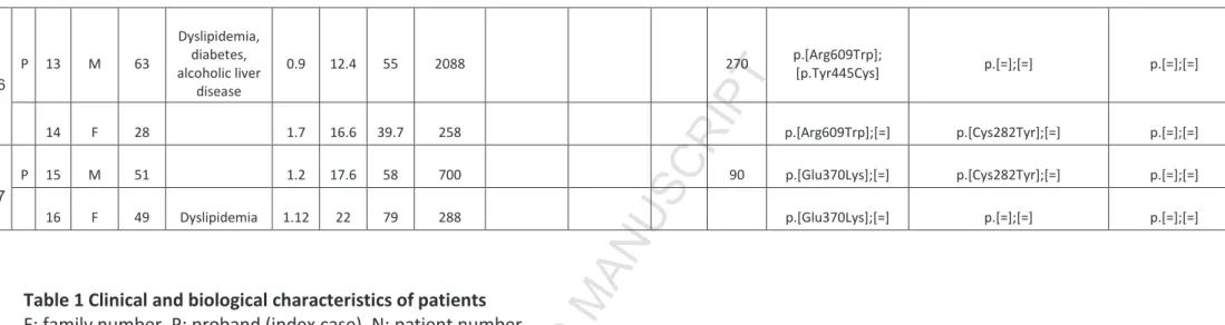
Table 1 Clinical and biological characteristics of patients
F: family number, P: proband (index case), N: patient number
TF: serum transferrin (g/l, N: 2-3,8), Iron : serum iron (μmol/l, N:12,5-25), TFsat: serum transferrin saturation (%, N <45), Ferritin: serum ferritin (µg/L, N<200 in women and <300 in men), Hepcidin: serum hepcidin level (nmol/l, N:4-30), hepcidin ferritin ratio (N:4-30), LIC: Liver Iron Content determined by magnetic resonance imaging (µmol/g, N<40). NTBI: Non transferrin bound iron (N<0,5 µmol/L). LPI: Labile Plasma Iron (N<0,5 µmol/L).
A
C
C
EPT
ED
M
A
N
U
SC
R
IPT
F P N Sex Age Comorbidity TF Iron TF
Sat Ferritin Hepcidin
Hepcidin /ferritin
Ratio
NTBI
/ LPI LIC Transferrin HFE HAMP
1
P 1 M 67 BMI 25 1.1 14.7 53 611 22.2 4 <0,5 170 p.[Val221Gly];[=] p.[=];[=] p.[Gly71Asp];[=]
2 F 76 High grade Non Hodgkin Lymphoma 0.95 10.4 43 1527 22.5 2.1 p.[Val221Gly];[=] p.[Cys282Tyr];[=] p.[=];[=] 3 F 64 1 16 64 325 16.4 5 <0,5 p.[Val221Gly];[=] p.[His63Asp];[Cys282Tyr] p.[=];[=] 4 M 73 bloodletting since 2004 1.2 15.5 52* (95) 96*
(2500) 2.1 2.2 p.[Val221Gly];[=] p.[His63Asp];[Cys282Tyr] p.[Gly71Asp];[=]
5 M 74 1.21 17.5 58 90 p.[Val221Gly];[=] p.[His63Asp];[Cys282Tyr] p.[=];[=] 2 P 6 M 52 1.3 17.9 55 886 170 p.[Tyr533X];[=] p.[=];[=] p.[=];[=] 3 P 7 F 70 1.07 20.4 76 143 p.[Tyr533X];[=] p.[His63Asp];[=] p.[=];[=] 8 F 47 1.3 25 75 154 p.[Tyr533X];[=] p.[His63Asp];[Cys282Tyr] p.[=];[=] 9 F 61 1.5 20.6 54.9 44 p.[Tyr533X];[=] p.[His63Asp];[=] p.[=];[=] 4 P 10 M 39 BMI 26 1.3 22.4 68 665 26.6 4.3 <0,5 75 p.[Cys421Arg];[=] p.[=];[=] p.[=];[=] 11 M 51 1.25 9.8 31.7 934 28.3 3.1 <0,5 70 p.[Cys421Arg];[=] p.[=];[=] p.[=];[=] 5 P 12 F 70 HTA, Dyslipidemia, BMI 32, regular blood donation 1.3 10.6 32 542 39 7.7 <0,5 c.1623-1G>T (splicing mutation) p.[=];[=] p.[=];[=]
A
C
C
EPT
ED
M
A
N
U
SC
R
IPT
6 P 13 M 63 Dyslipidemia, diabetes, alcoholic liver disease 0.9 12.4 55 2088 270 p.[Arg609Trp]; [p.Tyr445Cys] p.[=];[=] p.[=];[=] 14 F 28 1.7 16.6 39.7 258 p.[Arg609Trp];[=] p.[Cys282Tyr];[=] p.[=];[=] 7 P 15 M 51 1.2 17.6 58 700 90 p.[Glu370Lys];[=] p.[Cys282Tyr];[=] p.[=];[=] 16 F 49 Dyslipidemia 1.12 22 79 288 p.[Glu370Lys];[=] p.[=];[=] p.[=];[=]Table 1 Clinical and biological characteristics of patients F: family number, P: proband (index case), N: patient number
TF: serum transferrin (g/l, N: 2-3,8), Iron : serum iron (μmol/l, N:12,5-25), TFsat: serum transferrin saturation (%, N <45), Ferritin: serum ferritin (µg/L, N<200 in women and <300 in men), Hepcidin: serum hepcidin level (nmol/l, N:4-30), hepcidin ferritin ratio (N:4-30), LIC: Liver Iron Content determined by magnetic resonance imaging (µmol/g, N<40). NTBI: Non transferrin bound iron (N<0,5 µmol/L). LPI: Labile Plasma Iron (N<0,5 µmol/L).