ORIGINAL ARTICLE
Rapid and highly specific screening for
NPM1 mutations
in acute myeloid leukemia
Elisabeth Oppliger Leibundgut&Naomi A. Porret&
Marianne Bienz Muggli&Heidi Baumgartner&
Meike Dahlhaus&Gabriela M. Baerlocher
Received: 27 June 2012 / Accepted: 28 October 2012 / Published online: 17 November 2012 # Springer-Verlag Berlin Heidelberg 2012
Abstract NPM1 mutations, the most frequent molecular alterations in acute myeloid leukemia (AML), have become important for risk stratification and treatment decisions for patients with normal karyotype AML. Rapid screening for NPM1 mutations should be available shortly after diagnosis. Several methods for detecting NPM1 mutations have been described, most of which are technically challenging and require additional laboratory equipment. We developed and validated an assay that allows specific, rapid, and simple screening for NPM1 mutations. FAST PCR spanning exons 8 to 12 of the NPM1 gene was performed on 284 diagnostic AML samples. PCR products were visualized on a 2 % agarose E-gel and verified by direct sequencing. The FAST PCR screening method showed a specificity and sensitivity of 100 %, i.e., all mutated cases were detected, and none of negative cases carried mutations. The limit of detection was at 5–10 % of mutant alleles. We conclude that the FAST PCR assay is a highly specific, rapid (less than 2 h), and sensitive screening method for the detection of NPM1 muta-tions. Moreover, this method is inexpensive and can easily be integrated in the routine molecular diagnostic work-up of established risk factors in AML using standard laboratory equipment.
Keywords NPM1 . Acute myeloid leukemia . Mutation detection . Specificity . Sensitivity
Introduction
In acute myeloid leukemia (AML), an increasing number of genetic alterations have been described to be involved in the pathogenesis of the disease, and to determine the phenotype and the impact of the treatment [1]. As a consequence, the identification of molecular alterations by cytogenetic and/or molecular analysis is of great importance for risk stratification and treatment decisions. Molecular analyses are particularly important in AML patients with a normal karyotype who constitute about half of all AML cases [2].
NPM1 mutations represent the most frequent alterations in AML, occurring in approximately one third of adult patients with AML, and are associated with a normal karyotype in 85 % of cases [3]. In the 2008 World Health Organization (WHO) classification, AML with mutated nucleophosmin (NPM1) has been defined as a separate provisional entity, and there is increasing evidence that NPM1 mutations repre-sent founder genetic events in AML. Various studies have demonstrated the favorable prognostic impact of NPM1 muta-tions especially in the absence of a FLT3 ITD [4–8].
On the molecular level, NPM1 mutations are insertions that cluster within exon 11 and 12 of the NPM1 gene. The inser-tions typically consist of 1 or 2 tetranucleotides at nucleotides 956 to 971 (reference sequence NM_002520) corresponding to nucleotides c.860 through c.875 (HGVS nomenclature) [9]. The most prevalent mutation is the 960insTCTG (type A mutation) that is present in approximately 80 % of cases. Rare cases of non-tetrameric insertions and deletions/insertions have been described as well as single cases of 8-bp insertions in exon 11 leading to a truncated protein [10,11]. All muta-tions in exon 12 lead to a frame-shift and an elongated protein
E. Oppliger Leibundgut
:
N. A. Porret:
M. Bienz Muggli:
G. M. BaerlocherLaboratory of Molecular Diagnostics, Department of Hematology, University Hospital Bern and University of Bern,
Freiburgstrasse 4,
CH-3010 Bern, Switzerland
E. Oppliger Leibundgut (*)
:
H. Baumgartner:
M. Dahlhaus:
G. M. BaerlocherExperimental Hematology, Department of Clinical Research, University of Bern,
CH-3010 Bern, Switzerland e-mail: elisabeth.oppliger@insel.ch
with loss of the nucleolar localization domain and/or gain of a nuclear export signal resulting in an accumulation of the protein in the cytoplasm. A variety of molecular methods for detecting NPM1 mutations have been described, including PCR amplification and direct sequencing, high-resolution fragment analysis based on a fluorescence-labeled forward primer (Genescan), melting curve analysis on the LightCycler system, denaturing high-performance liquid chromatography (DHPLC), and LNA-mediated PCR clamping [12]. These methods are technically challenging and require well-trained personnel and additional laboratory equipment that might not be available in many molecular routine laboratories. Hence, since the detection of NPM1 mutations at diagnosis is impor-tant for risk stratification and treatment decision, molecular laboratories should be able to perform the initial analysis using their standard laboratory equipment. We therefore developed a rapid and simple screening assay based on FAST PCR ampli-fication and standardized agarose gel electrophoresis.
Materials and methods Patient samples
We validated our assay on 284 diagnostic samples from adult patients with AML referred to our laboratory, includ-ing 158 NPM1-mutated (NPM1-mt) samples and a series of 126 in-house NPM1-wild-type (NPM1-wt) samples. Patients were diagnosed according to the WHO criteria at the Univer-sity Hospitals of Bern, Basel, and Zürich and the District Hospitals of Lucerne, Bellinzona, St. Gallen, Thun, and Aarau. All patients were enrolled in the multicenter prospective trials of the HOVON/SAKK Study Group (HOVON AML proto-cols 42, 43, 81, 92, 102, 103) or treated according to the HOVON/SAKK protocols after informed consent according to the declaration of Helsinki. Patient characteristics are given in Table1. All samples of bone marrow or peripheral blood were collected at diagnosis.
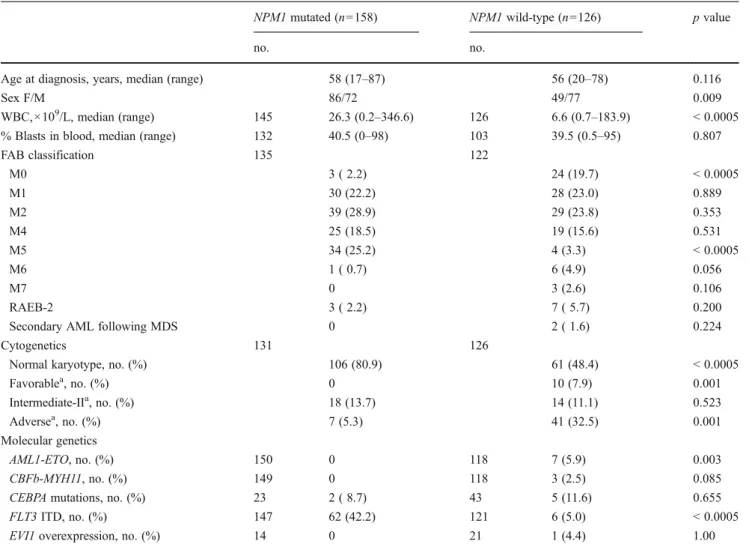
Table 1 Patient characteristics
NPM1 mutated (n0158) NPM1 wild-type (n0126) p value
no. no.
Age at diagnosis, years, median (range) 58 (17–87) 56 (20–78) 0.116
Sex F/M 86/72 49/77 0.009
WBC,×109/L, median (range) 145 26.3 (0.2–346.6) 126 6.6 (0.7–183.9) < 0.0005
% Blasts in blood, median (range) 132 40.5 (0–98) 103 39.5 (0.5–95) 0.807
FAB classification 135 122 M0 3 ( 2.2) 24 (19.7) < 0.0005 M1 30 (22.2) 28 (23.0) 0.889 M2 39 (28.9) 29 (23.8) 0.353 M4 25 (18.5) 19 (15.6) 0.531 M5 34 (25.2) 4 (3.3) < 0.0005 M6 1 ( 0.7) 6 (4.9) 0.056 M7 0 3 (2.6) 0.106 RAEB-2 3 ( 2.2) 7 ( 5.7) 0.200
Secondary AML following MDS 0 2 ( 1.6) 0.224
Cytogenetics 131 126
Normal karyotype, no. (%) 106 (80.9) 61 (48.4) < 0.0005
Favorablea, no. (%) 0 10 (7.9) 0.001 Intermediate-IIa, no. (%) 18 (13.7) 14 (11.1) 0.523 Adversea, no. (%) 7 (5.3) 41 (32.5) 0.001 Molecular genetics AML1-ETO, no. (%) 150 0 118 7 (5.9) 0.003 CBFb-MYH11, no. (%) 149 0 118 3 (2.5) 0.085
CEBPA mutations, no. (%) 23 2 ( 8.7) 43 5 (11.6) 0.655
FLT3 ITD, no. (%) 147 62 (42.2) 121 6 (5.0) < 0.0005
EVI1 overexpression, no. (%) 14 0 21 1 (4.4) 1.00
a
Cytogenetic groups according to ELN classification (13). Favorable: t(8;21)(q22;q22); inv(16)(p13.1;q22) or t(16;16)(p13.1;q22). Intermediate-II: t(9;11)(p22;q23); cytogenetic abnormalities not classified as favorable or adverse. Adverse: inv(3)(q21;q26.2) or t(3;3)(q21;q26.2); t(6;9)(p23;q34); t(v;11)(v;q23); -5 or del(5q); -7; abnl(17p); complex karyotype
Mutational analysis
Total RNA was extracted from Ficoll-separated mononuclear cells using the QIAmp RNA Blood Mini Kit (Qiagen, Hombrechtikon, Switzerland) and reverse transcribed by the Superscript II system (Invitrogen, Lucerne, Switzerland). For mutational analysis, a primer pair was designed to amplify exons 8 to 12 of the NPM1 gene (NPM663F 5 ′-GCGCCAGT-GAAGAAATCTATAC-3′ and NPM1109R 5′-GGACAA-CATTTATCAAACACGG-3′) yielding a 446-bp product. Two microliters of cDNA were amplified in a volume of 20μl containing 0.5 μM of each primer and 10 μl of 2× GeneAmp Fast PCR Master Mix (Applied Biosystems, Rotkreuz, Switzerland) on a 9800 or Veriti Fast Thermal Cycler (Applied Biosystems). Reaction conditions consisted of an initial denaturation step at 94 °C for 10 s followed by 35 cycles at 94 °C for 0 s, 60 °C for 5 s, and 72 °C for 20 s and a final elongation step at 72 °C for 30 s. PCR products were run for 40 min on a 2 % agarose E-gel (Invitrogen) where a double band indicates a mutation and a single band represents the wild-type sequence. All PCR products were verified by direct sequencing in both directions using BigDye Terminator Mix version 3 (Applied Biosystems, Rotkreuz, Switzerland) on an ABI3130xl automated sequencer. PCR products yielding a small smear above the wild-type band were sequenced with p r i m e r s N P M 7 9 3 F ( 5′-AAAACTCCTAAAACAC CAAAAGGACC-3′), NPM883F (5′-AAAGTGGAAGC CAAATTCATCAA-3′), NPM845R (5′-CACCTTTTTCTA TACTTGTTGCATTT-3′), and NPM964R (5′-TATTTTCT TAAAGAGACTTCCTCCA-3′).
With FAST PCR, thermal cycling reactions that normally take over an hour and a half can take as little as 25 min. The FAST PCR technique is performed on designated FAST PCR thermal cyclers and optimized reagents must be used. Alternatively, our PCR protocol was also run by standard
conditions on an ABI 9700 Thermal Cycler (Applied Biosystems) using Roche Taq polymerase (Roche, Rotkreuz, Switzerland) and buffer. Reaction conditions consisted of denaturation at 95 °C for 5 min, 35 cycles at 95 °C for 30 s, 60 °C for 30 s, and 72 °C for 45 s and a final elongation at 72 °C for 5 min. Results were consistent, but PCR products were somewhat fainter probably due to a lower PCR efficiency.
Statistical analysis
Statistical analyses were performed on SPSS 17.0 for Windows (SPSS Inc., Chicago, Illinois). The nonpara-metric Wilcoxon–Mann–Whitney test was used for con-tinuous data and the χ2 or Fisher exact (whenever an expected value was <5) test for categorical data to compare demographic, laboratory and molecular parameters between the patient groups. A p value of <0.05 was considered statistically significant.
Results
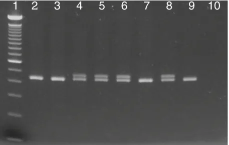
Of 284 AML samples included in the study, 158 were NPM1 mutated and 126 were wild-type as determined by FAST PCR screening. Mutated PCR products inva-riably showed two distinct bands on agarose gel elec-trophoresis, probably based on heteroduplex formation (Fig. 1). All mutated and wild-type PCR products were directly sequenced in both directions, and the results were 100 % concordant with the screening method. Mutations included 151 typical 4-bp insertions and 6 uncommon mutations in exon 12 (Table 2). In addition, a 19-bp deletion was detected in exon 9 outside the commonly mutated region leading to a frame-shift and a premature stop codon.
1 2 3 4 5 6 7 8 9 10
Fig. 1 After FAST PCR, PCR products are visualized on a 2 % agarose E-gel, where a double band indicates a muta-tion and a single band represents the wildtype sequence. Lanes 1 100 bp ladder, 2, 3 and 7 patient samples with wild-type NPM1, 4–6 patient samples with mutat-ed NPM1, 8 positive control cell line OCI-AML3, 9 wild-type control cell line HL60, 10 no template control
In 2 samples with mutated and 10 samples with wild-type NPM1, a novel 3-bp in frame-deletion in exon 8 (748delTCA) was identified resulting in a loss of serin 218 (p.Ser218del). We detected this sequence variation in samples of patients with other hematological malignancies and healthy donors at variable proportions (CML, 0/18; ALL, 3/18; CLL, 5/12; healthy donors, 14/15) suggesting to represent a common polymorphism. The frequency and potential relevance of this sequence variation is currently under investigation in our laboratory.
Specificity and sensitivity of FAST PCR screening
The specificity and diagnostic sensitivity of the FAST PCR screening method were 100 %, i.e., all positive samples carried mutations and none of the 126 negative cases were mutated (positive predictive value of 1.00 and negative predictive value of 1.00). The limit of detection (LOD) of the assay as assessed by serial dilution experiments of the cell line OCI-AML3 in the negative cell line HL60 was 5–10 % mutated alleles.
NPM1 mutations and biological and morphological features The incidence of NPM1 mutations was higher in women (NPM1-mt, women, n086 (54 %); men n072; NPM1-wt, women, n049 (39 %), men n077, p00.009). The median ages of patients with and without NPM1 mutations were 58 and 56 years, respectively. Patients with NPM1 mutations had
significantly higher median white blood cell counts than patients without NPM1 mutations (26.3 × 10⁹/L versus 6.6 × 10⁹/L; p < 0.0005). No difference was observed with regard to the peripheral blast count (NPM1-mt, median, 40.5; NPM1-wt, median, 39.5). NPM1 mutations were predominantly found in the French–American–British (FAB) subgroups M1, M2, M4, and M5, and were less frequent in M0, M6, and M7. When compared to the NPM1-wt group, NPM1 mutations were significantly less frequent in FAB M0 and more frequent in FAB M5 (FAB M0, 2.2 % versus 19.7 %; p < 0.0005; FAB M5, 25.2 % versus 3.3 %; p<0.0005).These data confirm the known features of many reports reviewed in [3].
NPM1 mutations and cytogenetic and molecular features Cytogenetic data were available for 257 of the 284 patients with AML. In accordance with results from other studies, NPM1 mutations were significantly associated with a nor-mal karyotype (NPM1-mt, 80.9 %; NPM1-wt, 48.4 %; p< 0.0005) [3]. Patients with an aberrant karyotype were clas-sified in cytogenetic risk groups according to the recom-mendations of the European LeukemiaNet [13]. We found significantly less patients with NPM1 mutations in the fa-vorable and adverse cytogenetic subgroups compared to patients without NPM1 mutations (favorable, 0 versus 7.9 %; p00.001; adverse, 5.3 % versus 32.5 %; p00.001); 13.7 % and 11.1 % of patients with and without NPM1 mutations carried chromosomal aberrations of type
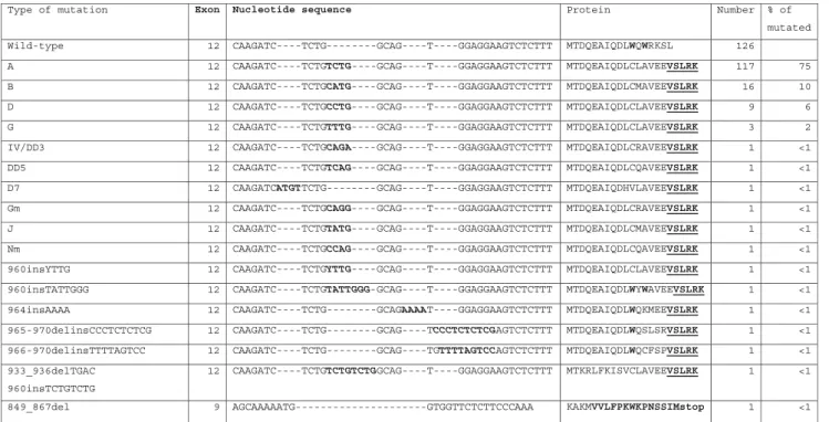
Table 2 NPM1 mutations detected by FAST PCR screening
Type of mutation Exon Nucleotide sequence Protein Number % of
mutated 2 1 e p y t -d l i
W CAAGATC----TCTG--------GCAG----T----GGAGGAAGTCTCTTT MTDQEAIQDLWQWRKSL 126
2 1
A CAAGATC----TCTGTCTG----GCAG----T----GGAGGAAGTCTCTTT MTDQEAIQDLCLAVEEVSLRK 117 75 2
1
B CAAGATC----TCTGCATG----GCAG----T----GGAGGAAGTCTCTTT MTDQEAIQDLCMAVEEVSLRK 16 10 2
1
D CAAGATC----TCTGCCTG----GCAG----T----GGAGGAAGTCTCTTT MTDQEAIQDLCLAVEEVSLRK 9 6
2 1
G CAAGATC----TCTGTTTG----GCAG----T----GGAGGAAGTCTCTTT MTDQEAIQDLCLAVEEVSLRK 3 2 2 1 3 D D / V
I CAAGATC----TCTGCAGA----GCAG----T----GGAGGAAGTCTCTTT MTDQEAIQDLCRAVEEVSLRK 1 <1
2 1 5
D
D CAAGATC----TCTGTCAG----GCAG----T----GGAGGAAGTCTCTTT MTDQEAIQDLCQAVEEVSLRK 1 <1 2
1 7
D CAAGATCATGTTCTG--------GCAG----T----GGAGGAAGTCTCTTT MTDQEAIQDHVLAVEEVSLRK 1 <1
2 1 m
G CAAGATC----TCTGCAGG----GCAG----T----GGAGGAAGTCTCTTT MTDQEAIQDLCRAVEEVSLRK 1 <1 2
1
J CAAGATC----TCTGTATG----GCAG----T----GGAGGAAGTCTCTTT MTDQEAIQDLCMAVEEVSLRK 1 <1 2
1 m
N CAAGATC----TCTGCCAG----GCAG----T----GGAGGAAGTCTCTTT MTDQEAIQDLCQAVEEVSLRK 1 <1
2 1 G T T Y s n i 0 6
9 CAAGATC----TCTGYTTG----GCAG----T----GGAGGAAGTCTCTTT MTDQEAIQDLCLAVEEVSLRK 1 <1 2 1 G G G T T A T s n i 0 6
9 CAAGATC----TCTGTATTGGG-GCAG----T----GGAGGAAGTCTCTTT MTDQEAIQDLWYWAVEEVSLRK 1 <1
2 1 A A A A s n i 4 6 9 CAAGATC----TCTG---GCAGAAAAT----GGAGGAAGTCTCTTT MTDQEAIQDLWQKMEEVSLRK 1 <1 965-970delinsCCCTCTCTCG 12 CAAGATC----TCTG---GCAG----TCCCTCTCTCGAGTCTCTTT MTDQEAIQDLWQSLSRVSLRK 1 <1 966-970delinsTTTTAGTCC 12 CAAGATC----TCTG---GCAG----TGTTTTAGTCCAGTCTCTTT MTDQEAIQDLWQCFSPVSLRK 1 <1
933_936delTGAC 960insTCTGTCTG 12 CAAGATC----TCTGTCTGTCTGGCAG----T----GGAGGAAGTCTCTTT MTKRLFKISVCLAVEEVSLRK 1 <1 A A A C C C T T C T C T T G G T G -G T A A A A A C G A 9 l e d 7 6 8 _ 9 4 8 KAKMVVLFPKWKPNSSIMstop 1 <1
intermediate II. Analysis of concomitant molecular aberra-tions confirmed the significant known association of NPM1 mutations and FLT3 ITD (42.2 % versus 5 %; p<0.0005). No AML1-ETO and CBFb-MYH11 fusions were found in the NPM1-mutated group. The frequencies of CEBPA muta-tions and EVI1 overexpression were not different in patients with and without NPM1 mutations.
Discussion
Given the favorable prognostic value of the NPM1+/ FLT3 ITD− genotype and the resulting therapeutic con-sequences, a rapid and simple method to assess the presence of NPM1 mutations is required. In this report, we show that FAST PCR and subsequent gel electro-phoresis is a specific and robust tool for the screening of NPM1 mutations and can be performed in less than 2 h. PCR products are run on a ready-to-use 2 % agarose gel that clearly separates the mutant from the wild-type band. The remaining PCR product can direct-ly be sequenced for accurate determination of the type of mutation. Although the prognostically favorable phe-notype of NPM1 mutations is independent of the type of mutation, it is important to identify the precise mutation for the design of real-time primers and probes for subsequent monitoring of minimal residual disease by qPCR.
In contrast to other methods, both the specificity and diagnostic sensitivity of the FAST PCR screening assay are 100 % (methods reviewed in [12]). The LOD of 5– 10 % mutated alleles equals the sensitivity of other methods such as capillary electrophoresis and is higher than for direct sequencing. For minimal residual disease detection, more sensitive methods such as allele-specific qPCR are appropriate, yet at the price of a limited diagnostic sensitivity that is restricted to one specific mutation per assay.
The FAST PCR technique has the advantage of reducing time-to-results by approximately a half to two thirds. Many but not all standard PCR protocols can be converted with little effort. The protocol presented in this report can also be run with standard conditions but yields the most PCR prod-uct by the FAST PCR mode.
We conclude that FAST PCR followed by agarose gel electrophoresis allows highly specific, rapid and sensi-tive screening for NPM1 mutations. More precisely, all mutations were detected and none was missed by this method. In addition, our assay is inexpensive and can easily be integrated in the routine molecular diagnostic work-up of established risk factors in AML using standard laboratory equipment.
Acknowledgments This work was in part supported by a grant from the SAKK (Swiss Group for Clinical Cancer Research) for translation-al research. We thank the Swiss members of the HOVON/SAKK Study Group for providing the samples and the technicians for performing the analyses. A special thank goes to Johanna A. Kremer Hovinga for statistical advice and to Jacqueline Schoumans, Jeroen Goede, Christian Kalberer, Pirmin Schmid, Marc Heizmann, Thomas Lehmann, Max Solenthaler, and Georg Stüssi for patient data.
Conflict of interest The authors declare that they have no conflict of interest.
References
1. Marcucci G, Haferlach T, Dohner H (2011) Molecular genetics of adult acute myeloid leukemia: prognostic and therapeutic implica-tions. J Clin Oncol 29:475–486
2. Mrozek K, Marcucci G, Paschka P et al (2007) Clinical relevance of mutations and gene-expression changes in adult acute myeloid leukemia with normal cytogenetics: are we ready for a prognostically prioritized molecular classification? Blood 109:431–448
3. Falini B, Martelli MP, Bolli N et al (2011) Acute myeloid leukemia with mutated nucleophosmin (NPM1): is it a distinct entity? Blood 117:1109–1120
4. Schnittger S, Schoch C, Kern W et al (2005) Nucleophosmin gene mutations are predictors of favorable prognosis in acute myeloge-nous leukemia with a normal karyotype. Blood 106:3733–3739 5. Dohner K, Schlenk RF, Habdank M et al (2005) Mutant
nucleo-phosmin (NPM1) predicts favorable prognosis in younger adults with acute myeloid leukemia and normal cytogenetics: interaction with other gene mutations. Blood 106:3740–3746
6. Verhaak RG, Goudswaard CS, van Putten W et al (2005) Mutations in nucleophosmin (NPM1) in acute myeloid leukemia (AML): association with other gene abnormalities and previously established gene expression signatures and their favorable prognostic significance. Blood 106:3747–3754
7. Thiede C, Koch S, Creutzig E et al (2006) Prevalence and prog-nostic impact of NPM1 mutations in 1485 adult patients with acute myeloid leukemia (AML). Blood 107:4011–4020
8. Schlenk RF, Dohner K, Krauter J et al (2008) Mutations and treat-ment outcome in cytogenetically normal acute myeloid leukemia. N Engl J Med 358:1909–1918
9. den Dunnen JT, Antonarakis SE (2000) Mutation nomenclature extensions and suggestions to describe complex mutations: a dis-cussion. Hum Mutat 15:7–12
10. Albiero E, Madeo D, Bolli N et al (2007) Identification and functional characterization of a cytoplasmic nucleophosmin leu-kaemic mutant generated by a novel exon-11 NPM1 mutation. Leukemia 21:1099–1103
11. Pitiot AS, Santamaria I, Garcia-Suarez O et al (2007) A new type of NPM1 gene mutation in AML leading to a C-terminal truncated protein. Leukemia 21:1564–1566
12. Wertheim G, Bagg A (2010) Nucleophosmin (NPM1) mutations in acute myeloid leukemia: an ongoing (cytoplasmic) tale of dueling mutations and duality of molecular genetic testing methodologies. J Mol Diagn 10:198–202
13. Dohner H, Estey EH, Amadori S et al (2010) Diagnosis and management of acute myeloid leukemia in adults: recommenda-tions from an international expert panel, on behalf of the European LeukemiaNet. Blood 115:453–474