DESCRIPTION OF THE LIFE STAGES OF MACROGLENES PENETRANS (KIRBY) (HYMENOPTERA: CHALCIDOIDEA, PTEROMALIDAE), A PARASITOID OF THE
WHEAT MIDGE, SITODIPLOSIS MOSELLANA (GEHIN) (DIPTERA: CECIDOMYIIDAE)
J.F. DOANE, R. DECLERCK-FLOATE, and A.P. ARTHUR
Agriculture Canada Research Station, Saskatoon, Saskatchewan, Canada S7N OX2
and F. AFFOLTER
C.A.B. International Institute of Biological Contro1;Chemin des Grillons 1, CH-2800 DelCmont, Switzerland
Abstract Can. Ent. 121: 1041-1048 (1989)
The life stages of Macroglenespenetrans (Kirby), an egg-larval parasitoid of the wheat midge, Sitodiplosis mosellana (GBhin), are described. The mean length and width, respectively, of 25 parasitoid eggs deposited in host eggs were 0.105 k 0.008 (SD) mm and0.041+0.006mm. Matureoocytes were0.131+0.008 mm long by 0.038k0.004 mm wide, slightly larger than laid eggs.
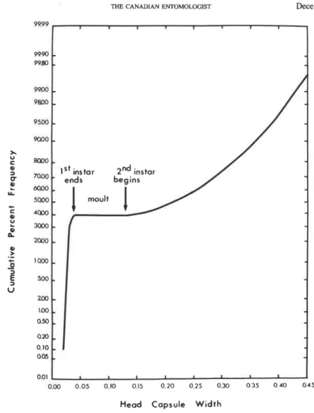
A frequency distribution of head width (HW) indicated two larval instars during the feeding period. During development, the HW of the first instar remains almost constant at about 0.03 mm whereas body length (BL) increases from about 0.15 to about 0.3 mm. Both HW and BL increase in the second instar from about 0.15 and 0.49 to 0.34 and 1.05 mm, respectively, for mature larvae. The third instar does not feed and is characterized by hook-like "pseudomandibles" and four incurving spines on the terminal segment. The pupa is adectious exarate. Adults are about 2 mm long, shiny bluish-black, and have translucent wings. Males have prominent rust-coloured eyes; eyes of females are less prominent and fuscous.
Resume
Les stades de dCveloppement de Macroglenes penetrans (Kirby), un parasitoide ovo- larvaire de la cCcidomyie orange du bl6, Sitodiplosis mosellana (GChin), sont dCcrits. Les oeufs pondus dans ceux de I'h6te mesurent 0,105
+
0,008 mm (moyenne r Ccart- type, n = 25) de long et 0,041 k 0,006 mm de large, alors que les oocytes matures sont 1Cgkrement plus grands avec respectivement 0,131+
0,008 mm et 0,038 k 0,004 mm. Pendant la periode d'alimentation, la distribution des frkquences de la largeur de la t6te du premier stade larvaire a une longueur constante d'environ 0,03 mm alors que la longueur de corps (BL) passe d'environ 0,15 mm a 0,3 mm en fin de croissance. Durant le deuxikrne stade larvaire, les deux pararnktres HW et BL passent respecti- vement de 0,15 mm a 0,34 mm et de 0,49 mm a 1,05 mm en fin de croissance. Le troisikme stade larvaire ne s'alimente pas et est caractens6 par des "pseudomandi- bules" unciformes et les quatre spinules incurvCes du segment terminal. La nymphe est du type libre. Les adultes, d'environ 2 mm de long, sont de couleur bleu-noir mktallique et posskdent des ailes translucides. Les mdles se distinguent par leur yeux proeminents de couleur rouille; les yeux des femelles sont moins proeminents et brun foncC.Introduction
In 1984 a chalcidoid wasp, later identified as Pirenepenetrans (Kirby), was observed emerging in some research plots used to study the wheat midge, Sitodiplosis mosellana (GChin), in northeastern Saskatchewan. This parasitoid was first described by Kirby in 1800 as Ichneumon penetrans. The imago of this insect was later described under the generic name Macroglenes by Johansson (1936) as reviewed by Peck (1963). Synonymy and taxonomy have been given by Graham (1969). Although the generic name Pirene has been preferred and widely used, Macroglenes Westwood has precedence (BouEek 1988) and will be used here. The species Macroglenes penetrans (Kirby) is widely distributed and usually common in Europe (Doeksen 1938; Speyer and Waede 1956; Barnes 1956). Its distribution in Canada and the United States is unknown but it was collected in Manitoba
1042 THE CANADIAN ENTOMOLOGIST December 1989 associated with the wheat midge outbreak that occurred there from 1954 to 1959 (Beirne 1971). Nine specimens from that collection were identified as P. penetrans by C. Yoshi- mot0 at the Biosystematics Research Centre (Ottawa, Canada). In a concurrent study in Switzerland, 15 specimens of M. penetrans were identified by Z. BouEek at the Com- monwealth Institute of Entomology, London.
A number of hymenopterous parasitoids are known to attack the wheat blossom midges S. mosellana and Contarinia tritici (Kirby) in Europe (Barnes 1956). Both midge species were thought to be hosts for the same complex of parasitoids (Johannson 1936; Doeksen 1938; Basedow and Schiitte 1982). However, a recent study indicates that S. mosellana and C . tritici have distinct complexes of parasitoids within the families Pteromalidae and Platygasteridae (Affolter 1987). Affolter (1987) lists parasitoids of S. mosellana as M.
penetrans, Platygaster sp., and Euxestonotus error (Fitch); those ascribed to C . tritici are
Piestopleura sp., Isostasius sp., and Leptacis tipulae (Kirby). Recently collected European specimens of E. error are considered to be conspecific with specimens described by Fitch in 1865 (Masner 1989). The rarity of E. error in North America suggests that it was accidently introduced by earlier settlers but failed to become well established (Masner 1989). Another platygasterid wasp (Inostemma horni Ashmead) was reported to be a par- asitoid of S. mosellana in Washington State by Reeher (1945).
Although M. penetrans is known as an important natural control agent of S. mosellana in Europe (Basedow and Schiitte 1982; Affolter 1987), no detailed studies on its biology have been made; its immature stages have not been described. The objective of this study was to describe the life stages of this parasitoid, primarily to enable the separation of the immature instars from those of other parasitoid species known to occur in Europe and which may occur in North America. The major portion of the work was done in Saskatch- ewan supplemented with observations on the immature instars of the parasitoid from a population in Switzerland.
Materials and Methods
Adult. Adult M. penetrans were reared from field-collected wheat midge cocoons. The
lengths of 50 M. penetrans males and 32 females were obtained.
Egg. Parasitoid females that emerged in the laboratory were held for a few days and then
dissected to obtain oocytes. Oocytes were stained in 1% Eosin in water to improve micro- scopic examination. Twenty-five oocytes were measured for each of five females. Six to seven oocytes also were obtained for each of three females from the Swiss population. Eggs of M. penetrans were obtained by inducing female parasitoids to oviposit into wheat midge eggs in the laboratory. The lengths and widths of 25 parasitoid eggs dissected from midge eggs were recorded. Eggs and all subsequent stages were measured with an ocular micrometer and compound microscope.
Larva. To obtain a range in size of parasitoid larvae, soil samples containing wheat midge
larvae were collected from the field each week from May to mid-October. An average of 25 (range 6-53) parasitoid larvae was dissected from the midge larvae each week to mea- sure for parasitoid head width (HW), head length (HL), and body length (BL). An average of 17 (range 4-30) parasitoid larvae dissected from midge larvae collected at weekly inter- vals from wheat heads from mid-July to mid-September also was measured. A frequency distribution of head widths from 693 parasitoid larvae was used to separate the larval instars.
Observations were made on living larvae and whole mounts to determine the presence of mandibles in different instars and to describe morphological differences that could be used to separate instars. Larvae considered to be in the early and late second instar were fixed in Bouin's fixative (Baker 1958) overnight, stained with Rhodamine B (in 50% ethyl
Volume 121 THE CANADIAN ENTOMOLOGIST 1043 alcohol) for 4 h during dehydration in a graded alcohol series, and then mounted in "Per- mount". Also, semi-permanent slides were made of late second-instar larvae that were cleared and mounted in de Faure's fluid under a cover slip.
Third-instar Larva and Pupa. Third-instar larvae and pupae were obtained by holding
parasitized midge larvae that had broken diapause on damp blotting paper and allowing the parasitoids to develop. Measurements of HW and BL were obtained for 10 third-instar larvae and BL and thoracic width on seven pupae.
Results
Adult. Both sexes of M. penetrans (Fig. 1A and B) are shiny bluish-black with iridescent
wings. Prominent rust-coloured eyes distinguish the males (Fig. 1 A), the lower one-quarter of each eyed being darker than the remainder. Eyes of the females (Fig. 1B) are less prominent than those of males and are brownish-black. The mean length
+
SD of 50 males and 32 females was 1.77+
0.119 and 1.80 ? 1.05 mm, respectively.Egg. Eggs of M. penetrans, an egg-larval parasitoid, are laid into the eggs of the host wheat midge (Fig. 1C). Oviposition was observed in the laboratory and parasitized eggs also were found in the field (unpublished data). Mean length ( + SD) and width ( + SD) of 25 eggs laid by M. penetrans was 0.105 ? 0.008 and 0.041
+
0.006 mm, respectively. The eggs are glistening creamy white, slightly translucent, and have no noticeable sculp- turing at magnifications of 100-200 times.Mature oocytes are milky white and have a stalk more prominent than on the laid egg (Fig. 2A). Mean length and width of 125 oocytes from the Saskatchewan population was 0.131
+
0.008 and 0.38 +0.004 mm, respectively. Mean length and width of 20 oocytes from the Swiss population was 0.1 15+
0.009 and 0.033+
0.003 mm, respectively, slightly smaller than those measured for the Saskatchewan population.Larval Instars. The cumulative frequency distribution of the HW clearly separated the
first and second instars (Fig. 3). Early and late development of each of these instars were distinguishable, so this designation was made for convenience in separating larvae of different maturity within instars. The third instar does not feed and is of short duration.
First-instar larva. Newly hatched M. penetrans larvae have a prominent head with
an elongated translucent body (Fig. 2B and C). The body widens slightly behind the head and tapers to the end of the abdomen. Eleven segments usually are visible in newly hatched larvae; a small spine or bristle marks each of the nine abdominal segments dorsally (Fig. 2B). A pair of terminal ventrally curving hooks are borne on the abdomen (Fig. 2B). Small sickle-shaped mandibles that move independently also are evident (Fig. 2C). Mean length and width of four mandibles from two individual larvae from the Swiss population were 8.36 and 3.93 Ihm, respectively. As the first-instar larva grows it becomes tear-drop-shaped (Fig. 2D). Although the body length increases from about 0.144 to 0.302 mm, the width of the head remains almost constant (Fig. 3). In late first-instar larvae segmentation and bristles on the dorsum are not evident; the caudal hooks are still present. The gut, filled with a pale yellowish substance, can be seen through the body wall. The large, more opaque larva is, therefore, more easily seen when dissected from the host. A pair of small internal lobes, probably brain lobes, are present behind the head abutting the gut (Fig. 2D).
Second-instar larva. Unlike the first instars, the head of second instqrs does not
protrude anteriorly and caudal hooks are not present (Fig. 2E and F). There is also a progressive increase in HW as development continues (Fig. 3). Mean HW, HL, and BL
of early second-instar M. penetrans dissected from midge larvae from overwintering cocoons in early May were 0.188
+
0.025 (n = 29), 0.074+
0.016 (n = 27), and 0.514+0.075 (n= 36) mm. Usually 13 body segments, including the head, are evident, with the thoracic segments being most prominent. The exuviae of first-instar larvae oftenI(*W THE CANADIAN ENTOMOLQGIST December 1989
FIG. 1. Adult male (A) and female (B) M. penetrans and (C) S . mosellana eggs containing eggs ( e ) of M. penerrans.
are observed attached to the end of the abdomen when the larva is dissected from the host (Fig. 2E). This detaches when the larva is placed in distilled water.
Through the body wall, the gut and what appear to be brain lobes can be seen. The gut has a central core filled with dense orange material. The esophagus often can be seen (Fig. 2F).
Volume 121 THE CANADIAN ENTOMOLOGIST' 1045
F I G . 2 . Oocyte, and larval stages of M. penetrans: A, stalked oocytes; B (lateral) and C (ventral), views of an early first-instar larva showing dorsal spines (s), terminal hooks (h), and mandibles (m); D, ventral view of late first-instar larva showing brain lobes (b) and gut (g); E (dorsal) and F (lateral), views of an early second-instar larva showing brain lobes (b), gut (g), exuviae of first instar (ex), and esophagus (es); G, dorsal view of late second-instar larva showing brain lobes (b) and gut (g) seen through body wall; H, 3-dimensional lateral view
of late second-instar larva with characteristic tail curve.
Older second-instar larvae are longer, wider, and more robust than younger ones. Fully mature larvae dissected from wheat midge larvae collected from the field about mid- June had attained HW's, HL's, and BL's of 0.338 k0.053 (n = 22), 0.261 k0.042 (n= 21), and 1.052 k 0.25 (n = 25) mm, respectively.
Through the body wall, orange gut and brain lobes are apparent (Fig. 2G). Imaginal discs of antennae and mouth parts are evident as more-or-less circular areas beneath the cephalic cuticle. Segmentation is more pronounced and the terminal segments of the abdo- men typically curve upward (Fig. 2H). No mandibles were observed in whole mounts of second-instar larvae but they were seen occasionally in living larvae during microscopic examination. They were unsclerotized and made noticeable only by their movement.
1046 THE CANADIAN ENTOMOLOGIST December 1989
Head Capsule Width
9999 9990 9980 PPM) 9 6 0 0 , 9SW m , >.
:
f w o - 0 HIOO P ?! 6mx, LL C m o $ 'MO' m - P, n 10, 0.-
* I M X ) , 0-
3E
"-
u 2 0 0 - 1 0 0 . O M ax, 0.10 Q05 001FIG. 3. Cumulative frequency distribution of head capsule width showing the end of the first instar and beginning of the second instar.
.
I 1 1 ,.
-
, l S t instar 2nd instar ends1
moult/
.
.
.
.
.i.
I I I I I-
000 0 0 5 010 015 020 025 030 0 3 5 0 4 0 0 4 5Third-instar larva. Parasitized larvae spin an elongate cocoon that appears to be identical to that spun by the unparasitized larvae. Within the cocoon the third-instar larva forms, enclosed by the second larval skin which is ruptured as the third instar develops. On moist blotting paper in the laboratory, parasitized and unparasitized larvae sometimes develop through the third instar and pupate without forming a cocoon. The third instar required only 2 days to complete development and transform to the pupa in the laboratory. Mean third-instar HW and BL (n = 10) were 0.330
+
0.086 and 1.403+
0.300 mm, respectively. Including the head, 13 segments were visible in dorsal view (Fig. 4A). Ante- riorly, the head has two protruberances that disappear during transformation from third instar to pupa (Fig. 4A and B). The mouth area is bordered by two hook-like structures that appear to be mandibles (Fig. 4C). However, as the third-instar larva transforms to the pupa, these hooks are seen to be situated just below the developing pupal eye and were not associated with the mouth region so have been termed "pseudomandibles". They were no longer observed after the complete transformation from the third instar to the pupa. AVolume 121 THE CANADIAN ENTOMOLOGIST 1047
FIG. 4. A (dorsal) and B (ventral), views of a third-instar larva; C, an enlarged ventral view of the head showing the precursal antennal discs (a) and eyes and pseudomandible @); D, terminal segment with incurving spines
(s); E (dorsal), F (lateral), and G (ventral), views of a pupa.
yellowish food pellet, taking up most of the gut region, is seen through the body wall (Fig. 4B). The terminal segment of the abdomen usually has four or occasionally five hook-like spines (Fig. 4D) which are also lost when the prepupa transforms to the pupa.
Pupa. The pupa of M. penetrans is adectious and exarate and is slightly larger than the third instar and slightly smaller than the adult. Mean length of seven specimens was 1.68
+
0.19 mm. During development, the pupa is initially light in colour and then the eyes, ocelli, wings, and legs begin to pigment, followed by the remainder of the body. In males, the ventral approximately one-quarter of the eye darkens first and this portion remains darker than the remainder of the eye in the adult.1048 THE CANADIAN ENTOMOLOCiW December 1989 Discussion
Kutter (1934) found that Pirene graminae Haliday, a species closely related to M.
penetrans, parasitized the larvae of its host, Contarinia pisi Winnertz, and not the egg. Otherwise the two species appear to be similar in the morphology of their immature stages. Freshly deposited eggs of P. graminae were slightly larger (0.16-0.17 mm long by 0.06- 0.08 mm wide) than those of M. penetrans. Pirene grarninae also has two distinct larval stages during the feeding period. Mandibles in first-instar P. graminae appear to be iden- tical in form with those of M. penetrans. Kutter illustrated internal mandibles in late sec- ond-instar P. graminae similar to those observed in M. penetrans. Also, Kutter describes a prepupae which Clausen (1940) considered to be a third-instar larva similar to the third- instar larva we describe for M. penetrans.
Acknowledgments
We thank L. Moffatt and T. Nelson for their assistance in laboratory and field work, and Y. Powell for drawings of the third instar and pupa. We are also grateful to C. Yosh- imoto, Biosystematics Research Centre, Ottawa, and Z. BouEek, Commonwealth Institute of Entomology, London, for identifications. This research was funded in part by a grant from the Saskatchewan Agriculture Development Fund. Research at the CIBC was sup- ported by Agriculture Canada and forms part of a Ph.D. thesis study by F. Affolter.
References
Affolter, F. 1987. Studies on the natural enemies of the wheat midge, Sitodiplosis mosellana. C.A.B. Interna- tional Institute of Biological Control, Annual Report 1987. p. 27.
Baker, J.R. 1958. Principles of Biological Microtechnique. Methusen & Co. Ltd., London.
Barnes, H.F. 1956. Gall Midges of Economic Importance Vol. 7: Gall Midges of Cereal Crops. Crosby, Lock- wood & Son Ltd., London.
Basedow, T., and F. Schutte. Die Populationsdynamik der Weizengallmucken Contarinia tritici (Kirby) und Sitodiplosis mosellana (Gkhin) (Dipt., Cecidomyidae) in zwei norddeutschen Weizenbaugebieten von 1969 bis 1976. Zool. Jahrb. Syst. 109: 33-82.
Beirne, B.P. 1971. Pest insects of annual crop plants in Canada. I. Lepidoptera. 11. Diptera. 111. Coleoptera. Mem. ent. Soc. Can. 78: 65-66.
BouEek, Z. 1988. Australasian Chalcidoidea (Hymenoptera). A Biosystematic Revision of Genera of Fourteen Families with a Reclassification of Species. C. A.B. International, Wallingford, U.K. 832 pp.
Clausen, C.P. 1940. Entomophagous Insects. McGraw Hill.
Doeksen, J. 1938. De tanvegalmuggen Contarinia tritici Kirby an Sitodiplosis mosellana (Gehin) (Diptera:
Cecidomyiidae) in Nederland. Versl. Tarwe Comm., Groningen 12: 237-296.
Fitch, A. 1865. Insects infesting grain crops. Injurious and Other Insects of the State of New York, 6th Rep. Graham, M.W.R. de V. 1969. The Pteromalidae of north-western Europe (Hymenoptera: Chalcidoidea). Bull.
Br. Mus. Nut. Hist. En?. Suppl. 16. 908 pp.
Johansson, E. 1936. Studier och forsok rorande vetemyggorna Contarinia tritici Kirby och Clinodiplosis mosel- l a m Geh. sarnt deras bekampande. IV Undersokning av Vetemyggomas parasiter: 1 Svalof och Weibullsh-
olm aren 1932-1935 antraffade arter Statens. Me&. VSirtskyddsanst. Stockh. 15. 19 pp.
Kirby, W. 1800. A continuation of the history of Tipula tritici in a letter to T. Marsham. Trans. Linn. Soc. Lond. (Zool.) 5: 9 6 1 11.
Kutter, H. 1934. Weiter Untersuchungen iiber Kakothrips robustus Uzel und Contarinia pisi Winn., sowie deren Parasiten, insbesondere Pirene graminae Hal. Mitt. Schweiz Ent. Gesell. 26: 1 4 6 .
Masner, L. 1989. Euxestonotus error (Fitch) - Palearctic or Nearctic? Agric. Can. Biocontrol News 2: 23-24. Peck, 0 . 1963. A catalogue of the Nearctic Chalcidoidea (Insecta: Hymenoptera). Can. Ent. Suppl. 30. Reeher, M.M. 1945. The wheat midge in the Pacific Northwest. U.S.D.A. Circular 732. 8 pp.
Speyer, W., and M. Waede. 1956. Feinde und Parasiten der Weizengallmiicken. Ang. Schiidlingsk PJlanzen- umweltsch. 19: 185-191.