2010/071
Effects of
m
CT radiation on tissue engineered bone-like
constructs
Thomas P. Kraehenbuehl1,a, Martin Stauber1, Martin Ehrbar2, Franz Weber2, Heike Hall3 and Ralph Mu¨ller1,
*
1Institute for Biomechanics, ETH Zurich, Zurich,
Switzerland
2Section Bioengineering and Department of
Craniomaxillofacial Surgery, University Hospital, Zurich, Switzerland
3Department of Materials, ETH Zurich, Zurich,
Switzerland
Abstract
High-resolution, non-destructive imaging with micro-com-puted tomography (mCT) enablesin situ monitoring of tissue
engineered bone constructs. However, it remains controver-sial, if the locally imposed X-ray dose affects bone devel-opment and thus could influence the results. Here, we developed a model system for mCT monitoring of tissue engineered bone-like constructs. We examined the in vitro
effects of high-resolutionmCT imaging on the cellular level by using pre-osteoblastic MC3T3-E1 cells embedded into three-dimensional collagen type I matrices. We found no sig-nificantly reduced cell survival 2 h after irradiation with a dose of 1.9 Gy. However, 24 h post-irradiation, cell survival was significantly decreased by 15% compared to non-irra-diated samples. The highest dose of 7.6 Gy decreased sur-vival of the pre-osteoblastic MC3T3-E1 cells by around 40% at 2 days post-irradiation. No significant increase of alkaline phosphatase (ALP) activity at 2 days post-irradiation was found with a dose of 1.9 Gy. However, ALP activity was significantly decreased after 7 days. Using our model system, the results indicate thatmCT imaging with doses as low as 1.9 Gy, which is required to obtain a reasonable image qual-ity, can induce irreparable damages on the cellular level.
Keywords: alkaline phosphatase activity (ALP); cell
surviv-al; collagen; MC3T3-E1 pre-osteoblastic cells; micro-com-puted tomography (mCT); tissue engineering.
Present address: Hoffmann-La Roche, Translational Research a
Sciences, Nutley, NJ 07110, USA.
*Corresponding author: Ralph Mu¨ller, PhD, Institute for Biomechanics, Wolfgang-Pauli-Strasse 10, 8093 Zurich, Switzerland
Phone: q41-44-63245 92 Fax: q 41-44-63212 14 E-mail: [email protected]
Introduction
In vitro engineering of functional tissue constructs has gained
importance in the past decade as the shortage of organs and tissues for transplantation increased w21x, but also for safety and efficacy testing of new therapeutics w8, 12, 15x. So far, quality testing of engineered tissue constructs relied on destructive techniques including reverse transcription poly-merase chain reaction to analyze gene expression or histol-ogy to assess organization and integrity of the tissue. Thus, monitoring over time and functional optimization in response to assessed changes in tissue morphology and functionality was not possible with these techniques. Other methods, such as magnetic resonance imaging or positron emission tomog-raphy, do not provide sufficient spatial resolution w10, 17x.
Micro-computed tomography (mCT) was shown to be use-ful as a non-destructive method in imaging three-dimensional (3D) bone microarchitecture at resolutions in the order of 10 mm in relatively short acquisition time (5–30 min) and at low cost w2, 3, 19x. In recent studies,mCT was used to monitor the development of 3Din vitro engineered bone-like
tissues w4, 13, 14x, cartilage-like tissues w23x or skin-like lay-ers w20x over time. However, one drawback of obtaining high-qualitymCT images can be the locally deposed radia-tion dose and its potential effect on cell survival, cell func-tionality and cell development w11x.
Several studies have examined the effects of ionizing radi-ation on bone cells in a two-dimensional (2D) environment at doses up to 8 Gy w5, 6, 9x. Although the proliferation of MC3T3-E1 pre-osteoblastic cells and osteoblast-like cells from the rat calvarium was not affected by a radiation dose of 0.4 Gy, a dose of 4.0 Gy significantly decreased prolif-eration in all studies, and in some cases significantly altered alkaline phosphatase (ALP) activity, collagen production and vascular endothelial growth factor secretion as compared to non-irradiated control samples. However, irradiation effects on 3D tissue engineered constructs, which mimic native 3D tissue and organ structures better than 2D environments w8, 12x, are not fully understood. Specifically, the critical dose, which can maximally be deposited onto the constructs with-out inducing cellular damages, is not clear. Because there is a big difference between a flat layer of cells and a complex 3D tissue w1x, it is important to investigate the irradiation effect in 3D tissues.
The aim of this study was to assess this maximum dose. We have developed a model system formCT monitoring of 3D tissue engineered constructs and to elucidate effects of the ionizing radiation applied bymCT imaging on the cel-lular level by using MC3T3-E1 pre-osteoblastic cells embed-ded in 3D collagen type I matrices mimicking bone-like tissue w7, 16x. We hypothesized that the locally deposed
X-ray dose by high-resolution mCT imaging can cause altera-tions on the cellular level in a dose- and time-dependent manner. This not only can affect the development of engi-neered 3D tissue constructs for clinical applications but also the results of pharmaceutical studies for testing new therapeutics.
Materials and methods
Dose measurement with thermoluminescent detectors
The radiation dose occurring with typical settings for mCT scanning was measured with thermoluminescent detectors (TLDs) made of lithium fluoride powder embedded in a glass tube of 12 mm length and 1.4 mm diameter (TLD-700, Hars-haw/Filtrol, Solon, OH, USA). For mCT measurement, the TLDs were placed in a cylindrical polymethyl methacrylate (PMMA) cylinder (inner diameter: 2 mm, outer diameter: 8 mm) to ensure corresponding radiation doses as applied to the cells embedded into the collagen matrix. The mCT (mCT40, Scanco Medical AG, Bru¨ttisellen, Switzerland) was operated at a peak voltage of 50 kV with an integration time of 100 ms in all possible scanning modes. The three primary scanning modes provided by the mCT manufacturer were ‘‘standard resolution’’ mode (250 projections/1808, 1024 samples per projection), ‘‘medium resolution’’ mode (500 projections/1808, 1024 samples), and ‘‘high resolution’’ mode (1000 projections/1808, 2048 samples). In each mode, five different nominal resolutions were available namely 12, 16, 20, 30, and 36 mm for the standard and medium reso-lution mode, and 6, 8, 10, 15, and 18mm for the high res-olution mode. These nominal resres-olution values correspond to the settings that can be chosen to run the scans and do not represent the actual spatial resolution, which is typically lower. The best spatial resolution (10% MTF) at 6mm voxel size is specified by the manufacturer to be 10 mm. After irradiation, the TLDs were read out with a photo-multiplier using a standard heating protocol.
For all cell experiments the medium resolution mode at 20 mm nominal resolution was used, but with a threefold increased integration time resulting in a calculated base dose of 1.9 Gy per measurement. To achieve the higher doses of 3.8, 5.7, and 7.6 Gy, the base measurement was repeated 2, 3, and 4 times, respectively. The total times for application of these radiation doses were 1.5 h, 3 h, 4.5 h, and 6 h, respectively. To examine potential differences in image qual-ity, a bovine trabecular bone sample was scanned withmCT in four different modes: in standard resolution mode at 30 mm and 16 mm nominal resolution, as well as in high resolution mode at 15mm and 8mm nominal resolution.
Culture of pre-osteoblastic MC3T3-E1 cells
A MC3T3-E1 subclone 24 (LGC Promochem Sarl, Mols-heim Cedex, France) was cultured between passage 6–10 in alpha-modified minimum essential medium containing 10% fetal bovine serum (both Invitrogen, Basel, Switzerland),
1mg/ml bone morphogenic protein-2 produced as previously shown w22x, and 25 mmol 4-(2-hydroxyethyl)-1-piperazine-ethanesulfonic acid (Sigma-Aldrich, Buchs, Switzerland). Preparation of 3D collagen matrices
Collagen type I matrices (Vitrogen 100, Cohesion, Palo Alto, CA, USA) of 2 mg/ml were used. Matrix formation was initiated by raising the pH to physiological conditions (pH 7.4) by adding 0.1 M NaOH to the collagen solution. To ensure homogeneous cell encapsulation, the matrices were presolidified at 378C for 5 min prior to the addition of 500,000 cells/ml collagen I. Matrices with a volume of 30ml were then filled into sterilized (70% ethanol, exposure to UV light) PMMA sample holders with a capacity of 35ml and incubated for 30 min at 378C to solidify. The sample holders containing the 3D cell cultures were then kept in 1 ml medi-um in a well of a 24-well plate under standard cell culture conditions.
Irradiation withmCT
A high-resolution micro-computed tomography system (mCT40, Scanco Medical AG, Bru¨ttisellen, Switzerland) was used to apply the ionizing radiation to the cells embedded in the 3D collagen type I matrices. The system was operated in all cell experiments at a peak voltage of 50 kV with an inte-gration time of 300 ms at a nominal resolution of 20 mm. The absolute dose delivered to the samples was assessed in an independent experiment using TLDs. In all experiments, cultured MC3T3-E1 cells embedded in 3D collagen type I matrices were exposed to radiation 24 h after seeding into the collagen matrix. For cell survival experiments, cells were irradiated with doses of 1.9, 3.8, 5.7, and 7.6 Gy. For DNA and ALP assays, a dose of 1.9 Gy was applied. Control sam-ples were placed inside themCT, shielded with a lead cyl-inder against potential irradiation. Temperature effects on cell survival and ALP expression during amCT run can thus be excluded. Survival experiments as well as ALP/DNA exper-iments were all compared with thismCT control.
Cell survival aftermCT irradiation
Cell survival after mCT irradiation was determined with a live/dead cytotoxicity kit (Molecular Probes, Eugene, OR, USA). This assay was performed 2 h and 24 h post-irradi-ation (per dose, and per time point: nsampless3, ncontrols9).
The samples were washed in phosphate buffered saline (PBS, Sigma Aldrich, Basel, Switzerland) and stained for 45 min with 200 ml live/dead staining solution (4 mM ethidium
homodimer-1, 2mMcalceinAM in PBS). The samples were then washed in PBS and fixed in 4% glutaraldehyde. Epi-fluorescence images were taken at five randomly selected locations (area of ;2=1 mm2) within the matrix by using
a fluorescence microscope (Zeiss Axiovert 135, Feldbach, Switzerland) and the fluorescence for calceinAM was deter-mined with an excitation wavelength of 495 nm and an emis-sion wavelength of 515 nm. For ethidium homodimer-1, an excitation wavelength of 495 nm and an emission
wave-length of 635 nm were used, respectively. The number of surviving cells per total number of cells (live cells plus dead cells, between 30 and 100 cells in total, per location) was determined by manual counting. The cell numbers of the irradiated samples were normalized to the non-irradiated control samples, which were placed into themCT with lead shielding.
ALP activity assay
Each sample was homogenized in 300 ml lysis solution (0.2% v/v Triton X-100 in 0.56M
2-amino-methyl-1-propa-nol, pH 10.0). Total number of samples/controls per time point: nsampless10, ncontrols10. From each sample an aliquot
was used to determine ALP activity. Briefly, 150ml of the lysed cell suspension was mixed with 20ml of p-nitrophenyl phosphate solution (74 mg/ml in 0.56 M
2-amino-methyl-1-propanol, pH 10.0) and incubated at 378C for 30 min. The reaction was stopped by addition of 85ml 2N NaOH. The ALP activity was then determined by measuring the absor-bance at 405 nm with a spectrometer (Spectronic Genesys 2PC, Thermo Fisher Scientific, Milford, MA, USA). DNA determination assay
To determine the DNA content per gel, 30ml of the lysed cell suspension was neutralized by addition of 270 ml TE buffer (100 mMTris-HCl, pH 7.6, 1 mMEDTA) and
centri-fuged for 15 min at 11,000=g. Then, 270ml of sample or DNA standard was mixed with 270ml Pico Green (Molec-ular Probes, Eugene, OR, USA) which was diluted 1:200 in TE buffer. Total number of samples/controls per time point: nsampless10, ncontrols10. Fluorescence was measured in
trip-licate using a spectrometer (Synergy HT, BioTek Instru-ments, Inc., Winooski, VT, USA) at 520 nm (excitation wavelength: 480 nm).
Statistical analysis
The cell survival experiments were performed at different doses and are shown as mean values"standard deviation (total number of samples: per dose and per time point: nsampless3, ncontrols9). Each sample point was normalized
by its corresponding control sample. Comparative analysis was performed with a two-way analysis of variance. To test for significant differences between the dose groups, a pair-wise t-test was applied with Holm correction for multiple testing, for both the 2 h and 24 h time groups. To examine significant differences between the 2 h and 24 h time groups within a dose group, we used the Student t-test. Significant differences were accepted for p-0.05.
The results of the DNA and ALP tests were statistically analyzed using a pairwise Wilcoxon rank sum test with Holm correction for multiple testing. Total number of samples/con-trols per time point: nsampless10, ncontrols10. Differences
between two data sets were considered statistically signifi-cant if p-0.05. For all statistical analyses we used ‘‘R’’, a language and environment for statistical computing (R Foun-dation for Statistical Computing, Vienna, Austria).
Results
Dose measurement and image quality
To investigate the relation between applied dose and image quality, a bovine trabecular bone sample was scanned with
mCT applying the doses 0.150 Gy and 0.528 Gy in the stan-dard resolution mode, and 0.458 Gy and 1.980 Gy in the high resolution mode (Figure 1A–C). Better image quality (higher resolution) resulted from higher applied doses (Fig-ure 1A). The gray level images show a better quality by increasing the resolution from 30 to 8mm, and with chang-ing the imagchang-ing mode from standard to high resolution. In addition, the binary images in high resolution mode were found to better display the ‘‘real’’ trabecular structure as compared to the binary images in the standard resolution mode. However, the differences are relatively small and depending on the application a low resolution mode can yield reasonable results at an acceptable dose. We show that the relation between dose and image quality, here represented as nominal resolution, can be modeled as a power law with exponents 1.74, 1.76, and 2.03 for the standard, medium, and high resolution mode, respectively (Figure 1B,C).
Cell survival aftermCT imaging is dose- and time-dependent
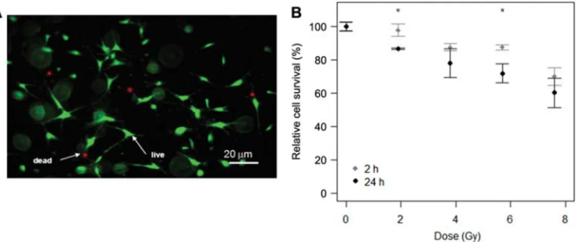
To examine the effects of high-resolutionmCT imaging on a cellular level, we first evaluated the survival rate 2 h and 24 h post-irradiation, with doses of 1.9, 3.8, 5.7, and 7.6 Gy, respectively, using custom-made PMMA sample holders. We found no significant increase in cell death for a dose of 1.9 Gy immediately (2 h) after irradiation as compared to the non-irradiated control group. However, 24 h post-irradi-ation, cell survival was significantly decreased by 15% over the control group. Doses after exposure to 3.8 Gy and 5.7 Gy resulted in survival rates of around 80%. Although we did not find a significant decrease in cell survival after exposure to 3.8 Gy between 2 h and 24 h post-irradiation, we dem-onstrate a significant decrease after exposure to 5.7 Gy by 15%. The highest dose of 7.6 Gy decreased the survival rate of the cells by around 40% 24 h post-irradiation as compared to non-irradiated control samples. Nevertheless, no signifi-cant difference between 2 h and 24 h post-irradiation was observed for this highest dose.
Time-dependent decrease of ALP activity per cell To further investigate the cellular effects of high-resolution
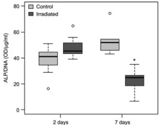
mCT imaging, we applied a dose of 1.9 Gy and analyzed ALP activity. When ALP activity was normalized with the DNA content of the same sample, we found no significant increase in ALP activity 2 days post-irradiation, but a sig-nificant decrease to approximately 60% at 7 days (Figure 2).
Discussion
This is one of the first studies investigating the effect of ionizing radiation of high-resolutionmCT measurements on
Figure 1 Relation ofmCT image quality and applied dose.
(A)mCT images of a bovine trabecular bone sample. Insets show higher magnifications and the corresponding binary images, respectively. These scans were performed in four different modes: standard resolution at 30mm and 16mm nominal resolution (voxel size), and high resolution at 15mm and 8mm nominal resolution. The nominal resolution corresponds to the scanner settings and not to the actual spatial resolution. (B) The relation between dose and image quality (here given as nominal resolution) can be modeled as a power law with exponents 1.74, 1.76, and 2.03, for standard, medium and high resolution mode, respectively. (C) Radiation dose in function of the imaging mode. For all cell experiments the medium resolution mode at 20mm nominal resolution was used, but with a threefold integration time resulting in a calculated base dose of 1.9 Gy per measurement. To achieve the higher doses of 3.8, 5.7, and 7.6 Gy, the base measurement was repeated 2, 3, and 4 times, respectively.
Figure 2 Time-dependent decrease of ALP activity after exposure to 1.9 Gy radiation dose.
Although no significant alteration in the 2 day post-irradiation group was found, the ALP activity was significantly decreased by 60% at 7 days post-irradiation. In these box plots, the box represents/con-tains 50% of the data, and the whiskers show the data range exclud-ing extreme values that are shown as sexclud-ingle data points. Differences between two data sets were considered statistically significant if p-0.05 and are indicated in the Figure by a star.
a cellular level by using a 3D tissue-like model system. Using our model system, the results indicate thatmCT imag-ing with doses as low as 1.9 Gy, which is required to obtain a reasonable image quality, can induce irreparable damages on the cellular level.
The dose applied by amCT measurement depends on the imaging mode and scanner settings, which are adapted to the features of interest. For instance, if bone volume density is of interest, a relatively low image quality could provide suf-ficiently good results. However, to capture alterations on the bone surface, a relatively good image quality with high res-olution is required, which could be needed to expose the tissue to doses of 2 Gy or even higher. In such cases, the applied dose is not negligible and could result in potential side effects on the cellular level as demonstrated in Figures 2 and 3.
Our results indicate that the effects of ionizing radiation on survival of the pre-osteoblastic MC3T3-E1 cells are dif-ferent in our 3D model system with collagen type I matrices mimicking natural bone-like tissue as compared to 2D cul-ture plates. We show considerably higher survival rates when similar doses are applied as previously reported for 2D stud-ies (Figure 3A,B). For instance, it was demonstrated that osteoblast-like cells from the calvarium of newborn rats sur-vived only by 60%, 35%, and less than 5% on a 2D surface when 2, 4, and 6 Gy, respectively, were applied to the culture w5x. The reason for this difference is unclear. We speculate that the 3D organization of the cells mimicking a more nat-ural tissue-like environment than a 2D culture could have a
protective effect, but also supports repair mechanisms more efficiently after irradiation damage. In addition, shielding effects of the PMMA sample holder might not be totally excluded.
Figure 3 Survival of pre-osteoblastic MC3T3-E1 cells in 3D collagen matrices aftermCT imaging.
(A) Representative image of pre-osteoblastic cells in 3D collagen matrices 24 h post-irradiation. (B) Dose- and time-dependent survival 2 h and 24 h after irradiation as assessed by quantifying live/dead cell staining. At a dose of 2 Gy, which is commonly applied forin vitro and in vivo imaging, the fraction of surviving cells 24 h after irradiation is significantly lower than at 2 h. The highest dose applied (8 Gy) still
resulted in survival of )60% both at 2 h and 24 h after irradiation. The survival rates at each dose were normalized by the survival rates of the control samples and are shown as mean values"standard deviation (nsampless3, ncontrolss9; images were taken at five randomly selected/representative locations within the matrix). Significant differences were accepted if p-0.05. * Indicates significantly different data between 2 h and 24 h.
Expression of ALP activity is a relevant marker in the early differentiation process of MC3T3-E1 pre-osteoblastic cells towards mature osteoblasts as it is upregulated with proceeding maturation w18x. We show here a time-dependent decrease of ALP activity after exposure to radiation (Figure 2). Although no significant alteration was found 2 days post-irradiation, the ALP activity was significantly decreased at 7 days by around 60%. We speculate that differentiation and maturation processes could be inhibited after irradiation. However, it is not clear by which mechanisms the ALP activ-ity is influenced by irradiation. It could be as a result of altered cytokine expression profiles, by the irradiation direct-ly or by combined processes w6x. Other authors found inhi-bition of differentiation, when exposing mature osteoblasts in a 2D environment to 4 Gy doses. In a study by Hagen-mu¨ller et al., ALP activity normalized to the DNA content was shown to be decreased by around 20% after 3 days of irradiation, around 60% after 6 days, and around 70% after 9 days. However, a study using 3D scaffolds prepared from silk fibroin and human mesenchymal stem cells did not dem-onstrate alterations in ALP activity after repeated high-res-olution mCT imaging over a period of 28 days w13x. The reasons for these controversial results on the ALP activity could include different study designs including different cell types, radiation doses, the different times between themCT imaging and the ALP activity assays, which can allow DNA repair processes in some cases, as well as the different bio-physical and biochemical properties of the 3D scaffolds used in the respective studies.
The direct transfer of the results obtained in this study into
in vivo experiments might not be possible; however, the in vitro model system applied here allows conclusions for in vitro tissue engineered constructs or bone chambers (i.e.,
critical dose). The critical dose forin vivo applications needs
to be assessed with the respectivein vivo models, specifically
the effects on bone tissue growth, healing, and turnover. Potential immune reactions, vascularization of the tissue, and potential shielding effects of the surrounding tissue can affect the critical dosein vivo.
Conclusion
We have established a model system allowing the monitoring of 3D tissue engineered constructs usingmCT imaging. Our results indicate significant impairments on the cellular level after imaging with high-resolutionmCT in a time- and dose-dependent manner. Results from this study indicate that imaging with high-resolution mCT with doses as low as 1.9 Gy, which is required to obtain a reasonable image qual-ity, can yield irreparable damages on the cellular level.
It is therefore recommended to use the lowest resolution necessary in monitoring studies of bone chambers and 3D tissue engineered bone-like constructs. Future studies could include other cells such as mesenchymal cells present in bone tissue and/or different scaffolds, as well as longitudinal monitoring. Longitudinal animal studies could be performed to assess a criticalin vivo dose to avoid potential damages
on the cellular level.
Acknowledgements
We thank Dr. George Raeber for helpful discussions on collagen I matrix formation and analysis of cell survival, and Paul Lu¨thi for manufacturing the PMMA sample holders. This project was sup-ported by the SNF Professorship in Bioengineering of the Swiss National Science Foundation (FP 620-58097.99 and PP-104317/1). T.P.K. was supported by the Swiss National Science Foundation (grant numbers 120938 and PBELP3-127902).
Author disclosure statement
The authors declare no competing financial interests.
References
w1x Abbott A. Cell culture: biology’s new dimension. Nature 2003; 424: 870–872.
w2x Arnold M, Kummermehr J, Trott KR. Radiation-induced impairment of osseous healing: quantitative studies using a standard drilling defect in rat femur. Radiat Res 1995; 143: 77–84.
w3x Bonse U, Busch F, Gu¨nnewig O, et al. 3D computed X-ray tomography of human cancellous bone at 8 microns spatial and 10(-4) energy resolution. Bone Miner 1994; 25: 25–38. w4x Cartmell S, Huynh K, Lin A, Nagaraja S, Guldberg R.
Quan-titative microcomputed tomography analysis of mineraliza-tion within three-dimensional scaffolds in vitro. J Biomed Mater Res A 2004; 69: 97–104.
w5x Dare A, Hachisu R, Yamaguchi A, Yokose S, Yoshiki S, Oka-no T. Effects of ionizing radiation on proliferation and dif-ferentiation of osteoblast-like cells. J Dent Res 1997; 76: 658–664.
w6x Dudziak ME, Saadeh PB, Mehrara BJ, et al. The effects of ionizing radiation on osteoblast-like cells in vitro. Plast Reconstr Surg 2000; 106: 1049–1061.
w7x Elsdale T, Bard J. Collagen substrata for studies on cell behavior. J Cell Biol 1972; 54: 626–637.
w8x Friedrich J, Seidel C, Ebner R, Kunz-Schughart LA. Sphe-roid-based drug screen: considerations and practical approach. Nat Protoc 2009; 4: 309–324.
w9x Gal TJ, Munoz-Antonia T, Muro-Cacho CA, Klotch DW. Radiation effects on osteoblasts in vitro: a potential role in osteoradionecrosis. Arch Otolaryngol Head Neck Surg 2000; 126: 1124–1128.
w10x Genant HK, Engelke K, Fuerst T, et al. Noninvasive assess-ment of bone mineral and structure: state of the art. J Bone Miner Res 1996; 11: 707–730.
w11x Goldwein JW. Effects of radiation therapy on skeletal growth in childhood. Clin Orthop Relat Res 1991; 262: 101–107. w12x Griffith LG, Swartz MA. Capturing complex 3D tissue
phys-iology in vitro. Nat Rev Mol Cell Biol 2006; 7: 211–224.
w13x Hagenmu¨ller H, Hofmann S, Kohler T, et al. Non-invasive time-lapsed monitoring and quantification of engineered bone-like tissue. Ann Biomed Eng 2007; 35: 1657–1667. w14x Hofmann S, Hagenmu¨ller H, Koch AM, et al. Control of in
vitro tissue-engineered bone-like structures using human mes-enchymal stem cells and porous silk scaffolds. Biomaterials 2007; 28: 1152–1162.
w15x Kirshner J, Thulien KJ, Martin LD, et al. A unique three-dimensional model for evaluating the impact of therapy on multiple myeloma. Blood 2008; 112: 2935–2945.
w16x Lanza RP, Langer R, Vacanti JP. Principles of tissue engi-neering. San Diego, CA: Academic Press 2007.
w17x Majumdar S, Genant HK, Grampp S, et al. Correlation of trabecular bone structure with age, bone mineral density, and osteoporotic status: in vivo studies in the distal radius using high resolution magnetic resonance imaging. J Bone Miner Res 1997; 1: 111–118.
w18x Rodan GA, Rodan SB. Expression of the osteoblastic phe-notype. In: Peck WA, editor. Advances in bone and mineral research. Annual II. Amsterdam: Excerpta Medica 1984: 244–285.
w19x Ru¨egsegger P, Koller B, Mu¨ller R. A microtomographic sys-tem for the nondestructive evaluation of bone architecture. Calcif Tissue Int 1996; 58: 24–29.
w20x Thurner P, Mu¨ller R, Raeber G, Sennhauser U, Hubbell JA. 3D morphology of cell cultures: a quantitative approach using micrometer synchrotron light tomography. Microsc Res Tech 2005; 66: 289–298.
w21x U.S. Department of Health and Human. U.S. Government web site for organ and tissue donation and transplantation: www.organdonor.gov.
w22x Weber FE, Eyrich G, Gra¨tz KW, Thomas RM, Maly FE, Sai-ler HF. Disulfide bridge conformers of mature BMP are inhib-itors for heterotopic ossification. Biochem Biophys Res Commun 2001; 286: 554–558.
w23x Zehbe R, Goebbels J, Ibold Y, Gross U, Schubert H. Three-dimensional visualization of in vitro cultivated chondrocytes inside porous gelatine scaffolds: a tomographic approach. Acta Biomater 2010; 6: 2097–2107.
Received September 27, 2009; accepted April 7, 2010; online first June 22, 2010