Abstract. Angiogenesis, i.e. the proliferation of new blood vessels from pre-existing ones, is an underlying process in many human diseases, including cancer, blinding ocular disorders and rheumatoid arthritis. The ability to selectively target and interfere with neovascu-larisation would potentially be useful in the diagnosis and treatment of angiogenesis-related diseases. This re-view presents the authors’ re-views on some of the most relevant markers of angiogenesis described to date, as well as on specific ligands which have been character-ised in pre-clinical animal models and/or clinical studies. Furthermore, we present an overview on technologies which are likely to have an impact on the way molecular targeting of angiogenesis is performed in the future.
Keywords: Angiogenesis – Targeting – Antibody –
Cancer – Chronic inflammation
Eur J Nucl Med Mol Imaging (2004) 31:1327–1341 DOI 10.1007/s00259-004-1648-0
Ligand-based targeting of disease: general concepts
Targeting
The vast majority of approaches for the treatment of dis-ease are limited by lack of specificity. This constraint holds true for many diseases, but it is most evident in the treatment of solid tumours, where most chemotherapeu-tic agents exhibit poor accumulation in the tumour mass owing to poor blood perfusion, irregular vasculature and
high interstitial pressure in the tumour environment [1, 2]. Moreover, multidrug resistance proteins may further decrease drug uptake. As a consequence, the develop-ment of therapeutic agents which preferentially accumu-late in solid tumours represents a main focus of modern anticancer research.
Since Paul Ehrlich first envisioned the possibility of selective delivery of antibodies to the tumour environment at the end of the nineteenth century, the concept of “magic bullets” capable of tumour targeting has been extended to other molecules (peptides, small organic molecules, etc.) capable of selective localisation in the tumour environ-ment. After some delay following the invention of hybri-doma technology [3], several monoclonal antibodies have been approved by the US Food and Drug Administration (http://www.fda.gov) and in Europe, both for the imaging of disease and for therapeutic applications.
The synergy between imaging opportunities and ther-apeutic applications is one of the main attractions of bio-medical approaches which rely on the selective delivery of bioactive molecules to the tumour environment. In principle, the same binding moiety (e.g. a monoclonal antibody) can be used for the delivery of agents which facilitate tumour detection (radionuclides, fluorophores) or agents capable of triggering a biocidal event (radionu-clides, photosensitisers, drugs, cytokines, pro-coagulant factors, etc.). Quantitative biodistribution studies in ani-mals and imaging studies in patients are invaluable tools for the characterisation of markers of diseases (e.g. tu-mour-associated antigens) and of the corresponding lig-ands (e.g. monoclonal antibodies). Indeed, ligand-based targeted therapeutic strategies may represent one of the few areas of pharmaceutical research in which the per-formance of a therapeutic strategy can be monitored at several stages of the development process. Whenever un-satisfactory targeting results are observed in biodistribu-tion or imaging studies, these data urge the researchers to use better target antigens and/or better ligands in their pharmaceutical strategy. Furthermore, imaging of the tar-get molecule responsible for therapy allows for individu-alised patient selection and early monitoring of the thera-peutic intervention.
Dario Neri (
✉
)Institute of Pharmaceutical Sciences,
Department of Chemistry and Applied Biosciences, Swiss Federal Institute of Technology Zurich, Winterthurerstrasse 190, 8057 Zurich, Switzerland e-mail: neri@pharma.ethz.ch
Tel.: +41-1-6356063, Fax: +41-1-6356886
Review article
Molecular targeting of angiogenesis for imaging and therapy
Simon S. Brack1, Ludger M. Dinkelborg2, Dario Neri1
1 Institute of Pharmaceutical Sciences, Department of Chemistry and Applied Biosciences, Swiss Federal Institute of Technology Zurich,
Zurich, Switzerland
2 Research Laboratories of Schering AG, Berlin, Germany
Published online: 5 August 2004 © Springer-Verlag 2004
The importance of antigen quality in ligand-based tar-geted imaging or therapeutic strategies cannot be over-stated. In theory, an ideal target should be specific, easily accessible, stable and abundant. In practice, few antigens exhibit all these properties, but (as we will see in “An-giogenesis-related disorders”) markers of angiogenesis may combine a number of attractive features, such as ex-cellent accessibility, low expression in normal adult tis-sues and over-expression in several relevant pathologies, including cancer.
Angiogenesis
Angiogenesis, i.e. the sprouting of new blood capillaries from existing vessels, is an essential process for embry-onic development. Once the vascular network is in place, endothelial cells remain quiescent and angiogenesis is triggered only locally and transiently during a number of physiological conditions like the female reproductive cy-cle, wound healing or hair growth [4]. Angiogenesis is a tightly controlled multi-step process, in which pro-angio-genic and anti-angiopro-angio-genic factors are in equilibrium to neutralise one another. Imbalance of this equilibrium, due to either upregulation of pro-angiogenic or downreg-ulation of anti-angiogenic mediators, induces angiogene-sis.
Angiogenesis is an important feature of a range of pathological conditions, cancer being one of the most prominent examples [5] (see “Angiogenesis-related dis-orders”). The growth of new capillaries is often triggered in conditions of pathological cellular proliferation, isch-aemia or chronic inflammation, where an increase in blood supply may compensate for hypoxia and insuffi-cient delivery of nutrient to the tissue [6, 7]. Unlike the situation in physiological conditions, blood vessels grow unabated in cancer and other pathologies, and tumour angiogenesis sustains the progression of the disease.
During angiogenesis, endothelial cells detach from the pre-existing destabilised vessel, migrate into the peri-vascular space and proliferate to finally mature and form new vascular structures. A number of growth factors, proteases, adhesion molecules and other angiogenic me-diators which enable endothelial cell migration or prolif-eration regulate this process. Vascular endothelial growth factor (VEGF) is considered one of the most important growth factors in angiogenesis [8]. It increases the per-meability of existing blood vessels and acts as an endo-thelial cell survival factor, as well as being a potent en-dothelial cell mitogen. The neutralising humanised monoclonal anti-VEGF antibody bevacizumab (Avastin, Genentech) has recently been approved for the treatment of colorectal cancer [9], but showed no survival benefit in patients with breast cancer [10].
Ligands to markers of angiogenesis and their applications in targeting
In this review, we will refer to “targeting” as the selec-tive delivery of a molecular agent to a site of disease. In most cases, this molecular agent will be a ligand specific to a target antigen (“marker”) which is over-expressed at the site of disease. At present, monoclonal antibodies are the only clinically proven class of high-affinity binding molecules which can be generated against virtually any marker of disease. However, as we will see in “Ligands for targeting applications”, other molecular species (such as peptides, aptamers, or small organic molecules) are also being considered for targeting applications.
Monoclonal antibodies exhibit slow elimination from the blood and accumulate predominantly in the liver. Rapidly clearing antibody fragments are typically pre-ferred for imaging applications in nuclear medicine. By contrast, intact immunoglobulins continue to represent the antibody format of choice for many therapeutic appli-cations [11], which rely on the antibody’s ability to inter-fere with signalling events and to activate antibody-de-pendent cellular cytotoxicity mechanisms or complement. The immunogenicity of rodent antibodies continues to be a concern for repeated administrations to humans, and the use of chimeric, humanised or fully human antibod-ies is generally preferred [11, 12].
Markers of angiogenesis can be located on the lumi-nal or ablumilumi-nal aspects of new blood vessels (Fig. 1). Components of the modified extracellular matrix are mostly found on the abluminal side of neovasculature. These antigens are often stable and abundant. Some ex-tracellular matrix components (such as oncofetal fi-bronectins and tenascins) display a restricted pattern of expression and may serve as useful antigens for tumour targeting applications. In spite of the fenestration of tu-mour blood vessels, the ligand-based targeting of ablu-minal antigens can be a slow process owing to the irreg-ular vasculature and high interstitial pressure of solid tu-mours, as well as to the fact that such ligands must ex-travasate in order to reach their target.
While markers of angiogenesis on the luminal side of new blood vessels are better accessible, it would be mis-leading to consider that they will necessarily lead to faster, more efficient ligand-based targeting applications. The targeting of such antigens, by means of a rapidly clearing ligand (e.g. an antibody fragment), can be viewed essentially as a single-compartment pharmaconetic problem. A small antibody fragment with a low ki-netic dissociation constant for the antigen will have two main possible fates: it will either bind to the accessible target or be cleared from the circulation via the renal and hepatobiliary route. In this model, the time required for semisaturation of the target antigen satisfies the follow-ing equation:
where kon is the kinetic association constant of the anti-body for the antigen and [Ab] is the initial antianti-body con-centration in blood, neglecting blood clearance (which obviously can only make the targeting process even less efficient).
From Eq. 1, it is obvious that an accessible luminal target can be saturated very rapidly if high concentra-tions of ligands are used (e.g. T1/2=0.692 s if kon=106M1s−1
and [Ab]=1 µM). However, such rapid targeting results
will correspond to only a minimal fraction of antibody being used for targeting (and will translate into very poor percent injected dose per gram of tissue), unless the tar-get antigen is abundantly expressed. In fact, saturation of target antigen leaves most of the remaining antibody in circulation (leading to poor tumour/blood ratios). Such a targeting scenario will not be satisfactory for applica-tions in nuclear medicine, as the performance of radiola-belled antibodies for imaging and radioimmunotherapy applications relies strongly on the achievement of high tumour/organ ratios.
Lowering the initial antibody concentration to levels that stoichiometrically match the amounts of accessible antigen does not help. Even with exceptionally high af-finity antibodies, the ultimate diffusion-controlled limits on kon values make it impossible to rapidly target low-abundance accessible antigens with low (e.g. subnanomo-lar) concentrations of ligands.
We anticipate that a combination of several parame-ters, such as blood circulation properties, vascular in-tegrity, antigen abundance, tumour size and ligand kon values, will crucially influence the targeting efficiency for markers located on the luminal aspect of new blood vessels.
Angiogenesis-related disorders
Cancer
Most of the current knowledge about angiogenesis stems from investigations on tumoural angiogenesis. A large
number of molecules involved in angiogenesis have been first identified in tumours and later confirmed in other pathological conditions.
Many tumours in humans persist in situ without being accompanied by angiogenesis [13, 14]. At that stage they tend to be clinically undetectable and are rarely larger than 1–2 mm in diameter because diffusion of oxygen and nutrients limits their size. The high rate of prolifera-tion in these tumours is compensated by abundant inter-nal apoptosis as a consequence of insufficient blood supply.
As the tumour adopts an angiogenic phenotype, the balance between pro-angiogenic and anti-angiogenic fac-tors is upset and angiogenesis is triggered. The tumour mass is allowed to overtake the apoptotic rate and ex-pands. This process is referred to as “angiogenic switch” [5, 15]. Not only is angiogenesis required for tumours to grow beyond a certain size, but it also enables tumour cells to migrate into surrounding tissue and to colonise distant sites, forming metastases. Metastases again can only grow to a threatening size if the metastatic cells are able to trigger angiogenesis [5].
Although the mechanisms eliciting the angiogenic switch are not entirely understood to date, it is believed that besides tumour-suppressor mutation and oncogene activation, hypoxia plays a pivotal role [16]. There are at least two hypoxia-dependent regulatory mechanisms which lead to VEGF expression. The first mechanism re-lies on the transcription factor hypoxia-inducible factor (HIF-1), which controls VEGF transcription [17]. The alpha subunit of HIF-1, HIF-1α, is degraded under nor-moxic conditions and stabilised under hypoxia [6, 18, 19]. Second, VEGF mRNA becomes stabilised under hypoxic conditions [20]. VEGF concentrations stimulate proliferation of endothelial cells, which in turn produce many unspecific angiogenic stimulators, including basic fibroblast growth factor (bFGF), acid fibroblast growth factor (aFGF), transforming growth factor α and β (TGFα and TGFβ) and platelet-derived endothelial cell growth factor (PD-ECGF). Additionally, tumour cells produce proteases, among which are matrix
metallopro-Fig. 1. Schematic
representa-tion of the tumour neovascula-ture as a target for biomedical intervention. Markers of angio-genesis can be located on the luminal (blue) or the abluminal (green) aspect of new blood vessels. Proteins of the extra-cellular matrix are mainly situ-ated on the abluminal side, pos-sibly impairing their accessibil-ity from the bloodstream. How-ever, while markers on the lu-minal side are better accessible, they may be less abundant and less stable
teinases (MMP) and serine proteases like urokinase plas-minogen activator (uPA) or tissue plasplas-minogen activator. Endothelial cells display cell adhesion molecules such as integrins αvβ3 and αvβ5which mediate interaction with the extracellular matrix. Laminin, type IV collagen and tenascin are synthesised to constitute the new basement membrane.
Reduced oxygen tension promotes angiogenesis not only by stimulating the production of inducers but also by reducing the production of inhibitors. Thrombospon-din-1 was the first angiostatic protein for which anoxia-triggered downregulation during tumourigenesis was demonstrated [21]. Since then, a number of endogenous angiogenesis inhibitors have been identified.
The tumour vessels differ from their normal counter-parts: architecturally, they are irregularly shaped, dilated and tortuous, and even contain dead ends [22]. Extensive fenestration, an abnormal basement membrane and un-usually wide gaps between adjacent endothelial cells make them leaky [23–25].
The treatment of cancer with an anti-angiogenic ap-proach was first proposed more than two decades ago [13]. Accordingly, various anti-angiogenic strategies have been investigated pclinically. This extensive re-search has culminated in the recent approval of beva-cizumab (Avastin, Genentech) as first-line treatment for metastatic colon carcinoma [9, 26].
Age-related macular degeneration (ARMD) and proliferative diabetic retinopathy
Ocular neovascularisation is associated with many ocular diseases and is responsible for the majority of cases of irreversible blindness in the developed world. Abnormal ocular angiogenesis may ultimately cause severe vitreous cavity bleeding, retinal detachment and glaucoma lead-ing to blindness. Proliferative diabetic retinopathy and age-related macular degeneration (ARMD), which are the two most common angiogenesis-related eye diseases, are therefore of high socio-economic impact [27].
The stimulus giving rise to diabetic retinopathy is un-known, but it is likely that hyperglycaemia leads to vascu-lar abnormalities and to ocuvascu-lar ischaemia, a process which probably involves leucocytes or platelets [28–30]. The hypoxic portions of the retina release VEGF, which in turn promotes pathological vasoproliferation in the retina [31, 32]. Recently, an angiogenic inhibitor responsible for the avascularity of the cornea and the vitreous was identified as pigment epithelium-derived factor and found to be defi-cient in diabetic retinopathy [33, 34]. Inhibition of VEGF with a soluble VEGF receptor chimeric protein was found to suppress retinal neovascularisation in a mouse model [35]. A number of strategies based on VEGF inhibition are currently undergoing clinical investigation.
ARMD is the main cause of blindness in elderly peo-ple. Macular degeneration refers to the breakdown of
cells in the centre of the retina. Ten percent of ARMD patients are affected by choroidal neovascularisation, which is responsible for most of the vision loss [27]. Vessels arising from the choroidal vasculature grow into the plane of the retinal pigment epithelium and sub-reti-nal space. This occurs nearly exclusively in the macular and perimacular regions of the retina. The newly formed vessels are structurally weak and lack integrity, which results in loss of photoreceptors. As with diabetic reti-nopathy, the causes of ARMD have not yet been eluci-dated. Besides local tissue hypoxia as a consequence of local ischaemia, inflammation has been implicated in ARMD [36].
Psoriasis
Psoriasis is a chronic inflammatory skin disease that af-fects approximately 1–3% of the Western population [37]. Clinically psoriasis appears as symmetrical, well-demarcated erythematous plaques topped with silvery white scales, most commonly on the scalp, knees, el-bows and trunk. Although rarely fatal, it severely im-pairs quality of life. The disease is characterised by hy-perproliferation of keratinocytes, infiltration of inflam-matory cells and increased cytokine levels. The last-mentioned are responsible for activation of keratinocytes and lymphocyte invasion by cytokine-mediated upregu-lation of adhesion molecules on endothelial cells.
Psoriasis is accompanied by expansion of the superfi-cial dermal microvasculature and elongation of capillary loops passing into the dermal papillae and the papillary tip [38]. Although angiogenesis may not be a primary event in psoriasis, it occurs at an early stage. Several an-giogenic stimuli have been implicated in psoriasis: pro-angiogenic VEGF and IL-8 expression was found to be upregulated while thrombospondin-1 was downregulated in epidermal keratinocytes [39–41]. Aberrant expression of a wide range of angiogenic molecules in psoriatic skin has been reported in the literature, these molecules in-cluding integrin αvβ3, angiopoietin-2, TGFα and IL-15 [42–45]. As early as 1972, it was recognised that psoria-sis therapy could rely on inhibition of angiogenepsoria-sis [46]. Current medications for psoriasis possess a certain anti-angiogenic activity, e.g. cyclosporin A or retinoids [47, 48].
Rheumatoid arthritis
Rheumatoid arthritis (RA) is a chronic destructive mus-culoskeletal autoimmune disorder associated with thick-ening of the synovial membrane lining the joints, inflam-mation, hyperproliferation of synovial cells and forma-tion of a proliferating pannus. It involves a pro-inflam-matory cytokine cascade, leucocyte invasion, damage of the affected cartilage and joints and bone erosion. The
extensive hyperplasia of the synovium requires a com-pensatory increase in the number of blood vessels to nourish and oxygenate the tissue. Hence, angiogenesis is central in the pathological course of RA.
Over-expression of a number of angiogenic factors is responsible for a pro-angiogenic imbalance in RA. VEGF is expressed in RA synovium and elevated in the serum of RA patients. Serum levels of VEGF further correlate with disease activity, and improvement in the clinical symptoms of RA is associated with a reduction in VEGF levels. Inactivation of VEGF has been exploit-ed as a therapeutic approach for RA therapy [49, 50].
Other pro-angiogenic factors like tumour necrosis factor-alpha (TNFα), IL-1, or TGFβ are elevated in the synovial fluid of RA patients, and their blockade has also evolved as a strategy for the treatment of RA [51–53]. The most promising therapies available nowa-days are TNFα-neutralising molecules. Anti-TNFα ther-apy has been found to ameliorate essentially all aspects of RA and, importantly, halts joint destruction; it has be-come the standard for RA therapy [54].
Markers of angiogenesis
Markers of angiogenesis for vascular targeting
A number of protein antigens expressed either in the ves-sel or in the adjacent matrix of the vesves-sel have been characterised as targets for the selective delivery of anti-bodies (or peptides) to the tumour neovasculature. For some of these markers, both clinical and pre-clinical data are already available. This section presents the authors’ views on the most promising tumour vascular targets which have been characterised in the recent past. The list of antigens includes proteins which are preferentially ex-pressed on the surface of endothelial cells in tumour blood vessels, as well as components of the modified ex-tracellular matrix which surrounds the tumour neovascu-lature.
Fibronectin extra-domain B
Fibronectin is a large glycoprotein, which is present in large amounts in plasma and tissues. The extra-domain B of fibronectin (EDB) is a domain of 91 amino acids which, in normal conditions, is not present in the fibro-nectin molecule [55]. However, the EDB domain is typi-cally inserted in the fibronectin molecules at sites of tis-sue remodelling by a mechanism of alternative splicing at the level of the primary transcript. The EDB domain has an identical sequence in mouse, rat, rabbit, dog, monkey and man. This sequence conservation greatly fa-cilitates the pre-clinical and clinical development of EDB targeting agents, as it allows the same EDB binding molecule to be used in different immunocompetent
syn-geneic animal models of pathologies and in patients. An-tibodies recognising a cryptic epitope on domain VII of fibronectin (which is adjacent to EDB) have been avail-able since the late 1980s [56, 57]. However, possibly be-cause of tolerance, the isolation of anti-EDB monoclonal antibodies using hybridoma technology has not been possible until now, and antisera to EDB have been shown to recognise EDB-containing fibronectin only af-ter N-glycanase treatment [58].
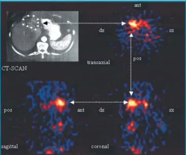
Using human antibody phage technology [59, 60] and other recombinant antibody technologies [61], our group (in collaboration with the group of L. Zardi in Genoa) has isolated a number of human monoclonal antibodies to EDB [62–64]. In particular, scFv(L19) is a human anti-body fragment with subnanomolar affinity to EDB [64], which has been shown to efficiently localise on tumoural and non-tumoural neovasculature both in animal models [65–67] and in patients with cancer [68]. Figure 2 shows single-photon emission computed tomography (SPECT) images from a patient with liver metastases of colorectal cancer. Selective uptake of the radiolabelled scFv(L19) in the lesions is clearly visible in the transaxial, coronal and sagittal projections. A careful biodistribution analysis of the L19 antibody in scFv, mini-antibody and IgG format has recently been reported [69].
A large number of derivatives of the L19 antibody have been produced and tested in pre-clinical animal models, including conjugates with photosensitisers [70], therapeutic radionuclides (unpublished), drugs (unpub-lished), liposomes [71], procoagulant agents [72], cy-tokines [73–77], enzymes [78] and other binding pro-teins [79, 80].
Fig. 2. SPECT images obtained 21 h after injection of
radioiodi-nated scFv(L19), showing the transaxial, sagittal and coronal pro-jections of the abdomen of a patient with liver metastases of colo-rectal cancer, were matched to the CT scan of the abdomen of the same patient. Adapted from [68]
EDB is essentially undetectable in most normal adult tissues, with the notable exceptions of the endometrium in the proliferative phase and some vessels of the ovaries. However, EDB is abundantly expressed in a va-riety of solid tumours [56, 62, 81–85] as well as in ocu-lar angiogenesis [70, 86], RA [87] and wound healing [88]. Typically, the pattern of EDB expression in tu-mours either is predominantly perivascular or exhibits a diffuse staining of the tumour stroma. In different condi-tions, tumour cells, fibroblasts and/or endothelial cells may contribute to the synthesis of EDB-containing fibro-nectin. In mice, the targeted deletion of the EDB exon resulted in transgenic mice which developed normally, healed bone fractures normally and which could develop tumours, thus suggesting that the function of EDB is re-dundant in mice.
Large tenascin-C isoforms
Several isoforms of tenascin-C can be generated as a re-sult of alternative splicing which may lead to the inclu-sion of (multiple) domains in the central part of this pro-tein, ranging from domain A1 to domain D [89, 90]. Tra-ditionally, one has referred to the large isoform of tenascin-C for tenascin molecules which would putative-ly contain all the extra domains, and to the small tenascin-C isoform whenever the extra domains were ab-sent. A strong over-expression of the large isoform of tenascin-C has been reported for a number of tumours [90], and two monoclonal antibodies specific for do-mains A1 and D, respectively [91–95], have been exten-sively characterised in the clinic.
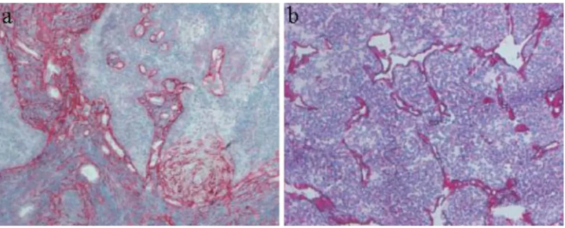
Figure 3 shows tumour sections stained with the L19 antibody specific to EDB and with a human monoclonal antibody specific to a large isoform of tenascin-C. In both cases, tumour vascular structures are strongly stained in red.
Recently, we have reported (in collaboration with the Zardi group) that the extra domain C of tenascin-C dis-plays a more restricted pattern of expression compared with the other extra domains of tenascin-C [96], with a
predominantly perivascular staining as depicted with im-munohistochemistry. The C domain of tenascin-C is un-detectable in most normal adult tissues, but is over-ex-pressed in high-grade astrocytomas [96] and other tu-mour types (unpublished).
Integrins αvβ3and αvβ5
Integrins αvβ3and αvβ5have been proposed as markers of angiogenesis [36, 97–99] and as targets both for anti-body-based inhibitory strategies and for the ligand-based delivery of therapeutics to the tumour neovasculature. While these integrins are over-expressed in several solid tumours, expression in normal tissues has been reported in immunohistochemical studies [100]. A high-affinity human monoclonal antibody to the integrin αvβ3 has been studied in an exploratory immunoscintigraphic clin-ical study, but yielded disappointing results for tumour imaging [101].
Prostate-specific membrane antigen (PSMA)
Prostate-specific membrane antigen (PSMA) is a mem-brane glycoprotein with hydrolytic activity, which is pre-dominantly expressed in the prostate and whose concen-trations have been found to be elevated in patients with prostate cancer [102]. PSMA is present in virtually every prostate cancer, and its levels are elevated in higher grade cancers, metastatic disease and hormone-refractory prostate cancer [103, 104]. The interest in vascular tar-geting applications of PSMA has been stimulated by the observation that PSMA is over-expressed in the neovas-culature of several solid tumour types [105, 106], while expression around blood vessels in normal tissues is lim-ited to breast, kidney, duodenum and prostate [107]. The monoclonal antibody J591 has been used in clinical im-munoscintigraphy studies for the imaging of progressing hormone-independent prostate cancer [108, 109].
Fig. 3. a Frozen tissue section
of a human squamous cell carci-noma of the tonsils, stained with an antibody specific to a large isoform of tenascin-C (red). Counterstaining was performed with Gill’s haematoxylin.
b Frozen tissue section of a
sub-cutaneously grown mouse F9 teratocarcinoma, stained with L19, a human antibody specific to the oncofetal fibronectin iso-form (red) and counterstained with Gill’s haematoxylin
Endoglin (CD105)
Endoglin (CD105) is a TGFβco-receptor which is over-expressed in tumour neovasculature [110, 111]. Even though recent immunohistochemical studies have shown the expression of endoglin in normal adult tissues [112, 113], monoclonal antibodies to endoglin have been used in biodistribution studies and for imaging purposes in animal models of cancer [114, 115].
VEGF and VEGF-receptor complex
The discovery of VEGFs (and especially VEGF-A) as prime mediators of angiogenesis has stimulated interest in the use of VEGFs, VEGF receptors and their com-plexes as antigens for the targeted delivery of antibody derivatives to the tumour neovasculature. The over-expression of VEGFs and VEGF receptors in tumours is well documented [116, 117]. The selective localisation of monoclonal antibodies to VEGF-A [118, 119], VEGF receptor 2 [120, 121] and VEGF-A/VEGF receptor 2 complex [122] has been documented previously. Howev-er, the targeting efficiencies reported until now have not been spectacular, probably reflecting the relatively low absolute amounts of antigen in the tumour.
CD44
The monoclonal antibody TES-23, specific to an isoform of CD44, has been associated with some of the most im-pressive tumour targeting performances in rodent models of cancer. Tumour values as high as 50–150% injected dose per gram (%ID/g) have been reported as early as 1 h after intravenous injection [123]. CD44 is a cell surface receptor of great molecular heterogeneity, due to both al-ternative splicing of at least ten out of 20 exons and ex-tensive post-translational modifications. CD44 is a ubiq-uitous antigen that was initially discovered as a surface antigen on T-lymphocytes and granulocytes and later im-plicated in various physiological and pathophysiological processes, including embryogenesis, haematopoiesis, in-flammation and tumour progression. TES-23 recognises a widely distributed form of CD44 lacking variant exons, termed CD44H. However, the epitope was shown to in-clude a post-translational modification which is found in an activated, tumour-associated form of CD44H [124].
CD44 splice variants which contain exon 6, CD44v6, are involved in tumour progression [125, 126]. Antibod-ies specific for CD44v6 (CD44 splice variants which contain exon 6) are currently undergoing clinical trials [127].
Phosphatidyl serine phospholipids
Phosphatidyl serine phospholipids (PS) are major com-ponents of the cell membrane which, in normal condi-tions, are confined to the inner leaflet of the lipid bilayer. However, under conditions of cellular stress and apopto-sis (and particularly in proliferating endothelial cells), the exposure of phosphatidyl serine to the outer leaflet of endothelial cell membranes (making it “visible” for targeting molecules) has been reported. Annexin V and monoclonal antibodies have been used to confirm the surface accessibility of the phosphatidyl serine moiety on endothelial cells in vitro (for example, after treatment with hydrogen peroxide) and in vivo [128]. The impres-sive microscopic analysis of tumour targeting perfor-mance by monoclonal antibodies to PS has not yet been complemented by a quantitative biodistribution analysis, but the 9D2 antibody displayed a potent anti-tumour ac-tivity even when used as naked antibody in rodent mod-els of cancer.
The surface display of PS on activated platelets has been reported previously [129, 130], and it remains to be seen whether this property may facilitate tumour target-ing applications (in view of the fact that blood coagula-tion often leads to the formacoagula-tion of a provisional extra-cellular matrix in solid tumours) or may hinder the selec-tive localisation of monoclonal antibodies to tumour neovasculature.
Magic roundabout (ROBO-4)
Over the past few years, the Bicknell group has devel-oped bioinformatic strategies that utilise the wealth of information now accessible in the public databases to identify novel endothelial-specific genes. One of the genes identified is magic roundabout (MR or ROBO-4) [131]. The roundabout family of genes comprises several closely related genes (three in man) that were previously thought to be present only in neuronal tissue and to be involved in axon guidance. Roundabouts have five IgG and three fibronectin-like extracellular domains. They are large transmembrane receptors for ligands known as slits (three in man). The discovery of an endothelial-spe-cific roundabout was quite unexpected. Analysis of MR expression by a combination of Northern blotting, in situ hybridisation and immunohistochemistry has shown it to be highly restricted. Thus, MR is absent from adult tis-sues except at sites of active angiogenesis, including tu-mours. It is highly expressed in the embryo and it is pre-sumed that it is a gene intimately involved in the devel-opment of the vasculature. This expression pattern is highly unusual and has previously been found only for delta4, an endothelial-specific member of the delta fami-ly. The pattern of MR expression makes it ideally suited to vascular targeting. The latter has been known for many years to be an effective strategy to eradicate large
solid tumours in mouse models but its development in man has been hindered by the lack of a suitable target. Biodistribution studies with radiolabelled ligands (e.g. monoclonal antibodies) are badly needed, in order to as-sess the real potential of ROBO-4 as a target for imaging and/or biomolecular therapeutic intervention.
Other markers: aminopeptidase N, annexin A1
Recent proteomic methodologies (such as in vivo pep-tide phage panning or silica bead-based recovery of en-dothelial cell proteins—see “Methodologies to discover novel markers of angiogenesis”)—have identified a vas-cular form of aminopeptidase N (CD13) [132] and an-nexin A1 [117] as markers of tumour angiogenesis, which are readily accessible from the bloodstream. For CD13 ligands, evidence of vascular targeting activity has been indirect until now, and was mainly based on immu-nohistochemical studies of tissue sections following in-travenous injection of phage peptide [132] or on the im-proved anticancer therapeutic index of TNF-peptide fu-sion proteins compared with native TNF [133].
By contrast, impressive tumour targeting data have recently been reported for antibodies specific to rat an-nexin A1 in a rat lung metastasis model of breast cancer. Values of 34%ID/g have been reported in rat tumours as early as 2 h after intravenous injection of the antibody, radiolabelled with125I. However, it is not clear from the
article whether one or more antibodies to rat annexin A1 were used for the tumour targeting experiment. More-over, the125I-labelled antibody was found to be
therapeu-tic in rats at relatively low doses (single injection of 50µCi radiolabelled antibody per rat).
Methodologies to discover novel markers of angiogenesis
Transcriptomic analysis of tumour endothelial cells The hypothesis that the tumour environment would trig-ger the over-expression of certain genes in the endotheli-al cells of tumour neovasculature triggered research ac-tivities in which the transcriptomic profile of endothelial cells in normal tissues and in tumours were compared. The first approaches relied on subtractive hybridisation techniques and led to the identification of H cadherin as a protein which was over-expressed in the neovascula-ture of certain tumours. The same antigen, however, was also present in the vasculature of some normal organs [134].
Shortly afterwards, St Croix and colleagues per-formed a genome-wide comparison of normal and tu-mour-derived endothelial cells using serial analysis of gene expression (SAGE) [135]. The analysis pinpointed genes (termed tumour endothelial markers, or “TEMs”)
which were preferentially found in tumour endothelial cells. For some of these TEMs, preferential expression in tumour endothelial cells was confirmed by in situ hy-bridisation. Since this seminal paper, a number of groups have worked on similar experimental approaches, im-proving the methodologies for endothelial cell purifica-tion and mRNA extracpurifica-tion, often preferring Affymetrix gene chip technology for the genome-wide comparison of transcriptomes.
As more and more transcriptomes of solid tumours and of endothelial cells have become available, re-searchers have started to compare these databases, with the aim of discovering endothelial cell-specific tumour-associated markers. Crossing of expression databases of well-defined in vitro cell culture models of angiogenesis with expression data from diagnostic samples of human diseased tissues, followed by further prioritisation, re-sulted in the identification of stanniocalcin as a putative endothelial marker of cancer [136]. In situ hybridisation analysis showed a striking over-expression of stanniocal-cin in the neovasculature of colorectal cancer. The anti-gen was undetectable in the normal colon mucosa. How-ever, expression of stanniocalcin in various adult organs (thyroid gland, ovary, prostate, kidney) at high levels has been reported [137], although some discrepancies in the literature about the tissue distribution of this protein have emerged [138]. The bioinformatics approach which has led to the identification of magic roundabout as a marker of tumour angiogenesis has been described in “Markers of angiogenesis for vascular targeting.”
Perfusion with silica beads
In principle, the most direct way to discover novel mark-ers of angiogenesis consists in the in vivo labelling of vascular structures, followed by recovery and compara-tive proteomic analysis. The group of Jan Schnitzer re-ported the use of colloidal silica for the in vivo coating of the vasculature [139, 140]. The physical modification enabled the enrichment of silica-coated structures such as luminal cell plasma membranes and their caveolae. Recently, 2D-PAGE analysis of silica-coated plasma membranes from rat lungs bearing breast adenocarcino-ma and from noradenocarcino-mal rat lungs led to the identification of a number of tumour-specific vascular proteins [117]. Apart from proteins which were known to be specific for the tumour vasculature (VEGF receptors, endoglin, aminopeptidase N and others), some proteins without a known association with the tumour vasculature have been identified, like annexin A1.
In vivo biotinylation
Our group has developed a methodology for the in vivo chemical labelling and identification of vascular proteins.
We perform terminal perfusion of tumour-bearing mice with a reactive ester of biotin (biotin-LC-sulfoNHS from Pierce) [(Rybak et al., Proteomics, 2004, in press)]. This compound covalently modifies primary amino groups present in proteins and phospholipids. The charged sul-phate moiety in the biotin derivative minimises the cross-ing of cell membranes, which results in a preferential la-belling of cell surface proteins that are readily accessible from the bloodstream. A similar approach has previously been used in an ex vivo system by De La Fuente and col-leagues [141].
Biotinylated proteins can be purified on a streptavidin column in the presence of SDS from homogenised tis-sue. For proteomic investigation, several methodologies can be considered, for instance 2D-PAGE, 1D-PAGE af-ter chromatographic pre-fractionation or gel-free spec-trometric analysis.
Ligand-based methodologies
Ruoslahti, Pasqualini and co-workers have pioneered the in vivo biopanning of phage peptide libraries in an at-tempt to identify binding specificities against different vascular addresses in different tissues and/or tumours [142, 143]. This approach has led to the discovery of peptides specific for aminopeptidase N and for integrins [132, 144]. In principle, binding specificities can be ob-tained against unknown vascular proteins which are ac-cessible from the luminal side of blood vessels. Binding peptides may facilitate the identification of the cognate antigen. Even though indirect evidence of tumour target-ing is available (e.g. ex vivo immunohistochemical anal-ysis from mice injected with phage peptides), biodistri-bution studies with pure peptides are badly needed to demonstrate the real potential of the technology and its general applicability.
Ligands for targeting applications
Antibodies
The invention of hybridoma technology for the genera-tion of monoclonal antibodies [3], followed by the iden-tification of disease-associated antigens, has stimulated myriad pre-clinical and clinical studies for the imaging and/or therapy of angiogenesis-related diseases. It is now well established that rodent antibodies are immunogenic in humans.
In 1986, the group of Greg Winter pioneered the gen-eration of humanised antibodies [145], obtained by trans-planting the complementarity determining regions (CDRs) of murine antibodies onto a human antibody framework. Several humanised antibodies are now ap-proved both in Europe and in the USA. Antibody hu-manisation was later complemented by the generation of
human antibodies by immunisation of transgenic animals carrying human immunoglobulin genes [146, 147] and by antibody phage technology [59].
The display of antibody fragments on the surface of filamentous phage allows the easy construction of large (>109 antibodies) libraries of human antibodies, from
which monoclonal antibodies can be isolated by panning the phage library onto an immobilised antigen. When re-quired, antibody affinity can be “matured” using combi-natorial mutagenesis of the antibody gene and stringent selection strategies [64, 148–151]. Antibody phage tech-nology directly yields antibody fragments (typically in scFv or Fab format). However, other antibody formats (e.g. IgG) can easily be obtained by transplanting the genes coding for the variable antibody domains into suit-able expression vectors.
Recently, ribosome display has been proposed as a fully in vitro avenue for the isolation and affinity matu-ration of human antibodies [152–154].
Peptides
A number of peptides have been suggested to be capable of selective localisation on tumour neovasculature. In addition to the phage peptides identified by the groups of Ruoslahti and Pasqualini [142–144, 155], peptides re-sulting from the degradation of extracellular matrix com-ponents have been shown to target tumour blood vessels using microscopic techniques [156, 157]. Quantitative biodistribution studies in animals or scintigraphic studies in patients are badly needed in order to confirm these initial promising observations. Biodistribution studies in animals with integrin-binding RGD-containing peptides have so far yielded disappointing tumour/organ ratios. Novel technologies for the isolation of high-affinity binding peptides are available [158], but in vivo stability of linear peptides remains a cause of concern. On the other hand, the validated use of cyclic peptides (such as somatostatin analogues) binding to internalising recep-tors for tumour targeting applications suggests that such molecular structures may also be suitable for the molecu-lar targeting of angiogenesis one day.
Aptamers
Besides antibodies, aptamers (single-stranded nucleic ac-ids capable of adopting a complex three-dimensional structure) are possibly the only other class of molecules from which specific binding molecules against a variety of target antigens can be isolated (reviewed in [159]). Aptamer technology relies on the fact that it is possible to generate large (>1012 members) libraries of
single-stranded nucleic acids, which can be panned for their binding to a target antigen. The nucleic acids (RNA or DNA molecules) captured in this procedure can then be
amplified using PCR-based techniques, and used to gen-erate single-stranded material for further cycles of pan-ning. The stability of RNA molecules can be improved using Spiegelmer technology [160].
Promising imaging studies of rodent models of cancer with radiolabelled aptamers specific to tenascin-C have been described [161]. The potential of aptamers for tu-mour targeting applications is now being investigated in the clinic.
Small organic drugs
In contrast to antibody technology, the isolation of high-affinity small organic binders to protein antigens can be a difficult task, which often fails when the epitopes to be recognised do not contain hydrophobic pockets [162]. An increasing amount of experimental evidence suggests that bidentate ligands, recognising adjacent but non-overlapping surfaces of the target protein, may display high binding affinity and specificity as a result of the chelate effect [163]. Methods for the identification of such bidentate ligands include “SAR by NMR” (where-by structure-activity relationships are obtained from nu-clear magnetic resonance [164]), dynamic combinatorial chemistry [165] and tethering approaches [166]. We have developed encoded self-assembling chemical libra-ry technology (ESACHEL) as an avenue for the con-struction of large DNA-coded libraries of bidentate com-pounds [167]. ESACHEL libraries are generated by the stable self-assembly of pharmacophores, forming higher order structures (dimers, trimers or tetramers). In a typi-cal implementation, organic molecules are linked to indi-vidual oligonucleotides, which mediate the self-assembly of the library and provide a code associated to each bind-ing moiety. The resultbind-ing library can be very large, as it originates by the combinatorial self-assembly of smaller sub-libraries. After the capture of the desired binding specificities on the target of interest, the “binding code” can be “decoded” by a number of experimental tech-niques (e.g. hybridisation on DNA chips, by a modified PCR technique followed by sequencing). We have de-scribed the isolation of ESACHEL-derived bidentate molecules with nanomolar affinity to carbonic anhydrase II [167]. A number of carbonic anhydrases are over-ex-pressed at sites of hypoxia in physiological and patho-logical conditions [168–170].
Concluding remarks
Interest in the study of pathological angiogenesis has grown steadily in the last three decades. The recognition that angiogenesis supports the development and mainte-nance of a wide range of serious diseases, together with the accessibility of markers of angiogenesis for pharma-ceutical agents in the bloodstream, has stimulated
re-search activities both for the identification of angiogene-sis-associated antigens and for the development of spe-cific binding molecules (antibodies, aptamers, peptides, etc.). The molecular targeting of angiogenesis is now re-cognised as a pharmaceutical strategy with considerable potential for the imaging and therapy of cancer and other angiogenesis-related diseases. However, only a small number of antigens have been subjected to a sufficiently detailed analysis in terms of immunohistochemical char-acterisation and biodistribution studies using well-char-acterised binding molecules (e.g. high-affinity antibod-ies). An even smaller fraction of ligands have entered clinical development programmes, making it difficult to evaluate the potential biomedical value of the cognate antigen. A more efficient translation of diagnostic and therapeutic approaches from the bench to the clinic will continue to be one of the main challenges in biomedical research in the coming years.
Our experience with monoclonal antibodies to the EDB domain of fibronectin has convinced us of the rele-vance of using (whenever possible) specific binding molecules capable of antigen recognition in different an-imal species. This cross-reactivity allows a careful eval-uation of targeting agents, which are specific for markers of angiogenesis, in a syngeneic setting and immunocom-petent animals, thereby offering more powerful predic-tion of the challenges and opportunities for clinical de-velopment activities.
In the past, monoclonal antibodies have been the sole class of binding molecules which could be generated against a wide variety of antigens, and they have there-fore dominated research activities aimed at the selective delivery of bioactive molecules to sites of angiogenesis. However, the emergence of novel technologies (ap-tamers, encoded self-assembling chemical libraries, etc.) suggests that in the not too distant future it will be possi-ble to perform targeting experiments with a variety of molecular agents. Perhaps we will then be able to assess the relative merits of different targeting technologies, as well as the relative value of different classes of molecules for the imaging and therapy of angiogenesis-related diseases.
Evaluation of the biomedical potential of markers of angiogenesis located on either the luminal or the ablumi-nal aspect of new blood vessels represents one of the most controversial areas in pathological angiogenesis re-search. While in the past most of the best-studied anti-gens have been components of the modified extracellular matrix, modern technologies such as silica bead perfu-sion or in vivo biotinylation allow the identification of accessible markers of angiogenesis located in the lumi-nal aspect of new blood vessels. Quantitative biodistri-bution studies with well-characterised binding molecules (e.g. antibodies) are urgently needed to shed light on the pharmaceutical potential of these classes of antigen.
References
1. Bosslet K, Straub R, Blumrich M, et al. Elucidation of the mechanism enabling tumor selective prodrug monotherapy. Cancer Res 1998; 58:1195–201.
2. Jain RK. Delivery of molecular and cellular medicine to solid tumors. Adv Drug Deliv Rev 2001;46:149–68.
3. Kohler G, Milstein C. Continuous cultures of fused cells secreting antibody of predefined specificity. Nature 1975; 256:495–7.
4. Bischoff J. Approaches to studying cell adhesion molecules in angiogenesis. Trends Cell Biol 1995; 5:69–74.
5. Folkman J. Angiogenesis in cancer, vascular, rheumatoid and other disease. Nat Med 1995; 1:27–31.
6. Pugh CW, Ratcliffe PJ. Regulation of angiogenesis by hyp-oxia: role of the HIF system. Nat Med 2003;9:677–84. 7. Carmeliet P. Angiogenesis in health and disease. Nat Med
2003; 9:653–60.
8. Ferrara N, Gerber HP, LeCouter J. The biology of VEGF and its receptors. Nat Med 2003; 9:669–76.
9. Hurwitz H, Fehrenbacher L, Novotny W, et al. Bevacizumab plus irinotecan, fluorouracil, and leucovorin for metastatic colorectal cancer. N Engl J Med 2004; 350:2335–42. 10. Cobleigh MA, Langmuir VK, Sledge GW, et al. A phase I/II
dose-escalation trial of bevacizumab in previously treated metastatic breast cancer. Semin Oncol 2003; 30:117–24. 11. Brekke OH, Sandlie I. Therapeutic antibodies for human
dis-eases at the dawn of the twenty-first century. Nat Rev Drug Discov 2003; 2:52–62.
12. Hudson PJ, Souriau C. Engineered antibodies. Nat Med 2003; 9:129–34.
13. Folkman J. Tumor angiogenesis: therapeutic implications. N Engl J Med 1971; 285:1182–6.
14. Folkman J. Anti-angiogenesis: new concept for therapy of solid tumors. Ann Surg 1972;175:409–16.
15. Hanahan D. A flanking attack on cancer. Nat Med 1998; 4:13–4.
16. Bergers G, Benjamin LE. Tumorigenesis and the angiogenic switch. Nat Rev Cancer 2003; 3:401–10.
17. Forsythe JA, Jiang BH, Iyer NV, et al. Activation of vascular endothelial growth factor gene transcription by hypoxia-in-ducible factor 1. Mol Cell Biol 1996; 16:4604–13.
18. Jaakkola P, Mole DR, Tian YM, et al. Targeting of HIF-alpha to the von Hippel–Lindau ubiquitylation complex by O2 -reg-ulated prolyl hydroxylation. Science 2001; 292:468–72. 19. Ivan M, Kondo K, Yang H, et al. HIFalpha targeted for
VHL-mediated destruction by proline hydroxylation: implications for O2sensing. Science 2001; 292:464–8.
20. Dibbens JA, Miller DL, Damert A, et al. Hypoxic regulation of vascular endothelial growth factor mRNA stability re-quires the cooperation of multiple RNA elements. Mol Biol Cell 1999; 10:907–19.
21. Tenan M, Fulci G, Albertoni M, et al. Thrombospondin-1 is downregulated by anoxia and suppresses tumorigenicity of human glioblastoma cells. J Exp Med 2000; 191:1789–98. 22. Konerding MA, Fait E, Gaumann A. 3D microvascular
archi-tecture of pre-cancerous lesions and invasive carcinomas of the colon. Br J Cancer 2001; 84:1354–62.
23. Roberts WG, Palade GE. Neovasculature induced by vascu-lar endothelial growth factor is fenestrated. Cancer Res 1997; 57:765–72.
24. Jain RK. Transport of molecules across tumor vasculature. Cancer Metastasis Rev 1987; 6:559–93.
25. Hashizume H, Baluk P, Morikawa S, et al. Openings between defective endothelial cells explain tumor vessel leakiness. Am J Pathol 2000; 156:1363–80.
26. Ferrara N, Hillan KJ, Gerber HP, Novotny W. Discovery and development of bevacizumab, an anti-VEGF antibody for treating cancer. Nat Rev Drug Discov 2004; 3:391–400. 27. Lee P, Wang CC, Adamis AP. Ocular neovascularization: an
epidemiologic review. Surv Ophthalmol 1998; 43:245–69. 28. Ishida S, Yamashiro K, Usui T, et al. Leukocytes mediate
ret-inal vascular remodeling during development and vaso-oblit-eration in disease. Nat Med 2003; 9:781–8.
29. McLeod DS, Lefer DJ, Merges C, Lutty GA. Enhanced ex-pression of intracellular adhesion molecule-1 and P-selectin in the diabetic human retina and choroid. Am J Pathol 1995; 147:642–53.
30. Diacovo TG, Puri KD, Warnock RA, Springer TA, von An-drian UH. Platelet-mediated lymphocyte delivery to high en-dothelial venules. Science 1996; 273:252–5.
31. Malecaze F, Clamens S, Simorre-Pinatel V, et al. Detection of vascular endothelial growth factor messenger RNA and vascular endothelial growth factor-like activity in prolifera-tive diabetic retinopathy. Arch Ophthalmol 1994; 112:1476– 82.
32. Miller JW, Adamis AP, Shima DT, et al. Vascular endothelial growth factor/vascular permeability factor is temporally and spatially correlated with ocular angiogenesis in a primate model. Am J Pathol 1994; 145:574–84.
33. Dawson DW, Volpert OV, Gillis P, et al. Pigment epithelium-derived factor: a potent inhibitor of angiogenesis. Science 1999; 285:245–8.
34. Ogata N, Nishikawa M, Nishimura T, Mitsuma Y, Matsumu-ra M. Unbalanced vitreous levels of pigment epithelium-de-rived factor and vascular endothelial growth factor in diabet-ic retinopathy. Am J Ophthalmol 2002; 134:348–53.
35. Aiello LP, Pierce EA, Foley ED, et al. Suppression of retinal neovascularization in vivo by inhibition of vascular endothelial growth factor (VEGF) using soluble VEGF-receptor chimeric proteins. Proc Natl Acad Sci U S A 1995; 92:10457–61. 36. Friedlander M, Theesfeld CL, Sugita M, et al. Involvement of
integrins alpha v beta 3 and alpha v beta 5 in ocular neovascu-lar diseases. Proc Natl Acad Sci U S A 1996; 93:9764–9. 37. Greaves MW, Weinstein GD. Treatment of psoriasis. N Engl
J Med 1995; 332:581–8.
38. Creamer D, Sullivan D, Bicknell R, Barker J. Angiogenesis in psoriasis. Angiogenesis 2002; 5:231–6.
39. Detmar M, Brown LF, Claffey KP, et al. Overexpression of vascular permeability factor/vascular endothelial growth factor and its receptors in psoriasis. J Exp Med 1994; 180:1141–6. 40. Nickoloff BJ, Mitra RS, Varani J, Dixit VM, Polverini PJ.
Aberrant production of interleukin-8 and thrombospondin-1 by psoriatic keratinocytes mediates angiogenesis. Am J Pa-thol 1994; 144:820–8.
41. Detmar M. The role of VEGF and thrombospondins in skin angiogenesis. J Dermatol Sci 2000; 24(Suppl 1):S78–84. 42. Creamer D, Allen M, Sousa A, Poston R, Barker J. Altered
vascular endothelium integrin expression in psoriasis. Am J Pathol 1995; 147:1661–7.
43. Kuroda K, Sapadin A, Shoji T, Fleischmajer R, Lebwohl M. Altered expression of angiopoietins and Tie2 endothelium re-ceptor in psoriasis. J Invest Dermatol 2001; 116:713–20. 44. Elder JT, Fisher GJ, Lindquist PB, et al. Overexpression of
transforming growth factor alpha in psoriatic epidermis. Sci-ence 1989; 243:811–4.
45. Villadsen LS, Schuurman J, Beurskens F, et al. Resolution of psoriasis upon blockade of IL-15 biological activity in a xe-nograft mouse model. J Clin Invest 2003; 112:1571–80. 46. Folkman J. Angiogenesis in psoriasis: therapeutic
implica-tions. J Invest Dermatol 1972; 59:40–3.
47. Hernandez GL, Volpert OV, Iniguez MA, et al. Selective in-hibition of vascular endothelial growth factor-mediated an-giogenesis by cyclosporin A: roles of the nuclear factor of activated T cells and cyclooxygenase 2. J Exp Med 2001; 193:607–20.
48. Diaz BV, Lenoir MC, Ladoux A, et al. Regulation of vascular endothelial growth factor expression in human keratinocytes by retinoids. J Biol Chem 2000; 275:642–50.
49. Ballara S, Taylor PC, Reusch P, et al. Raised serum vascular endothelial growth factor levels are associated with destruc-tive change in inflammatory arthritis. Arthritis Rheum 2001; 44:2055–64.
50. Grosios K, Wood J, Esser R, Raychaudhuri A, Dawson J. Angiogenesis inhibition by the novel VEGF receptor tyrosine kinase inhibitor, PTK787/ZK222584, causes significant anti-arthritic effects in models of rheumatoid arthritis. Inflamm Res 2004; 53:133–42.
51. Szekanecz Z, Szegedi G, Koch AE. Angiogenesis in rheuma-toid arthritis: pathogenic and clinical significance. J Investig Med 1998; 46:27–41.
52. Koch AE. Review: angiogenesis: implications for rheuma-toid arthritis. Arthritis Rheum 1998; 41:951–62.
53. Maini RN, Taylor PC. Anti-cytokine therapy for rheumatoid arthritis. Annu Rev Med 2000; 51:207–29.
54. Feldmann M, Brennan FM, Paleolog E, et al. Anti-TNFalpha therapy of rheumatoid arthritis: what can we learn about chronic disease? Novartis Found Symp 2000; 256:53–69; discussion 69–73, 106–111, 266–9.
55. Zardi L, Carnemolla B, Siri A, et al. Transformed human cells produce a new fibronectin isoform by preferential alter-native splicing of a previously unobserved exon. EMBO J 1987; 6:2337–42.
56. Carnemolla B, Balza E, Siri A, et al. A tumor-associated fi-bronectin isoform generated by alternative splicing of mes-senger RNA precursors. J Cell Biol 1989; 108:1139–48. 57. Carnemolla B, Leprini A, Allemanni G, Saginati M, Zardi L.
The inclusion of the type III repeat ED-B in the fibronectin molecule generates conformational modifications that un-mask a cryptic sequence. J Biol Chem 1992; 267:24689– 92.
58. Peters JH, Trevithick JE, Johnson P, Hynes RO. Expression of the alternatively spliced EIIIB segment of fibronectin. Cell Adhes Commun 1995; 3:67–89.
59. Winter G, Griffiths AD, Hawkins RE, Hoogenboom HR. Making antibodies by phage display technology. Annu Rev Immunol 1994; 12:433–55.
60. Viti F, Nilsson F, Demartis S, Huber A, Neri D. Design and use of phage display libraries for the selection of antibodies and enzymes. Methods Enzymol 2000; 326:480–505. 61. Giovannoni L, Viti F, Zardi L, Neri D. Isolation of
anti-an-giogenesis antibodies from a large combinatorial repertoire by colony filter screening. Nucleic Acids Res 2001;29:E27. 62. Carnemolla B, Neri D, Castellani P, et al. Phage antibodies
with pan-species recognition of the oncofoetal angiogenesis marker fibronectin ED-B domain. Int J Cancer 1996; 68:397–405.
63. Neri D, Carnemolla B, Nissim A, et al. Targeting by affinity-matured recombinant antibody fragments of an angiogenesis
associated fibronectin isoform. Nat Biotechnol 1997; 15:1271–5.
64. Pini A, Viti F, Santucci A, et al. Design and use of a phage display library. Human antibodies with subnanomolar affinity against a marker of angiogenesis eluted from a two-dimen-sional gel. J Biol Chem 1998; 273:21769–76.
65. Viti F, Tarli L, Giovannoni L, Zardi L, Neri D. Increased binding affinity and valence of recombinant antibody frag-ments lead to improved targeting of tumoral angiogenesis. Cancer Res 1999; 59:347–52.
66. Tarli L, Balza E, Viti F, et al. A high-affinity human antibody that targets tumoral blood vessels. Blood 1999; 94:192–8. 67. Demartis S, Tarli L, Borsi L, Zardi L, Neri D. Selective
tar-geting of tumour neovasculature by a radiohalogenated hu-man antibody fragment specific for the ED-B domain of fi-bronectin. Eur J Nucl Med 2001; 28:534–9.
68. Santimaria M, Moscatelli G, Viale GL, et al. Immunoscinti-graphic detection of the ED-B domain of fibronectin, a mark-er of angiogenesis, in patients with cancmark-er. Clin Cancmark-er Res 2003; 9:571–9.
69. Borsi L, Balza E, Bestagno M, et al. Selective targeting of tu-moral vasculature: comparison of different formats of an an-tibody (L19) to the ED-B domain of fibronectin. Int J Cancer 2002; 102:75–85.
70. Birchler M, Viti F, Zardi L, Spiess B, Neri D. Selective tar-geting and photocoagulation of ocular angiogenesis mediated by a phage-derived human antibody fragment. Nat Biotech-nol 1999; 17:984–88.
71. Marty C, Odermatt B, Schott H, et al. Cytotoxic targeting of F9 teratocarcinoma tumours with anti-ED-B fibronectin scFv antibody modified liposomes. Br J Cancer 2002; 87:106–12. 72. Nilsson F, Kosmehl H, Zardi L, Neri D. Targeted delivery of
tissue factor to the ED-B domain of fibronectin, a marker of angiogenesis, mediates the infarction of solid tumors in mice. Cancer Res 2001; 61:711–6.
73. Carnemolla B, Borsi L, Balza E, et al. Enhancement of the antitumor properties of interleukin-2 by its targeted delivery to the tumor blood vessel extracellular matrix. Blood 2002; 99:1659–65.
74. Halin C, Niesner U, Villani ME, Zardi L, Neri D. Tumor-tar-geting properties of antibody-vascular endothelial growth factor fusion proteins. Int J Cancer 2002; 102:109–16. 75. Halin C, Rondini S, Nilsson F, et al. Enhancement of the
an-titumor activity of interleukin-12 by targeted delivery to neo-vasculature. Nat Biotechnol 2002; 20:264–9.
76. Borsi L, Balza E, Carnemolla B, et al. Selective targeted de-livery of TNFalpha to tumor blood vessels. Blood 2003; 102:4384–92.
77. Halin C, Gafner V, Villani ME, et al. Synergistic therapeutic effects of a tumor targeting antibody fragment, fused to inter-leukin 12 and to tumor necrosis factor alpha. Cancer Res 2003; 63:3202–10.
78. Heinis C, Alessi P, Neri D. Engineering a thermostable hu-man prolyl endopeptidase for antibody-directed enzyme pro-drug therapy. Biochemistry 2004;43:6293–303.
79. Niesner U, Halin C, Lozzi L, et al. Quantitation of the tumor-targeting properties of antibody fragments conjugated to cell-permeating HIV-1 TAT peptides. Bioconjug Chem 2002; 13:729–36.
80. Melkko S, Halin C, Borsi L, Zardi L, Neri D. An antibody-calmodulin fusion protein reveals a functional dependence be-tween macromolecular isoelectric point and tumor targeting performance. Int J Radiat Oncol Biol Phys 2002; 54:1485–90.
81. Castellani P, Viale G, Dorcaratto A, et al. The fibronectin isoform containing the ED-B oncofetal domain: a marker of angiogenesis. Int J Cancer 1994; 59:612–8.
82. Kaczmarek J, Castellani P, Nicolo G, et al. Distribution of oncofetal fibronectin isoforms in normal, hyperplastic and neoplastic human breast tissues. Int J Cancer 1994; 59:11–6. 83. Castellani P, Borsi L, Carnemolla B, et al. Differentiation
be-tween high- and low-grade astrocytoma using a human re-combinant antibody to the extra domain-B of fibronectin. Am J Pathol 2002; 161:1695–700.
84. Birchler MT, Milisavlijevic D, Pfaltz M, et al. Expression of the extra domain B of fibronectin, a marker of angiogenesis, in head and neck tumors. Laryngoscope 2003; 113:1231–7. 85. Kosmehl H, Berndt A, Strassburger S, et al. Distribution of
laminin and fibronectin isoforms in oral mucosa and oral squamous cell carcinoma. Br J Cancer 1999; 81:1071–9. 86. Nicolo M, Biro A, Cardillo-Piccolino F, et al. Expression of
extradomain-B-containing fibronectin in subretinal choroidal neovascular membranes. Am J Ophthalmol 2003; 135:7– 13.
87. Kriegsmann J, Berndt A, Hansen T, et al. Expression of fi-bronectin splice variants and oncofetal glycosylated fibronec-tin in the synovial membranes of patients with rheumatoid arthritis and osteoarthritis. Rheumatol Int 2004; 24:25–33. 88. Ffrench-Constant C, Van de Water L, Dvorak HF, Hynes RO.
Reappearance of an embryonic pattern of fibronectin splicing during wound healing in the adult rat. J Cell Biol 1989; 109:903–14.
89. Carnemolla B, Borsi L, Bannikov G, Troyanovsky S, Zardi L. Comparison of human tenascin expression in normal, simian-virus-40-transformed and tumor-derived cell lines. Eur J Biochem 1992; 205:561–7.
90. Borsi L, Carnemolla B, Nicolo G, et al. Expression of differ-ent tenascin isoforms in normal, hyperplastic and neoplastic human breast tissues. Int J Cancer 1992; 52:688–92.
91. Paganelli G, Magnani P, Zito F, et al. Pre-targeted immun-odetection in glioma patients: tumour localization and single-photon emission tomography imaging of [99m Tc]PnAO-bi-otin. Eur J Nucl Med 1994; 21:314–21.
92. Riva P, Arista A, Franceschi G, et al. Local treatment of ma-lignant gliomas by direct infusion of specific monoclonal an-tibodies labeled with131I: comparison of the results obtained in recurrent and newly diagnosed tumors. Cancer Res 1995; 55:5952s–6s.
93. Riva P, Arista A, Sturiale C, et al. Treatment of intracranial human glioblastoma by direct intratumoral administration of 131I-labelled anti-tenascin monoclonal antibody BC-2. Int J Cancer 1992; 51:7–13.
94. Reardon DA, Akabani G, Coleman RE, et al. Phase II trial of murine131I-labeled antitenascin monoclonal antibody 81C6 administered into surgically created resection cavities of pa-tients with newly diagnosed malignant gliomas. J Clin Oncol 2002; 20:1389–97.
95. Bigner DD, Brown MT, Friedman AH, et al. Iodine-131-la-beled antitenascin monoclonal antibody 81C6 treatment of patients with recurrent malignant gliomas: phase I trial re-sults. J Clin Oncol 1998; 16:2202–12.
96. Carnemolla B, Castellani P, Ponassi M, et al. Identification of a glioblastoma-associated tenascin-C isoform by a high affin-ity recombinant antibody. Am J Pathol 1999; 154:1345–52. 97. Friedlander M, Brooks PC, Shaffer RW, et al. Definition of
two angiogenic pathways by distinct alpha v integrins. Sci-ence 1995; 270:1500–2.
98. Sipkins DA, Cheresh DA, Kazemi MR, et al. Detection of tu-mor angiogenesis in vivo by alphaVbeta3-targeted magnetic resonance imaging. Nat Med 1998; 4:623–6.
99. Brooks PC, Montgomery AM, Rosenfeld M, et al. Integrin alpha v beta 3 antagonists promote tumor regression by in-ducing apoptosis of angiogenic blood vessels. Cell 1994; 79:1157–64.
100. Max R, Gerritsen RR, Nooijen PT, et al. Immunohistochemi-cal analysis of integrin alpha vbeta3 expression on tumor-as-sociated vessels of human carcinomas. Int J Cancer 1997; 71:320–4.
101. Posey JA, Khazaeli MB, DelGrosso A, et al. A pilot trial of Vitaxin, a humanized anti-vitronectin receptor (anti alpha v beta 3) antibody in patients with metastatic cancer. Cancer Biother Radiopharm 2001; 16:125–32.
102. Horoszewicz JS, Kawinski E, Murphy GP. Monoclonal anti-bodies to a new antigenic marker in epithelial prostatic cells and serum of prostatic cancer patients. Anticancer Res 1987; 7:927–35.
103. Wright GL Jr, Grob BM, Haley C, et al. Upregulation of prostate-specific membrane antigen after androgen-depriva-tion therapy. Urology 1996; 48:326–34.
104. Xiao Z, Adam BL, Cazares LH, et al. Quantitation of serum prostate-specific membrane antigen by a novel protein biochip immunoassay discriminates benign from malignant prostate disease. Cancer Res 2001; 61:6029–33.
105. Liu H, Moy P, Kim S, et al. Monoclonal antibodies to the ex-tracellular domain of prostate-specific membrane antigen also react with tumor vascular endothelium. Cancer Res 1997; 57:3629–34.
106. Chang SS, O’Keefe DS, Bacich DJ, et al. Prostate-specific membrane antigen is produced in tumor-associated neovas-culature. Clin Cancer Res 1999; 5:2674–81.
107. Chang SS, Reuter VE, Heston WD, et al. Five different anti-prostate-specific membrane antigen (PSMA) antibodies con-firm PSMA expression in tumor-associated neovasculature. Cancer Res 1999; 59:3192–8.
108. Bander NH, Trabulsi EJ, Kostakoglu L, et al. Targeting meta-static prostate cancer with radiolabeled monoclonal antibody J591 to the extracellular domain of prostate specific mem-brane antigen. J Urol 2003; 170:1717–21.
109. Bander NH, Nanus DM, Milowsky MI, et al. Targeted sys-temic therapy of prostate cancer with a monoclonal antibody to prostate-specific membrane antigen. Semin Oncol 2003; 30:667–76.
110. Wang JM, Kumar S, Pye D, et al. A monoclonal antibody de-tects heterogeneity in vascular endothelium of tumours and normal tissues. Int J Cancer 1993; 54:363–70.
111. Burrows FJ, Derbyshire EJ, Tazzari PL, et al. Up-regulation of endoglin on vascular endothelial cells in human solid tu-mors: implications for diagnosis and therapy. Clin Cancer Res 1995; 1:1623–34.
112. Balza E, Castellani P, Zijlstra A, et al. Lack of specificity of endoglin expression for tumor blood vessels. Int J Cancer 2001; 94:579–85.
113. Matsubara S, Bourdeau A, terBrugge KG, Wallace C, Letarte M. Analysis of endoglin expression in normal brain tissue and in cerebral arteriovenous malformations. Stroke 2000; 31:2653–60.
114. Bredow S, Lewin M, Hofmann B, Marecos E, Weissleder R. Imaging of tumour neovasculature by targeting the TGF-beta binding receptor endoglin. Eur J Cancer 2000; 36:675– 81.