HAL Id: hal-01983749
https://hal-univ-bourgogne.archives-ouvertes.fr/hal-01983749
Submitted on 15 May 2020
HAL is a multi-disciplinary open access
archive for the deposit and dissemination of
sci-entific research documents, whether they are
pub-lished or not. The documents may come from
teaching and research institutions in France or
abroad, or from public or private research centers.
L’archive ouverte pluridisciplinaire HAL, est
destinée au dépôt et à la diffusion de documents
scientifiques de niveau recherche, publiés ou non,
émanant des établissements d’enseignement et de
recherche français ou étrangers, des laboratoires
publics ou privés.
Distributed under a Creative Commons Attribution| 4.0 International License
Gilles Houvenaeghel, Eric Lambaudie, Jean-Marc Classe, Chafika Mazouni,
Sylvia Giard, Monique Cohen, Christelle Faure, Pierre Charitansky, Roman
Rouzier, Emile Daraï, et al.
To cite this version:
Gilles Houvenaeghel, Eric Lambaudie, Jean-Marc Classe, Chafika Mazouni, Sylvia Giard, et al..
Lymph node positivity in different early breast carcinoma phenotypes: a predictive model. BMC
Cancer, BioMed Central, 2019, 19 (1), pp.45. �10.1186/s12885-018-5227-3�. �hal-01983749�
R E S E A R C H A R T I C L E
Open Access
Lymph node positivity in different early
breast carcinoma phenotypes: a predictive
model
Gilles Houvenaeghel
1,14*, Eric Lambaudie
1,14, Jean-Marc Classe
2, Chafika Mazouni
3, Sylvia Giard
4, Monique Cohen
1,
Christelle Faure
5, Hélène Charitansky
6, Roman Rouzier
7, Emile Daraï
8, Delphine Hudry
9, Pierre Azuar
10,
Richard Villet
11, Pierre Gimbergues
12, Christine Tunon de Lara
13, Marc Martino
1, Jean Fraisse
9, François Dravet
2,
Marie Pierre Chauvet
4and Jean Marie Boher
1Abstract
Background: A strong correlation between breast cancer (BC) molecular subtypes and axillary status has been shown. It would be useful to predict the probability of lymph node (LN) positivity. Objective: To develop the performance of multivariable models to predict LN metastases, including nomograms derived from logistic regression with clinical, pathologic variables provided by tumor surgical results or only by biopsy.
Methods: A retrospective cohort was randomly divided into two separate patient sets: a training set and a validation set. In the training set, we used multivariable logistic regression techniques to build different predictive nomograms for the risk of developing LN metastases. The discrimination ability and calibration accuracy of the resulting nomograms were evaluated on the training and validation set.
Results: Consecutive sample of 12,572 early BC patients with sentinel node biopsies and no neoadjuvant therapy. In our predictive macro metastases LN model, the areas under curve (AUC) values were 0.780 and 0.717 respectively for pathologic and pre-operative model, with a good calibration, and results with validation data set were similar: AUC respectively of 0.796 and 0.725.
Among the list of candidate’s regression variables, on the training set we identified age, tumor size, LVI, and molecular subtype as statistically significant factors for predicting the risk of LN metastases.
Conclusions: Several nomograms were reported to predict risk of SLN involvement and NSN involvement. We propose a new calculation model to assess this risk of positive LN with similar performance which could be useful to choose management strategies, to avoid axillary LN staging or to propose ALND for patients with high level probability of major axillary LN involvement but also to propose immediate breast reconstruction when post mastectomy radiotherapy is not required for patients without LN macro metastasis.
Keywords: Breast cancer, Sentinel node, Risk prediction, Nomogram, Molecular subtype
* Correspondence:[email protected];
1
Institut Paoli Calmettes et CRCM, 232 boulevard de Sainte Marguerite, 13009 Marseille, France
14Aix-Marseille University, Unité Mixte de Recherche S912, Institut de
Recherche pour le Développement, 13385 Marseille, France Full list of author information is available at the end of the article
© The Author(s). 2019 Open Access This article is distributed under the terms of the Creative Commons Attribution 4.0 International License (http://creativecommons.org/licenses/by/4.0/), which permits unrestricted use, distribution, and reproduction in any medium, provided you give appropriate credit to the original author(s) and the source, provide a link to the Creative Commons license, and indicate if changes were made. The Creative Commons Public Domain Dedication waiver (http://creativecommons.org/publicdomain/zero/1.0/) applies to the data made available in this article, unless otherwise stated.
Houvenaeghelet al. BMC Cancer (2019) 19:45 https://doi.org/10.1186/s12885-018-5227-3
Synopsis
A retrospective cohort of 12.572 early BC patients with SN biopsies was randomly divided into two separate patient sets to develop, validate and compare different predictive nomograms for the risk of developing LN me-tastases from clinical and pathologic variables provided by tumor surgical results or by biopsy.
Background
In breast cancer (BC), nodal status is a major prognostic factor that determines therapeutic decisions to a large extent. Sentinel lymph node biopsy (SLNB) provides a reliable assessment of the axilla status in early clinically node-negative BC [1]. Since it also causes less morbidity than axillary lymph node dissection (ALND), it is now considered as a standard of care procedure. The omis-sion of completion ALND in patients with negative sen-tinel lymph nodes (SLN) has been recognized as a reasonable attitude since the publication of the NSABP B-32 results [2]. Moreover, it is likely that it can be safely expanded to patients with minimal SLN involvement (isolated tumor cells and micro metastases), with regard to survival outcomes [3, 4]. Indeed, 40 to 70% of these patients do not have metastatic non-sentinel lymph nodes (NSLN) [5]. Main predictors of LN metastases are tumor size, grade, lymphovascular invasion (LVI), age at diagnosis, extracapsular extension of the positive SLN, and hormonal and HER2 receptor status [6–10]. In addition, a strong correlation between BC molecular subtypes and /or tumor phenotypes on the one hand (determined by hormonal receptor and HER2 status) and axillary status on the other hand has been shown in numerous studies [11–16].
The determination of the risk of positive axillary LN can significantly contribute to therapeutic decisions. However, this risk cannot be immediately induced from the results of multivariate analyses that provide broad statistical information. Only an appropriate prediction tool, using a nomogram, can indicate the individual risk of a given patient. These nomograms can also be used to compare populations from different studies. A large co-hort is necessary to reliably determine the probability of positive SN, particularly for less frequent tumor pheno-types. Reyal et al. published such a nomogram predictive of the risk of developing SN metastases in 2011 [11], built on a training set made of 1543 early-stage BC patients, and validated on two cohorts of 615 and 496 patients respectively. This model was further validated in a cohort of 755 consecutive patients treated at Institut Curie in 2009 [17].
The aim of our study was to develop and compare the performance of multivariable models to predict LN metastases, including nomograms derived from logistic regression with clinical, pathologic variables provided by
tumor surgical results or only provided by biopsy as explanatory variables.
Methods
Patients
Our cohort consisted of 12,572 consecutive patients with small (≤ 30 mm based on clinical and radiologic fin-dings), clinically node-negative invasive BC, who did not receive neoadjuvant therapy, and underwent SLNB be-tween 1999 and 2012 at 13 French centers. HER2 status was determined for all patients. During the first years of the study, ALND was systematically performed in some sites; thereafter, ALND was performed only in case of SN involvement, this attitude being homogeneous within all the participating sites.
Evaluation
The following data were retrieved: characteristics of pa-tients (age at the time of SLNB), and tumors [size, clinical stage, histological type, estrogen (ER), progesterone (PR) and HER2 status, LVI, Scarff-Bloom-Richardson (SBR) grade], description of ALND (number of LN sampled and involved), and results of the pathological examination of surgical resection specimens. Tumor size was determined on the results of pathological examination but could be evaluated pre operatively by mammography, sonography and in selected cases by MRI (clinical T stage). LVI was detected on surgical specimen.
Tumor phenotype was defined by the combination of ER, PR and HER2 status, evaluated by immuno-histochemistry (IHC) and confirmed by FISH in case of IHC-HER2 2+. Positivity for ER and PR was determined according to French guidelines (≥ 10% of cancer cells expressing ER/PR). Five molecular subtypes were defined according to clinico-pathological criteria [18]. Because information on Ki-67 was not available, we used grade to capture cell pro-liferation, as described by von Minckwitz et al [19] The fol-lowing definitions were used: triple-negative (basal-like, HER2-/HR-), HER2 positive (non-luminal, HER2+/HR-), and luminal (HR+), divided into luminal A (HR+/HER2 −/grade1 or 2), luminal B-HER2-negative like (HR+/HER2 −/grade 3), and luminal B-HER2-positive like (HR+/HER2+ all grades).
Although the methods used for histological examin-ation were not standardized in the protocol, all sites pro-ceeded similarly: serial sections were performed every 200μm and stained with standard hematoxylin and eosin. The number of sections was six to ten, or pursued until node exhaustion in case of large SN. Additional IHC analysis was done in case of negative results at standard examination. For additional nodes identified by completion ALND, routine HE analysis was performed.
Five categories of LN status were defined: negative LN (pN0i-), isolated tumor cells (pN0(i+): < 0.2 mm), detected
either by hematoxylin and eosin (HE) staining or by cyto-keratin IHC, micro metastases (pN1mi: > 0.2 mm and < 2 mm), and macro metastases (> 2 mm), divided into single and multiple macro metastases [20].
Statistical methods
Our main objective was to create prediction models for the risk of LN positivity and the risk of LN macroscopic metastases from clinical and pathologic variables pro-vided by tumor surgical results or by biopsy, and evalu-ate their performance with respect to three main features: discrimination (i.e. whether the relative ranking of individual predictions is in the correct order), calibra-tion (i.e. agreement between observed outcomes and predictions) and clinical utility defined as proportions of patients classified into risk categories using predefined cutoff values (< 10%, between 10 and 20%, between 20 and 30%, between 30 and 40%, and > = 40%). Our main evaluation criteria were based on the final status of LN metastases (pN0(i+), pN1mi or pN1ma) as the result of SLNB alone or the final result of both SLNB and ALND. LN positivity was defined as the presence of isolated tumor cells, micro or macro LN metastases. We used lo-gistic regression models [21] including age (<=40, 41–
75,> 75), tumor size (<=20, 20–30, > = 30 mm) or clinical T stage (T0-T1, T2, T3-T4), tumor grade, histology type, LVI, and molecular subtypes as predictor factors to predict each individual risks. The list of predictor factors was set beforehand, based on the investigator’s experience and some reference papers [6–11, 13–15, 17]. No additional procedure was used in regression analysis to reduce the list of only 5 or 6 predictor factors identified beforehand. Prior to analysis, we randomly divided our initial cohort (N = 12,572) in two separate sub-cohorts: a large training cohort (N = 8381) to create prediction models and a con-firmatory cohort (N = 4191) to evaluate their individual’s prediction performance. A split-sample approach was adopted in order to estimate unbiasedly the model performance, as these estimates are known to be biased upwards when regression parameters are estimated on the same dataset [22]. First we performed a descriptive analysis using the following criteria: patient’s age at SN bi-opsy, clinical and pathological tumor size, tumor grade and histology type, lymphovascular invasion or not (LVI), presence of estrogen (ER), progesterone (PR) and hormo-nal receptors (RH), Her2 positivity, tumor subtype, num-ber of SN removed and final LN status. The evaluation of each model was assessed in the training sample and the confirmatory sample. Differences in patient’s and tumor’s characteristics were compared using Chi Square or exact Fisher test, Student or Wilcoxon rank sum tests as appro-priate. The discrimination ability was evaluated by the area under the ROC (Receiver Operating Characteristic) curve (AUC). We used the functions roc and pROC
implemented in R to estimate AUC with 95%CI and test for difference in AUCs along the Delong’s method in the confirmatory sample [23]. Empirical distributions of AUC observed after re-fitting a model on bootstrap replicates (B = 2000) were used to estimate AUC and difference in AUCs with 95% Ci in the training sample. Model calibra-tion was evaluated using Hosmer goodness-of-fit test [24]. All statistical analyses were conducted in the R Language and Environment for Statistical Computing version 3.2.5 (The R Foundation, Vienna, Austria).
Results
Patients’ characteristics
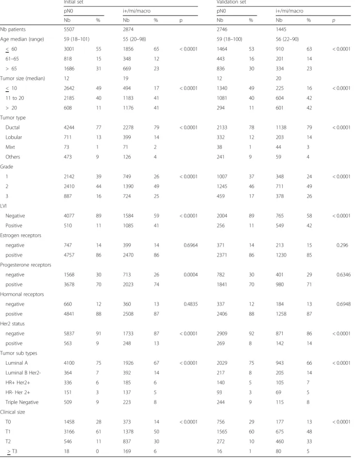
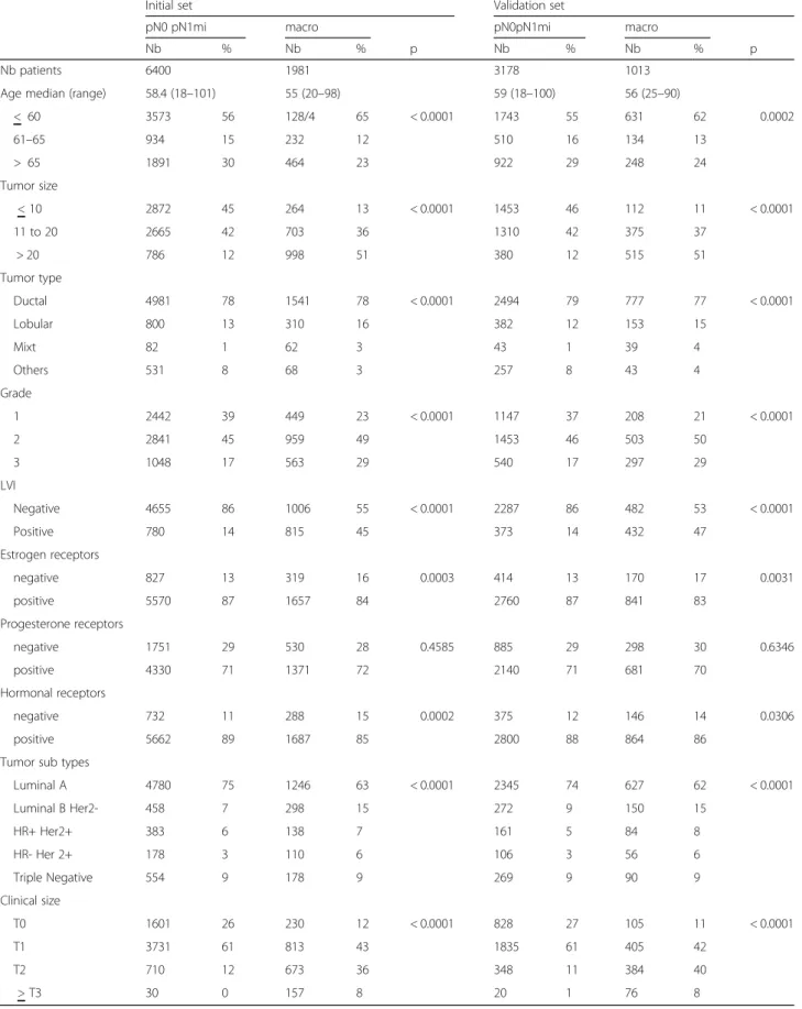
Patients’ main characteristics are summarized in Table1. SBR grade was 1, 2 and 3 in 34, 46 and 20% of cases re-spectively. Hormone receptor-positive tumors (ER+ and/ or PR+) accounted for 88% of cases (11,013 patients). Final LN status, taking into account ALND results when performed, was: pN0(i-) in 8253 patients (66%), pN0(i+) in 355 (3%), pN1mi in 970 (8%) and macro metastasis in 2994 (24%). The comparison between patients with posi-tive and negaposi-tive final LN status, and between patients with LN macro metastases versus pN0 or pNo(i+) or pN1mi showed statistically significant differences with regard to age, pathologic tumor size, SBR grade, LVI, histological type and distribution of molecular subtypes (Tables2and3).
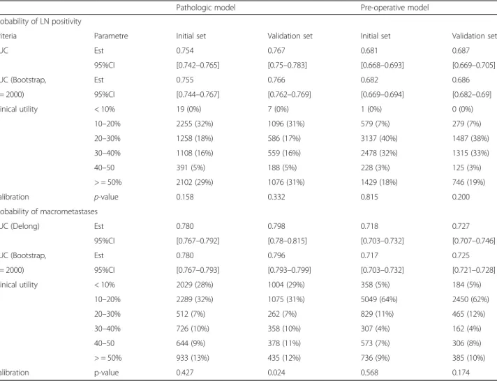
We first predicted the individual probabilities of final LN positivity and of detecting LN macro metastases from selected clinico-pathologic predictor factors provided by tumor surgical results. The model AUCs with 95% CIs for confirmatory and training samples were respectively 0.767 [0.750–0.783] and 0.755 [0.744–0.767]. Calibration plot and Hosmer-Lemeshow test revealed that the calibration is adequate (p = 0.332 in confirmatory sample, p = 0.158 in training sample). With respect to clinical utility in con-firmatory and training samples, the probability of positive LN were respectively below 10% for 7 patients (< 1%) and 19 patients (< 1%), between 10 and 20% for 1096 (31%) and 2255 (32%), and≥ 20% for 2409 (68.6%) and 4859 (68.1%) patients (Table 4) (Fig.1A, Additional file 1: Fig-ure S1A and Additional file 2: Figure S2A). The second pathological model estimated the probability of detecting LN macro metastases only. The AUC values for confirma-tory and training samples were respectively 0.798 (0.780– 0.815) and 0.780 [0.767–0.790]. Clinical utility measures, estimated the probability of LN macro metastases re-spectively in confirmatory and training samples below 10% for 1004 patients (29%) and 2029 patients (28%), between 10 and 20% for 1075 patients (31%) and 2289 patients (32%), and > 20% for 1433 patients (41%) and 2815 patients (39.4%). The Hosmer-Lemeshow test re-vealed a poor calibration of the model (p = 0.024 in
confirmatory sample, p = 0.427 in training sample) (Table4 and Additional file1: Table S1) (Fig. 1B, Add-itional file2: Figure S1B, Additional file3: Figure S2B).
We evaluated the loss in discrimination ability in pre-operative prediction models omitting the information about LVI and substituting pathological tumor size informa-tion by clinical T stage. For the overall probability of LN posi-tivity, the AUC values for confirmatory and training samples were respectively 0.687 [0.669–0.705] and 0.682 (0.669– 0.694). For the probability of detecting LN macro metastases, the observed AUC results for confirmatory and training sam-ples were respectively 0.727 [0.707–0.746] and 0.717 (0.703– 0.732). The calibration of both pre-operative models was found satisfactory. (Table 4) (Fig.1 C-D, Additional file 2: Figure S1C-D, Additional file3: Figure S2C-D).
The change in AUCs between pathological and per-operative model were found statistically significantly decreased (p < 0.001). We also evaluated in the con-firmatory sample the discrimination ability of the predic-tion models obtained when treating the variable age and tumor size as continuous. The AUC values for predicting LN positivity and the presence of LN metastases were respectively 0.774 [0.758, 0.79] and 0.805 [0.789–0.823]. The observed increases were significantly (p = 0.041 and p = 0.026), but the results in terms of calibration were judged inadequate (Hosmer-Lemeshow p value < 0.001).
Discussion
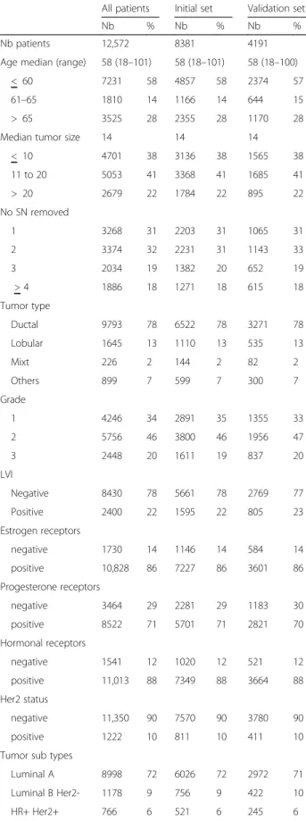
The aim of this study was to better understand the relationships between tumor characteristics and the probability of axillary LN positivity. The large cohort used in our study is appropriate for less frequent tumor phenotypes (namely Her2+ and HR-Her2-). We distin-guished between various histological tumor types, showing Table 1 Population: all patients and patients according to initial
data set or validation set
All patients Initial set Validation set
Nb % Nb % Nb %
Nb patients 12,572 8381 4191
Age median (range) 58 (18–101) 58 (18–101) 58 (18–100)
< 60 7231 58 4857 58 2374 57
61–65 1810 14 1166 14 644 15
> 65 3525 28 2355 28 1170 28
Median tumor size 14 14 14
< 10 4701 38 3136 38 1565 38 11 to 20 5053 41 3368 41 1685 41 > 20 2679 22 1784 22 895 22 No SN removed 1 3268 31 2203 31 1065 31 2 3374 32 2231 31 1143 33 3 2034 19 1382 20 652 19 > 4 1886 18 1271 18 615 18 Tumor type Ductal 9793 78 6522 78 3271 78 Lobular 1645 13 1110 13 535 13 Mixt 226 2 144 2 82 2 Others 899 7 599 7 300 7 Grade 1 4246 34 2891 35 1355 33 2 5756 46 3800 46 1956 47 3 2448 20 1611 19 837 20 LVI Negative 8430 78 5661 78 2769 77 Positive 2400 22 1595 22 805 23 Estrogen receptors negative 1730 14 1146 14 584 14 positive 10,828 86 7227 86 3601 86 Progesterone receptors negative 3464 29 2281 29 1183 30 positive 8522 71 5701 71 2821 70 Hormonal receptors negative 1541 12 1020 12 521 12 positive 11,013 88 7349 88 3664 88 Her2 status negative 11,350 90 7570 90 3780 90 positive 1222 10 811 10 411 10
Tumor sub types
Luminal A 8998 72 6026 72 2972 71
Luminal B Her2- 1178 9 756 9 422 10
HR+ Her2+ 766 6 521 6 245 6
Table 1 Population: all patients and patients according to initial data set or validation set (Continued)
All patients Initial set Validation set
Nb % Nb % Nb % HR- Her 2+ 450 4 288 3 162 4 Triple Negative 1091 9 732 9 359 9 pN final pN0(i-) 8253 66 5507 66 2746 66 pN0(i+) 355 3 233 3 122 3 pN1mi 970 8 660 8 310 7 Macro 2994 24 1981 24 1013 24 Clinical size T0 2764 23 1831 23 933 23 T1 6784 57 4544 57 2246 56 T2 2115 18 1383 17 732 18 > T3 283 2 187 2 96 2
Table 2 Initial data set and validation set results according to axillary nodal involvement
Initial set Validation set
pN0 i+/mi/macro pN0 i+/mi/macro
Nb % Nb % p Nb % Nb % p
Nb patients 5507 2874 2746 1445
Age median (range) 59 (18–101) 55 (20–98) 59 (18–100) 56 (22–90)
< 60 3001 55 1856 65 < 0.0001 1464 53 910 63 < 0.0001
61–65 818 15 348 12 443 16 201 14
> 65 1686 31 669 23 836 30 334 23
Tumor size (median) 12 19 12 20
< 10 2642 49 494 17 < 0.0001 1340 49 225 16 < 0.0001 11 to 20 2185 40 1183 41 1081 40 604 42 > 20 608 11 1176 41 294 11 601 42 Tumor type Ductal 4244 77 2278 79 < 0.0001 2133 78 1138 79 < 0.0001 Lobular 711 13 399 14 332 12 203 14 Mixt 73 1 71 2 38 1 44 3 Others 473 9 126 4 241 9 59 4 Grade 1 2142 39 749 26 < 0.0001 1007 37 348 24 < 0.0001 2 2410 44 1390 49 1245 46 711 49 3 887 16 724 25 459 17 378 26 LVI Negative 4077 89 1584 59 < 0.0001 2004 89 765 58 < 0.0001 Positive 510 11 1085 41 256 11 549 42 Estrogen receptors negative 747 14 399 14 0.6964 371 14 213 15 0.296 positive 4757 86 2470 86 2371 86 1230 85 Progesterone receptors negative 1568 30 713 26 0.0004 782 30 401 29 0.6346 positive 3678 70 2023 74 1841 70 980 71 Hormonal receptors negative 660 12 360 13 0.4835 337 12 184 13 0.6948 positive 4841 88 2508 87 2406 88 1258 87 Her2 status negative 5837 91 1733 87 < 0.0001 2909 92 871 86 < 0.0001 positive 563 9 248 13 269 8 142 14
Tumor sub types
Luminal A 4100 75 1926 67 < 0.0001 2029 75 943 66 < 0.0001 Luminal B Her2- 364 7 392 14 217 8 205 14 HR+ Her2+ 336 6 185 6 140 5 105 7 HR- Her 2+ 151 3 137 5 93 3 69 5 Triple Negative 509 9 223 8 244 9 115 8 Clinical size T0 1458 28 373 14 < 0.0001 756 29 177 13 < 0.0001 T1 3166 61 1378 50 1565 60 675 48 T2 546 11 837 30 272 10 460 33 > T3 18 0 169 6 16 1 80 5
Table 3 Initial data set and validation set results according to axillary nodal macro metastasis involvement
Initial set Validation set
pN0 pN1mi macro pN0pN1mi macro
Nb % Nb % p Nb % Nb % p
Nb patients 6400 1981 3178 1013
Age median (range) 58.4 (18–101) 55 (20–98) 59 (18–100) 56 (25–90)
< 60 3573 56 128/4 65 < 0.0001 1743 55 631 62 0.0002 61–65 934 15 232 12 510 16 134 13 > 65 1891 30 464 23 922 29 248 24 Tumor size < 10 2872 45 264 13 < 0.0001 1453 46 112 11 < 0.0001 11 to 20 2665 42 703 36 1310 42 375 37 > 20 786 12 998 51 380 12 515 51 Tumor type Ductal 4981 78 1541 78 < 0.0001 2494 79 777 77 < 0.0001 Lobular 800 13 310 16 382 12 153 15 Mixt 82 1 62 3 43 1 39 4 Others 531 8 68 3 257 8 43 4 Grade 1 2442 39 449 23 < 0.0001 1147 37 208 21 < 0.0001 2 2841 45 959 49 1453 46 503 50 3 1048 17 563 29 540 17 297 29 LVI Negative 4655 86 1006 55 < 0.0001 2287 86 482 53 < 0.0001 Positive 780 14 815 45 373 14 432 47 Estrogen receptors negative 827 13 319 16 0.0003 414 13 170 17 0.0031 positive 5570 87 1657 84 2760 87 841 83 Progesterone receptors negative 1751 29 530 28 0.4585 885 29 298 30 0.6346 positive 4330 71 1371 72 2140 71 681 70 Hormonal receptors negative 732 11 288 15 0.0002 375 12 146 14 0.0306 positive 5662 89 1687 85 2800 88 864 86
Tumor sub types
Luminal A 4780 75 1246 63 < 0.0001 2345 74 627 62 < 0.0001 Luminal B Her2- 458 7 298 15 272 9 150 15 HR+ Her2+ 383 6 138 7 161 5 84 8 HR- Her 2+ 178 3 110 6 106 3 56 6 Triple Negative 554 9 178 9 269 9 90 9 Clinical size T0 1601 26 230 12 < 0.0001 828 27 105 11 < 0.0001 T1 3731 61 813 43 1835 61 405 42 T2 710 12 673 36 348 11 384 40 > T3 30 0 157 8 20 1 76 8
a lower LN positivity rate in tumors other than ductal, lobular or mixt, as previously reported for BC with favorable histology (tubular, mucinous, papillary, medul-lary, adenoid cystic and secretory) that are associated with a very low LN positivity rate [25].
In our model, we used the same independent variables as Reyal et al. [11], namely age, tumor size, molecular subtypes and LVI, and we added grade and histological type. However, age intervals were different, as well as tumor phenotype definitions (ER only in the Reyal model) and tumor size description (continuous variable in the Reyal model). We obtained different odds ratios for the same variables and clinical utility results were different and higher for low probability of positive lymph node, particularly for macro metastases in our popula-tion for both models. Clinical utility results for low probability of positive lymph node could be contributive to avoid surgical axillary staging by sentinel lymph node biopsy or axillary lymph node dissection.
The models were less reliable when information about LVI was missing. LVI could be detected on pre-operative
biopsies but the difference in accuracy is obviously large in comparison with surgical specimen analysis.
The HER2 status was unknown in old studies [8] and others studies were based on small number of patients. We found that HER2 negative tumors were associated with LN positivity less frequently than HER2 positive tumors (22.9% vs. 31.9%). Lu et al. published that the lowest probability of node metasta-sis was for ER- / HER2- tumors [12]. Similarly in our study, triple negative tumors had the lowest probabil-ity of node metastasis, while HR- / Her2+ tumors had the highest probability. Reyal et al. hypothesized that the axillary LN metastatic process is predomin-antly related to intrinsic biological properties in ER-negative and HER2-negative BC, while tumor size, proliferation rate and LVI are the main determinants in the ER positive or HER2 positive breast cancers. However, positive axillary lymph nodes in triple nega-tive BC were pejoranega-tive prognostic factors for sentinel node macro-metastases but also for occult sentinel node involvement (pN0(i+) and pN1mi) [26].
Table 4 Discrimination, calibration and clinical utility measures of pathologic and pre-operative prediction models
Pathologic model Pre-operative model
Probability of LN positivity
Criteria Parametre Initial set Validation set Initial set Validation set
AUC Est 0.754 0.767 0.681 0.687
95%CI [0.742–0.765] [0.75–0.783] [0.668–0.693] [0.669–0.705]
AUC (Bootstrap, Est 0.755 0.766 0.682 0.686
B = 2000) 95%CI [0.744–0.767] [0.762–0.769] [0.669–0.694] [0.682–0.69] Clinical utility < 10% 19 (0%) 7 (0%) 1 (0%) 0 (0%) 10–20% 2255 (32%) 1096 (31%) 579 (7%) 279 (7%) 20–30% 1258 (18%) 586 (17%) 3137 (40%) 1487 (38%) 30–40% 1108 (16%) 559 (16%) 2478 (32%) 1315 (33%) 40–50 391 (5%) 188 (5%) 228 (3%) 125 (3%) > = 50% 2102 (29%) 1076 (31%) 1429 (18%) 746 (19%) Calibration p-value 0.158 0.332 0.815 0.200 Probability of macrometastases
AUC (Delong) Est 0.780 0.798 0.718 0.727
95%CI [0.767–0.792] [0.78–0.815] [0.703–0.732] [0.707–0.746]
AUC (Bootstrap, Est 0.780 0.796 0.717 0.725
B = 2000) 95%CI [0.767–0.793] [0.793–0.799] [0.703–0.732] [0.721–0.728] Clinical utility < 10% 2029 (28%) 1004 (29%) 358 (5%) 184 (5%) 10–20% 2289 (32%) 1075 (31%) 5049 (64%) 2450 (62%) 20–30% 512 (7%) 262 (7%) 829 (11%) 465 (12%) 30–40% 726 (10%) 358 (10%) 307 (4%) 162 (4%) 40–50 644 (9%) 378 (11%) 573 (7%) 306 (8%) > = 50% 933 (13%) 435 (12%) 736 (9%) 385 (10%) Calibration p-value 0.427 0.024 0.568 0.174
A reliable predictive model of LN positivity, based on pathologic parameters, can be used to compare populations from different studies, particularly for trials with or without axillary surgical procedure. Above all, it might allow avoid-ing SN biopsy when the probability of positivity is very low (< 10%). Some authors already suggested that SN biopsy could be omitted in tumors with good-prognosis subtypes [25] or that axillary dissection is useless in older patients [27]. We believe that these criteria lack accuracy and we prefer a decision-making approach, based on molecular subtypes. However, we must be aware of the risk of insuffi-cient treatment in small tumors with favorable prognostic factors, in which LN status is a major determinant of adju-vant chemotherapy and regional radiotherapy. Moreover, the model is less reliable when LVI is not documented, which is usually the case before surgery. Ultra-sonography of the axilla and percutaneous biopsy is a growing practice. These clinical predictive tools may be helpful relative to the use of axillary ultra-sonography with percutaneous LN bi-opsy for patients with high level risk of axillary LN involvement.
These models can also be contributive in order to de-termined indications of post mastectomy radiotherapy for patients with axillary lymph nodes macro-metastases [28], particularly when immediate breast reconstruction can be proposed.
Conclusions
We reported a reliable predictive model of LN positivity according to different early breast carcinoma phenotypes in a large cohort. The determination of the risk of positive axillary LN can significantly contribute to therapeutic de-cisions. These models, with or without LVI results, can also be used to determine the risk of positive axillary LN or the risk of LN macro-metastasis. Before surgery, clinical models can be used to propose SLNB or not according to LN involvement probability. After surgery, in case of SLNB omission, if LN involvement probability is high, with eventually modifications of adjuvant treatment indi-cations according to LN status, a re-operation can be pro-posed (SLNB or cALND). Thus clinical and pathologic models should be helpful in surgical planning, in the set-ting of a clinical trial and in clinical practice to avoid SLNB for very low risk of LN involvement and to avoid re-operation in case of SLNB omission or to propose ALND for patients with high level probability of major ax-illary LN involvement but also to propose immediate breast reconstruction when PMRT is not required for. A
B
C
D
Fig. 1 Nomograms. 1a: Nomogram predictive of LN Involvement– Pathologic model. 1b: Nomogram predictive of LN macro metastases– Pathologic model. 1c: Nomogram predictive of LN Involvement– Clinical model. 1d: Nomogram predictive of LN macro metastases– Clinical model.
Additional files
Additional file 1: Table S1. Logistic regression results. (DOCX 22 kb)
Additional file 2:Figure S1. Calibration plots of our models. 1A: Calibration of predictive LN Involvement for validation set– Pathologic model. 1B: Calibration of predictive LN macro metastases for validation set– Pathologic model. 1C: Calibration of predictive LN Involvement for validation set– Clinical model. 1D: Calibration of predictive LN macro metastases for validation set– Clinical model. (DOCX 45 kb)
Additional file 3:Figure S2. ROC curves of our models. 2A: ROC curves of predictive LN Involvement– Pathologic model. 2B: ROC curves of predictive LN macro metastases– Pathologic model. 2C: ROC curves of predictive LN Involvement– Clinical model. 2D: ROC curves of predictive LN macro metastases– Clinical model. (DOCX 44 kb)
Acknowledgments Not applicable. Funding
This research did not receive any specific grant from funding agencies in the public, commercial, or not-for-profit sectors.
Availability of data and materials
The datasets generated and analyzed during the current study are available in theClinicalTrials.gov(Identifier: NCT02869607) repository,https:// clinicaltrials.gov/ct2/show/NCT02869607?cond=Breast
+Cancer&cntry=FR&city=Marseille&rank=2
Authors’ contributions
All authors have participated to the data collection: acquisition, analysis and interpretation.
All authors been involved in drafting the manuscript. GH, EL, JMB and MC have written this document.
All authors have read and approve the final version of paper for submission in BMC CANCER. They assure that the manuscript is not, and will not be, under simultaneous consideration by any other publication. The paper reports previously unpublished work. All authors given final approval of the version to be published.
Ethics approval and consent to participate
This work was approved by our institutional review board (IPC - Comité d’Orientation Stratégique). All procedures performed in this study involving human participants were done in accordance with the French ethical standards and with the 2008 Helsinki declaration. All included patients provided written informed consent.
Consent for publication Not applicable. Competing interests
The authors declare that they have no competing interests.
Publisher’s Note
Springer Nature remains neutral with regard to jurisdictional claims in published maps and institutional affiliations.
Author details
1
Institut Paoli Calmettes et CRCM, 232 boulevard de Sainte Marguerite, 13009 Marseille, France.2Institut René Gauducheau, Site Hospitalier Nord, St
Herblain, France.3Institut Gustave Roussy, 114 rue Edouard Vaillant, Villejuif,
France.4Centre Oscar Lambret, 3 rue Frédéric Combenal, Lille, France. 5
Centre Léon Bérard, 28 rue Laennec, Lyon, France.6Centre Claudius Regaud, 20-24 rue du Pont St Pierre, Toulouse, France.7Centre René Huguenin, 35
rue Dailly, Saint Cloud, France.8Hôpital Tenon, 4 rue de la Chine, Paris,
France.9Centre Georges François Leclerc, 1 rue du Professeur Marion, Dijon,
France.10Hôpital de Grasse, Chemin de Clavary, Grasse, France.11Hôpital des Diaconnesses, 18 rue du Sergent Bauchat, Paris, France.12Centre Jean Perrin,
58 rue Montalembert, Clermont Ferrand, France.13Institut Bergonié, 229
Cours de l’Argonne, Bordeaux, France.14Aix-Marseille University, Unité Mixte
de Recherche S912, Institut de Recherche pour le Développement, 13385 Marseille, France.
Received: 24 January 2018 Accepted: 16 December 2018 References
1. Lyman GH, Giuliano AE, Somerfield MR, Benson AB 3rd, Bodurka DC, Burstein HJ, et al. American Society of Clinical Oncology guideline recommendations for sentinel lymph node biopsy in early-stage breast cancer. J Clin Oncol. 2005;23:7703–20.
2. Krag DN, Anderson SJ, Julian TB, Brown AM, Harlow SP, Costantino JP, et al. Sentinel-lymph-node resection compared with conventional axillary-lymph-node dissection in clinically axillary-lymph-node-negative patients with breast cancer: overall survival findings from the NSABP B-32 randomised phase 3 trial. Lancet Oncol. 2010;11:927–33.
3. Giuliano AE, Hunt KK, Ballman KV, Beitsch PD, Whitworth PW. Blumencranz PWet al. Axillary dissection vs no axillary dissection in women with invasive breast cancer and sentinel node metastasis: a randomized clinical trial. JAMA. 2011;305:569–75.
4. Galimberti V, Cole BF, Zurrida S, Viale G, Luini A, Veronesi P, et al. Axillary dissection versus no axillary dissection in patients with sentinel-node micrometastases (IBCSG 23-01): a phase 3 randomised controlled trial. Lancet Oncol. 2013;14:297–305.
5. Friedman D, Gipponi M, Murelli F, Meszaros P, Solari N, Massa M, et al. Predictive factors of non-sentinel lymph node involvement in patients with invasive breast cancer and sentinel node micrometastases. Anticancer Res. 2013;33:4509–14.
6. Rivadeneira DE, Simmons RM, Christos PJ, Hanna K, Daly JM, Osborne MP. Predictive factors associated with axillary lymph node metastases in T1a and T1b breast carcinomas: analysis in more than 900 patients. J Am Coll Surg. 2000;191:1–6 discussion 6-8.
7. González-Vela MC, Garijo MF, Fernández FA, Buelta L, Val-Bernal JF. Predictors of axillary lymph node metastases in patients with invasive breast carcinoma by a combination of classical and biological prognostic factors. Pathol Res Pract. 1999;195:611–8.
8. Fehm T, Maul H, Gebauer S, Scharf A, Baier P, Sohn C, et al. Prediction of axillary lymph node status of breast cancer patients by tumor biological factors of the primary tumor. Strahlenther Onkol. 2005;181:580–6. 9. Yip CH, Taib NA, Tan GH, Ng KL, Yoong BK, Choo WY. Predictors of axillary
lymph node metastases in breast cancer: is there a role for minimal axillary surgery? World J Surg. 2009;33:54–7.
10. Bader AA, Tio J, Petru E, Bühner M, Pfahlberg A, Volkholz H, et al. T1 breast cancer: identification of patients at low risk of axillary lymph node metastases. Breast Cancer Res Treat. 2002;76:11–7.
11. Reyal F, Rouzier R, Depont-Hazelzet B, Bollet MA, Pierga JY, Alran S, et al. The molecular subtype classification is a determinant of sentinel node positivity in early breast carcinoma. PLoS One. 2011;6:e20297. 12. Lu X, Lu X, Wang ZC, Iglehart JD, Zhang X, Richardson AL. Predicting
features of breast cancer with gene expression patterns. Breast Cancer Res Treat. 2008;108:191–201.
13. Crabb SJ, Cheang MC, Leung S, Immonen T, Nielsen TO, Huntsman DD, et al. Basal breast cancer molecular subtype predicts for lower incidence of axillary lymph node metastases in primary breast cancer. Clinical Breast Cancer. 2008;8:249–56.
14. Van Calster B, Vanden Bempt I, Drijkoningen M, Pochet N, Cheng J, Van Huffel S, et al. Axillary lymph node status of operable breast cancers by combined steroid receptor and HER-2 status: triple positive tumours are more likely lymph node positive. Breast Cancer Res Treat. 2009;113:181–7. 15. Lee JH, Kim SH, Suh YJ, Shim BY, Kim HK. Predictors of axillary lymph node
metastases (ALNM) in a Korean population with T1-2 breast carcinoma: triple negative breast cancer has a high incidence of ALNM irrespective of the tumor size. Cancer Res Treat. 2010;42:30–6.
16. Kim MJ, Ro JY, Ahn SH, Kim HH, Kim SB, Gong G. Clinicopathologic significance of the basal-like subtype of breast cancer: a comparison with hormone receptor and Her2/neu-overexpressing phenotypes. Hum Pathol. 2006;37:1217–26.
17. Ngo C, Mouttet D, De Rycke Y, Reyal F, Fourchotte V, Hugonnet F, et al. Validation over time of a nomogram including HER2 status to predict the sentinel node positivity in early breast carcinoma. Eur J Surg Oncol. 2012;38:1211–7.
18. Goldhirsch A, Wood WC, Coates AS, Gelber RD, Thürlimann B, Senn HJ, et al. Strategies for subtypes--dealing with the diversity of breast cancer: highlights of the St. Gallen international expert consensus on the primary therapy of early breast Cancer 2011. Ann Oncol. 2011;22:1736–47. 19. von Minckwitz G, Untch M, Blohmer JU, Costa SD, Eidtmann H, Fasching PA,
et al. Definition and impact of pathologic complete response on prognosis after neoadjuvant chemotherapy in various intrinsic breast cancer subtypes. J Clin Oncol. 2012;30:1796–804.
20. Houvenaeghel G, Goncalves A, Classe JM, Garbay JR, Giard S, Charytensky H, et al. Characteristics and clinical outcome of T1 breast cancer: a multicenter retrospective cohort study. Ann Oncol. 2014.
21. Harrell FE. Regression Modeling Strategies with Applications to Linear Models, Logistic Regression and Survival Analysis. New York: Springer-Verlag; 2001. p. 568. ISBN 0–387–95232-2.
22. Gerds TA, Cai T, Schumacher M. The performance of risk prediction models. Biom J. 2008;(4):457–79.
23. Robin X, Turck N, Hainard A, Tiberti N, Lisacek F, Sanchez J-C, Müller M. pROC: an open-source package for R and S+ to analyze and compare ROC curves. BMC Bioinformatics. 2011;12:77.
24. Hosmer DW Jr., Lemeshow S. Applied logistic regression, Second Edition. New York: Wiley; 2000.
25. Mendez JE, Fey JV, Cody H, Borgen PI, Sclafani LM, et al. Can sentinel lymph node biopsy be omitted in patients with favorable breast cancer histology? Ann Surg Oncol. 2005;12:24–8.
26. Houvenaeghel G, Sabatier R, Reyal F, Classe JM, Giard S, Charitansky H, Rouzier R, Faure C, Garbay JR, Daraï E, Hudry D, Gimbergues P, Villet R, Lambaudie E. Axillary lymph node micrometastases decrease triple-negative early breast cancer survival. Br J Cancer. 2016;115(9):1024–31.
27. Martelli G, Miceli R, Daidone MG, Vetrella G, Cerrotta AM, Piromalli D, et al. Axillary dissection versus no axillary dissection in elderly patients with breast cancer and no palpable axillary nodes: results after 15 years of follow-up. Ann Surg Oncol. 2011;18:125–33.
28. Forissier V, Tallet A, Cohen M, Classe JM, Reyal F, Chopin N, Mazouni C, Gimbergues P, Daraï E, Colombo PE, Azuar P, Lambaudie E, Houvenaeghel G. Is post-mastectomy radiation therapy contributive in pN0-1mi breast cancer patients? Results of a French multi-centric cohort. Eur J Cancer. 2017; 87:47–57.