HAL Id: inserm-00722738
https://www.hal.inserm.fr/inserm-00722738
Submitted on 3 Aug 2012
HAL is a multi-disciplinary open access
archive for the deposit and dissemination of
sci-entific research documents, whether they are
pub-lished or not. The documents may come from
teaching and research institutions in France or
abroad, or from public or private research centers.
L’archive ouverte pluridisciplinaire HAL, est
destinée au dépôt et à la diffusion de documents
scientifiques de niveau recherche, publiés ou non,
émanant des établissements d’enseignement et de
recherche français ou étrangers, des laboratoires
publics ou privés.
Hypoxia-inducible factor (HIF1α) gene expression in
human shock states.
Julien Textoris, Nathalie Beaufils, Gabrielle Quintana, Amin Lassoud,
Laurent Zieleskiewicz, Sandrine Wiramus, Valéry Blasco, Nathalie Lesavre,
Claude Martin, Jean Gabert, et al.
To cite this version:
Julien Textoris, Nathalie Beaufils, Gabrielle Quintana, Amin Lassoud, Laurent Zieleskiewicz, et al..
Hypoxia-inducible factor (HIF1α) gene expression in human shock states.. Critical Care, BioMed
Central, 2012, 16 (4), pp.R120. �10.1186/cc11414�. �inserm-00722738�
R E S E A R C H
Open Access
Hypoxia-inducible factor (HIF1a) gene expression
in human shock states
Julien Textoris
1,2*, Nathalie Beaufils
3, Gabrielle Quintana
1, Amin Ben Lassoud
3, Laurent Zieleskiewicz
1,
Sandrine Wiramus
1, Valéry Blasco
1, Nathalie Lesavre
4, Claude Martin
1, Jean Gabert
3and Marc Leone
1,2Abstract
Introduction: Hypoxia-inducible factor-1 (HIF1) controls the expression of genes involved in the cellular response to hypoxia. No information is available on its expression in critically ill patients. Thus, we designed the first clinical study in order to evaluate the role of HIF1a as a prognosis marker in patients suffering from shock.
Methods: Fifty consecutive adult patients with shock and 11 healthy volunteers were prospectively enrolled in the study. RNA was extracted from whole blood samples and expression of HIF1a was assessed over the first four hours of shock. The primary objective was to assess HIF1a as a prognostic marker in shock. Secondary objectives were to evaluate the role of HIF1a as a diagnostic and follow-up marker. Patient survival was evaluated at day 28. Results: The causes of shock were sepsis (78%), hemorrhage (18%), and cardiac dysfunction (4%). HIF1a expression was significantly higher in the shock patients than in the healthy volunteers (121 (range: 72-168) versus 48 (range: 38-54) normalized copies, P <0.01), whatever the measured isoforms. It was similar in non-survivors and survivors (108 (range 84-183) versus 121(range 72-185) normalized copies, P = 0.92), and did not significantly change within the study period.
Conclusions: The present study is the first to demonstrate an increased expression of HIF1a in patients with shock. Further studies are needed to clarify the potential association with outcome. Our findings reinforce the value of monitoring plasma lactate levels to guide the treatment of shock.
Introduction
Shock states are defined by an acute circulatory failure leading to prolonged and intense tissue hypoxia that may lead to death. Tissue hypoxia is accompanied by a decreased production of ATP in the mitochondria. Hypoxia-inducible factor-1 (HIF1) is a heterodimer made of two sub-units (a and b) [1,2]. The gene coding for HIF1a is on chromosome 14 (14q21-q24) [3]. HIF1a protein concentration is correlated to cellular oxygen concentration [4]. In hypoxemic conditions, HIF1a is not degraded and accumulates in the cellular nucleus [5]. The effects of HIF1a are stimulation of erythropoi-esis, glycolysis, angiogenerythropoi-esis, and vasodilation [1]. In normoxic conditions, HIF1a and its messenger RNA (mRNA) have a very short half-life of five minutes [6,7].
This suggests that HIF1a is an immediate surrogate marker of cellular oxygenation.
In human shock states, plasma lactate is routinely used as a marker of tissue hypoxia. This marker has been vali-dated for the detection of shock states as well as the pre-diction of patient outcomes [8,9]. However, plasma lactate concentrations are influenced by both the produc-tion and clearance of lactate. This can be a limitaproduc-tion for the interpretation of plasma lactate concentrations at the bedside. The main objective of the present study was to evaluate the potential prognostic role of HIF1a in ICU patients with shock states. Secondary objectives were to evaluate the role of HIF1a as a detection marker and its correlation with plasma lactate concentrations.
Materials and methods
Patients
The study received approval of the Ethics Committee (n° 2009-A00105-52) and was conducted in a 15-bed ICU of a teaching hospital (928 beds). Inclusion criteria were
* Correspondence: [email protected]
1
Service d’anesthésie et de réanimation, Hôpital Nord, Assistance Publique-Hôpitaux de Marseille, Chemin des bourrely, 13915, Marseille, France Full list of author information is available at the end of the article
© 2012 Textoris et al.; licensee BioMed Central Ltd. This is an open access article distributed under the terms of the Creative Commons Attribution License (http://creativecommons.org/licenses/by/2.0), which permits unrestricted use, distribution, and reproduction in any medium, provided the original work is properly cited.
≥18 years of age and ≤80 years of age, and shock. Shock was defined as follows: hypotension requiring fluid infu-sion and use of vasopressors, and plasma lactate concen-trations >2 mmol/L. The shock should be related to sepsis, bleeding, or cardiac dysfunction. The exclusion criteria were pregnancy and patients without a social security number or deprived of freedom. After a next of kin gave informed consent, the patients with shock were prospectively included.
The patients had to be enrolled within six hours after admission to the ICU. All enrolled patients were equipped with a central line and an arterial catheter. Blood samples for HIF1a (italic refers to mRNA throughout the manuscript) measurements were col-lected on PaxgeneTM tubes (BD, Franklin Lakes, NJ, USA) and stored at -80°C until RNA extraction. Sam-pling was performed at the time of shock detection (H0), and after 1 hour (H1), 3 hours (H3), and 4 hours (H4). These time points were selected to follow the early steps of the interventions in patients with shock.
The following variables were collected: age, sex, body mass index, and admission simplified acute physiology score (SAPS) II [10], sequential organ failure assessment (SOFA) score [11], vital signs, type of shock, type of ventilation, drugs needed for the treatment of shock states, biochemical variables, blood cell count and coa-gulation variables. In addition, arterial blood gas and plasma lactate concentrations were measured at each time point. Mortality was evaluated at day 28. Data on the duration of mechanical ventilation, vasopressor infu-sion, and ICU stay were also obtained. Treatment goals were based on most available guidelines [8]. Briefly, mean arterial pressure (MAP) was targeted at ≥65 mmHg, urine flow ≥0.5 ml/kg/hour, and central venous oxygen saturation (ScvO2) ≥70%, as described elsewhere
[12].
RNA extraction and quantification of HIF1a variants
Total RNA was isolated using the PAXgene™ Blood RNA Kit (Qiagen, Courtaboeuf, France) according to the manufacturer’s instructions. A total of 1µg of RNA was reverse transcribed with 200 UI MMLV Reverse Tran-scriptase following the EAC (Europe Against Cancer) protocol [13]. The cDNA was diluted in a final volume of 50 µl. Amplification and quantification of HIF1a variants were performed as previously described with some modi-fications of primers and probe sequences (Additional file 1, Figure S1) [14]. Transcripts of the gene coding for TBP (TATA box-binding protein) were also quantified as the endogenous RNA control. Final HIF1a mRNA concen-trations were expressed in normalized copy numbers as previously described [14].
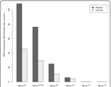
The gene HIF1a is composed of 15 exons, resulting in a principal transcript (HIF1aWT) [3,15] and seven
alternative splice variants which have been reported in human cell lines [16-20]. Amplification of HIF1aWT
showed that it was expressed by circulating blood cells, as well as the splicing variants HIF1aTAGand HIF1a736 (Figure 1). HIF1a516and HIF1a557splice variants tested two isoforms coding for negative dominants. These two isoforms were not or were poorly expressed by circulat-ing blood cells. Relative expression of different isoforms was similar between patients and volunteers (Figure 1).
In a subgroup of six patients, at H0, blood was col-lected from both arterial and venous lines. With regard to the expression of HIF1a, no difference was found between the venous and arterial blood samples (data not shown). Then, a group of 11 healthy volunteers, non-smokers, was evaluated for HIF1a expression and used as controls.
Statistical analysis
From previous studies [14,21], 44 patients with shock were required to achieve a predictive value of 90% with a bias <5% and a 5% risk a. Data were analyzed using the software SPSS and R. Quantitative variables are expressed as median and interquartile range. Qualitative variables are expressed as absolute counts and percen-tages. Differences between groups were tested using non-parametric tests (Mann-Whitney and Kruskall-Wallis tests). A P level of 0.05 or less was considered significant.
Results
Patient characteristics
Fifty patients with shock (average age 57 (range: 18 to 80 years) and 11 healthy volunteers (average age 50 (range: 29 to 70 years) were prospectively included. Women
Figure 1 Expression of different HIF1a variants in shock patients (black bars) and controls (grey bars).
Textoris et al. Critical Care 2012, 16:R120 http://ccforum.com/content/16/4/R120
represented 25% of the cohort of patients and 27% of the cohort of healthy volunteers. The HIF1a expression was unaffected by sex (P = 0.7) or age (P = 0.8). The causes of shock were sepsis, bleeding, and cardiac dysfunction in 39 (78%), 9 (18%), and 2 (4%) cases, respectively (Table 1). Plasma bilirubin concentration and SOFA score differed significantly in survivors and non-survivors (Table 1).
HIF1a expression
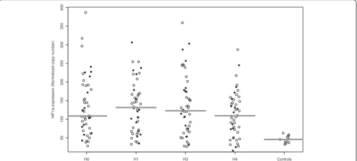
At any time points of the study period, the expression of HIF1a was significantly increased in the patients with shock (Figure 2). At H0, 121 (range: 72 to 168) normalized copies were found in patients with shock, as compared with 46 (range: 38 to 54) normalized copies in healthy volunteers (P <0.01). The detailed values for each time point are presented in Table 2. Of note, the expression of HIF1a did not differ according to the type of shock (data not shown). We did not find a relation between the expression of HIF1a and the absolute number of white blood cells (data not shown).
HIF1a expression, plasma lactate levels, and outcome
Independently of time points, the HIF1a expression did not differ in the survivors and non-survivors (Table 2). In contrast, with the exception of H0, plasma lactate levels were higher in the non-survivors than in the sur-vivors (Table 3). A weak positive correlation was found between HIF1a expression and plasma lactate concen-trations (r2= 0.1; P = 2.10-5).
No correlation was found between the HIF1a expres-sion and admisexpres-sion SAPS 2, shock duration, use of mechanical ventilation, SOFA score, and length of ICU stay. The changes in HIF1a expression between H0 and H4 were not predictive of outcome (Figure 2). The HIF1aexpression was not correlated with hemoglobin, PaO2, and PaO2/FiO2 ratio.
Expression of HIF1a and response to shock treatment
The expression of HIF1a was significantly higher in 24 patients who received more than two liters of fluid expansion: 124 (range: 100 to 168) normalized copies versus 87 (range: 44 to 141) normalized copies (P = 0.02). No difference was found according to the type of administered fluid (crystalloid versus colloids). The HIF1a expression was not correlated with the dose of vasopressors.
Discussion
The present study is the first to show an increased expression of HIF1a in patients with shock, as com-pared with healthy volunteers. The changes in HIF1a expression over time were not correlated with the patient outcome or their treatment responses. Especially, according to our findings, HIF1a expression cannot serve to determine the true level of tissue oxygenation.
A significant increase in HIF1a expression was observed in the patients who received more than two liters of fluid expansion. Nevertheless, no correlation was found with markers of severity, such as MAP, SAPS2 and SOFA score. This finding invites us to hypothesize that this increase was related to a specific effect of fluid infu-sion. Among several hypotheses, one may consider that large fluid resuscitation can impair tissue oxygenation [22]. Another explanation would be that fluid administra-tion was related to the severity of vasodilaadministra-tion, which in turn may be related to tissue-hypoxia. Further investiga-tions are needed to clarify this issue. Larger groups of patients should be evaluated in order to elucidate such a specific effect.
HIF1a has an ultra-short half-life [23,24]. One interest-ing point of the present study is that durinterest-ing the four hours of the study period, the expression of HIF1a was stable. Our initial hypothesis was that due to its ultra-short
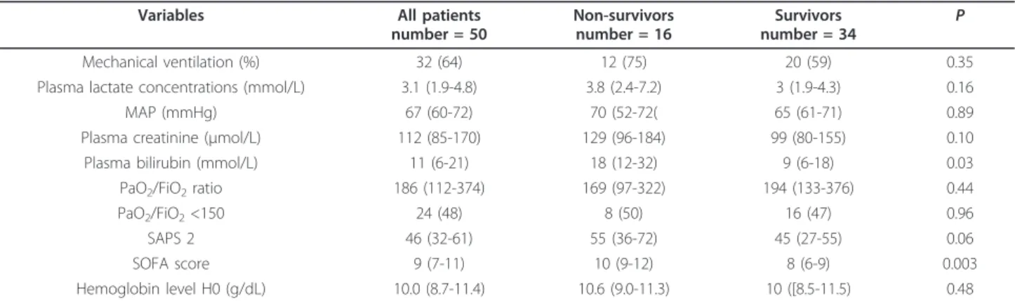
Table 1 Characteristics of the patients according to their survival (day 28).
Variables All patients number = 50 Non-survivors number = 16 Survivors number = 34 P Mechanical ventilation (%) 32 (64) 12 (75) 20 (59) 0.35 Plasma lactate concentrations (mmol/L) 3.1 (1.9-4.8) 3.8 (2.4-7.2) 3 (1.9-4.3) 0.16 MAP (mmHg) 67 (60-72) 70 (52-72( 65 (61-71) 0.89 Plasma creatinine (µmol/L) 112 (85-170) 129 (96-184) 99 (80-155) 0.10 Plasma bilirubin (mmol/L) 11 (6-21) 18 (12-32) 9 (6-18) 0.03 PaO2/FiO2ratio 186 (112-374) 169 (97-322) 194 (133-376) 0.44
PaO2/FiO2<150 24 (48) 8 (50) 16 (47) 0.96
SAPS 2 46 (32-61) 55 (36-72) 45 (27-55) 0.06
SOFA score 9 (7-11) 10 (9-12) 8 (6-9) 0.003
Hemoglobin level H0 (g/dL) 10.0 (8.7-11.4) 10.6 (9.0-11.3) 10 ([8.5-11.5) 0.48
MAP, mean arterial pressure; PaO2/FiO2,partial pressure in oxygen related to the inspired fraction of oxygen; SAPS 2, Simplified Acute Physiology Score; SOFA , sequential organ failure assessment.
half-life, HIF1a could provide an immediate reflection of tissue oxygenation. We failed to demonstrate this effect or tissue oxygenation remained unaffected by time and treat-ment steps. A persistent HIF1a expression has already been demonstrated in cases of lipopolysaccharide stimula-tion and during a sustained inflammatory response [25-28]. Such situations are obviously present in the study patients. A major inflammatory response is present in patients with shock due to the ischemia-reperfusion induced by the treatment of shock [23,29-33]. Because plasma lactate concentrations are not only dependent on production but also on its metabolism, we hypothesized that HIF1a would be a better marker. Our study clearly shows that, at the bedside, lactate remains a better marker of shock than HIF1a. The quantity of protein may be a more accurate marker than the gene expression. Future studies need to clarify this point.
HIF1a is a biomarker of states of cellular hypoxia. Its interest as a marker of outcomes in patients with shock
has never been evaluated before. Nevertheless, our results show that, despite attractive speculations about biomarkers, clinical trials are crucial to evaluate their actual role [4,5]. In the present study, the expression of HIF1a is markedly increased during shock states. The observed increase could be related to the tissue ischemia of shock states or to the inflammatory response. No relation was found between HIF1a expression and oxy-genation variables. However, our results show a trend toward an increased expression in patients with low levels of hemoglobin (Hb >8 g/dL: 109 (range: 84 to 174) HIF1a copies versus Hb <8 g/dL: 161 (range: 74 to 270) HIF1a copies; P = 0.4). Larger samples of patients would be required in order to validate this trend. The expression of HIF1a was wider than expected in our rationale. The wide dispersion of the values may be explained by the ultra-short half-life of HIF1a. This may have affected the power of the study.
In our study, HIF1a mRNA expression seems to fail to reflect hypoxia. Several hypotheses may explain this result. First, we measured the expression of HIF1a mRNA in plasma. Actually, in the case of hypoxia, its
Figure 2 Expression of HIF1a over time in survivors (white circles) and non survivors (black circles). The horizontal bar indicates the median for each group.
Table 2 Normalized copies of HIF1a in patients with shock according to their survival (day 28) and controls.
Time Patients number = 50 Non-survivors number = 16 Survivors number = 34 P H0 109 (78-185) 108 (84-183) 121 (72-185) 0.92 H1 132 (69-169) 123 (99-189) 142 (68-167) 0.88 H3 123 (72-177) 120 (81-180) 123 (68-157) 0.57 H4 109 (73-150) 114 (75-140) 106 (68-150) 0.97 Controls number = 10 baseline 46 (38-54)*
*P < 0.01 as compared with patients at H0.
Table 3 Plasma lactate (mmol/L) according to the survival of patients at day 28.
Time All patients number = 50 Non-survivors number = 16 Survivors number = 34 P H0 3.1 (1.9-4.8) 3.6 (2.4-7.2) 3.0 (1.9-4.3) 0.16 H1 2.8 (1.8-3.6) 3.4 (2.4-7.3) 2.4 (1.8-3.2) 0.04 H3 2.5 (1.7-3.2) 3.4 (2.3-6.3) 2.3 (1.7-2.7) 0.03 H4 2.5 (1.9-3.4) 3.7 (2.5-5.3) 2.4 (1.6-3.0) 0.01 Textoris et al. Critical Care 2012, 16:R120
http://ccforum.com/content/16/4/R120
expression may be more accurate in tissue than in blood. However, regarding our study goals, the collec-tion of tissue biopsy was irrelevant. Second, we may hypothesize that the protein of HIF1a may better reflect tissue hypoxia than its mRNA expression. However, the determination of the protein levels is time consuming, whereas that of RNA levels can be performed in a short time. Our study was aimed at providing an early marker in real-life clinical practice. Finally, divergently from HIF1a, plasma lactate levels may reflect pyruvate accu-mulation rather than cell hypoxia in sepsis and injury [9]. The evaluation of HIF1a values beyond four hours may also bring new evidence of its role in patients with shock. Future studies are needed to determine whether its expression during late phases of shock may be related to early interventions. Finally, it is important to consider that the present study focused on blood deter-minations. This could not reflect with enough accuracy the state of ischemia at the tissue level [21].
Conclusions
The present study is the first to show the increased expression of HIF1a, a transcription factor that controls genes implied in the response to cellular ischemia, in patients with shock. Within the limitations of the study, HIF1aexpression was not correlated with the outcome of patients. Further studies including larger groups of patients are warranted to clarify this issue.
Key messages
• Hypoxia-inducible factor 1 alpha (HIF1a) is a tran-scription factor that controls the expression of genes in response to cellular hypoxia
• HIF1a mRNA is elevated in patients with shock, as compared to healthy volunteers
• HIF1a expression was not correlated to patient outcome
• HIF1a expression over the first hours of shock management was independent of clinical evolution and outcome
• To assess patients with shock, plasma lactate levels seem better than HIF1a expression
Additional material
Additional file 1: Figure S1. RT-PCR primers pairs location. Schematic representation of the location of the various pairs of primers used to amplify several splicing variant of HIF1a.
Abbreviations
HIF1α: Hypoxia Inducible Factor 1 alpha ; SAPS: Simplified Acute Physiological Score ; SOFA: Sequential Organ Failure Assessment.
Acknowledgements
This work was supported by the Grant ‘Appel d’Offre de Recherche Clinique-Assistance Publique des Hôpitaux de Marseille’ n°2009-A00364-53, Marseille, France.
Author details
1
Service d’anesthésie et de réanimation, Hôpital Nord, Assistance Publique-Hôpitaux de Marseille, Chemin des bourrely, 13915, Marseille, France.
2URMITE, CNRS U7278, INSERM U1095, Faculté de médecine Timone,
Aix-Marseille Univ, Boulevard Jean Moulin, 13385, Aix-Marseille, France.3Laboratoire de Biochimie et Biologie Moléculaire, Hôpital Nord, Assistance Publique-Hôpitaux de Marseille, Chemin des bourrely, 13915, Marseille, France.4Centre
d’Investigation Clinique, Hôpital Nord, Assistance Publique-Hôpitaux de Marseille, Chemin des bourrely, 13915, Marseille, France.
Authors’ contributions
JT, GQ, SW, LZ and VB were involved in the enrollment of patients, the completion of chart report forms, and the collection blood samples. NL and GQ were involved in the enrollment of the healthy donors, the completion of chart report forms, and the collection of blood samples. AB and NB handled the blood samples and performed the molecular analysis. JT, GQ, CM, JG, ML wrote the manuscript. ML, JG, CM designed the study. All authors have read and approved the final manuscript.
Competing interests
The authors declare that they have no competing interests.
Received: 4 May 2012 Revised: 14 June 2012 Accepted: 10 July 2012 Published: 10 July 2012
References
1. Semenza GL, Agani F, Iyer N, Jiang BH, Leung S, Wiener C, Yu A: Hypoxia-inducible factor 1: from molecular biology to cardiopulmonary physiology. Chest 1998, 114:40S-45S.
2. Hellwig-Bürgel T, Rutkowski K, Metzen E, Fandrey J, Jelkmann W: Interleukin-1beta and tumor necrosis factor-alpha stimulate DNA binding of hypoxia-inducible factor-1. Blood 1999, 94:1561-1567. 3. Semenza GL, Rue EA, Iyer NV, Pang MG, Kearns WG: Assignment of the
hypoxia-inducible factor 1alpha gene to a region of conserved synteny on mouse chromosome 12 and human chromosome 14q. Genomics 1996, 34:437-439.
4. Schumacker PT: Hypoxia-inducible factor-1 (HIF-1). Crit Care Med 2005, 33: S423-425.
5. Maxwell PH: Hypoxia-inducible factor as a physiological regulator. Exp Physiol 2005, 90:791-797.
6. Berra E, Richard DE, Gothié E, Pouysségur J: HIF-1-dependent transcriptional activity is required for oxygen-mediated HIF-1alpha degradation. FEBS Lett 2001, 491:85-90.
7. Wang GL, Jiang BH, Rue EA, Semenza GL: Hypoxia-inducible factor 1 is a basic-helix-loop-helix-PAS heterodimer regulated by cellular O2 tension. Proc Natl Acad Sci USA 1995, 92:5510-5514.
8. Dellinger RP, Levy MM, Carlet JM, Bion J, Parker MM, Jaeschke R, Reinhart K, Angus DC, Brun-Buisson C, Beale R, Calandra T, Dhainaut J-F, Gerlach H, Harvey M, Marini JJ, Marshall J, Ranieri M, Ramsay G, Sevransky J, Thompson BT, Townsend S, Vender JS, Zimmerman JL, Vincent J-L: Surviving Sepsis Campaign: international guidelines for management of severe sepsis and septic shock: 2008. Intensive Care Med 2008, 34:17-60. 9. Levy B: Lactate and shock state: the metabolic view. Curr Opin Crit Care
2006, 12:315-321.
10. Le Gall JR, Lemeshow S, Saulnier F: A new Simplified Acute Physiology Score (SAPS II) based on a European/North American multicenter study. JAMA 1993, 270:2957-2963.
11. Vincent JL, Moreno R, Takala J, Willatts S, De Mendonça A, Bruining H, Reinhart CK, Suter PM, Thijs LG: The SOFA (Sepsis-related Organ Failure Assessment) score to describe organ dysfunction/failure. On behalf of the Working Group on Sepsis-Related Problems of the European Society of Intensive Care Medicine. Intensive Care Med 1996, 22:707-710. 12. Leone M, Blidi S, Antonini F, Meyssignac B, Bordon S, Garcin F, Charvet A,
nonsurvivors than in survivors after early resuscitation of septic shock. Anesthesiology 2009, 111:366-371.
13. Gabert J, Beillard E, van der Velden VHJ, Bi W, Grimwade D, Pallisgaard N, Barbany G, Cazzaniga G, Cayuela JM, Cavé H, Pane F, Aerts JLE, De Micheli D, Thirion X, Pradel V, González M, Viehmann S, Malec M, Saglio G, van Dongen JJM: Standardization and quality control studies of ‘real-time’ quantitative reverse transcriptase polymerase chain reaction of fusion gene transcripts for residual disease detection in leukemia - a Europe Against Cancer program. Leukemia 2003, 17:2318-2357. 14. Dales J-P, Beaufils N, Silvy M, Picard C, Pauly V, Pradel V,
Formisano-Tréziny C, Bonnier P, Giusiano S, Charpin C, Gabert J: Hypoxia inducible factor 1alpha gene (HIF-1alpha) splice variants: potential prognostic biomarkers in breast cancer. BMC Med 2010, 8:44.
15. Iyer NV, Leung SW, Semenza GL: The human hypoxia-inducible factor 1alpha gene: HIF1A structure and evolutionary conservation. Genomics 1998, 52:159-165.
16. Gothié E, Richard DE, Berra E, Pagès G, Pouysségur J: Identification of alternative spliced variants of human hypoxia-inducible factor-1alpha. J Biol Chem 2000, 275:6922-6927.
17. Chun YS, Choi E, Yeo EJ, Lee JH, Kim MS, Park JW: A new HIF-1 alpha variant induced by zinc ion suppresses HIF-1-mediated hypoxic responses. J Cell Sci 2001, 114:4051-4061.
18. Chun Y-S, Choi E, Kim T-Y, Kim M-S, Park J-W: A dominant-negative isoform lacking exons 11 and 12 of the human hypoxia-inducible factor-1alpha gene. Biochem J 2002, 362:71-79.
19. Lee K-H, Park J-W, Chun Y-S: Non-hypoxic transcriptional activation of the aryl hydrocarbon receptor nuclear translocator in concert with a novel hypoxia-inducible factor-1alpha isoform. Nucleic Acids Res 2004, 32:5499-5511.
20. Depping R, Hägele S, Wagner KF, Wiesner RJ, Camenisch G, Wenger RH, Katschinski DM: A dominant-negative isoform of hypoxia-inducible factor-1 alpha specifically expressed in human testis. Biol Reprod 2004, 71:331-339.
21. Leone M, Boutière B, Camoin-Jau L, Albanèse J, Horschowsky N, Mège J-L, Martin C, Dignat-George F: Systemic endothelial activation is greater in septic than in traumatic-hemorrhagic shock but does not correlate with endothelial activation in skin biopsies. Crit Care Med 2002, 30:808-814. 22. Legrand M, Mik EG, Balestra GM, Lutter R, Pirracchio R, Payen D, Ince C:
Fluid resuscitation does not improve renal oxygenation during hemorrhagic shock in rats. Anesthesiology 2010, 112:119-127. 23. Hellwig-Bürgel T, Stiehl DP, Wagner AE, Metzen E, Jelkmann W: Review:
hypoxia-inducible factor-1 (HIF-1): a novel transcription factor in immune reactions. J Interferon Cytokine Res 2005, 25:297-310. 24. Görlach A, Diebold I, Schini-Kerth VB, Berchner-Pfannschmidt U, Roth U,
Brandes RP, Kietzmann T, Busse R: Thrombin activates the hypoxia-inducible factor-1 signaling pathway in vascular smooth muscle cells: role of the p22(phox)-containing NADPH oxidase. Circ Res 2001, 89:47-54. 25. Koury J, Deitch EA, Homma H, Abungu B, Gangurde P, Condon MR, Lu Q,
Xu D-Z, Feinman R: Persistent HIF-1alpha activation in gut ischemia/ reperfusion injury: potential role of bacteria and lipopolysaccharide. Shock 2004, 22:270-277.
26. Frede S, Stockmann C, Freitag P, Fandrey J: Bacterial lipopolysaccharide induces HIF-1 activation in human monocytes via p44/42 MAPK and NF-kappaB. Biochem J 2006, 396:517-527.
27. Blouin CC, Pagé EL, Soucy GM, Richard DE: Hypoxic gene activation by lipopolysaccharide in macrophages: implication of hypoxia-inducible factor 1alpha. Blood 2004, 103:1124-1130.
28. Peyssonnaux C, Cejudo-Martin P, Doedens A, Zinkernagel AS, Johnson RS, Nizet V: Cutting edge: essential role of hypoxia inducible factor-1alpha in development of lipopolysaccharide-induced sepsis. J Immunol 2007, 178:7516-7519.
29. Shih SC, Claffey KP: Role of AP-1 and HIF-1 transcription factors in TGF-beta activation of VEGF expression. Growth Factors 2001, 19:19-34. 30. Richard DE, Berra E, Pouyssegur J: Nonhypoxic pathway mediates the
induction of hypoxia-inducible factor 1alpha in vascular smooth muscle cells. J Biol Chem 2000, 275:26765-26771.
31. Feldser D, Agani F, Iyer NV, Pak B, Ferreira G, Semenza GL: Reciprocal positive regulation of hypoxia-inducible factor 1alpha and insulin-like growth factor 2. Cancer Res 1999, 59:3915-3918.
32. Laughner E, Taghavi P, Chiles K, Mahon PC, Semenza GL: HER2 (neu) signaling increases the rate of hypoxia-inducible factor 1alpha
(HIF-1alpha) synthesis: novel mechanism for HIF-1-mediated vascular endothelial growth factor expression. Mol Cell Biol 2001, 21:3995-4004. 33. Nizet V, Johnson RS: Interdependence of hypoxic and innate immune
responses. Nat Rev Immunol 2009, 9:609-617.
doi:10.1186/cc11414
Cite this article as: Textoris et al.: Hypoxia-inducible factor (HIF1a) gene expression in human shock states. Critical Care 2012 16:R120.
Submit your next manuscript to BioMed Central and take full advantage of:
• Convenient online submission
• Thorough peer review
• No space constraints or color figure charges
• Immediate publication on acceptance
• Inclusion in PubMed, CAS, Scopus and Google Scholar
• Research which is freely available for redistribution
Submit your manuscript at www.biomedcentral.com/submit
Textoris et al. Critical Care 2012, 16:R120 http://ccforum.com/content/16/4/R120