Distribution and Metabolism of Antibodies and Macromolecules in
Tumor Tissue
By Greg M. Thurber B.S. Chemical Engineering
Minor Biochemistry and Molecular Biology The Pennsylvania State University, 2003
Submitted to the Department of Chemical Engineering in Partial Fulfillment of the Requirements for the Degree of
DOCTOR OF PHILOSOPHY In Chemical Engineering
At the
Massachusetts Institute of Technology May 2008
[aucse 2@ti
OF TEOHINOLOGY
J
UN 0 6 2008
LIBRARIES
© 2008 Massachusetts Institute of TechnologyAll rights reserved
Signature of Author
Greg M. Thurber Department of Chemical Engineering May 2008
Certified by__,
SbK. Dane Wittrup
C.P. Dubbs Professor of Chemical Engineering and Biological Engineering Thesis Advisor
Accepted by
William M. Deen Professor of Chemical Engineering Chairman, Committee for Graduate Students
Distribution and Metabolism of Antibodies and Macromolecules in Tumor Tissue
By Greg M. Thurber
Submitted to the Department of Chemical Engineering on May 22, 2008 in Partial Fulfillment of the Requirements for the Degree of Doctor of Philosophy in Chemical Engineering
ABSTRACT
Tumor targeting drugs that selectively treat cancerous tissue are promising agents for lowering the morbidity and mortality of cancer. Within this field, antibody treatments for cancer are currently
being developed for both imaging and therapeutic applications. A major limitation with this class of drugs is the poor distribution and low uptake in tumor tissue. Poor distribution leaves some cells completely devoid of treatment, while others experience marginally toxic concentrations that could foster drug resistance. The low overall uptake in vascularized tumors constrains the therapeutic index
and lowers signal to noise ratios for imaging applications. Since antibody therapies are currently used to treat both bulk tumors and residual disease, an understanding of the limitations in targeting prevascular metastases and vascularized tumors is required.
In order to circumvent the current limitations with antibody therapies, the underlying causes must first be determined. In this thesis, the various steps in tumor localization of antibodies are analyzed in order to determine which steps are limiting uptake and distribution. Mathematical models are developed that indicate the distance antibodies and other binding macromolecules will penetrate into tumors and micrometastases. These models can also estimate the maximum uptake and time course of antibody concentration in tumors. The experimental distribution of a CEA binding antibody is measured in tumor spheroids and a mouse xenograft system to validate the model predictions.
Using dimensional analysis of the fundamental transport rates that occur in tumors and micrometastases, two main groups determine the distance antibodies will penetrate in tumor tissue. The clearance modulus indicates whether antibody persistence in the blood is sufficient to allow the
drug to reach all cells in the micrometastasis or vascularized tumor. The Thiele modulus, defined for antibody transport in tumors, relates the internalization and catabolism of bound antibodies on cancer cells to the maximum distance the antibodies will reach in the tissue. These groups are related to the overall time course and maximum uptake in tumors, indicating when all cells will be targeted, and what factors determine this limit. These models can aid in experimental design, data interpretation, and strategies to improve uptake.
Thesis Supervisor: Dr. K. Dane Wittrup
Acknowledgements
First, I would like to thank my advisor for his excellent advice and expertise over the course of this work. His terrific intellectual input, suggestions, questions, and challenges have guided the project, taught me how to do research, and made this thesis possible. Dane has put together a great lab, where
it's both exciting and enjoyable to work. His enthusiasm and patience during the ups and downs of research have been highly motivating and really facilitated the research progress. Finally, beyond the research, I want to thank him for the lessons and advice that will serve for years to come.
I want to thank all the members of the Wittrup lab who have helped me during my time here.
From the simple act of loaning material to insightful suggestions, their contributions pervade the following pages. Countless discussions with them have taught me a lot over my graduate career, and it
is these people who have made it such a friendly and encouraging atmosphere. This includes former students and postdocs that have helped me early on in my research: Stefan Zajic, Dave Colby, Andy
Yeung, Shaun Lippow, Jeff Swers, Balaji Rao, Katarina Midelfort, Andy Rakestraw, Ginger Chao, Wai Lau, Dasa Lipovsek, Andrea Piatesi, Jennifer Cochran, and Pankaj Karande. And the current students, who have helped in the past and will carry on the work, include Shanshan Wu Howland, Steve Sazinsky, Mike Schmidt, Margaret Pawlowski, Annie Gai, Eileen Higham, Ben Hackel, Kelly Davis, David Liu, Chris Pirie, Jordi Mata-Fink, Jamie Spangler, and John Rhoden.
I would especially like to thank my thesis committee members Prof. Bill Deen and Prof. Peter So. I always looked forward to my thesis committee meetings and their constructive input, and their insight
constantly provided scope and perspective to the project.
Hyuk-Sang Kwon in Prof. So's lab was a tremendous help with gathering the microscopy data, and Prof. Sangeeta Bhatia and her lab, especially Todd Harris, have been instrumental in making the in
vivo work happen. Todd in particular was a great help in providing advice and training for the animal
studies.
I also want to thank my undergraduate research advisor, Dr. Wayne Curtis. He was the one who
plucked me out of the classroom and got me into the laboratory. Without that motivation, I may have never realized how much I enjoy research.
My friends, both at MIT and elsewhere, have provided tremendous support over the years.
Whether it's lunchtime coffee in the amphitheatre or a late night break with my roommates, their laughter and support has been refreshing time and time again. Weekend trips and in-town visits from my college friends and those back home have always kept things interesting and new.
My family has been incredible. My mom and dad have been there for me long before there was
even the possibility of a thesis, and since then, they have supported me until the end. I also want to thank my older brother, John, for paving the way for me. And for always letting me know that.
My fiancee Melissa has been spectacular over the years with her caring and support. I want to
thank her for her love, patience, and support she gave and continues to give each day. And lastly, I want to give thanks to God, who amazes me every day with His creation.
To my parents, John and Sandy, for their unconditional love and support. Without them, none of this would have been possible.
Table of Contents
Chapter 1- Introduction 11
1.1 Background 11
1.2 Antibodies as Drugs 12
1.3 Tumor Physiology 13
Chapter 2 - Theory of Antibody Spatial Distribution in Tumor Tissue 23
2.1 Importance of Spatial Distribution in Imaging and Therapy 23
2.2 Model Development 23
2.3 Previous Models and Current Approach 29
2.4 Transport 31
2.5 Antibody Clearance 37
2.6 Limitations to Antibody Localization 39
2.7 Micrometastases versus Vascularized Tumors 50
2.8 Effects of Affinity on Targeting 54
2.9 Buckingham Pi Analysis 62
2.10 Methods to Improve Distribution 65
Chapter 3 - Experimental Distribution of Antibodies in Tumor Spheroids 79
3.1 Introduction 79
3.2 Previous Spheroid Studies 80
3.3 Carcinoembryonic Antigen 81
3.4 Materials and Methods for Tracking anti-CEA scFvs in LS174T Spheroids 82
3.5 Anti-CEA scFv Results 88
3.6 Discussion of anti-CEA scFvs 102
Chapter 4 - Theory of the Time Course of Antibody Concentration in Tumors 111
4.1 Importance of the Time Course of Antibody Concentration in Tumors 111
4.2 Background on Vascular Distribution and Antibody Uptake 111
4.3 Model Development 112
4.4 Compartmental Model Results 123
4.5 Compartmental Model Validation 130
4.6 Discussion 137
Chapter 5 - Experimental Uptake in Mouse Xenografts 147
5.2 Methods 150
5.3 Results 151
5.4 Discussion 165
Appendix - Mathematical Derivations 173
A.1 List of Symbols 174
A.2 Equations for Microscopic Distribution of Antibodies 176
A.3 Binding Modulus 182
A.4 Clearance Modulus 183
A.5 Thiele Modulus 192
A.6 Biot Number and Other Boundary Conditions 203
A.7 Shrinking Core Derivation with Catabolism - Spherical Geometry 210
A.8 Low Affinity Antibodies 212
A.9 Buckingham Pi Analysis 227
A.10 Pretargeting Number 230
A.11 Solutions without Closed Forms 231
A.12 Convection-Diffusion Model 247
A.13 Compartmental Model 251
A.14 Effective Endocytosis Rate with Recycling 267
A.15 Compartmental Model - Internalization versus Degradation 268
Chapter 1- Introduction
1.1 Background
Cancer is a leading cause of mortality worldwide(1), and the second leading cause of death in the United States(2). Despite intense research, the prognosis for many forms of cancer is still very poor. The disease strikes when cells in the body begin growing uncontrollably, interfering with normal
function. Because these cells originate from the host, it is difficult to treat the disease without affecting healthy tissues.
Standard treatment for cancer typically involves surgery to remove the tumor, radiation, and chemotherapy. If the tumor has not spread to the rest of the body (metastasized), surgery of the primary tumor is typically curative. However, when cancer cells disseminate in the vascular and/or lymphatic system, the disease is much more difficult to treat. Radiation and surgery both require knowledge of the location of the disease, but current imaging methods are only capable of detecting masses roughly one centimeter or larger in diameter(3-5). Therefore, chemotherapy is the only option for treating disseminated disease.
Traditional chemotherapeutics are small molecule drugs that are delivered systemically. Most of them work by disrupting DNA replication or other machinery required for cell division, thereby selectively killing rapidly dividing cells. However, normal tissues also undergo cell division, and the chemotherapeutics damage these cells as well, often causing severe side effects, such as nausea, alopecia (hair loss), and extreme fatigue. Due to their poor selectivity, intense, near-lethal courses of drugs are delivered to obtain maximum effect against the neoplastic tissue, followed by breaks in therapy of several weeks to allow the body to recover(6).
The poor selection for cancer cells has led to a variety of research trying to find ways of
selectively targeting the cancerous tissue for more effective therapy. This includes, but is not limited to, targeting signaling pathways that are disrupted during the multiple steps of tumorigenesis(7), for example oncogenes such as RAS(8), tumor suppressor genes like p53(9, 10), and angiogenesis receptors(11). Tumor selectivity can also be achieved by enhanced uptake from abnormal tumor physiology(12), eliciting an immune response due to abnormal protein expression(13), and targeting surface antigens that are mal-expressed or up-regulated on cancer cells(14, 15). This thesis focuses on the use of antibodies that specifically bind cancer cell antigens on the surface of tumor cells.
1.2 Antibodies as Drugs
Antibodies are proteins produced by the immune system that recognize and associate with foreign proteins and molecules in the body. While there are several classes, the most prevalent in the blood stream is immunoglobulin G (IgG), an approximately 150 kDa 'Y-shaped' protein that contains two identical binding domains and a constant (Fc) region that recruits immune effector cells(16).
Interest in this class of drugs was motivated with the development of monoclonal
antibodies(17), allowing the mass production of antibodies with identical specificity. Further progress in protein engineering has allowed researchers to make a variety of smaller fragments of this molecule(18) as well as engineer particular binding specificity, affinity, dissociation rate, stability, reduced
immunogenicity, and biological effects(19-22). These techniques have allowed researchers to develop antibodies that bind specifically to antigens that are over-expressed or expressed incorrectly on the surface of cancer cells. This class of drugs then has the ability to bind and concentrate locally in cancer tissue while lowering effects in the normal tissue. There is also flexibility in the type of therapy
administered. Current FDA-approved drugs include naked antibodies that can block cell signaling and recruit immune effector cells(14, 23, 24), carry chemical or biological toxins(25), or deliver a
radioisotope that emits local radiation to kill the cell(26).
Although much excitement has been created over the use of these molecules to specifically target cancer without the harmful side effects of small molecule drugs, this type of therapy has not fully lived up to its potential. One of the major problems with antibody therapies is their distribution in tumor tissue. Because of their large size (150 kDa for an IgG), the transport of these drugs is severely limited in tumors. Examination of tumors after the systemic delivery of an antibody drug shows large spatial heterogeneities with perivascular and peripheral localization(27-30). Large areas within the tumor contain cancer cells where very little or no drug is present. Not only is the spatial distribution a problem, but the total amount that enters the tumor is also low. The fraction of the injected dose that localizes within a tumor rarely reaches above 0.01% per gram of tissue in humans(31).
Tumor therapy with antibodies is a complex problem, with both pharmacokinetic as well as pharmacodynamic considerations. However, a clear example where uptake is a limiting factor comes from studies on radioimmunotherapy (RIT). In this type of therapy, a radioisotope is conjugated directly to the antibody, and following uptake in tumor sites, the isotope emits radiation locally, giving a higher
systemically, as opposed to external beam radiation, so it can target disseminated disease. With short range isotopes, dosage to the surrounding tissue is minimized. The effect of radiation on normal and tumor tissue is well studied(32), and given a sufficient dose, the cancer cells will be killed. However, due to low uptake within solid tumors, results have been poor(33). Full IgG molecules have long circulation times in the plasma, and the dose to bone marrow, which is very sensitive to radiation, is limiting. The use of fragments allows more rapid clearance from the normal tissue, but this results in even lower uptake. A more in depth analysis of clearance versus uptake is discussed in chapters 2 and 4. The challenge with this therapy is delivering sufficient doses to kill the most resistant cancer cells while sparing the most sensitive normal tissues.
A more detailed understanding of the transport of antibodies and antibody fragments is needed
to help improve imaging and therapy. Due to the complexity of tumor physiology and targeting, mathematical models are vital to understanding the behavior of these types of drugs. One of the criticisms of this type of modeling is the large heterogeneity found in tumors. While many tumor properties (and and therefore model inputs) are heterogeneous, this analysis focuses on order of magnitude estimates for spatial and temporal distribution. Local values can vary considerably, but an overall understanding of the driving forces and limitations on uptake can be obtained by modeling analysis. Many of the concepts are based on rates that differ by several orders of magnitude (e.g. 100-to 1000-fold differences). Even with the considerable heterogeneity present in tumors, many of these relationships will hold true for most relevant targeting scenarios. From an imaging or therapeutic design perspective, these trends are more useful in identifying ways of circumventing targeting problems, rather than analyzing specific cases that may not be found in other areas of tissue or different tumors.
1.3 Tumor Physiology
In order to understand the basis of the modeling analysis, assumptions, and when these assumptions fail, a brief overview in tumor physiology is necessary. First, there are two different targeting scenarios that will be examined, a micrometastasis that is not yet vascularized, and a vascularized solid tumor. Antibody therapies are being used for both cases, i.e. bulk disease and minimal residual disease(34), so it is important to understand some of the fundamental differences between these clinical targets.
Metastasis occurs when cancerous cells from the primary tumor lose adhesion with the surrounding tissue and travel to a different location within the body. The most common form of metastasis is through the lymph system, and this is used in staging many cancers. Alternatively, cancer cells can enter the circulation and travel a significant distance away from the primary tumor(35). While the lymph system is more amenable to cancer cell survival and local spread(36), metastasis via the vasculature is more difficult to treat, since this route forms distant metastases which cannot be removed by taking larger surgical margins around the primary tumor or removing local lymph nodes.
When a cancer cell begins to grow in a new site, it initially forms a cluster of new cells imbedded in the surrounding normal tissue. These micrometastases continue to divide and grow, fed by the vasculature in the surrounding normal tissue, until their size becomes limited by oxygen transport. The
process of angiogenesis has been extensively studied, but some of the earlier work was done in the
Rip-TAG mouse model. In this model, oncogenes are expressed by the
13
cells in the islets of the pancreas. Although the cells become tumorigenic, they cannot grow beyond 100 to 200 VIm in diameter until they have recruited their own blood supply by the process of angiogenesis(37). Larger clusters of cells can exist, but they suffer from central necrosis due to these transport limitations, so only cells near the surface remain viable(38).Normal tissues in the body have well organized vascular and lymphatic structures. This efficient transport system provides adequate exposure of most tissues to systemically delivered drugs, including the tissue surrounding a metastasis. Fluid balance in the tissue prevents edema (swelling) and ensures that the interstitial pressure remains close to zero (Figure 1.1 A, left). According to Starling's hypothesis, fluid is filtered from the capillary due to a higher sum of hydrostatic and osmotic pressures (effective pressure) at the arterial end (Figure 1.1 B, left). Hydrostatic pressure decreases along the length of the capillary, and the low permeability of vessels to proteins causes an increase in osmotic pressure. Towards the venous end, the effective capillary pressure is less than the interstitial pressure, so fluid is
reabsorbed into the vessel lumen(39, 40). The remaining fluid is taken up by local lymph vessels, which often contain a negative pressure. These closed end vessels are held open by attachment to the surrounding cells and are highly permeable to macromolecules and cells of the immune system(41). Capillaries and lymph vessels are adequately spaced so that delivery of oxygen and nutrients and removal of waste products is efficiently carried out in the tissue. Although capillaries are not highly permeable to macromolecules, the pressure differences allow convection between the tissue and vessels, which is typically more important than diffusion for these large molecules.
A more extensive look at the convective and diffusive contributions to macromolecular
permeability of capillaries is given by Rippe and Haraldsson(42). Here, the authors use a two-pore model to analyze the transport of proteins and fluid across the capillary wall. The capillary membrane is assumed to consist of a series of small pores, roughly 4-6 nm in radius, and large pores, approximately 20-40 nm. The large pores are thought to have a low reflection coefficient, so that the hydrostatic
pressure drives filtration of fluid and macromolecules across these pores. Osmotic absorption of fluid through the more selective small pores counterbalances some of this fluid flow. These conditions allow the circulation of fluid across the capillary wall even in situations of zero fluid filtration. The good fit
between two-pore experimental predictions and data is used to argue that active transcytosis across capillary walls is insignificant compared to the passive mechanisms of transport from the blood to tissue.
The physiology of solid tumors is very different than the well organized structure of normal tissue. While much of this lies outside the scope of this thesis, several factors are important in
understanding the unique state of convection and diffusion within tumors as well as the origin of some of the heterogeneity present in this type of tissue.
The biology of cancer is exceedingly complex, but it is important to understand how this can impact the distribution of antibodies. A limited overview of some of the major tumor cell types and biological effects can illustrate the complexity in tumor tissue composition and signaling. Cancerous tissue consists of multiple cell types, namely the cancer itself and many types of supporting stromal
cells. These include endothelial cells that form the vascular structure, inflammatory cells such as macrophages, and connective cells such as fibroblasts. In extreme cases, cancer cells are in the minority, such as Hodgkin's disease, where they make up 1% of the tumor. There is immense complexity in the signaling, cross-talk, and interaction of tumor tissue, essentially encompassing all healthy cell signaling pathways in a pathological manner. Many of these pathways operate during wound healing, but are constitutively activated. For example, carcinoma cells may release platelet derived growth factor (PDGF), which recruits fibroblasts and monocytes. The monocytes differentiate in the tissue to become macrophages (tumor associated macrophages or TAMs). In hypoxic areas, these
cells, along with some tumor cells, release vascular endothelial growth factor (VEGF) to recruit endothelial cells. PDGF also causes some fibroblasts (carcinoma associated fibroblasts or CAFs) to
release matrix metalloproteases (MMPs), which degrade extracellular matrix (ECM), releasing more growth factors and allowing movement of cells within the tumor. Recruitment and differentiation of fibroblasts into myofibroblasts leads to the increased secretion of ECM components, and increased
collagen can lead to 'hard' desmoplastic tumors. ((37) and references therein) Some of these variations, such as heterogeneity of diffusion through different ECM compositions or variations in vascular density, will directly affect antibody targeting, but others have a smaller impact. While these different cell types have a large biological influence on the tumor, they essentially behave as antigen negative cells from an antibody targeting perspective. This is advantageous, since it may result in fewer differences between antibody pharmacokinetics in animal models and the clinic. In contrast, small molecule drugs that act by inhibiting one or several of these pathways are highly dependent on the other activated pathways in the tumor, and animal models like tumor xenografts are notoriously easier to treat than clinical tumors. These differences will have to be kept in mind when studying antibody pharmacodynamics, however, since there may be major differences in tumor cell response to the antibody, immune effector cell recruitment, tumor growth, metastasis, etc.
Although the presence of these cell types and many of the biological interactions do not have a direct impact on targeting, the interaction with the vasculature has major implications. Several signaling
molecules increase the vascular permeability, which is useful in wound healing by increasing the local amount of clotting factors and providing access to white blood cells. One of the most important players is vascular endothelial growth factor (VEGF), also known as vascular permeability factor. This molecule has a dual role of recruiting endothelial cells for angiogenesis and inducing vascular leak(43). The role in angiogenesis has a direct effect on targeting of antibodies, since the vasculature is the major source for antibody entering the tumor. The effects of increased permeability, however, are more complicated.
VEGF increases permeability of fluid and small solutes by inducing the formation of
fenestrations in the blood vessel wall and disrupting cell-cell adhesion, which allows large molecules through intercellular junctions(43). The increased fluid permeability causes large amounts of fluid to leave the vessel due to the higher hydrostatic pressure inside. By allowing increased passage of large molecules, the normal osmotic gradient across the vessel is lowered, so re-absorption by the vessel is significantly reduced. Normally, excess fluid in the interstitium is removed by the lymphatic system, but tumors largely lack functional lymphatics(36). Fluid pressure builds up within the interstitium until the
pressure gradient across the blood vessel wall is low enough that little fluid exits the vessels(44). This elevated interstitial pressure has profound implications for antibody targeting (Figure 1.1).
0.5
Normal Tissue
B
Figure 1.1 - Elevated Interstitial Pressure in Solid Tumors. A) The normalized interstitial pressure
gradient is plotted for the dotted line below the graph that crosses through the normal tissue and center of the tumor. When the normalized pressure gradient is equal to one (in the tumor), the hydrostatic and osmotic pressure gradients inside and outside the capillaries are in balance, so there is no net fluid flow across the vessel wall. The pressure gradient at the tumor periphery can lie within or outside the edges of the tumor depending on conditions at the tumor boundary. This elevated interstitial pressure is a result of the local fluid balance in the tissue. B) In normal tissues, a large hydrostatic pressure gradient at the beginning of capillaries causes net fluid filtration. As the proteins in the plasma become more concentrated and the pressure drops along the length of the capillary, reabsorption of interstitial fluid occurs. The remainder of the fluid that is not reabsorbed is taken up by low pressure, closed-end lymph vessels (left). In solid tumors, permeability factors increase the fluid flow across the vessel wall and lower the osmotic pressure difference. With increased fluid extravasation and decreased
absorption, fluid builds up in the tissue. The only escape for fluid is from the tumor periphery, but poor hydraulic conductivity in tumors from high cell and extracellular matrix densities allows this only at the tumor periphery(45). The interstitial fluid pressure is elevated in the remainder of the tissue, preventing more fluid extravasation from the vessels.
Elevated interstitial pressure is present in most solid tumors and is correlated with poor prognosis(44). It has also been implicated in blood vessel collapse, which reduces the number of capillaries that can deliver drug to the tumor. Since the source of the elevated pressure is from the hydrostatic pressure in the blood vessel, it is unlikely that elevated interstitial directly causes tumor
blood vessel collapse. However, the lack of higher pressure in the vessel keeping it open may make it more susceptible to collapse, and solid stress from rapidly dividing tumor cells surrounding the capillary can close the vessel. This leads to large necrotic regions cut-off from the vascular supply. If the cell density is reduced, for example with radiation therapy, these vessels may reopen, increasing tumor perfusion(46). This mechanism may also be why low-pressure, functional lympatics cannot operate in tumors. The increase in interstitial pressure means fluid does not flow from the vessel to interstitium, so convection across the vessel wall is severely diminished. Large molecules are typically transported by convection due to their low diffusion coefficients, so the lack of convection severely restricts their
transport. These molecules therefore rely primarily on diffusion to cross the vessel wall and transport through the tumor interstitium(47).
High permeability also contributes to the disruption of blood flow in tumor capillaries. In healthy tissue, a steady pressure gradient from the arterial end to the venous end directs flow consistently along the length of the vessel. High permeability allows cross-talk between the interstitial and capillary
pressures, which may slow blood flow and divert it to the tumor periphery(48). This poor flow is exacerbated by the poorly formed vessel structures, including bifurcations, dead-ends, and loop structures(49). The inflammatory response may cause leukocytes to stick to the vessel walls,
temporarily blocking flow(50), and malfunctioning arteriole shunting may also temporarily cut off the blood supply to certain areas(51). Finally, some vessels may operate in a partially collapsed state(52), with minor fluctuations causing transient opening and closing of the vessels. This all leads to transient hypoxia in the tumor, where certain regions are cut-off from the blood supply for varying lengths of time. Chronic hypoxia also occurs from extensive collapse of vessels as discussed above, as well as the poor recruitment of vessels initially (lack of sufficient angiogenesis). This insufficient access to the blood supply leads to hypoxic regions in the tumor, which are more resistant to radiation and some
chemotherapy(53). Lack of oxygen also causes pH gradients within the tumor and changes in metabolism(54, 55). Regions far from active blood vessels cannot survive, and larger tumors often contain extensive regions of necrosis.
Antibodies have the potential to significantly improve cancer therapy by selectively treating cancer cells while reducing exposure in the normal tissues of the body. There are a variety of obstacles to their efficient uptake and distribution throughout tumors caused by the unique physiology in
malignant tissue. The following chapters describe a mechanistic modeling approach that can be used to understand antibody distribution in tumors and how it can be improved.
References
1. Cancer Fact Sheet: World Health Organization; 2006. Report No.: N0297.
2. Heron M. Deaths: Leading Causes for 2004: Center for Disease Control; 2007.
3. Heicappell R, Muller-Mattheis V, Reinhardt M, et al. Staging of pelvic lymph nodes in neoplasms of the bladder and prostate by positron emission tomography with 2- F-18 -2-deoxy-D-glucose.
European Urology 1999;36:582-7.
4. Kumar R, Alavi A. Clinical applications of fluorodeoxyglucose-positron emission tomography in the management of malignant melanoma. Current Opinion in Oncology 2005;17:154-9.
5. Saisho H, Yamaguchi T. Diagnostic imaging for pancreatic cancer - Computed tomography, magnetic resonance imaging, and positron emission tomography. Pancreas 2004;28:273-8.
6. Katzung B, editor. Basic and Clinical Pharmacology. 9 ed. New York: McGraw-Hill Companies; 2004.
7. Hanahan D, Weinberg RA. The hallmarks of cancer. Cell 2000;100:57-70.
8. Friday BB, Adjei AA. K-ras as a target for cancer therapy. Biochimica Et Biophysica Acta-Reviews on Cancer 2005;1756:127-44.
9. Roth JA. Adenovirus p53 gene therapy. Expert Opinion on Biological Therapy 2006;6:55-61. 10. Ventura A, Kirsch DG, McLaughlin ME, et al. Restoration of p53 function leads to tumour regression in vivo. Nature 2007;445:661-5.
11. Shih T, Lindley C. Bevacizumab: An angiogenesis inhibitor for the treatment of solid malignancies. Clinical Therapeutics 2006;28:1779-802.
12. Bartlett DW, Su H, Hildebrandt Ii, Weber WA, Davis ME. Impact of tumor-specific targeting on the biodistribution and efficacy of siRNA nanoparticles measured by multimodality in vivo imaging. Proceedings of the National Academy of Sciences of the United States of America 2007;104:15549-54. 13. Yee C, Greenberg P. Modulating cell immunity to tumours: New strategies for monitoring T-cell responses. Nature Reviews Cancer 2002;2:409-19.
14. Plosker GL, Figgitt DP. Rituximab - A review of its use in non-Hodgkin's lymphoma and chronic lymphocytic leukaemia. Drugs 2003;63:803-43.
15. Albanell J, Baselga J. Trastuzumab, a humanized anti-HER2 monoclonal antibody, for the treatment of breast cancer. Drugs of Today 1999;35:931-46.
16. Kindt TJ, Goldsby RA, Osborne BA. Kuby Immunology. 6th ed. New York: W. H. Freeman and Company; 2007.
17. Kohler G, Milstein C. Continuous cultures of fused cells secreting antibody of predefined specificity. Nature 1975;256:495-7.
18. Wu AM, Senter PD. Arming antibodies: prospects and challenges for immunoconjugates. Nature Biotechnology 2005;23:1137-46.
19. Gai SA, Wittrup KD. Yeast surface display for protein engineering and characterization. Current Opinion in Structural Biology 2007;17:467-73.
20. Boder E, Wittrup KD. Yeast surface display for screening combinatorial polypeptide libraries. Nature Biotechnology 1997;15:553-7.
21. Maynard J, Georgiou G. Antibody engineering. Annual Review of Biomedical Engineering
2000;2:339-76.
22. Skerra A. Alternative non-antibody scaffolds for molecular recognition. Current Opinion in Biotechnology 2007;18:295-304.
23. Carter P, Presta L, Gorman CM, et al. Humanization of an Anti-P185her2 Antibody for Human Cancer-Therapy. Proceedings of the National Academy of Sciences of the United States of America
24. Clynes RA, Towers TL, Presta LG, Ravetch JV. Inhibitory Fc receptors modulate in vivo cytoxicity against tumor targets. Nature Medicine 2000;6:443-6.
25. Kratz F, Abu Ajaj K, Warnecke A. Anticancer carrier-linked prodrugs in clinical trials. Expert Opinion on Investigational Drugs 2007;16:1037-58.
26. Hernandez M, Knox S. Radiobiology of radioimmunotherapy: targeting CD20 B-cell antigen in non-Hodgkin's lymphoma. International Journal of Radiation Oncology Biology Physics 2004;59:1274-87.
27. Shockley TR, Lin JK, Nagy JA, Tompkins RG, Yarmush ML, Dvorak HF. SPATIAL-DISTRIBUTION OF
TUMOR-SPECIFIC MONOCLONAL-ANTIBODIES IN HUMAN-MELANOMA XENOGRAFTS. Cancer Research 1992;52:367-76.
28. Adams G, Schier R, McCall A, et al. High Affinity Restricts the Localization and Tumor Penetration of Single-Chain Fv Antibody Molecules. Cancer Research 2001;61:4750-5.
29. Dennis MS, Jin HK, Dugger D, et al. Imaging tumors with an albumin-binding Fab, a novel tumor-targeting agent. 2007;67:254-61.
30. Juweid M, Neumann R, Paik C, et al. Micropharmacology of Monoclonal-Antibodies in Solid Tumors - Direct Experimental-Evidence for a Binding-Site Barrier. 1992;52:5144-53.
31. Welt S, Divgi CR, Real FX, et al. QUANTITATIVE-ANALYSIS OF ANTIBODY LOCALIZATION IN
HUMAN METASTATIC COLON CANCER - A PHASE-I STUDY OF MONOCLONAL ANTIBODY-A33. Journal of
Clinical Oncology 1990;8:1894-906.
32. Hall E, Giaccia A. Radiobiology for the Radiologist. 6th ed: Lippincott Williams and Wilkins; 2006.
33. Koppe MJ, Bleichrodt RP, Oyen WJG, Boerman OC. Radioimmunotherapy and colorectal cancer. British Journal of Surgery 2005;92:264-76.
34. Minchinton Al, Tannock IF. Drug penetration in solid tumours. Nature Reviews Cancer
2006;6:583-92.
35. Pantel K, Brakenhoff RH. Dissecting the metastatic cascade. Nature Reviews Cancer
2004;4:448-56.
36. Swartz MA, Skobe M. Lymphatic function, lymphangiogenesis, and cancer metastasis. Microscopy Research and Technique 2001;55:92-9.
37. Weinberg RA. The Biology of Cancer. 1st ed: Garland Science; 2006.
38. Sutherland R. Cell and Environment Interactions in Tumor Microregions: The Multicell Spheroid Model. Science 1988;240:177-84.
39. Michel CC. One hundred years of Starling's hypothesis. News in Physiological Sciences
1996;11:229-37.
40. Silverthorn DU, Ober WC, Garrison CW, Silverthorn AC. Human Physiology: Prentice Hall; 2001. 41. Swartz MA. The physiology of the lymphatic system. Advanced Drug Delivery Reviews
2001;50:3-20.
42. Rippe B, Haraldsson B. Fluid and protein fluxes across small and large pores in the microvasculature. Application of two-pore equations. Acta Physiol Scand 1987;131:411-28.
43. Weis SM, Cheresh DA. Pathophysiological consequences of VEGF-induced vascular permeability. 2005;437:497-504.
44. Heldin CH, Rubin K, Pietras K, Ostman A. High interstitial fluid pressure - An obstacle in cancer therapy. Nature Reviews Cancer 2004;4:806-13.
45. Baxter L, Jain RK. Transport of Fluid and Macromolecules in Tumors: 1. Role of Interstitial Pressure and Convection. Microvascular Research 1989;37:77-104.
46. Hilmas D, Gillette E. MORPHOMETRIC ANALYSES OF THE MICROVASCULATURE OF TUMORS
DURING GROWTH AND AFTER X-IRRADIATION. Cancer 1974;33:103-10.
47. Pluen A, Boucher Y, Ramanujan S, et al. Role of tumor-host interactions in interstitial diffusion of macromolecules: Cranial vs. subcutaneous tumors. Proceedings of the National Academy of Sciences of the United States of America 2001;98:4628-33.
48. Baish JW, Netti PA, Jain RK. Transmural coupling of fluid flow in microcirculatory network and interstitium in tumors. Microvascular Research 1997;53:128-41.
49. Ahlstrom H, Christofferson R, Lorelius L. Vascularization of the continuous human colonic cancer cell line LS 174 T deposited subcutaneously in nude rats. APMIS 1988;96:701-10.
50. Honess DJ, Kitamoto Y, Rampling MR, Bleehen NM. Nicotinamide and pentoxifylline increase human leucocyte filterability: A possible mechanism for reduction of acute hypoxia. 1996;74:S236-S40.
51. Dewhirst MW, Kimura H, Rehmus SWE, et al. Microvascular studies on the origins of perfusion-limited hypoxia. British Journal of Cancer 1996;74:S247-S51.
52. Galie M, D'Onofrio M, Montani M, et al. Tumor vessel compression hinders perfusion of ultrasonographic contrast agents. Neoplasia 2005;7:528-36.
53. Vaupel P, Thews O, Hoeckel M. Treatment resistance of solid tumors - Role of hypoxia and anemia. Medical Oncology 2001;18:243-59.
54. Helmlinger G, Yuan F, Dellian M, Jain RK. Interstitial pH and p02 gradients in solid tumors in vivo:
High-resolution measurements reveal a lack of correlation. Nature Medicine 1997;3:177-82. 55. Helmlinger G, Sckell A, Dellian M, Forbes N, Jain RK. Acid Production in Glycolysis-impaired Tumors Provides New Insights into Tumor Metabolism. Clinical Cancer Research 2002;8:1284-91.
Chapter 2 - Theory of Antibody Spatial Distribution in Tumor Tissue 2.1 Importance of Spatial Distribution in Imaging and Therapy
The heterogeneous localization of systemically delivered antibodies in tumor tissue has been recognized for over twenty years(1). This chapter focuses on mathematical models that describe the transport of antibodies from the circulation to the tumor tissue, highlighting the reasons for this uneven
distribution. Once the underlying causes are obtained, a discussion on methods to circumvent these limitations is presented.
The presence of spatial heterogeneity in tumors is detrimental to the therapeutic outcome. Not only will some cells completely escape therapy, able to repopulate the tumor once treatment is finished, but some regions will contain marginally toxic concentrations, able to foster the development of drug resistance. Any treatment where cell killing is faster than cell growth can have a positive therapeutic outcome, i.e. shrinking of the tumor. A more effective treatment will be able to reach a larger number of cells, reducing the treatment time, lowering the chance of spread during this period, and cutting the chances of developing drug resistance.
The resolution of imaging modalities such as PET and SPECT are lower than the roughly 100 gim
distance between tumor blood vessels, so spatial heterogeneity has less of a direct impact on tumor imaging. However, it is important to understand the impact spatial heterogeneity has on total uptake within a vascularized tumor, which determines the signal to noise ratio that is achievable. Also, understanding uptake in small tumors is important for developing more sensitive imaging modalities, ones which are potentially able to locate tumors smaller than the current roughly 1 cm limit of detection(2-4).
2.2 Model Development
Mathematical modeling is a critical tool in understanding the distribution of antibody drugs in tumors. The complexity of antibody targeting is readily apparent from the multiple transport steps involved and variation in the factors that determine uptake. Systemically delivered antibody must distribute in the plasma volume, flow to the site of the tumor, extravasate across the blood vessel wall, diffuse between the cells, and bind to the target antigen. During this time it is being cleared from the
plasma, diffusing or flowing by convection from the surface of the tumor, and being internalized by the cancer cells themselves. Many of these factors vary between systems by several orders of magnitude, such as the dose, plasma clearance rate, and antigen internalization rate, making it difficult to generalize from a few experiments. It is virtually impossible to accurately control conditions with in vivo models, so experimentally varying parameters to determine their effect on uptake is impractical. Intra-tumor and inter-tumor variability blurs trends and masks some of the general principles that dictate localization. While it is important to be mindful of the variability and heterogeneity present in tumors that affect the
uptake, this should not stand as an insurmountable barrier to analysis. Mathematical models provide quantitative insight into the fundamental factors controlling the distribution of these drugs. Complete control over simulation parameters allows in-silico experiments to determine trends and trade-offs.
Even the variability present in tumors can be assessed by using a range of parameter values to see the effect on the outcome. These model simulations are a powerful tool to use in conjunction with
experimental investigation.
Throughout the development of these models, the term antibody will be used to describe any binding macromolecule. This includes IgG molecules, antibody conjugates, antibody fragments, and smaller binding proteins. Although the parameter values change for the different sizes, the basic principles are the same. With some further adjustments, the approach could be adapted for analyzing the diffusion, binding, and cellular processing of many small molecule drugs and larger nanoparticles, which also show heterogeneous distribution in solid tumors(5, 6).
This modeling exercise will examine two contrasting cases of antibody targeting in order to develop a general theory for the uptake and distribution of antibodies in tumor tissue. The first case applies to residual disease, where micrometastases are present that are imbedded in normal tissue. These metastases are at the prevascular stage, where oxygen and nutrients must diffuse inwards from the surrounding tissue. The second case is for a vascularized tumor. Once a primary tumor or
metastasis develops the ability to recruit its own vascular supply, the size of the mass can grow beyond its previous surface transport limited size. The tumor vasculature is able to supply oxygen and nutrients to an expanding mass of cancerous cells.
a. Micrometastases
The micrometastasis will be treated as a spherical clump of tumor cells (figure 2.1). The transport of all molecules occurs from the surface of the metastasis, and the lack of vasculature means
diffusion is the only mode of transport. For simplicity, the tissue is treated as a pseudo-homogenous medium. Diffusion around and between the cells is handled by measuring the effective diffusion coefficient in the tissue. The accessible volume is factored in by the void fraction, which adjusts for the interstitial concentrations that determine the binding and dissociation rates. Despite the
heterogeneities found in tumor tissue, diffusion has been found to be consistent with experimental data of transport in vitro and in vivo(7-10).
Micrometastasis
Cellular
Level
Solid Tumor
Figure 2.1 - Geometry of the model system for antibody distribution. The spherical
representation of a micrometastasis is illustrated on the left, while the cross section of a capillary shows the cylindrical analysis for vascularized tumors on the right. Within the tissue for both models, antibody diffuses and binds antigen. Some of the major rates used in the modeling are clearance from the plasma
(1), extravasation from blood vessels (2), normal tissue clearance by lymphatic drainage (3), diffusion in
the tumor tissue (4), binding (5), internalization (6), recycling (7), and degradation (8). The distance antibody must travel is based on the radius of the micrometastasis or around a tumor blood vessel (R).
The equations describing this transport are presented below and in the appendix (section 2).
a[Ab] DV2[Ab] - o [Ab][Ag] + ko [B] at e a[B] [B]= ko_[Ab][Ag] k -k,[B]-k,[B] at e 2.1 2.2
a[Ag]
k-R,
[Ab][Ag]+kf[B] - ke[Ag]
2.3
at e
Where [Ab] is the free antibody concentration, [B] is the bound antibody-antigen complex
concentration, [Ag] is the free antigen concentration, D is the diffusion coefficient in tumor tissue, V2 is the Laplacian, kon is the binding rate, E is the void volume, kff is the dissociation rate constant, ke is the endocytosis rate, and Rs is the antigen synthesis rate. A list of all the symbols used in the thesis can be
found in the Appendix, Section 1.
The transport is considered symmetrical except for variations in the radial distribution. A no-flux (Neumann) boundary condition is used at the center of the spheroid. The surface of the
micrometastasis will have a defined concentration, i.e. a Dirichlet boundary condition, given by the concentration in the normal tissue. This is given by a two-compartment model for transport in the body.
The validity of assuming a constant concentration at the surface of micrometastases has been analyzed by Banerjee and colleagues(11). They argue that a Robin boundary condition more accurately represents the surface boundary condition. Their simulations show that the source term and diffusion in normal tissue are not fast enough to yield a constant concentration at the surface, and the
concentration is actually depleted by diffusion into the metastasis. While this more sophisticated boundary condition likely better represents the actual conditions at the surface, the magnitude of the uptake rate is similar in both cases. The more recent estimates for diffusion coefficients, which are the faster of the two rates used in their paper, yield less of a difference than older estimates of this rate. Convection in the surrounding normal tissue is also ignored, but convection in the radial direction would lower this transport limitation at the surface.
b. Vascularized Tumors
The above equations and boundary conditions describe the model for micrometastases. The transport in solid tumors must be analyzed in a different manner. Antibodies in solid tumors localize primarily around the tumor blood vessels, the transport system for the tumor. Here, the model will focus primarily on the distribution around these blood vessels. Due to the shape of tumor capillaries, a cylindrical model will be used, similar to a Krogh cylinder. An inner cylinder represents the tumor capillary, where the concentration inside is equivalent to the plasma concentration. A cylindrical
annulus of tissue surrounding the capillary is used to represent the cancer cells. The outer radius represents half of the intercapillary distance, where cells beyond this radius are supplied by an adjacent
capillary. The concentration of antibody is a function of the radius only, so it follows the same equations as given above (where the Laplacian is for a cylinder, not a sphere). Justification for ignoring variations along the length of the capillary is given later (Chapter 4). A no-flux boundary condition is used at the outer radius so no antibody crosses this boundary (figure 2.1).
The Krogh cylinder was first used to analyze the transport of oxygen in tissue. August Krogh noted the regular, parallel spacing of blood vessels in muscle tissue, and used this to justify the cylindrical arrangement of vessels. In fact a series of these cylinders, stacked more like a hexagonal array, describes the spatial orientation quite well to first approximation. Using this cylindrical annulus to describe the geometry of the system, he analyzed the diffusion rates of oxygen and the distance it must travel in order to adequately supply the muscle tissue. For reference, he found that in warm-blooded animals, the outer radius, R, was 10-15 microns, such that all cells are within 1-2 cell diameters of a capillary in this type of tissue(12).
The Krogh cylinder model has been criticized as overly simplifying the vascular structure. Most tissues lack such a regular array of vessels, and exchange with larger vessels is completely ignored. The no flux condition used at the outer radius isolates each cylinder so that exchange between vessels is not allowed. The vascular network in solid tumors is even more disorganized and heterogeneous than normal tissue. The stochastic distribution of tumor blood vessels results from their haphazard recruitment by tumors cells as they continue to grow uncontrollably beyond the limits imposed by oxygen diffusion. In solid tumors, there is a wide range in the intercapillary distance(13), half of which is the maximum distance cells are found from the nearest vessel. Given the heterogeneous nature of tumor blood vessels, the use of a Krogh cylinder must be justified.
Histological examination of antibody distribution after systemic delivery shows perivascular distribution. The antibody rarely reaches beyond 1 or 2 cell diameters away from the capillary(14-16). In these situations, the no-flux boundary condition, also known as a non-interacting boundary condition, is justified since antibody never reaches the outer radius, and the boundary condition does not affect the distribution. Low affinity or non-binding antibodies may be able to reach the outer radius since they are not slowed by binding. However, in the center of a tumor, the blood vessel is surrounded by other capillaries. The transport from the surrounding capillaries occurs in a similar manner, and antibody that reaches half the intercapillary distance will be met by antibody originating from the adjacent capillary. The symmetry assures that the net-flux across this boundary is zero, which is identical to the no flux condition; this is also known as a mirror boundary condition. In some regions, the intercapillary distance
may be large, but this can be taken into account by using a larger outer radius, R. In the case of low affinity or non-binding antibodies near the surface of the tumor, the no-flux condition across the outer radius does not hold. This will be treated with a separate term for surface effects found in Chapter 4.
With large doses, a significant portion of the tumor may become saturated, and the above assumptions may fail. Areas that have a high vascular density will become saturated before adjacent areas with few blood vessels. Antibody will then diffuse from these areas into the avascular regions.
However, as will be shown later in the compartmental model, this will not greatly affect the total uptake in the tumor when extravasation, not diffusion, is limiting. For now, the no-flux boundary condition at the outer surface will be used with knowledge of the above caveats.
c. Effects of Tumor Physiology
The unique physiology of tumors described in Chapter 1 has several implications on antibody uptake. Some of these properties directly affect targeting, while others have minimal influence, so they must be discussed in more detail.
Tumor blood flow can affect targeting in several ways. First, poor flow limits the amount of blood that perfuses the vascularized tumor. However, studies show that the amount of antibody exiting the vessels is so small that the concentration within the plasma is not affected (17, 18). Transient cessation of flow, such as that cause by arteriole shunting(19), temporary vessel collapse(20, 21), or blockage from leukocytes (22), would limit blood flow to certain vessels. However, as long as the antibody is not cleared from the plasma at a rate much faster than this transient hypoxia (usually on the order of several minutes(23, 24)), the temporary cessation of flow has little impact. Vessels would fill with the plasma concentration while they were open, and the low extravasation rate would ensure little depletion occurred within the vessel while it was not flowing. Vessels that are closed for long periods of time, on the same order as clearance from the blood, would not be effective at delivering antibody. In this case, the vessel can be treated as non-functional, and cells would have to be targeted by adjacent functional vessels. This increases the distance, R, between functional capillaries that must be reached in order to target all cells. Permanently disabled vessels cannot deliver oxygen, which would cause local
necrosis. From a practical standpoint, only the viable cells need to be targeted for effective therapy.
The non-cancerous cell types present in tumors have profound biological effects, but since they lack target antigen on their surface, they are not bound by the antibody drug. Since binding reduces the amount of free antibody available to transport through the tissue, slowing targeting, the antigen
negative cells in the tumor actually lack this impediment to transport and have little direct impact on targeting. If they are completely antigen negative and present in large amounts, they could reduce the overall antigen concentration, thereby increasing the distance antibodies can target.
In summary of the model development, micrometastases will be analyzed as a spherical mass of cells, imbedded in normal tissue. Antibody localization occurs only by diffusion from the surrounding normal tissue. The distribution in solid tumors is analyzed around the tumor capillaries using a Krogh cylinder. Antibody is supplied by the blood plasma but does not cross the outer radius of the cylinder. Due to uniformly elevated interstitial pressure, transport in the cylinder is treated by diffusion only.
2.3 Previous Models and Current Approach
Several models have been used previously to examine the transport of antibodies in tumor tissue(17, 25-29). Fujimori and colleagues showed that binding alone can cause heterogeneous distribution of antibodies on length scales less than 100 microns (microdistribution)(26). They coined the term 'binding site barrier' to describe the phenomena where antibodies are immobilized close to their site of entry. This 'barrier' is often misrepresented, however, and it is not the result of any physical or structural impedance to antibody transport. Rather, it is a dynamic boundary that is caused by a balance of factors, including affinity, diffusion, and internalization. Baxter and Jain(17) simulated distribution on a macroscopic scale in 1 cm diameter tumors. They examined the role of elevated interstitial pressure including tumor convection and spatial variation in extravasation. In a subsequent model(29), they showed binding and internalization are further detrimental to antibody distribution. Rippley and Stokes(27) used a microscopic model to analyze the effects of cellular interactions on the distribution around blood vessels. The well-studied kinetics of transferrin were used as the model therapeutic, and binding, internalization, and recycling were shown to have a large influence on
distribution. Using a similar model and measuring parameters for transferrin-toxin conjugates, Wenning and Murphy(28) quantitatively described cell killing in multicell tumor spheroids. They went on to generalize conditions where spheroid penetration is limited by internalization and describe trade-offs between affinity and dose. Graff and Wittrup(25) described a reductionist model of penetration, binding, and retention. Simple analytical expressions demonstrated the interplay between dose, affinity, diffusivity, antigen concentration, clearance, and internalization.
For each of the models, the relevant differential equations were solved numerically, and the solutions for various cases were presented graphically. These simulations are a valuable resource for
exploring system behavior by in silico experimentation. However, these simulations can only practically capture several base case scenarios. Given a system with different parameters, more simulations must be run to determine the behavior. This is not ideal for lending insight into the fundamental behavior of the system. The simulations do not give principles and concepts that can easily be extended to different scenarios. Analytical solutions are more suited for these types of results, where the outcome is an explicit function of the system properties. Unfortunately, even with assuming ideal spherical and cylindrical geometry, the targeting scenarios are too complex for analytical solutions. The series of coupled, non-linear, partial differential equations cannot be solved in most cases. Even with simplifying assumptions, the resulting infinite series solutions are complex and require a computer to visualize, defeating their purpose. Section 11.b and c of the appendix present two such solutions. The first
solution gives the distribution of a low affinity antibody in a sphere with no catabolism, and the second provides the solution for uptake in a Krogh cylinder with a constant plasma concentration (constant infusion) and no catabolism. Unfortunately, these series solutions provide little insight into the general behavior of antibody distribution.
Given the limited utility of analytical solutions and difficulty in generalizing the numerical simulations, a different approach was chosen to analyze the distribution of antibodies in tumor tissue. Dimensional analysis is a powerful technique that can provide insight into complex systems by conceptually comparing and contrasting the behavior of the system in different regimes and yielding quantitative results. As an example, one of the most common dimensionless numbers in the field of engineering is the Reynolds number, which describes flow as being either laminar or turbulent given a small or large magnitude. The current theory develops several dimensionless numbers that describe the qualitative distribution of antibodies in tumor tissue. From a practical standpoint, two of these numbers
in particular describe which cells in a tumor will be targeted by antibody, and the parameters required to evaluate the numbers can be estimated from the literature or obtained by in vitro experiments. In this way, experiments can be designed and analyzed with a better understanding of the expected
results.
In order to reduce the complex problem of antibody distribution to its simplest form, it will be parsed into two components: 1) transport; and 2) clearance. The mechanisms and timescales of each are fundamental in controlling the distance an antibody will permeate through tumor tissue under a given set of conditions. It is the balance between these processes of uptake and loss that governs how far antibody penetrates into a tumor.
2.4 Transport a. Diffusion
Diffusion is the dominant mode for antibody transport in tumor tissue. Although fluid flow in the blood vessel lumen carries the drug inside the tumor or to the surrounding normal tissue, once it extravasates from the vessel, random Brownian motion dominates and causes a net flow of molecules from high concentration to low concentration. Due to uniformly elevated interstitial pressure, diffusion is the dominant mode of transport in solid tumors(30). Convection is explicitly ignored in both the micrometastases and tumors. Models(17) and measurements(31) of convection in tumors show that it is very slow, on the order of 0.17 Im/s at its highest value in the tumor periphery. A convection-diffusion model examining transport around a capillary shows this rate is slow enough that it does not affect the distribution on the intercapillary length scale(32). Therefore, transport in both the
micrometastasis and vascularized tumor models will only consist of diffusion of the antibody.
b. Convection
While convection is slow enough that it does not affect the movement within the interstitium to a significant extent, it can increase the rate of extravasation from the blood vessels. Baxter and Jain(17) showed increased peripheral uptake caused by fluid filtration around the outer edge of the tumor using a convection-diffusion model. This occurrence will be examined closer to illustrate the effect this has on transport.
The high permeability and lack of functional lymphatics in tumors causes a large amount of fluid to build up in the tissue. This fluid must exit through the tumor surface, causing a net outward
movement of fluid in the tissue. For tumors isolated in a body cavity, such as the lumen of the intestine, the pressure at the surface is roughly zero. The pressure gradient drops from near vascular pressure to zero in a relatively short distance close to the surface. In the tumor center, the elevated interstitial pressure causes little fluid to extravasate, whereas near the periphery, the lower interstitial pressure allows fluid to escape. However, for tumors surrounded by normal tissue, the lymphatics are not able to completely drain this fluid immediately at the tumor surface, and the interstitial pressure does not drop until a distance outside of the tumor(17). In this case, the interstitial pressure is elevated at the tumor surface, there is no increase in extravasation within the tumor, and convection occurs only in the surrounding stroma. In fact, the pressure profiles measured within a subcutaneous tumor show the pressure gradient occurs almost entirely in the surrounding skin(33).
The rate of extravasation via convection versus extravasation from diffusion is known as the Peclet number. In the Baxter and Jain convection-diffusion model(17), the Peclet number was much greater than 1 near the surface. However, they could not measure the rate of convection versus diffusion, so based on estimates of Rippe and Haraldsson(34), they assumed that permeability measurements made in a rabbit ear chamber(35) were 0%, 1%, or 10% diffusive. Rippe and
Haraldsson's estimate is looking at a two-pore model, where convection can occur across large pores even in the absence of net filtration by reabsorption of fluid through small pores. This occurs via the difference in the reflection coefficient across the large and small pores. Fluid flow is determined by a
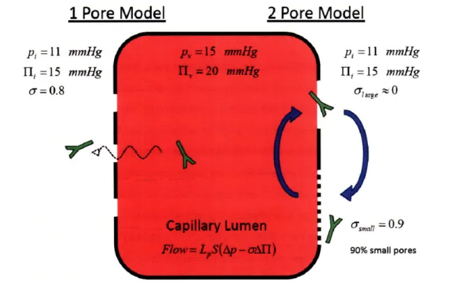
balance between the hydrostatic and osmotic pressure gradients across the vessel wall. Typically, the oncotic pressure is higher inside the capillary due to the high concentration of plasma proteins. In a two-pore model, the large pores in the capillary are not able to maintain this oncotic pressure gradient, so the larger hydrostatic pressure in the vessel forces fluid out of the large pores. This extra fluid is then reabsorbed by the small pores. These smaller pores are more prevalent in number, and the oncotic pressure gradient is greater than the hydrostatic pressure gradient due to the large reflection coefficient and high concentration of plasma proteins in the blood. This volume circulation occurs in the absence of net flow (figure 2.2). The convection across the large pores carries macromolecules with it, which dominates the transport in a two pore model. In this case, diffusion only makes a minor contribution. Convection can therefore occur in the center of a tumor even in the absence of net fluid filtration. However, the Baxter and Jain model uses a one pore model that only treats convection from the net movement of fluid from the vasculature to the interstitium. This results in the overall extravasation rate
in the center of the tumor in the Baxter and Jain model being 0%, 1%, or 10% of what was measured in
the rabbit ear chamber. Their early model may therefore overestimate the contribution of extravasation from fluid flow exiting the vessels (convective extravasation).
1 Pore Model
2 Pore Model
p, = 11 mmHg l, = 15 Olare mmHg S0 70V 0sal = 0.9 90% small poresFigure 2.2 - Models of capillary permeability. Using a single pore model, the larger hydrostatic pressure within the vessel is completely balanced by the higher osmotic pressure inside the capillary. In this case, there is no net flow, and macromolecules must diffuse in and out of the vessel (left). As will be shown later, a lower interstitial pressure near the surface of a tumor may allow convection from the vessels in this location. For a two pore model, the reflection coefficient varies between the large and small pores. The lack of an osmotic pressure gradient over the large pores causes fluid to exit the large pores, carrying macromolecules with it. The large osmotic pressure gradient over the small pores causes the
reabsorption of fluid, yielding no net fluid filtration. For the two pore model, convection over the large pores dominates the transport. Macromolecules cannot easily cross the small pores, resulting in a bias for macromolecules exiting the vessel. The current model assumes diffusive transport (left), where the increased permeability of tumor capillaries lowers the osmotic pressure gradient, and transport is not
biased in either direction.
For the current model, the values measured in the center of the tumor are assumed to be diffusive and occur even in the absence of net fluid flow. In a subsequent model using a dorsal skin chamber(36), Baxter and Jain argued that due to the experimental setup, where measurements are taken near the center of the tumor window that is impermeable to fluid, the permeability values are
primarily diffusive. In addition, the assumptions with pore sizes and osmotic pressure gradients for the two pore model may not be justified given the increased permeability from VEGF signaling and collapse of the osmotic pressure gradient(20). Assuming the permeability measurements are mostly convective inflates the Peclet number 10 to 100-fold (in the case of assuming 10% or 1% of the measured
p, =11 l, =15 o= 0.8 mmHg mmHg
OP
permeability is from diffusion) or an infinite amount when assuming no diffusion occurs. Therefore, diffusion likely dominates for many tumors, as will be discussed in chapter 4.
There are probably cases where net fluid extravasation dictates the uptake in tumors, as described by the Baxter and Jain model. However, given the parameter values above, diffusive
extravasation, or at least convection occurring in the absence of net fluid flow, dominates in the tumor. For the current analysis, the values measured in the center of the dorsal skin chamber are used as the permeability in the absence of net fluid extravasation. It is also assumed that transport is equal in both directions, given its diffusive nature. This final assumption may not be universally valid if the two pore model best describes the situation. Intravasation or extravasation may be biased under certain conditions. For example, although a bias has little effect on high affinity binders that are cleared by internalization, unbound macromolecules may not enter the tumor vasculature as easily due to outward convection across the large pores. In this case, there would be increased nonspecific uptake as unbound antibody takes longer to enter smaller pores or leave the tumor surface, which may be some distance away.
c. High Affinity Antibodies
The transport of antibodies is affected by their binding. As these molecules diffuse between cells, they associate with their particular antigen, immobilizing them on the surface of the cell. This depletes the number of free antibodies and slows penetration into the tumor mass. Antibody affinity is determined by the association rate and dissociation rate to the target. For monovalent interactions, the
Kd is simply:
Kd
2.4
kOn
There are four qualitatively different behaviors of transport and binding, depending on the rate of binding and dissociation. These four scenarios are outlined in figure 2.3, and they will be discussed throughout the chapter. The most common scenario for high affinity binders is a fast-on, slow-off rate which is often enhanced by avidity (multiple binding sites). The two moderate affinity cases are the fast-on, fast-off binders and the slow-fast-on, slow-off case. Finally, for molecules with a slow-on and fast-off