Advance Access publication December 11, 2006 doi:10.1093/molehr/gal105
© The Author 2006. Published by Oxford University Press on behalf of the European Society of Human Reproduction and Embryology. All rights reserved. For
Status of p53 in first-trimester cytotrophoblastic cells
M.Cohen, A.Meisser, L.Haenggeli, I.Irminger-Finger and P.Bischof
1
Department of Obstetrics and Gynaecology, Maternity, Laboratory of Hormonology, University of Geneva, Geneva, Switzerland
1To whom correspondence should be addressed at: Laboratoire d’Hormonologie, 32 Boulevard de la Cluse, 1211 Genève 14, Switzerland.
E-mail: paul.bischof@hcuge.ch
p53 has been called the cellular gatekeeper of the genome because it can induce cell-cycle arrest in G1, apoptosis or affect DNA
replication in response to DNA damage. As p53 has been observed in first-trimester cytotrophoblastic cells (CTB), but its
expres-sion in normal cells is generally not detectable because of its short half-life, p53 could play an important role in cellular
differenti-ation and/or in the control of the invasion of trophoblastic cells; therefore, p53 status was investigated in these cells. Using
different antibodies recognizing different epitopes of p53 protein, abundant p53 expression was observed both in nuclear and in
cytoplasmic compartments of first-trimester CTB. Whereas p53 was detected in the nuclei of few trophoblastic cells with an
anti-body recognizing the N-terminal epitope of the protein, high expression level of p53 in the cytoplasm of CTB was detected with an
antibody recognizing the middle part of p53. The lack of immunoreactivity of p53 with antibodies recognizing the epitopes located
at the N-terminus of p53 and the high level of p53 protein observed in the cytoplasm of CTB suggest that the N-terminus of p53 is
involved in the formation of complexes. These cytoplasmic complexes were detected under non-reducing conditions in western
blot analysis and had apparent molecular weights (MW) of 195, 167 or 125 kDa. These complexes could prolong the half-life of
p53 in the cytoplasm of CTBs. By contrast, in the nuclei of CTBs, p53 seems to be present as a tetramer.
Key words: complex/CTB/cytoplasm/nucleus/p53
Introduction
The transcription factor p53 activates the transcription of its target
genes by binding to a specific consensus DNA sequence consisting of
two copies of a 10-bp DNA motif 5
′-PuPuPuC(A/T)(T/A)GPyPyPy-3′
separated by 0–13 bp (El-Deiry et al., 1992). Wild-type (wt) p53, but
not mutants of p53, can efficiently bind the p53-binding element. By
binding to DNA, p53 activates genes involved in DNA repair, thus
avoiding DNA damage to be carried over to daughter cells. p53 is thus a
tumour suppressor. As stated by Levine (1997), p53 is the ‘gatekeeper’
of the genome. When p53 is mutated or otherwise altered in its
expres-sion, as in many cancers, it looses this tumour-suppressor activity and
becomes oncogenic (Matsumuto et al., 2006). The status of p53 in a cell
is thus directly related to the acquisition of an invasive phenotype. A
previous study suggested that p53 might regulate trophoblast invasion
by up-regulating the expression of matrix metalloproteinase-2 (MMP-2)
(Bian and Sun, 1997), but p53 is known to be a multifunctional protein
that exerts different effects. p53 could also play a role in cellular
differ-entiation in various cell types (Almog and Rotter, 1997).
Immunohistochemical studies of first-trimester trophoblast have
shown that p53 is detectable in the nucleus of cytotrophoblasts and
faintly in syncytiotrophoblasts (Haidacher et al., 1995; Marzusch
et al., 1995; Quenby et al., 1998). Wt p53 is generally not detectable by
immunohistochemistry because of its short half-life; consequently, its
presence in cytotrophoblasts suggests that p53 could be overexpressed
in these cells to control excessive trophoblastic proliferation in normal
placentation (Marzusch et al., 1995). Three hypotheses have already
been presented to account for the accumulation of p53 in cancer cells:
the presence of p53 mutant (Royds and Iacopetta, 2006), the presence
of spliced variants (Wu et al., 1994; Flaman et al., 1996; Courtois
et al., 2002; Yin et al., 2002; Maier et al., 2004; Bourdon et al., 2005)
and the sequestration of p53 in cytoplasm (Aladjem et al., 1998; Zaika
et al., 1999). Another type of regulation of the p53 protein level is its
rate of degradation, which is known to be mediated by the Mdm-2–
ubiquitin–proteasome degradation pathway (Haupt et al., 1997;
Kubbutat et al., 1997). The aim of this study was to understand why
p53 is overexpressed in cytotrophoblastic cells (CTB).
Materials and methods
Reagents
Dulbecco’s modified Eagle’s medium (DMEM), Hanks’ balanced salts and antibiotics mixture (penicillin and streptomycin) were products of Invitrogen (Basel, Switzerland). Fetal bovine serum (FBS) was from Biochrom AG (Oxoid AG, Basel, Switzerland). Complete mini cocktail inhibitor tablets were from Roche (Basel, Switzerland). BIO-RAD protein assay and Trans-Blot transfer medium were from Bio-Rad (Munich, Germany). Hybond-N+ mem-brane, rainbow-stained protein molecular weight (MW) markers and enhanced chemiluminescence (ECL) western blotting detection system were from Amer-sham Biosciences (Buckinghamshire, UK). Goat polyclonal glyceraldehyde-3-phosphatedehydrogenase (GAPDH)-specific antibody was from Santa Cruz (CA, USA). Mouse monoclonal wt p53-specific antibody clone Pab1620, mouse monoclonal p53 antibody clone Pab240, DO-1 and PAb421 were from Oncogene (Stehelin, Basel, Switzerland). Mouse monoclonal vimentin-specific antibody clone V9 and mouse monoclonal cytokeratin 7 clone OV-TL 12/30 were from Dako (Dako Schweiz AG, Baar, Switzerland). ‘Seize primary mammalian immunoprecipitation’ kit was from Pierce (Pierce, Rockford, USA). The proteasome inhibitor MG132 was from Sigma (Sigma-Aldrich, Switzerland).
Immunohistochemistry
First-trimester trophoblasts (n = 10) were obtained from patients undergoing a legal abortion and who gave their written informed consent. Breast cancer
tissue (used as positive control) was obtained as anonymized paraffin sections from our pathology department and used as positives controls. This study was approved by our departmental ethics committee. Tissues were rapidly washed with 0.1 mol/l phosphate buffer at pH 7.4 and fixed for 4–12 h in 4% buffered formalin at 4°C. The specimens were then dehydrated in ethanol and embed-ded in paraffin wax.
Serial sections of tissue were deparaffinized and rehydrated through graded ethanol. Antigen retrieval was performed by microwave pretreatment in 0.01 mol/l citrate buffer (pH 6.0) for 5 min four times, followed by cooling in a cold water bath. Non-specific binding was blocked with 5% (v/v) bovine serum albumin (BSA) in phosphate-buffered saline (PBS) for 20 min at room temper-ature. The sections were incubated with different primary antibodies specific for p53 (dilution 1/10) in 1% BSA–PBS overnight at 4°C. Sections were then washed with PBS (5 min), and p53 was visualized by further incubations with biotinylated anti-mouse secondary antibody (dilution 1/250), alkaline phosphatase-labelled conjugate (dilution 1/2) and Fast Red substrate (Dako, USA). Counterstaining was performed with hemalun.
CTB purification
Placental tissue was obtained from patients undergoing a legal abortion during the first trimester (7–12 weeks of gestation). Informed written consent was obtained from all patients before their inclusion in the study, for which approval was obtained from the local ethics committee. CTBs were isolated from first-trimester placentas and immunopurified (by negative adsorption on immobilized CD45 antibodies) as described elsewhere (Bischof et al., 1995) and grown in DMEM high glucose/F-12 containing 10% FBS and antibiotics (100 U/ml of penicillin and 100 μg/ml of streptomycin) at 37°C in a humidi-fied 5% CO2 atmosphere. Purity of the final cell preparation was evaluated by immunocytochemistry using cytokeratin-7 as a marker of CTBs and vimentin as a marker of non-epithelial cells. Less than 5% of the cells stained for vimen-tin, and 95% were cytokeratin-7-positive. Cells were treated or not with MG132 in serum-free medium for 24 h before lysis. Protein concentration was assayed with the Biorad assay.
Subcellular fractionation
Plated CTBs were rinsed in Hanks’ balanced salts, trypsinized and collected by centrifugation (800 g, 10 min). The pelleted cells were rinsed in ice-cold PBS buffer (0.01 M sodium phosphate, 138 mM NaCl and 2.7 mM KCl, pH 7.4) and collected by centrifugation. The pellets were resuspended in 20 vol-umes of 10 mM Hepes buffer containing 1.5 mM MgCl2, 10 mM NaCl and 0.5 mM dithiothreitol (DTT) and a Roche inhibitor cocktail tablet pH 7.9. Cell suspensions were incubated on ice for 30 min and collected by centrifu-gation. The pellet was resuspended in 10 volumes of 10 mM Hepes contain-ing 1.5 mM MgCl2, 10 mM NaCl and 0.5 mM DTT, 0.5% nonidet P40 and homogenized gently by passing the suspension at least five times through a 20-gauge needle fitted to a syringe. Nuclear fraction was obtained by centri-fugation at 1000 g for 10 min. The supernatant was collected for cytosolic analysis. Nuclei were resuspended by gentle homogenization in 0.88 M sucrose and 3 mM MgCl2 and centrifuged at 2500 g for 20 min to remove cell debris. The pellet was resuspended in PBS buffer and stored at –80°C until use. Assay for the cytoplasmic marker enzyme lactate dehydrogenase (LDH) was performed on each nuclear fraction to determine cytoplasmic contamination (Graham, 1993). No LDH activity was detected in our fraction.
LDH assay
Nuclear fraction (4 μg of protein) was incubated in sodium phosphate buffer, 0.1 M, pH 7, with sodium pyruvate (0.125 mg) and NADH (0.125 mg) for 30 min. The assay measured the rate of NADH absorbance decrease at 340 nm.
Immunoprecipitation
Seize primary mammalian immunoprecipitation kit was used for the elimina-tion of antigen contaminaelimina-tion which could interfere with the detecelimina-tion of p53. It was performed according to the manufacturer’s instructions. The antibodies PAb240, PAb-1620 and DO-1 were used to immunoprecipitate p53 from CTB cell extracts.
Western blot
Proteins were reduced or not (sample buffer containing or not DTT) and dena-tured by boiling at 100°C for 10 min. Samples were then subjected to sodium dodecyl sulphate–polyacrylamide gel electrophoresis (SDS–PAGE) using a 10% running gel. Rainbow-stained MW markers were used as standards. Pro-teins (40 μg) were electro-transferred to nitrocellulose membranes. Non-specific binding was blocked for 30 min at 37°C with 5% powdered milk in 0.2% NP40 buffer. p53-specific antibodies (diluted 1/1000) were incubated over-night with the nitrocellulose membrane. After washing, the membranes were incubated with the appropriate horse-radish peroxidase (HRP)-linked second-ary antibody (2 h, room temperature). After washing, the bands were revealed by chemiluminescence (ECL detection kit). Films were scanned with an Epson Perfection 1 200 Photo scanner, and the surface of the bands was measured by the Kodak 1D Image analysis software (Kodak, Rochester, NY, USA).
Cloning and sequencing of trophoblastic p53
RNA was extracted from CTBs purified from three separate placentas, and RT–PCR was performed. Primers were directed against ATG and Stop codon, amplifying the entire coding region. Amplicons were purified and cloned into pGEM. Inserts were sequenced from three individual clones, and sequences were compared with known human TP53 sequence.
Results
We immunolocalized p53 in first-trimester trophoblast using different
antibodies recognizing different epitopes of the p53 protein (Figure 1).
As shown in Figure 2, the cytoplasm but not the nuclei of villous and
extravillous CTB (Figure 2A) and breast cancer tissue (Figure 2C) is
highly positive for p53 protein when Pab240, specific for a mutated
p53 conformation, and Pab421 (results not shown) antibodies were
used. By contrast, PAb 1620, an antibody specific for the wt
confor-mation of p53 protein, only few nuclei of CTBs were positive (Figure 2B),
whereas in breast cancer specimens used as control, most nuclei and
some cytoplasmic compartments (Figure 2D) were positive.
To verify the status of p53 in CTB, we cloned and sequenced p53
cDNA of CTB. The observed p53 sequence corresponded to the one
published for the wt p53 (results not shown).
To study putative p53 protein isoforms, we immunoblotted cell
lysates and subcellular fractionations of CTB using different
antibod-ies recognizing different epitopes of the p53 protein. As shown in
Figure 3, all antibodies used recognized four main protein bands (84,
64, 53 and 35 kDa) in whole cell lysates. Intensities of the bands
varied with the antibodies.
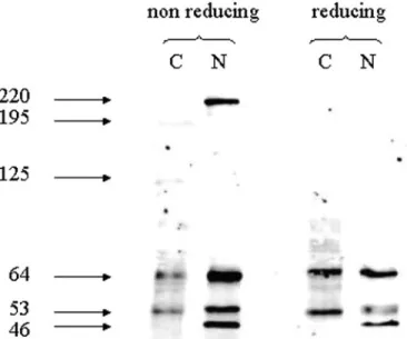
p53 was then immunolocalized with the DO-1 antibody in subcellular
fractionation of CTB. As shown in Figure 4, under reducing conditions,
p53 is present both in cytoplasmic and in nuclear fractions in CTB.
Sim-ilar results were obtained with CTBs from 7, 10 and 11 weeks of
gesta-tion (data not shown). The predominant protein isoforms in the
cytoplasm were 64- and 53-kDa proteins, whereas the most abundant
proteins in the nuclear fraction were 53- and 35-kDa proteins. When
western blots were performed under non-reducing conditions, three
more high-MW p53 isoforms were observed at 125 and 195 kDa in the
cytoplasm and at 220 kDa in the nuclear fraction of CTB.
Immunoprecipitation of p53 from CTB cell extracts with PAb-240,
PAb-1620 and DO-1 and immunoblotting with the same p53
antibod-ies were performed under non-reducing conditions. As shown in
Figure 5, immunoprecipitation of p53 with PAb240 revealed two
complexes of p53 with apparent MWs of 220 and 195 kDa
irrespec-tive of the anti-p53 used to visualize it, whereas only one complex
with an apparent MW at 220 kDa was detected when p53 was
immu-noprecipitated with PAb1620 or DO-1 antibodies. Two minor p53
complexes could be detected when p53 was immunoprecipitated with
PAb240 and immunoblotted with PAb-1620 and PAb-240. These
complexes had apparent MWs of 125 and 167 kDa.
As p53 is overexpressed in CTB, whereas its expression is
gener-ally not detectable because of its short half-life, we next examined the
activity of the p53 degradation pathway in CTBs using a proteasome
inhibitor. As shown in Figure 6, the degradation pathway of p53 is
present and active in CTBs because inhibition of the proteasome
increases the p53 levels.
Discussion
Localization studies of p53 in trophoblasts have so far been performed
with N-terminus-specific antibodies. As described previously (Haidacher
et al., 1995; Marzusch et al., 1995; Quenby et al., 1998), we confirm
here that p53 is immunohistochemically detectable in human
first-trimester trophoblast. However, while staining is found only in a few
nuclei of CTBs with N-terminus-specific p53 antibodies, intense
staining in the cytoplasm of villous and extravillous trophoblast was
obtained, with the PAb240 antibody recognizing an epitope located at
amino acids 213–217 and characteristic of the mutated conformation
of the protein. These observations would suggest that two different
forms of p53 exist in CTB. Furthermore, because antibodies
recogniz-ing the N-terminus of p53 did not localize p53 in the cytoplasm of
tro-phoblasts, but did detect p53 in breast cancer samples, one could
conclude that the N-terminal epitope of cytoplasmic p53 might be
absent or masked in CTB. However, western blot analysis of cellular
extracts of CTB, performed under reducing conditions with four
dif-ferent p53 antibodies recognizing difdif-ferent domains of the protein,
revealed four identical proteins with apparent MWs of 84, 64, 53 and
35. By comparison with a western blot performed on SAOS-2 cells
that do not contain p53 mRNA, background staining in different cells
was observed with different p53 antibodies (pAb240, 246, 1620, 421,
1801 and to a lesser extent Do-1) at
∼80 kDa (Bonsing et al., 1997).
So it is probable that the protein with an apparent MW of 84 kDa
observed in western blot of CTBs is non-specific to p53. Several p53
protein isoforms have been described in different cell types (Wu et al.,
1994; Flaman et al., 1996; Courtois et al., 2002; Yin et al., 2002;
Maier et al., 2004; Bourdon et al., 2005) but never in first-trimester
CTB. All described p53 proteins are truncated at the N- or C-terminus
(Figure 1). Because all antibodies used here recognized a protein with
an apparent MW of 35 kDa, it is unlikely to be an isoform of p53 or a
proteolytic fragment as described by Courtois et al. (2004). A 35-kDa
protein band was detected in three different p53-free yeast strains and
thus considered as non-specific (Nickels et al., 1997). Although in our
hand cloning and sequencing of p53 cDNA from CTB revealed only
wt p53 in these cells, our experimental conditions cannot rule out the
presence of quantitatively less important splice variants.
A p53 protein with an apparent MW of 64 kDa has already been
described as ADP-ribosylated p53 (Wesierska-Gadek et al., 1996).
ADP-ribosylation could modulate the balance between folded and
unfolded species and could stabilize and regulate the biological activity
of p53.
Immunoblotting of subcellular fractionations of CTB revealed that
p53 (both 53- and 64-kDa p53 proteins) can be observed both in
nuc-lear and in cytoplasmic fractions of these cells. Moreover, under
non-reducing conditions, the presence of p53 complexes with an apparent
MW of 220 kDa was found in the nuclei of CTB and might represent
tetramers of p53 because these complexes disappear under reducing
conditions. Tetramerization of p53 seems to be important for the
bind-ing of p53 to the DNA-bindbind-ing elements of its target genes (Weinberg
et al., 2004). Two other complexes were found in the cytoplasmic and
not in the nuclear fractions of CTBs with apparent MWs of 195 and
125 kDa. After immunoprecipitation of p53 under non-reducing
con-ditions, the same complexes were detected in the cellular extracts of
CTB using the PAb240 antibody. Because, under these conditions, no
protein with an apparent MW of 50 kDa was detected, it is suggested
that, in CTB, most p53 protein is complexed. The antibody specific
for the N-terminus of p53 did not recognize the 195-kDa complex and
two minor complexes (167 and 125 kDa), suggesting that the
N-ter-minal epitope of p53 might be masked by the formation of a protein
complex. Other proteins might be involved in these high-MW
com-plexes and could sequester p53 in cytoplasm of CTBs as described in
neuroblastoma cells or in embryonic stems cells (Aladjem et al., 1998;
Zaika et al., 1999). Modifications in the p53 structure (conformational
Figure 1. Structure of wild-type (wt) and published isoforms of p53 (Bourdon et al., 2005) and epitope specificity of p53 antibodies (DO-1, PAb1620, PAb240 and PAb421).W
M
i
b
A
N
D
n
d
i
n
g
d
o
m
a
i
n
O
l
i
g
o
m
e
r
i
s
a
t
i
o
n
n
o
i
t
a
v
i
t
c
a
s
n
a
r
T
3
5
p
t
W
N
C
53kD1
-o
D
3
5
p
β
46kD3
5
p
γ
46kD D k 1 4 D k 5 2 D k 8 4Δ
4
0
p
5
3
Δ
4
0
p
5
3
β
41kDΔ
4
0
p
5
3
γ
Δ
1
3
3
p
5
3
35kDΔ
1
3
3
p
5
3
β
25kD C N E K Q F S T Q D R L D L L M WCYFLINSS C N E K Q F S T Q D R L D L L M WCYFLINSS R L D L L M WCYFLINSS C N E K Q F S T Q DΔ
1
3
3
p
5
3
γ
0
2
6
1
b
A
P
P
A
b
2
4
0
P
A
b
4
2
1
modification or interaction with other proteins) have already been
described during differentiation of cells (Almog and Rotter, 1997).
Because the use of proteasome inhibitors in CTB showed that the
degradation pathway of p53 is active in these cells, one would
specu-late that the observed complexes in the cytoplasm could prolong the
half-life of p53 in the cytoplasm of CTB.
In conclusion, abundant p53 expression was observed both in
nuc-lear and in cytoplasmic compartments of first-trimester CTB. In
nor-mal cells, wt p53 protein is present at very low concentration because
of its rapid degradation mainly directed by MDM2 (Harris and
Levine, 2005). The high expression level of p53 in the cytoplasm of
CTBs and the lack of immunoreactivity of p53 with antibodies
recog-nizing the epitope located at the N-terminus of p53 suggest that, in
CTB, the N-terminus of p53 is involved in the formation of complexes
with apparent MWs of 195, 167 or 125 kDa that are located in the
Figure 2. Immunohistochemistry for p53 expression [A and C clone: PAb240, p53-specific antibody; B and D clone: PAb1620, wild-type (wt) p53-specific anti-body] in first-trimester trophoblast (A, B) and breast cancer tissue (C, D). Magnification ×400.
Figure 3. Western blots of whole cytotrophoblastic (CTB) lysates using DO-1, PAb421, PAb240 and PAb1620 antibodies under reducing conditions.
cytoplasm. These complexes could prolong the half-life of p53 in the
cytoplasm of CTB. By contrast, in the nuclei of CTB, p53 seems to be
present as a tetramer and is recognized by other antibodies than those
recognizing the cytoplasmic form. The overexpression of p53, its
par-ticular localization, and the presence of p53 high-MW complexes in
CTBs suggest that p53 could regulate the invasion of trophoblast.
Indeed, MMP-2 is a target gene for p53 (Bian and Sun, 1997), and this
enzyme is instrumental in trophoblast invasion during implantation
and placentation (Cohen et al., 2006). However, the influence of these
cytoplasmic p53 high-MW complexes is unknown so far.
Acknowledgements
The authors express their gratitude to the Swiss National Science Foundation for their financial support through grants to P.B. and I.I.-F.
References
Aladjem MI, Spike BT, Rodewald LW, Hope TJ, Klemm M, Jaenisch R and Wahl GM (1998) ES cells do not activate p53-dependent stress responses and undergo p53-independent apoptosis in response to DNA damage. Curr Biol 8,145–155.
Almog N and Rotter V (1997) Involvement of p53 in cell differentiation and development. Biochim Biophys Acta 1333,F1–F27.
Bian J and Sun Y (1997) Transcriptional activation by p53 of the human type IV collagenase (gelatinase A or matrix metalloproteinase 2) promoter. Mol Cell Biol 17,6330–6338.
Bischof P, Martelli M, Campana A, Itoh Y, Ogata Y and Nagase H (1995) Importance of matrix metalloproteinases in human trophoblast invasion. Early Pregnancy 1,263–269.
Bonsing BA, Corver WE, Gorsira MC, Van Viet M, Oud PS, Cornelisse CJ and Fleuren GJ (1997) Specificity of seven monoclonal antibodies against p53 evaluated with western blotting, immunohistochemistry, confocal laser scanning microscopy, and flow cytometry. Cytometry 28,11–24.
Bourdon JC, Fernandes K, Murray-Zmijewski F, Liu G, Diot A, Xirodimas DP, Saville MK and Lane DP (2005) p53 isoforms can regulate p53 transcrip-tional activity. Genes Dev 19,2122–2137.
Cohen M, Meisser A and Bischof P (2006) Metalloproteinases and human placental invasiveness. Placenta 27,783–793.
Courtois S, Verhaegh G, North S, Luciani MG, Lassus P, Hibner U, Oren M, Hainaut P (2002) DeltaN-p53, a natural isoform of p53 lacking the first transactivation domain, counteracts growth suppression by wild-type p53. Oncogene 21,6722–6728.
Courtois S, De Fromentel CC and Hainaut P (2004) p53 protein variants: structural and functional similarities with p63 and p73 isoforms. Oncogene 23,631–638. El-Deiry WS, Kern S, Pietenpol JA, Kinzler KW and Vogelstein B (1992)
Definition of a consensus binding site for p53. Nat Genet 1,45–49. Flaman JM, Waridel F, Estreicher A, Vannier A, Limacher JM, Gilbert D, Iggo R
and Frebourg T (1996) The human tumour suppressor gene p53 is alterna-tively spliced in normal cells. Oncogene 12,813–818.
Graham JM (1993) The identification of subcellular fractions from mammalian cells. Methods Mol Biol 19,1–18.
Haidacher S, Blaschitz A, Desoye G and Dohr G (1995) Immunohistochemical evidence of p53 protein in human placenta and choriocarcinoma cell lines. Hum Reprod 10,983–988.
Harris SL and Levine AJ (2005) The p53 pathway: positive and negative feed-back loops. Oncogene 24,2899–2908.
Haupt Y, Maya R, Kazaz A and Oren M (1997) Mdm2 promotes the rapid deg-radation of p53. Nature 387,296–299.
Kubbutat MH, Jones SN and Vousden KH (1997) Regulation of p53 stability by Mdm2. Nature 387,299–303.
Levine AJ (1997) p53, the cellular gatekeeper for growth and division. Cell 88,323–331.
Maier B, Gluba W, Bernier B, Turner T, Mohammad K, Guise T, Sutherland A, Thorner M and Scrable H (2004) Modulation of mammalian life span by the short isoform of p53. Gene Dev 18,306–319.
Marzusch K, Ruck P, Horny HP, Dietl J and Kaiserling E (1995) Expression of the p53 tumour suppressor gene in human placenta: an immunohistochemi-cal study. Placenta 16,101–104.
Matsumuto M, Furihata M and Ohtsuki Y (2006) Posttranslational phos-phorylation of mutant p53 protein in tumor development. Med Mol Morphol 39,79–87.
Nickels A, Bureik M, Montenarh M, Pfreundschuh M and Koch B (1997) p53 antibodies: call for quality. Int J Cancer 73,613–614.
Quenby S, Brazeau C, Drakeley A, Lewis-Jones DI and Vince G (1998) Onco-gene and tumour suppressor Onco-gene products during trophoblast differentiation in the first trimester. Mol Hum Reprod 4,477–481.
Royds JA and Iacopetta B (2006) p53 and disease: when the guardian angel fails. Cell Death Differ 13,1017–1026.
Figure 4. Western blot of cellular fractionations of cytotrophoblastic cells (CTB) performed under non-reducing and reducing conditions and probed with DO-1. C, cytoplasmic fraction; N, nuclear fraction.
Figure 5. Western blot of immunoprecipitated p53 from cytotrophoblastic cells (CTB) using PAb240 (1), PAb1620 (2) and DO-1 (3) under non-reducing conditions. Western blots were probed with PAb240, PAb1620 and DO-1. IP, immunoprecipitated.
Figure 6. Western blots of cytotrophoblastic (CTB) cell lysates treated or not with MG132 were probed with DO-1 anti-p53 antibody and samples run under reducing conditions. Glyceraldehyde-3-phosphatedehydrogenase (GAPDH) control is shown.
Weinberg RL, Freund SM, Veprintsev DB, Bycroft M and Fersht AR (2004) Regulation of DNA binding of p53 by its C-terminal domain. J Mol Biol 342,801–811.
Wesierska-Gadek J, Schmid G and Cerni C (1996) ADP-ribosylation of wild-type p53 in vitro: binding of p53 protein to specific p53 consensus sequence prevents its modification. Biochem Biophys Res Commun 224, 96–102. Wu Y, Liu Y, Lee L, Miner Z and Kulesz-Martin M (1994) Wild-type
alterna-tively spliced p53: binding to DNA and interaction with the major p53 protein in vitro and in cells. EMBO J 13,4823–4830.
Yin Y, Stephen CW, Luciani MG and Fahraeus R (2002) p53 stability and activity is regulated by Mdm2-mediated induction of alternative p53 transla-tion products. Nat Cell Biol 4,462–467.
Zaika A, Marchenko N and Moll UM (1999) Cytoplasmically “Sequestered” wild type p53 protein is resistant to Mdm2-mediated degradation. J Biol Chem 274,27474–27480.
Submitted on October 2, 2006; resubmitted on October 27, 2006; accepted on November 6, 2006
![Figure 2. Immunohistochemistry for p53 expression [A and C clone: PAb240, p53-specific antibody; B and D clone: PAb1620, wild-type (wt) p53-specific anti- anti-body] in first-trimester trophoblast (A, B) and breast cancer tissue (C, D)](https://thumb-eu.123doks.com/thumbv2/123doknet/14908473.657309/4.918.79.800.69.807/figure-immunohistochemistry-expression-specific-antibody-specific-trimester-trophoblast.webp)