Improvement of the human intestinal flora by ingestion of the probiotic strain
Lactobacillus johnsonii La1
Toshihiko Yamano
1, Hisakazu Iino
2, Mamiko Takada
2, Stephanie Blum
3, Florence Rochat
3and
Yoichi Fukushima
1*
1Nestle´ Japan Ltd, Nutrition Business Group, NYK Tennoz Building, 2-2-20, Higashi-Shinagawa, Shinagawa-ku, Tokyo, 140-0002,
Japan
2Showa Women’s University, 1 – 7 Taishidou, Setagayaku, Tokyo, 154-8533, Japan
3Nestle´ Research Centre, Route du Jorat 57, Vers-Chez-Les-Blanc, 1000 Lausanne 26, Switzerland
(Received 30 May 2004 – Revised 6 April 2005 – Accepted 21 April 2005)
To exert beneficial effects for the host, for example, improving the intestinal microflora, a probiotic must reach the intestine as a viable strain. These properties must be demonstrated by in vitro as well as in vivo methods. However, only a few well-designed human clinical studies have shown these properties. Lactobacillus johnsonii La1 has been shown to give many beneficial effects for the host, but it is unclear whether a viable strain of L. john-sonii La1 has the effect of improving host intestinal microflora. In the present study, a randomised double-blind placebo-controlled cross-over trial was conducted to elucidate the effect of L. johnsonii La1 on human intestinal microflora. Twenty-two young healthy Japanese women were randomly divided into two groups, and either received fermented milk with L. johnsonii La1 or a fermented milk without L. johnsonii La1 (placebo) daily for 21 d. Con-sumption of the fermented milk: (a) increased total Bifidobacterium and Lactobacillus, and decreased lecithinase-positive Clostridium in the faeces; (b) increased the faecal lactic acid concentrations; (c) decreased the faecal pH; (d) increased the defecation frequency. These changes were stronger than those observed with the placebo. L. johnsonii La1 was identified in all subjects only after the consumption of the fermented milk. These results suggest that L. johnsonii La1 can contribute to improve intestinal microflora with probiotic properties.
Double-blind placebo-controlled trial: Lactobacillus johnsonii La1: Probiotics: Intestinal microflora: Viable strains
In order to protect against invasion of pathogens and maintain body homeostasis, man has developed body defence systems. Since the intestine is continuously exposed to potential patho-gens (partially taken up with the diet), which can invade the host, natural defence systems have been developed in the intestine, such as mucosal mucins and lysozyme. The intesti-nal microflora also plays an important role to contribute to the overall defence system in the intestine. It is known that germ-free animals have a higher incidence of infection than conventional animals (Collins & Carter, 1978), and intestinal intra-epithelial lymphocytes of axenic animals increase after bacterial colonisation (Imaoka et al. 1996). The intestinal microflora is significantly related to the health and diseases of the host (Benner et al. 1981; Sudo et al. 1997; Cebra et al. 1998); in particular, Bifidobacterium and Lactobacillus contribute to beneficial defensive effects of the host. They exert antimicrobial effects against pathogens by releasing anti-microbial substances (Corthier et al. 1985; Gopal et al. 2001) or prevent adhesion of pathogenic bacteria onto epithelial cells (Chan et al. 1985). They activate natural killer cells (Haller et al. 2000a; Nagao et al. 2000), enhance IgA synthesis (Fukushima et al. 1998; Ibnou-Zekri et al. 2003), modulate cytokine production (Marin et al. 1997; Haller et al. 2000b)
and contribute to the maintenance of body homeostasis (Hata et al. 1996; Kiessling et al. 2002). They also aid nutri-tional absorption (McDonough et al. 1983) and vitamin syn-thesis (Gibson & Roberfroid, 1995). On the other hand, harmful bacteria such as Clostridium perfringens are associ-ated with various diseases and inflammatory responses (Del-neste et al. 1998; Matzinger, 1998; Haller et al. 2000b). Considering these facts, it is quite useful to improve the intes-tinal microflora; that is, to increase the indigenous beneficial bacteria and, by this, decrease harmful bacteria, for both host defence and nutritional benefits.
Recently, probiotic products containing certain strains of Bifidobacterium and Lactobacillus (Fuller, 1989; Lee & Sal-minen, 1995), which are normal inhabitants in the human adult gastrointestinal tract (GIT; Dubos et al. 1965; Lidbeck & Nord, 1993), have been shown to give beneficial effects on the host by improving the intestinal microflora. However, most studies, which show the impact of probiotics on the intestinal microflora, were performed without placebo or were non-blinded. Only a few studies show that either the strain(s) in the product or the product components have an effect to improve the intestinal microflora by means of double-blind placebo-controlled design.
Corresponding author:Dr Yoichi Fukushima, fax þ 81 3 5769 6290, email [email protected]
Abbreviations:cfu, colony-forming units; GIT, gastrointestinal tract; IEC, intestinal epithelial cells; MRS, de Man, Rogosa and Sharpe. qThe Authors 2006
Lactobacillus johnsonii La1 (NCC533; Nestle´ Culture Collection, Lausanne, Switzerland), which formerly belonged to the L. acidophilus group (Johnson et al. 1980; Fujisawa et al. 1992), was selected as a candidate of probiotics under the hypothesis that bacteria with adherence properties on intes-tinal epithelial cells (IEC) would more efficiently strengthen the host defence system than non-adherent strains. Since the intes-tinal epithelium is a target for pathogenic encounter and start site of infection, some well-adherent probiotic strains have the potential to prevent infection by competitive exclusion of pathogens in vitro. In addition, the modulation of the host immune system through the induction of strain-specific signals by L. johnsonii La1 to the IEC has been shown using the IEC – peripheral blood mononuclear cells co-culture model. It was also shown that L. johnsonii La1 had stronger adherence on the human Caco-2 and HT-29 cells than other Lactobacillus strains in vitro (Johnson et al. 1980; Bernet et al. 1994; Gopal et al. 2001). Finally L. johnsonii La1 was also shown to contrib-ute to the reinforcement of the host defence systems. Antimicro-bial activities against pathogens (Bernet-Camard et al. 1997; Blum et al. 1999), anti-pylori effects (Tuomola et al. 1999; Felley et al. 2001) and activation of both natural and specific immunity by the fermented milk containing L. johnsonii La1 have been reported (Link-Amster et al. 1994; Marteau et al. 1997; Donnet-Huges et al. 1999; Michetti et al. 1999). How-ever, the effect of L. johnsonii La1 on the intestinal microflora composition remains unclear.
The following criteria for probiotics have been suggested (Lee & Salminen, 1995; Schiffrin et al. 1995; Salminen et al. 1996): human origin; safe for consumption; adhesive property onto human intestinal epithelium; survival in the GIT, transient colo-nisation of the intestine; production of antimicrobial substances; antagonism against pathogenic bacteria; demonstrated ben-eficial effects on human health. Especially, survival ability in the GIT is considered to be of great importance for exerting pro-biotic effects on the heart. Although L. johnsonii La1 is highly resistant against gastric juice and bile acid in vitro (Prasad et al. 1998), this does not mimic exactly the conditions in situ.
In the present study, a randomised double-blind placebo-controlled cross-over trial was conducted to elucidate the strain-specific effect of L. johnsonii La1 on the intestinal microflora in young healthy Japanese women. We also deter-mined the ability of L. johnsonii La1 to adhere to Caco-2 cells and survive under GIT conditions in vitro.
Material and methods In vitro test
Adhesion test. L. johnsonii La1 and L. acidophilus La3, La4,
La5, La7, La10 and La18 (Nestle´ Culture Collection) were used. L. johnsonii La1 and L. acidophilus La5 and La7 are able to colonise gnotobiotic mice associated with human faecal flora, while other strains (L. acidophilus La3; Benner et al. 1981; Gibson & Roberfriod, 1995; Haller et al. 2000a) do not (, unpublished results). The adherence of bacteria onto Caco-2 cells was examined as described previously (Bernet et al. 1994). Briefly, Caco-2 cell monolayers were prepared on glass coverslips placed in six-well tissue culture plates (Corning Glass Works, Corning, NY, USA). Cells were
seeded at 1·4 £ 104 cells per cm2. Cells were maintained at
378C in a CO2– air atmosphere (10:90, v/v). Lactobacilli
(1 ml; 4 £ 108bacteria/ml) in the bacterial supernatant fraction
or fresh de Man, Rogosa and Sharpe (MRS) broth (Becton, Dickinson and Company, Franklin Lakes, NJ, USA) were added to 1 ml of the cell line culture medium. This suspension (2 ml) was added to each well of the tissue culture plate.
Plates were incubated at 378C in CO2– air (10:90, v/v) for 1 h.
After the incubation, monolayers were washed with sterile PBS, fixed with methanol, stained with Gram stain, and exam-ined with a microscope. Each adhesion assay was conducted in triplicate over three successive passages of intestinal cells. For each monolayer on a glass coverslip, the number of adherent bacteria was evaluated in twenty random microscopic areas. Two different technicians evaluated adhesions to eliminate bias. Resistance against simulated gastric juice
L. johnsonii La1, L. reuteri, L. gasseri, L. amylovorus, L. plan-tarum, Bifidobacterium longum and B. breve (Nestle´ Culture Collection) were used for in vitro GIT transit tolerance test. To simulate gastric juice, 0·3 % (w/v) pepsin in 0·5 % (w/v) sterile
saline at pH 2·0 was prepared and bacteria (1·0 ml; 4 £ 108
colony-forming units (cfu)/ml) were suspended into the simu-lated gastric juice. They were gently mixed and incubated at 378C for 3 h. Samples of 0·1 ml were removed 0, 1 and 3 h after anaerobic incubation and decimal dilution series with saline were prepared. From an appropriate dilution, 50 ml of samples were plated onto MRS agar media. After anaerobic incubation at 378C for 48 h, total viable bacteria were counted. Resistance against bile acid
L. johnsonii La1, L. reuteri, L. gasseri, L. amylovorus, L.
plan-tarum, B. longum and B. breve (1·0 ml; 4 £ 108cfu/ml) were
suspended in 1 % inoculum in MRS broth with 0·1 % bile acid (Becton, Dickinson and Company) at 378C for 15 h. Samples of 0·1 ml were removed 0 and 15 h after anaerobic incubation, and total viable bacteria were counted in the same manner as described earlier.
Resistance against simulated gastrointestinal tract conditions
L. johnsonii La1 (1 ml; 1 £ 107cfu) was incubated with
simu-lated gastric juice for 30 min and 0·1 % bile acid for 60 min. Samples of 0·1 ml were removed before and after the two-step incubation, total viable bacteria were counted in the same manner as described earlier.
Fermented milks
Fermented milks (120 g) contained Streptococcus
thermophi-lus (1 £ 108cfu) with (test) or without (placebo) L. johnsonii
La1 (1 £ 109cfu). The composition of both fermented
pro-ducts was as follows: energy, 427 kJ; protein, 4·6 g; lipids, 3·6 g; carbohydrates, 12·9 g; Na, 60 mg; Ca, 128 mg. Final pH was below 4·45. Their taste and texture were maximally adapted at the factory level.
Subjects and study design
Twenty-two young Japanese women from 20 to 22 years of age were selected as subjects under the declaration of Helsinki.
Informed consent was obtained from all volunteers before starting the study.
The study was conducted with a double-blind
placebo-con-trolled cross-over design shown inFig. 1. Subjects were
ran-domly divided into two groups. Following the observation period without any fermented milks administration for 21 d, either the test or the placebo-fermented milk was admini-strated daily for 21 d from day 1 to day 21. All subjects were requested to stop the consumption of other fermented milks and diets that might have effects on the intestinal micro-flora, such as oligosaccharide or dietary fibre. Antibiotics and cathartics were restricted from being prescribed except at the time of necessity. Subjects recorded a daily questionnaire on the number of defecations, diet, prescription and health remarks during the entire study period.
Faecal flora analysis
Faecal samples were collected at day 2 20, 2 7, 0, 11, 22, 50, 61, and 72. Whole faecal samples were stored anaerobically
(Anaero Packw
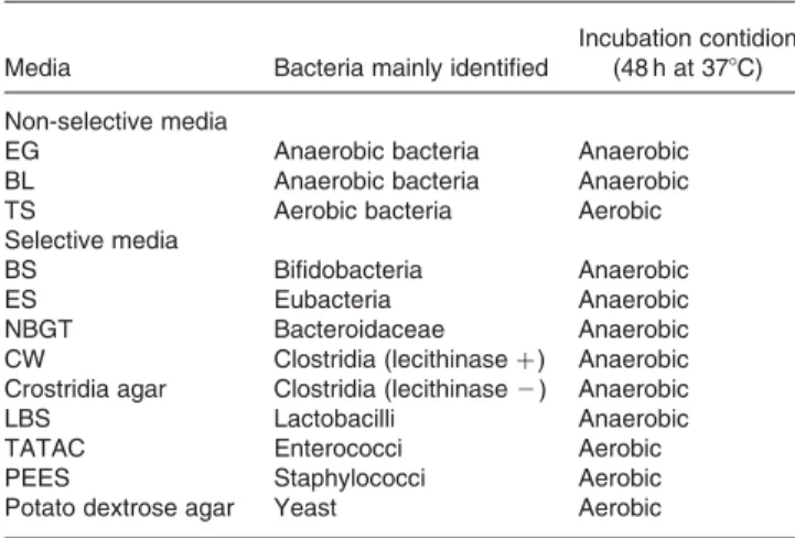
Kenki; Mitsubishi Gas Chemical Company Inc, Tokyo, Japan) at 48C and provided for faecal flora anal-ysis within 24 h after defecation. Decimal dilution series of faecal samples into the saline were prepared. From an appro-priate dilution, 50 ml of samples were plated onto the media and bacteria were analysed according to the Mitsuoka’s method (Mitsuoka et al. 1976). Three non-selective media (Eggerth-Gagnon, blood liver, trypcase sulfite agar media) and nine selective media (bifidobacteria-selective, enhanced selectivity, neomycin brilliant green taurocholate, Clostridium welchii, Clostridia, lactobacilli-selective, triphenyltetrazolium chloride-acridine, potato dextrose, phenylalcohol – egg yolk
suspension agar media) were used (Table1). Bacterial species
were identified by colonisation, Gram staining, morphology, lecithinase reaction, aerobic growth and sporulation. The ratio of bifidobacteria was calculated by dividing the number of bifidobacteria by the number of total bacteria.
Identification of L. johnsonii La1
The Lactobacillus colony isolated from lactobacilli-selective agar media was transferred onto MRS agar media with anti-biotics phosphomycine (0·8 mg/ml; Sigma-Aldrich Co. Ltd, St Louis, MO, USA), sulfamethoxazole (0·93 mg/ml; Wako Pure Chemical Industries Ltd, Osaka, Japan) and
tripetho-prime (0·05 mg/ml; Wako Pure Chemical Industries Ltd), and anaerobically incubated at 378C for 48 h. Grown bacteria
were washed with 1 ml Ringer buffer with 0·5 g L-cysteine
hydrochloride/l (Wako Pure Chemical Industries Ltd) in the Eppendorf tube and centrifuged at 4000 rpm for 5 min. The supernatant fraction was removed and the pellet was frozen at 2 208C until use. For DNA amplification, 1 ml bacterial pellet, 18 ml sterilised water and 5 ml proteinase K (10 mg/ ml; Amersham Biosciences Corp., Piscataway, NJ, USA) were mixed and heated at 558C for 30 min with Gene Amp PCR System 9600-R (Perkin Elmer Co., Ltd, Tokyo, Japan). Heated bacteria (1 ml) were added into the tubes premixed
with 1 ml 50 mM-DNA primer 50
(GGTTGGGTGAGAATTG-CACG)30 (Microsynth GmbH, Balgach, Switzerland), 1·25 ml
10 mM-deoxynucleotide triphosphate (Amersham Biosciences
Corp.), 0·75 ml 50 mM-MgCl2 (Amersham Biosciences
Corp.), 1·25 ml PCR buffer (Invitrogen Co. Ltd, Tokyo, Japan) and 8·25 ml sterilised water, and incubated at 988C for 10 min, then 1·25 ml PCR buffer, 1 U plantinum Taq DNA polymerase (Invitrogen Co. Ltd, Tokyo, Japan) and 11 ml sterilised water were added (reaction mix). Following the initial step of incubation at 948C for 5 min, reaction mix
Fig. 1. Study design. A randomised double-blind placebo-controlled cross-over trial was conducted. After 21 d of the non-intake period, either fermented milk con-taining Lactobacillus johnsonii La1 (1 £ 109colony-forming units/120 g; test fermented milk) or placebo fermented milk was administered daily for 21 d.
Table 1. Media and conditions used for faecal bacteria identification*
Media Bacteria mainly identified
Incubation contidion (48 h at 378C) Non-selective media
EG Anaerobic bacteria Anaerobic
BL Anaerobic bacteria Anaerobic
TS Aerobic bacteria Aerobic
Selective media
BS Bifidobacteria Anaerobic
ES Eubacteria Anaerobic
NBGT Bacteroidaceae Anaerobic
CW Clostridia (lecithinase þ ) Anaerobic
Crostridia agar Clostridia (lecithinase 2 ) Anaerobic
LBS Lactobacilli Anaerobic
TATAC Enterococci Aerobic
PEES Staphylococci Aerobic
Potato dextrose agar Yeast Aerobic
EG, Eggerth-Gagnon; BL, blood liver; TS, trypcase sulfite; BS, bifidobacteria-selec-tive; ES, eubacterium selecbifidobacteria-selec-tive; NBGT, neomycine-brilliant green-taurocholate; CW, Clostridium welchii; LBS, lactobacilli-selective; TATAC, triphenyltetrazolium chloride-acridine; PEES, phenylalcohol– egg yolk suspension.
* Mitsuoka’s method was used for faecal bacteria identification (Mitsuoka et al. 1976).
was provided to the amplification reaction for thirty cycles: denaturation (1 min at 958C), annealing (1 min at 408C), and extension (4 min at 728C). Amplification products were elec-trophoresed at 70 V for 2 h on 0·8 % (w/v) agarose gel with ethidium bromide (0·3 mg /ml) and visualised under UV illumination.
Faecal organic acid concentrations and pH
Faecal samples were homogenised and centrifuged. Obtained supernatant fractions were filtrated through chromatodisc and provided to a SCFA analysis set (Shodex OA; Shouwa-denkou K.K., Tokyo, Japan). Formic acid, acetic acid, lactic acid, propionic acid, butyric acid, isobutyric acid, valeric acid and isovaleric acid were identified.
Faecal pH was measured with a pH meter (Twin; Horiba Ltd, Kyoto, Japan) by touching the pH meter to faeces directly.
Statistic analysis
The results are expressed as mean values and standard deviations.
Data were analysed with software SPSS 11.0 J (SPSS Japan, Tokyo, Japan). Statistical differences between the test and pla-cebo groups, and between pre- and post-administration were examined with the Wilcoxon rank sum test. Statistical differ-ence on the appearance ratio of faecal bacteria was examined
with the x2test. The difference between means was considered
significant at P, 0·05.
Results
Adhesion ability onto Caco-2 cell in vitro
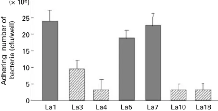
Fig. 2 shows the adhesion ability of L. johnsonii La1, L.
acid-ophilus La3, La4, La5, La7, La10 and La18 onto Caco-2 cells. Bacteria with the property of colonisation (L. johnsonii La1, L. acidophilus La5, La7) showed better adhesion ability onto Caco-2 cells than bacteria with poor colonisation property (L. acidophilus La3, La4, La10, La18). L. johnsonii La1 showed the best adhesion ability among the tested bacteria.
Survival ability in the gastrointestinal tract in vitro
Table2 shows the survival ability of seven strains of
Bifidobac-terium and Lactobacillus in bile acid and the simulated gastric juice. L. johnsonii La1, L. reuteri, L. amylovorus, L. plantarum, and B. breve showed survival ability after 15 h incubation in 0·1 % bile acids. L. johnsonii La1 showed the best survival abil-ity against the simulated gastric juice among the test bacteria and more than 10 % of tested L. johnsonii La1 survived after 3 h incubation. In the two-step incubation with simulated gastric juice for 30 min and bile acid for 60 min, L. johnsonii La1 was
added at 1 £ 107cfu and about 4 £ 106cfu of L. johnsonii La1
showed survival after the incubation. Intestinal microflora
Table3 shows the effect of the fermented milks on the intestinal
microflora. During the test period, the average number of
Bifido-bacterium in the faeces increased from 109·3(SD100·2) cfu/g (day
0) to 109·4(SD100·2) cfu/g (day 11; P, 0·05) and 109·7(SD100·8)
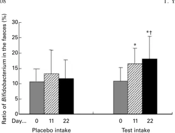
cfu/g (day 22; P, 0·001). The ratio of Bifidobacterium in the
faeces increased from 11·0 (SD4·3) % (day 0) to 16·6 (SD4·9)
% (day 11; P, 0·005) and 18·0 (SD7·4) % (day 22; P, 0·005)
(Fig. 3). Both the number and the ratio of Bifidobacterium at
day 22 were high compared with those during the placebo period (P, 0·001 and P, 0·01, respectively). The average
number of Lactobacillus increased from 105·3(SD 101·1) cfu/g
(day 0) to 106·5(SD101·3) cfu/g (day 11; P, 0·001) and 107·1
(SD100·2) cfu/g (day 22; P, 0·001) and they were high compared
with those during the placebo period (P, 0·005 and P, 0·001, respectively). The average number of lecithinase-positive
Clostridium decreased from 105·5 (SD 101·8) cfu/g (day 0) to
103·2 (SD 100·7) cfu/g (day 22; P, 0·005) and the number at
day 22 was low compared with that during the placebo period (P, 0·05). The appearance ratio of lecithinase-positive Clostri-dium decreased from 55 % (day 0) to 14 % (day 22; P, 0·005). During the placebo period, no significant changes were observed in either number or the ratio of Bifidobacterium and Lactobacillus. The average number of lecithinase-positive
Clostridium decreased from 105·3 (SD 100·7) cfu/g (day 0) to
104·3 (SD 101·4) cfu/g (day 14; P, 0·05), and the appearance
ratio of lecithinase-positive Clostridium decreased from 55 % (day 0) to 14 % (day 11; P, 0·01) and 23 % (day 22; P, 0·05). No significant changes were observed in the count of total bacteria, Bacteroidaceae or Eubacterium during both the test and placebo period.
Fig. 2. Adhesion property of lactobacilli. The number of adhering bacteria onto Caco-2 cell is shown. Lactobacillus johnsonii La1, L. acidophilus La3, La4, La5, La7, La10 and La18 were used. ( ), Bacteria with a property to colonise in gnotobiotic mice associated with human faecal flora; ( ), bacteria with a property to colonise in gnotobiotic mice associated with human faecal flora; cfu, colony-forming units.
Table 2. Survival ability against bile acid (0·1 % for 15 h) and simulated gastric juice
Gastric juice
Strain Bile acid 1 h 3 h
LactobacillusL. Johnsonii La1 þ þ þ þ þ
L. reuteri þ þ 2 L. gasseri 2 þ 2 L. amylovorus þ þ þ þ L. plantarum þ þ 2 BifidobacteriumB. longum 2 þ 2 B. breve þ þ 2
þ , More than 1/1000 of the tested bacteria survived; þ þ, more than 1/10 of the tested bacteria survived; 2 , no bacterial growth.
Table 3 . Effect of fermented milk containing Lactobacillus johnsonii La1 o n faecal flora (Mean v alues a nd standard d eviations) Placebo p eriod (n 22) Test p eriod (n 22) Day 0 Day 11 Day 2 2 D ay 0 D ay 11 Day 2 2 BC BC BC BC BC BC Mean SD AR Mean SD AR Mean SD AR Mean SD AR Mean SD AR Mean SD AR Total 10 ·3 0·3 1 00 10·3 0 ·1 1 00 10 ·3 0·1 100 10·3 0 ·2 1 00 10·3 0 ·1 100 10·4 0·2 100 Total anaerobes 10 ·3 0·3 1 00 10·3 0 ·1 1 00 10 ·3 0·1 100 10·3 0 ·2 1 00 10·3 0 ·1 100 10·4 0·2 100 Total aerobes 8 ·1 0·1 1 00 8 ·2 0 ·4 100 8 ·2 0·3 100 8·2 0 ·3 100 8 ·4 1 ·3 100 8·4 0 ·9 100 Bacteroidaceae 9 ·8 1 ·1 100 9 ·6 0 ·4 1 00 9 ·5 0 ·2 100 9·4 0 ·2 100 9 ·4 0 ·2 100 9·6 0 ·6 100 Bifidobacteria 9 ·3 0 ·4 100 9 ·4 0 ·3 1 00 9 ·3 0 ·2 100 9·3 0 ·2 100 9 ·4* 0 ·2 100 9·7****†††† 0·8 100 Eubacteria 9 ·6 1 ·0 100 9 ·6 0 ·5 1 00 9 ·8 0 ·9 100 9·4 0 ·4 100 9 ·4 0 ·2 100 9·7 0 ·9 100 Clostridia (lecithinase-positive) 5 ·3 0 ·7 55 4 ·3* 1 ·4 14** 5 ·3 1·3 2 3* 5·5 0 ·8 55 5·1 0 ·4 41 10·3***† 0·7 1 4*** Clostridia (lecithinase-negative) 8 ·1 0 ·2 100 8 ·2 0 ·3 1 00 8 ·1 0 ·2 100 7·3 0 ·5 100 7 ·8 1 ·7 100 8·1 100 Lactobacilli 5 ·7 1 ·2 91 6 ·3 1 ·4 86 5 ·3 5 ·3 86 5·3 1 ·1 100 6 ·5****††† 1 ·3 100 7·1****†††† 0·2 100 L. johnsonnii La1 N D 0 ND 0 N D 0 ND 0 6 ·3****†††† 0 ·5 1 00****†††† 6·9****†††† 1·4 1 00****†††† Streptococci 6 ·5 1 ·1 100 7 ·1 0 ·3 1 00 6 ·5** 0·6 100 7·3 1 ·1 100 7 ·2 0 ·4 100 8·2 0 ·9 100 Staphylococci 6 ·6 1 ·2 100 5 ·2**** 0 ·4 9 1 5 ·3**** 0·9 9 5 6 ·1 0 ·3 1 00 5·6 1 ·3 95 7·2* 0·9 9 1 Enterobacteriaceae 7 ·4 0 ·5 100 8 ·0* 0 ·3 1 00 7 ·6 1 ·1 100 7·3 0 ·5 100 7 ·4 0 ·5 100 7·4 0 ·4 100 Bacilli 7 ·8 1 ·1 100 7 ·9 2 ·1 1 00 8 ·1 0 ·2 100 8·1 0 ·2 100 7 ·9 1 ·0 100 7·9 1 ·2 100 Yeasts 3 ·5 1 ·1 64 3 ·7 1 ·3 59 6 ·7 2 ·2 41 3·6 1 ·4 100 4 ·2 1 ·7 4 1 3 ·6 1·2 4 5 BC , bacteria l coun t (log 10 col ony-forming units/g); AR , appea rance rate (%); ND, not detected (, 10 3 colony-forming units/g). Me an val ues w e re sig n ificantly different from those before intake (day 0): *P , 0·05, ** P , 0·01, ** * P , 0 ·005 , *** P , 0 ·00 1. Me an val ues w e re sig n ificantly different b e tween placebo and test per iod: † P , 0· 0 5 , † † P , 0 ·01, ††† P , 0 ·005 , †††† P , 0 ·0 01.
Identification of Lactobacillus johnsonii La1
Fig. 4 shows the changes of L. johnsonii La1 in the faeces. L.
johnsonii La1 was identified in all subjects after the adminis-tration of the test fermented milk only during the test period but not at day 0. L. johnsonii La1 was identified neither during the observation nor placebo period. The number of L.
johnsonii La1 was 106·3 (SD 100·5) cfu/g (day11) and 106·9
(SD 101·4) cfu/g (day 22) during the test period. L. johnsonii
La1 occupied more than 90 % of identified total lactobacillus at day 22. The maximum number of identified L. johnsonii
La1 was 7·5 £ 109cfu/d. More than the ingested number of
L. johnsonii La1 (1 £ 109cfu/d) was identified from nine out
of twenty-two subjects. No more L. johnsonii La1 was ident-ified 4 d after stopping the test fermented milk in all subjects.
Faecal organic acid concentrations and faecal pH
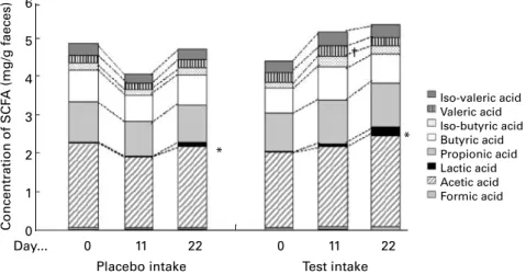
Fig. 5 shows the effect of the fermented milks on faecal lactic
acid and SCFA concentrations. During the test period, lactic
acid increased from 0·031 (SD 0·063) mg/g (day 0) to 0·208
(SD 0·300) mg/g (day 22; P, 0·05) on average. Faecal pH
decreased from 7·66 (SD 0·71) (day 0) to 7·17 (SD0·62) (day
11; P, 0·05) and 7·28 (SD 0·61) (day 22; P, 0·05) (Fig. 6).
During the placebo period, average lactic acid concentration
increased from 0·025 (SD 0·048) mg/g (day 0) to 0·113 (SD
0·234) mg/g (day 22; P, 0·05). No significant changes were observed on faecal pH during the placebo period.
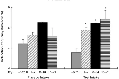
Defecation frequency
Fig. 7 shows the effect of the fermented milks on defecation
frequency in the subjects with mild constipation at less than
seven defecations/week during the observation period.
Twenty out of twenty-two subjects had mild constipation. During the test period, defecation frequency in the subjects
with mild constipation increased from 3·8 (SD 1·4) times/
week (days 2 6 to 0) to 4·9 (SD 1·9) times/week (days 1 – 7;
P, 0·05), to 5·2 (SD 2·8) times/week (days 8 – 14; P, 0·05)
and to 5·7 (SD 2·7) times/week (days 15 – 21; P, 0·05).
During the placebo period, weekly defecation frequency tended to increase but not significantly. No changes were observed in two subjects with normal defecation frequency (more than seven defecations/week during the observation period) during both the test and placebo periods.
Discussion
Since most of the probiotic strains exert their effects in the gut, it is very important for the evaluation of probiotics to show the capability of the given strain to reach the intestine viable and in high numbers. In the in vitro test, we evaluated the capability of L. johnsonii La1 to attach onto Caco-2 cells and survive in simulated GIT conditions. In the adhesion assay using Caco-2 cells, L. johnsonii La1 showed the strongest adhesion among seven other bacteria tested. It was recently shown that the lipoteichoic acid of L. johnsonii La1 and the gangliotri- and gangliotetra-osylceramides (asialo-GM1) of Caco-2 cells are involved in the mediation of adhesion (Gran-ato et al. 1999; Neeser et al. 2000). We also found a positive correlation between adhesion and colonisation. Bacteria with ability to colonise in axenic mice associated with human faecal flora demonstrated a better adhesion property on Caco-2 cells, suggesting that strains with colonisation proper-ties in vitro are likely to exert adhesion in situ. Although there is no direct evidence that probiotic bacteria with adhesion properties in vitro can also attach to the intestinal epithelium in vivo, the fact that L. johnsonii La1 has properties to adhere to other human intestinal cell-lines (29 and HT-29MTX) suggests a potential of L. johnsonii La1 to colonise the human intestine (Bernet et al. 1994; Blum et al. 1999). In the GIT survival test, 0·1 % bile acid is likely in human GIT, pH of human gastric juice is approximately 2·0 and salt content is not less than 0·5 % (w/v). These concentrations of bile acid and gastric juice were used to simulate human GIT conditions. L. johnsonii La1 showed survival in both simulated gastric juice and bile acid. Moreover, L. johnsonii La1 showed the best survival ability among seven tested bacteria, which have been reported to have beneficial effects on health (Mogensen et al. 2002). Although these long-time incubation tests are often used to evaluate the potential of probiotic
Fig. 3. Effect of fermented milk on faecal Bifidobacterium ratio. Data are expressed as mean values (n 22), with standard deviations represented by vertical bars. * The ratio of Bifidobacterium in the faeces was significantly increased compared with day 0 (P, 0·005). † The ratio of Bifidobacterium at day 22 was significantly higher compared with that during the placebo period (P, 0·01).
Fig. 4. Effect of the fermented milks on faecal total Lactobacillus ( ) and Lactobacillus johnsonii La1 ( ). Data are expressed as mean values (n 20), with standard deviations represented by vertical bars. L. johnsonii La1 was identified neither during the observation nor placebo period. * The number of L. johnsonii La1 was significantly higher during the test period compared with that during the placebo period (P, 0·001). cfu, Colony-forming units.
strains (Goldin et al. 1992; Charteris et al. 1998; Prasad et al. 1998), these situations probably do not occur in the body, and no studies have reported the survival ability of bacteria by simulating real human GIT conditions for bacteria (tempera-ture, pH, salts, passage-time and speed, etc). We mimicked human GIT conditions by a two-step incubation with simu-lated gastric juice for 30 min and bile acid for 60 min, and more than 40 % of tested L. johnsonii La1 survived in these conditions.
The presented in vitro data suggest that ingested L. johnso-nii La1 can survive in the GIT and reach the intestine in a viable state. To confirm these results in vivo, a randomised double-blind placebo-controlled cross-over trial was con-ducted. By this the effect of L. johnsonii La1 on the intestinal microflora composition was investigated.
As seen inTable3 andFig. 3, increases in the number and
the ratio of Bifidobacterium in the faeces were observed after the ingestion of the test fermented milk. It is reported that ingestion of the fermented milk containing Bifidobacterium and Lactobacillus increases the indigenous Bifidobacterium (Gilliland et al. 1978; Fukushima et al. 1997; Fuller &
Gibson, 1997; Yaeshima et al. 1997). Since more than 90 % of identified Lactobacillus were L. johnsonii La1 after the test fermented milk ingestion, the increase of Bifidobacterium observed in the present study is possibly due to the intestinal pH decrease induced by lactic acid or other fermented pro-ducts produced by L. johnsonii La1. However, as the volume of lactic acid in the faeces is too small, it is doubtful that such a small volume of lactic acid can decrease the intes-tinal pH. There are bacteria that utilise lactic acid such as Megasphaera spp., Deslfolvibrio spp. and Propionibacterium spp. (Stewart & Bryant, 1988). They might convert lactic acid to other SCFA and induce faecal pH decrease. Although we did not examine the changes in bacterial counts utilising lactic acid in the present study, we did observe a tendency of the other SCFA to increase after ingestion of the test fer-mented milk. L. johnsonii La1 secretes non-bacteriocin anti-microbial substances, which have a broad anti-pathogenic spectrum (Salmonella typhimurium, Listeria monocytogenes, Pseudomonas aeruginosa, etc), but are not effective against Bifidobacteria and Lactobacillus (Bernet-Camard et al. 1997). Although we did not demonstrate the production of antimicrobial substances in vivo, it might be speculated that these antimicrobial substances also contribute to the change in the intestinal environment advantageous for the growth of Bifidobacterium. Intestinal pH decrease inhibits the growth of harmful bacteria (Eklund, 1983). In accordance with this, we observed decreases in the number and the appearance ratio of lecithinase-positive Clostridium, which is associated with carcinogenic enzyme production. It is also known that ingestion of L. johnsonii La1 decreases the carcinogenic enzymes nitroreductase and azoreductase in human subjects (F. Rochat, unpublished results).
SCFA in the intestine, especially butyric acid, are utilised for energy by the intestine and promote peristaltic movements of the intestine (Yokokura et al. 1977; Young & Gibson, 1995). Poor peristalsis causes defecation frequency decrease, so-called constipation. Chronic constipation is related to var-ious diseases such as colon cancer. Thus, it is important to
pre-vent constipation to improve quality of life. As seen inFig. 7,
increase in defecation frequency was observed in the subjects
Fig. 5. Effect of the fermented milk on faecal SCFA concentrations. Data are expressed as mean values (n 22), with standard deviations represented by vertical bars. * Lactic acid significantly increased from day 0 to day 22 during the test period and during the placebo period (P, 0·05). † Valeric acid significantly increased from day 0 to day 11 during the test period (P, 0·05).
8·0 7·5 F aecal pH 7·0 6·5 0 Day... 11
Placebo intake Test intake
22 0
* *
11 22
Fig. 6. Effect of the fermented milk on faecal pH. Data are expressed as mean values (n 22), with standard deviations represented by vertical bars. * Faecal pH was significantly decreased compared with day 0 (P, 0·05).
with mild constipation (less than seven times defecation/week during the observation period) after the ingestion of the test fermented milk. A tendency of increased butyric acid and total organic acid concentration was also observed. Hence, increased organic acids might be utilised for energy by the intestine and promote peristaltic movements, and then increase defecation frequency. These data suggest that the fermented milk containing L. johnsonii La1 has beneficial effects to improve the intestinal microflora and defecation frequency.
During the placebo period, a decrease in the number of lecithinase-positive Clostridium number and tendency of increased defecation frequency were observed. Although the placebo-fermented milk does not contain L. johnsonii La1, it contains lactose and fermented products that have intrinsic effects to improve the intestinal microflora. It is likely that the placebo-fermented milk used in the present study may have impact on the intestinal microflora. However, no changes were observed on Bifidobacterium, Lactobacillus and faecal pH. These findings indicate that the observed improvements of intestinal microflora and defecation frequency by the test fermented milk are apparently stronger than those by the pla-cebo fermented milk. According to the subjects’ question-naires, the amounts of ingested nutrients, such as dietary fibre, did not change so much during the study period and the nutritional components were the same between the test and placebo fermented milk. The only difference between the test and placebo fermented milk is the presence of L. john-sonii La1, suggesting that L. johnjohn-sonii La1 has an essential role for intestinal microflora improvement.
In order to exert the beneficial effects on health, it is import-ant for probiotics to access the intestine as a viable strain, i.e. survive the human GIT, but no reports have shown the survi-val ability of L. johnsonii La1 in human GIT using a strain-specific DNA primer for L. johnsonii La1. In the present study, we examined the survival ability of L. johnsonii La1 in the human GIT by identifying L. johnsonii La1 in the faeces with a RAPD-PCR method (Welsh & McClelland, 1991; Johansson et al. 1995) using a specific primer for L. johnsonii La1 (Ventura & Zink, 2002). L. johnsonii La1 was
identified in all subjects who ingested the test fermented milk only during the test period. More than the ingested
number of L. johnsonii La1 (1 £ 109cfu/d) was identified
from nine out of twenty-two subjects. L. johnsonii La1 was identified within 3 d after stopping the test fermented milk. Since the turnover cycle of IEC is 3 – 4 d, colonised L. johnso-nii La1 in the intestine might disappear followed by the IEC turnover. These results suggest that L. johnsonii La1 has an ability to reach the intestine as a viable bacterium, and to pro-liferate and colonise temporarily in the intestine. The fact that L. johnsonii La1 disappeared within 4 d after stopping the test fermented milk is also important to show the safety of L. john-sonii La1. Thus, the probiotic properties of L. johnjohn-sonii La1 to reach the intestine as a viable strain and colonise in the intes-tine seems to work favourably in the improvement of the intestinal microflora.
In conclusion, the strain-specific effect of L. johnsonii La1 to improve the intestinal microflora as a viable strain was elu-cidated in the present study. L. johnsonii La1 is of human origin, safe for consumption (Shu et al. 1999), adheres to human intestinal cell lines, survives in the human GIT and colonises temporarily in the intestine, produces antimicrobial substances, antagonises the pathogenic bacteria, and has ben-eficial effects on human health. Therefore, L. johnsonii La1 can be a useful tool to promote human health as a probiotic.
References
Benner R, van Oudenaren A, Haaijman JJ, Slingerland-Teunissen J, Wostmann BS & Hijmans W (1981) Regulation of the ‘spon-taneous’ (background) immunoglobulin synthesis. Int Arch Allergy Appl Immunol 66, 404 – 415.
Bernet MF, Brassart D, Neeser JR & Servin AL (1994) Lactobacillus acidophilus LA 1 binds to cultured human intestinal cell lines and inhibits cell attachment and cell invasion by enterovirulent bac-teria. Gut 35, 483 – 489.
Bernet-Camard MF, Lievin V, Brassart D, Neeser JR, Servin AL & Hudault S (1997) The human Lactobacillus acidophilus strain La1 secretes a nonbacteriocin antibacterial substance(s) active in vitro and in vivo. Appl Environ Microbiol 63, 2747 – 2753.
Fig. 7. Effect of the fermented milks on the defecation frequency in the subjects with mild constipation (less than seven defecations/week during the observation period). Data are expressed as mean values (n 20), with standard deviations represented by vertical bars. * Weekly defecation frequency in the subjects with mild constipation was significantly higher compared with at days 2 6 to 0 (P, 0·05).
Blum S, Reniero R, Schiffrin EJ, et al. (1999) Adhesion studies for probiotics: need for validation and refinement. Trends Food Sci Technol 10, 405 – 410.
Cebra JJ, Periwal SB, Lee G, Lee F & Shroff KE (1998) Development and maintenance of the gut-associated lymphoid tissue (GALT): the roles of enteric bacteria and viruses. Dev Immunol 6, 13 – 18. Chan RCY, Reid G, Irvin RT, Bruce AW & Costerton JW (1985)
Competitive exclusion of uropathogens from human uroepithelial cells by Lactobacillus whole cells and cell wall fragments. Infect Immun 47, 84 – 89.
Charteris WP, Kelly PM, Morelli L & Collins JK (1998) Develop-ment and application of an in vitro methodology to determine the transit tolerance of potentially probiotic Lactobacillus and Bifi-dobacterium in the upper human gastrointestinal tract. J Appl Microbiol 84, 759 – 768.
Collins FM & Carter PB (1978) Growth of Salmonellae in orally infected germ-free mice. Infect Immun 21, 41 – 47.
Collins JK, Thornton G & Sullivan GO (1998) Selection of probiotics strains for human application. Int Dairy J 8, 487 – 490.
Corthier G, Dubos F & Raibaud P (1985) Modulation of cytotoxin production by Clostridium difficile in the intestinal tracts of gnoto-biotic mice inoculated with various human intestinal bacteria. Appl Environ Microbiol 49, 250 – 252.
Delneste Y, Donnet-Hughes A & Schiffrin EJ (1998) Functional foods: mechanisms of action on immunocompetent cells. Nutr Rev 156, S93 – S98.
Donnet-Huges A, Rochat F, Serrant P, Aeschlimann JM & Shiffrin EJ (1999) Modulation of nonspecific mechanisms of defense by lactic acid bacteria: effective dose. J Dairy Sci 82, 863 – 869.
Dubos R, Schaedler RW, Costello R & Hoet P (1965) Indigenous, normal and autochthonous flora of the gastrointestinal tract. J Exp Med 122, 67 – 76.
Eklund T (1983) The antimicrobial effect of dissociated and undisso-ciated sorbic acid at different pH levels. J Appl Bacteriol 54, 383 – 389.
Felley CP, Corthesy-Theulaz I, Rivero JL, et al. (2001) Favourable effect of an acidified milk (LC-1) on Helicobacter pylori gastritis in man. Eur J Gastroenterol Hepatol 13, 25 – 29.
Fujisawa T, Benno Y, Yaeshima T & Mitsuoka T (1992) Taxonomic study of the Lactobacillus acidophilus group, with recognition of Lactobacillus gallinarum sp. nov. and Lactobacillus johnsonii sp.nov. and synonymy of Lactobacillus acidophilus group A3 (Johnson et al. 1980) with the type strain of Lactobacillus amylo-vorus (Nakamura 1981). Int J Syst Bacteriol 42, 487 – 491. Fukushima Y, Kawata Y, Hara H, Terada A & Mitsuoka T (1998)
Effect of probiotic formula on intestinal immunoglobulin A pro-duction in healthy children. Int J Food Microbiol 42, 39 – 44. Fukushima Y, Li Shou-Tou Hara H, Terada A & MitsuokaT (1997)
Effect of follow-up formula containing bifidobacteria (NAN BF) on fecal flora and fecal metabolites in healthy children. Biosci Microflora 16, 65 – 72.
Fuller R (1989) Probiotics in man and animals. J Appl Bacteriol 66, 365 – 378.
Fuller R & Gibson GR (1997) Modification of the intestinal micro-flora using probiotics and prebiotics. Scand J Gastroenterol 222, Suppl., 28 – 31.
Gibson GR & Roberfroid MB (1995) Dietary modulation of the human colonic microbiota: introducing the concept of prebiotics. J Nutr 125, 1401 – 1412.
Gilliland SE, Speck ML, Nauyok GF Jr & Giesbrecht FG (1978) Influence of consuming fermented milk containing Lactobacillus acidophilus on fecal flora of healthy males. J Dairy Sci 61, 1 – 10. Goldin B, Gorbach S, Saxelin M, Barakat S, Gualtieri L & Salminen S (1992) Survival of Lactobacillus GG in human gastrointestinal tract. Dig Dis Sci 37, 121 – 128.
Gopal PK, Prasad J, Smart J & Gill HS (2001) In vitro adherence properties of Lactobacillus rhamnosus DR20 and Bifidobacterium
lactis DR10 strains and their antagonistic activity against an enterotoxigenic Escherichia coli. Int J Food Microbiol 67, 207 – 216.
Granato D, Perotti F, Masserey I, Rouvet M, Golliard M, Servin A & Brassart D (1999) Cell surface-associated lipoteichoic asid acts as an adhesion factor for attachment of Lactobacillus johnsonii La1 to human enterocyte-like Caco-2 cells. Appl Environ Microbiol 65, 1071 – 1077.
Haller D, Blum S, Bode C, Hammes WP & Shiffrin EJ (2000a) Acti-vation of human peripheral blood mononuclear cells by nonpatho-genic bacteria in vitro: evidence of NK cells as primary targets. Infect Immun 68, 752 – 759.
Haller D, Bode C, Hammes WP, Pfeifer A, Schiffrin EJ & Blum S (2000b) Non-pathogenic bacteria elicit a differential cytokine response by intestinal epithelial cell/leucocyte co-cultures. Gut 47, 79 – 87.
Hata Y, Yamamoto M, Ohni M, Nakajima K, Nakamura Y & Takano T (1996) A placebo-controlled study of the effect of sour milk on blood pressure in hypertensive subjects. Am J Clin Nutr 64, 767 – 771.
Ibnou-Zekri N, Blum S, Schiffrin EJ & von der Weid T (2003) Diver-gent patterns of colonization and immune response elicited from two intestinal Lactobacillus strains that display similar properties in vitro. Infect Immun 71, 428 – 436.
Imaoka A, Matsumoto S, Setoyama H, Okada Y & Umesaki Y (1996) Proliferative recruitment of intestinal intraepithelial lymphocytes after microbial colonization of germ-free mice. Eur J Immunol 26, 945 – 948.
Johansson ML, Quednau M, Molin G & Ahrne S (1995) Randomly amplified polymorphic DNA (RAPD) for rapid typing of Lacto-bacillus plantrum strains. Lett Appl Microbiol 21, 155 – 159. Johnson JL, Phelps CF, Cummins CS, London J & Gasser F (1980)
Taxonomy of Lactobacillus acidophilus group. Int J Syst Bacteriol 30, 53 – 68.
Kiessling G, Schneider J & Jahreis G (2002) Long-term consumption of fermented dairy products over 6 months increases HDL choles-terol. Eur J Clin Nutr 56, 843 – 849.
Lee YK & Salminen S (1995) The coming age of probiotics. Trends Food Sci Technol 6, 241 – 245.
Lidbeck A & Nord CE (1993) Lactobacilli and the normal human anaerobic microflora. Clin Infect Dis 16, S181 – S187.
Link-Amster H, Rochat F, Saudan KY, Mignot O & Aeschlimann JM (1994) Modulation of a specific humoral immune response and changes in intestinal flora mediated through fermented milk intake. FEMS Immunol Med Microbiol 10, 55 – 64.
McDonough F, Wells P, Wong N, Hitchins A & Bodewell C (1983) Role of vitamins and minerals in growth stimulation of rats fed with yogurt. Fed Proceed 42, 556 – 558.
Marin ML, Lee JH, Murtha J, Ustunol Z & Pestka JJ (1997) Differ-ential cytokine production in clonal macrophage and T-cell lines cultured with bifidobacteria. J Dairy Sci 80, 2713 – 2720. Marteau P, Vaerman JP, Dehennin JP, Bord S, Brassart D, Pochat P,
Desjeux JP & Rambaud JC (1997) Effects of intrajejunal perfusion and chronic ingestion of Lactobacillus johnsonii strain La1 on serum concentrations and jejunal secretions of immunoglobulins and serum proteins in healthy humans. Gastroenterol Clin Bikol 21, 293 – 298.
Matzinger P (1998) An innate sense of danger. Semin Immunol 10, 399 – 415.
Michetti P, Dorta G, Wiesel PH, et al. (1999) Effect of whey-based culture supernatant of Lactobacillus acidophilus (johnsonii) La1 on Helicobacter pylori infection in humans. Digestion 60, 203 – 209.
Mitsuoka T, Ohno K, Benno Y, Suzuki K & Nanba K (1976) The fecal flora of man. Communication of newly developed method with the old conventional method for the analysis of intestinal flora. Zentral Bakteriol Hyg Abt Orig A 234, 219 – 233.
Mogensen G, Salminen S, O’Brien J, et al. (2002) Food microorgan-isms – health benefits, safety evaluations and strains with docu-mented history of use in foods. IDF Bull 377, 10 – 19.
Nagao F, Nakayama M, Muto T & Okumura K (2000) Effects of fer-mented milk drink containing Lactobacillus casei strain Shirota on the immune system in healthy human subjects. Biosci Biotechnol Biochem 64, 2706 – 2708.
Neeser JR, Granato D, Rouvet M, Servin A, Teneberg S & Karlsson EA (2000) Lactobacillus johnsonii La1 shares carbohydrate-bind-ing specificities with several enteropathogenic bacteria. Glycobiol-ogy 10, 1193 – 1199.
Prasad J, Gill H, Smart J & Gopal PK (1998) Selection and charac-terization of Lactobacillus and Bifidobacterium strains for use as probiotics source. Int Dairy J 8, 993 – 1002.
Salminen S, Laine M, Von Wright A, Vuopio-Varkila J, Korhonen T & Mattila-Sandholm T (1996) Development of selection criteria for probiotics strains to assess their potential in functional foods. A Nordic and European approach. Biosci Microflora 15, 61 – 67.
Schiffrin EJ, Rochat F, Link-Amster H, Aeschlimann JM & Donnet-Huighes A (1995) Immunomodulation of human blood cells fol-lowing the ingestion of lactic acid bacteria. J Dairy Sci 78, 491 – 497.
Shu Q, Zhou JS, Rutherfurd KJ, Birtles MJ, Prasad J, Gopal PK & Gill HS (1999) Probiotic lactic acid bacteria (Lactobacillus acido-philus HN017, Lactobacillus rhamnosus HN001and Bifidobacter-ium lactis HN019) have no effects on the health of mice. Int Dairy J 9, 831 – 836.
Stewart CS & Bryant MP (1988) The rumen bacteria. In The Rumen Microbial Ecosystem, pp. 21 – 75 [PN Hobson, editor]. London: Elsevier Science.
Sudo N, Sawamura S, Tanaka K, Aiba Y, Kudo C & Koga Y (1997) The requirement of intestinal bacterial flora for the development of an IgE production system fully susceptible to oral tolerance induc-tion. J Immunol 159, 1739 – 1745.
Tuomola EM, Ouwehand AC & Salminen SJ (1999) The effect of probiotic bacteria on the adhesion of pathogens to human intestinal mucus. FEMS Immunol Med Microbiol 26, 137 – 142.
Ventura M & Zink R (2002) Specific identification and molecular typing analysis of Lactobacillus johnsonii by using PCR-based method and pulsed field gel electrophoresis. FEMS Microbiol Lett 217, 141 – 154.
Welsh J & McClelland M (1991) Genomic fingerprinting using arbi-trarily primed PCR and a matrix of pairwise combinations of pri-mers. Nucleic Acids Res 19, 5275 – 5279.
Yaeshima T, Takahashi S, Matsumoto N, Ishibashi N, Hayasawa H & Iino H (1997) Effect of yogurt containing Bifidobacterium BB536 on the intestinal environment, fecal characteristics and defecation frequency: a comparison with standard yogurt. Biosci Microflora 16, 73 – 77.
Yokokura T, Yajima T & Hashimoto S (1977) Effect of organic acid on gastrointestinal motility of rat in vitro. Life Sci 21, 59 – 62. Young GP & Gibson PR (1995) Butyrate and the human cancer cell.
In Physiological and Clinical Aspects of Short-Chain Fatty Acids, pp. 319 – 335 [JH Cummings, JL Rombeau and T Sakata, editors]. Cambridge, UK: Cambridge University Press.