Images in cardio-thoracic surgery
Scimitar syndrome in association with intrapulmonary sequestration
Michel Gonzalez
a,*
, Pierre Bize
b, Hans-Beat Ris
a, Thorsten Krueger
aa
Service of Thoracic and Vascular Surgery, Centre Hospitalier Universitaire Vaudois Lausanne, Switzerland bDepartment of Radiology, Centre Hospitalier Universitaire Vaudois Lausanne, Switzerland
Received 3 September 2010; received in revised form 9 November 2010; accepted 11 November 2010; Available online 11 January 2011
Keywords: Pulmonary sequestration; Scimitar syndrome
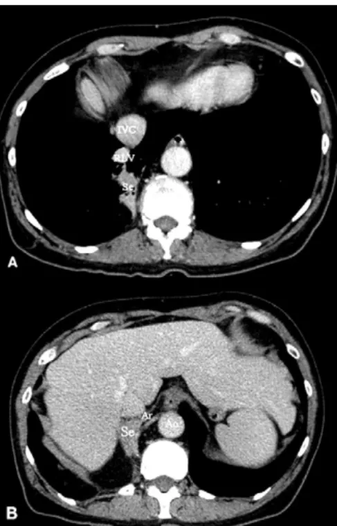
A 62-year-old woman was referred for repetitive
hemop-tysis. Chest computed tomography scan revealed
intrapul-monary sequestration of the right lower lobe with an
aberrant artery arising from the sus-diaphragmatic
descend-ing aorta (
Fig. 1
(A) and (B)). In addition, the right lower lobe
showed an anomalous venous drainage directly in the inferior
vena cava (Scimitar syndrome) (
Fig. 2
(A) and (B)) (
3D video
).
Appendix A. Supplementary data
Supplementary data associated with this article can be found, in the online version, atdoi:10.1016/j.ejcts.2010.11.033.
www.elsevier.com/locate/ejcts
European Journal of Cardio-thoracic Surgery 40 (2011) 273
[()TD$FIG]
Fig. 1. (A) Chest computed tomography scan showed intrapulmonary seques-tration (Se) of the right lower lobe. The right lower vein (RLV) is anterior to the sequestration with drainage directly in the inferior vena cava. (B) Vascular-isation of the sequestration by an aberrant artery (Ar) arising from the sus-diaphragmatic descending aorta (Ao).
[()TD$FIG]
Fig. 2. (A) Three dimensional reconstructed computed tomography image showing the venous drainage of the right lower lobe directly in the inferior vena cava (white arrow). (B) Intra-operative view with aberrant artery of the intrapulmonary sequestration (Ar) and venous drainage of the right lower lobe (RLV) directly in the inferior vena cava (IVC) just above the diaphragm. The patient underwent right lower lobectomy with uneventful recovery.
* Corresponding author. Address: Thoracic and Vascular Surgery Service, Centre Hospitalier Universitaire Vaudois, Rue du Bugnon 46, 1011-Lausanne, Switzerland. Tel.: +41 79 556 38 20; fax: +41 21 314 23 58.
E-mail address:[email protected](M. Gonzalez).
1010-7940/$ — see front matter # 2010 European Association for Cardio-Thoracic Surgery. Published by Elsevier B.V. All rights reserved. doi:10.1016/j.ejcts.2010.11.033