M A J O R A R T I C L E
Sustained Domestic Vector Exposure Is
Associated With Increased Chagas
Cardiomyopathy Risk but Decreased Parasitemia
and Congenital Transmission Risk Among
Young Women in Bolivia
Michelle Kaplinski,1,aMalasa Jois,2,aGerson Galdos-Cardenas,3,4Victoria R. Rendell,5Vishal Shah,6Rose Q. Do,7 Rachel Marcus,8Melissa S. Burroughs Pena,9Maria del Carmen Abastoflor,10Carlos LaFuente,10Ricardo Bozo,11 Edward Valencia,12Manuela Verastegui,12Rony Colanzi,4Robert H. Gilman,3and Caryn Bern13; for the Working Group on Chagas Disease in Bolivia and Peru
1Department of Cardiology, Children’s Hospital of Philadelphia, Pennsylvania;2Division of Internal Medicine, Brown University, Providence, Rhode Island; 3Department of International Health, Johns Hopkins Bloomberg School of Public Health, Baltimore, Maryland;4Universidad Católica Boliviana, Santa Cruz,
Plurinational State of Bolivia;5Duke Global Health Institute, Duke University, Durham, North Carolina;6Saint Louis University School of Medicine,
Missouri;7Department of Cardiology, University of Colorado, Denver;8Department of Cardiology, Washington Hospital Center, Washington, District of
Columbia;9Department of Cardiology, Duke University, Durham, North Carolina;10Hospital Japonés, Santa Cruz, and11Camiri Municipal Hospital, Camiri,
Plurinational State of Bolivia;12Laboratorio de Investigación en Enfermedades Infecciosas, Universidad Peruana Cayetano Heredia, Lima, Peru; and 13Department of Epidemiology and Biostatistics, University of California, San Francisco School of Medicine
Background. We studied women and their infants to evaluate risk factors for congenital transmission and cardiomyopathy inTrypanosoma cruzi–infected women.
Methods. Women provided data and blood for serology and quantitative polymerase chain reaction (PCR). Infants of infected women had blood tested at 0 and 1 month by microscopy, PCR and immunoblot, and serology at 6 and 9 months. Women underwent electrocardiography (ECG).
Results. Of 1696 women, 456 (26.9%) were infected; 31 (6.8%) transmitted T. cruzi to their infants. Women who transmitted had higher parasite loads than those who did not (median, 62.0 [interquartile range {IQR}, 25.8–204.8] vs 0.05 [IQR, 0–29.6]; P < .0001). Transmission was higher in twin than in singleton births (27.3% vs 6.4%; P = .04). Women who had not lived in infested houses transmitted more frequently (9.7% vs 4.6%;P = .04), were more likely to have positive results by PCR (65.5% vs 33.9%;P < .001), and had higher parasite loads than those who had lived in infested houses (median, 25.8 [IQR, 0–64.1] vs 0 [IQR, 0–12.3]; P < .001). Of 302 infected women, 28 (9.3%) had ECG abnormalities consistent with Chagas cardiomyopathy; risk was higher for older women (odds ratio [OR], 1.06 [95% confidence interval {CI}, 1.01–1.12] per year) and those with vector exposure (OR, 3.7 [95% CI, 1.4–10.2]). We observed a strong dose-response relationship between ECG abnormalities and reported years of living in an infested house.
Conclusions. We hypothesize that repeated vector-borne infection sustains antigen exposure and the consequent in-flammatory response at a higher chronic level, increasing cardiac morbidity, but possibly enabling exposed women to control parasitemia in the face of pregnancy-induced Th2 polarization.
Keywords. Chagas disease; Trypanosoma cruzi; infectious disease transmission; vertical; cardiomyopathy. In the Americas,Trypanosoma cruzi causes the highest
disease burden of any parasite, accounting for 7 times
more disability-adjusted life years lost than malaria [1]. An estimated 6 million people are infected, of whom 20%–30% will develop potentially life-threatening Received 14 January 2015; accepted 2 June 2015; electronically published 9
June 2015.
a
M. K. and M. J. contributed equally to this work.
Correspondence: Caryn Bern, MD, MPH, Department of Epidemiology and Biosta-tistics, University of California, San Francisco, 550 16th St, San Francisco, CA 94158 (caryn.bern2@ucsf.edu).
Clinical Infectious Diseases® 2015;61(6):918–26
© The Author 2015. Published by Oxford University Press on behalf of the Infectious Diseases Society of America. All rights reserved. For Permissions, please e-mail: journals.permissions@oup.com.
heart disease [2]. Chagas cardiomyopathy is characterized by a chronic inflammatory process [3]. The earliest signs are usually conduction system abnormalities, followed by atrio-ventricular blocks, atrio-ventricular arrhythmias, sinus node dysfunction, and progressive dilated cardiomyopathy [4,5]. The determinants of progression to cardiomyopathy are not well understood, but may include repeatedT. cruzi superinfec-tion, parasite virulence and tissue tropism, and host immune response [3].
Although vector exposure remains the most frequent infec-tion route,T. cruzi can also be transmitted by transfusion or transplantation, and from mother to child [6]. The Southern Cone Initiative has made major advances in blood screening and control of the principal vector,Triatoma infestans [7]. With the decrease in other routes, congenital transmission has become proportionately more important, accounting for an estimated 22% of new infections in 2010 [2]. In meta-analyses, the median transmission rate fromT. cruzi–infected women is estimated at 5% [8], but rates vary widely, from 1% to >15% across study populations [9–12].
From 2010 to 2013, we conducted a study of women and their infants in 2 sites in Santa Cruz Department, Bolivia. The ability to study congenitalT. cruzi transmission and cardiac status in the same women enabled us to examine the epidemi-ology of these 2 phenomena simultaneously. Our aims were to evaluate the incidence of vertical transmission and prevalence of early cardiomyopathy inT. cruzi–infected women, and to assess risk factors for both of these outcomes.
MATERIALS AND METHODS Ethics Statement
The protocol was approved by the institutional review boards of the Johns Hopkins Bloomberg School of Public Health, Hospi-tal Universitario Japonés, Universidad Católica Boliviana, Universidad Peruana Cayetano Heredia, Asociación Benéfica Proyectos en Informatica, Salud, Medicina y Agricultura, and the Centers for Disease Control and Prevention. Each woman provided written informed consent for herself and her infant. Study Population and Data and Specimen Collection
The study was conducted in Hospital Universitario Japonés in urban Santa Cruz (city population 1.7 million) and Hospital Municipal Camiri in Camiri (city population 30 191) (www. ine.gob.bo) [13,14]. Both hospitals are referral centers with a large proportion of cesarean deliveries. Although vector-borne T. cruzi transmission is absent in urbanized areas, both cities receive migrants from rural areas with intense transmission. Ca-miri is the capital of Cordillera province ( population 111 171) in the Bolivian Chaco and located a few miles from villages where 50%–100% of houses are heavily infested, with rapid
reinfestation after insecticide application [14,15]. The estimated nationalT. cruzi prevalence is 6% [2], but in rural areas of the Chaco, the all-age prevalence is 50%, and adult infection prev-alence reaches≥80% [14,16].
Trained study nurses enrolled women presenting for delivery and collected data, including a record of all houses lived in throughout the woman’s life, duration of residence, construc-tion materials, and observed vector infestaconstruc-tion. The study in-strument was developed and validated in earlier studies in Santa Cruz [13,17]; 2 nurses involved in the earlier validation supervised the interviews. Blood was collected, centrifuged, and immediately screened by 2 rapid tests,Trypanosoma Detect (InBios, Seattle, Washington) and Polychaco indirect hemag-glutination assay (IHA) at a single dilution of 1:16 (Lemos Laboratories, Santiago del Estero, Argentina). Sera were later tested in Santa Cruz by IHA with multiple dilutions and Cha-gatest lysate enzyme-linked immunosorbent assay (ELISA), with Recombinante 3.0 ELISA as a tie-breaker (both from Wie-ner Laboratories, Rosario, Argentina). A confirmed infection required positive results by≥2 conventional tests [18]. For infected women, a standard 12-lead electrocardiogram (ECG) was performed during a follow-up visit.
Management of CongenitalT. cruzi Infection
A study nurse attended the delivery of each rapid test–positive woman to collect cord blood. Infants of infected women had follow-up blood collection at 1, 6, and 9 months of age. Cord blood and 1-month specimens were examined by micromethod, the standard technique to diagnose congenitalT. cruzi infection in thefirst months of life in most Latin American healthcare fa-cilities [19]. Blood is collected in 4–6 heparinized microhema-tocrit tubes, sealed, and processed within 24 hours by centrifugation (12 000 rpm for 7 minutes) followed by micros-copy. Six- and 9-month specimens were centrifuged, and serum samples were tested by immunoglobulin G assays as described above for maternal testing.
Western blots were performed in our Lima laboratory to detect immunoglobulin M (IgM) to trypomastigote excreted-secreted antigens (IgM TESA-blot) in infant specimens follow-ing published methods [20]. Blots with clear bands at the expected molecular weights were considered positive; blots with weak or ambiguous bands were repeated. Ladder-like bands at 130–200 kDa on IgM TESA-blot demonstrate antibod-ies to shed acute-phase antigens, indicating acute or congenital infection [20]. In our earlier analysis, the TESA-blot showed 67% sensitivity and 100% specificity [13].
Quantitative polymerase chain reaction (PCR) was per-formed on maternal and infant specimens in Lima. For the first 598 women and the first 1000 infants in the study, phe-nol-chloroform extractions were performed from blood clot [13]. DNA was amplified using the primer set Cruzi1/Cruzi2
following published methods [13,21]. Parasite loads were calcu-lated based on the standard curve included in each run; the threshold of detection was estimated at 1 parasite equivalent/ mL. For later specimens, extractions were conducted using the automated Qiacube system with Qiagen kits (Qiagen N.V., Hilden, Germany). The assay was restandardized following this change, but analysis showed that the amount of DNA ex-tracted was much lower, and sensitivity, especially for maternal specimens whose parasite loads were relatively low, dropped significantly. For this reason, analyses related to maternal parasite load are limited to the specimens extracted by phe-nol-chloroform. Parasite loads were much higher in infant spec-imens, and the diagnosis of congenital infection was based on multiple samples and assays (Supplementary Table 1). We are therefore confident that the infections included in this analysis are true infections, and believe that few, if any, congenital infec-tions were missed based on the methodological change.
A neonatologist (M. C. A.) managed infected infants follow-ing Bolivian Control Program guidelines, which require positive results by micromethod or serology at 6–9 months [22]. For this analysis, we considered an infant to have congenital infection based on positive results by >1 assay or positive serology at 6– 9 months confirmed by IHA and ELISA. Infants with border-line serological results at 6 months were asked to return for repeat testing at 9 months.
Electrocardiographic Readings
Two cardiologists blinded to each other’s reading evaluated each ECG and recorded abnormalities following standardized methods [23]. We considered the following to be consistent with Chagas cardiomyopathy: complete right or left bundle branch block, left anterior or posterior fascicular block, complex or multiform ventricular premature beats, atrioventricular blocks in absence of drugs slowing AV conduction, sinus brady-cardia <45 beats/minute or sinus pauses >3.0, atrialfibrillation, junctional rhythm, or complex ventricular arrhythmias (multi-form, couplets, or nonsustained ventricular tachycardia). In-complete right bundle branch block was not considered to meet the criteria, because thisfinding can be a normal variant in young individuals [24].
Data Analysis
Variables related to vector exposure were derived from the list-ing of residences, construction material, and infestation. Statis-tical analyses were performed using SAS software for Windows 9.0. Categorical variables were compared byχ2or Fisher exact test as appropriate. Continuous variables were analyzed using the Wilcoxon rank-sum test. Multivariable models were con-structed using forward stepwise logistical regression, testing var-iables withP < .10 in univariate analyses. Model fit was assessed by the Akaike information criterion.
RESULTS
A total of 1696 women were screened for Chagas disease, 1213 in Santa Cruz between 1 June 2010 and 31 May 2013, and 483 in Camiri between 8 October 2012 and 5 March 2013. Women who delivered in Camiri were more likely to haveT. cruzi infec-tion than those in Santa Cruz (47.4% vs 18.7%, respectively; P < .0001; Table1). The difference in prevalence was significant within every maternal age group (P < .001 for 3 younger age groups;P < .01 for women 40–46 years old) (Figure1). Women delivering in Camiri were slightly younger than those in Santa Cruz, and more likely to report having lived in infested houses and in houses with mud walls and earthfloors, materials asso-ciated with vector infestation.
Thirty-four infants from 31 women were diagnosed with con-genitalT. cruzi infection (Supplementary Table 1). Congenital transmission occurred to 3 sets of twins; all 6 babies were infected. Three of 7 dichorionic twin births had transmission compared with 0 of 4 monochorionic twin births (P = .24). Infection status was in-conclusive for 7 infants, 2 with borderline 6-month serology who were treated by the national program before results could be repeat-ed, and 5 with a single positive result by PCR with threshold cycle close to the cutoff who failed to return for subsequent testing. The mothers of these 7 infants were excluded from further analyses.
Women who transmitted to their infants were significantly more likely to have positive results by PCR and had higher par-asite loads compared to those without transmission (Table2). Other significant predictors included younger maternal age and twin births. Mothers of infected infants were significantly less likely to report residence in an infested house, with an in-verse relationship between residence duration and transmission risk. Variables associated with domestic infestation such as mud walls showed similar associations.
Women without reported residence in an infested house had a higher transmission rate than those with this reported expo-sure (20/207 [9.7%] vs 11/241 [4.6%];P = .04). Women with no history of living in an infested house were more likely to have positive results by PCR (36/55 [65.5%] vs 21/62 [33.9%]; P < .001) and had higher parasite loads than those with a history of residence in an infested house (median, 25.8 [interquartile range {IQR}, 0–64.1] vs 0 [IQR, 0–12.3]; P < .001). The multi-variable model failed to converge because all women who trans-mitted had positive PCR results.
Of 302T. cruzi–infected women with ECG data, 28 (9.3%) showed at least 1 abnormality consistent with Chagas cardio-myopathy (Supplementary Table 2). The most common abnor-malities were bundle branch block and bradycardia. One woman had right bundle branch block, intermittent complete atrioventricular block, bradycardia, and junctional escape rhythm; she developed hypotension requiring vasopressors and received a pacemaker shortly after delivery [25].
Compared to women with normal ECGs, women with abnor-mal ECGs were older and more likely to report living in an
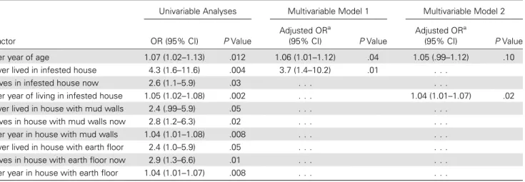
infested house at any time and at the time of study (Table3). The median duration of residence in an infested house was sig-nificantly longer for women with abnormal ECGs than those with normal ECGs (17.5 vs 1.0 years;P = .001). Other variables associated with house infestation showed similar associations. Women with ECG abnormalities were less likely than those with normal ECGs to transmit to their infants, but this associ-ation was not statistically significant (1/28 [3.6%] vs 22/274 [8.0%];P = .40). There was no significant difference in parasite load for those with and without ECG abnormalities. In a mul-tivariable model, increasing maternal age and residence in an infested house were associated with 5% and 4% increased risk, respectively, of ECG abnormalities per year of exposure (Table4). A second multivariable model estimated a 3.7-fold increased risk of ECG abnormalities among women who ever lived in an infested house vs those who had not.
The presence of ECG abnormalities demonstrated a strong dose-response relationship with increasing years of living in an infested house, whereas congenital transmission showed a strong dose-response relationship for maternal parasite load (Table5). Congenital transmission risk was significantly lower
for women in older age strata and those with longer duration of residence in an infested house.
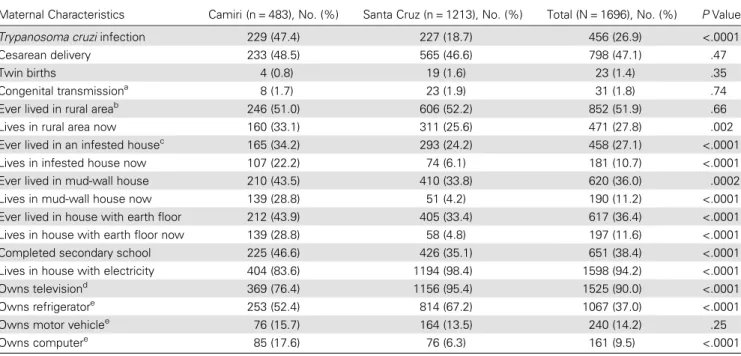
Table 1. Characteristics of Women Presenting for Delivery in Hospital Municipal Camiri, Camiri, and Hospital Japonés, Santa Cruz, Bolivia
Maternal Characteristics Camiri (n = 483), No. (%) Santa Cruz (n = 1213), No. (%) Total (N = 1696), No. (%) P Value Trypanosoma cruzi infection 229 (47.4) 227 (18.7) 456 (26.9) <.0001 Cesarean delivery 233 (48.5) 565 (46.6) 798 (47.1) .47 Twin births 4 (0.8) 19 (1.6) 23 (1.4) .35 Congenital transmissiona 8 (1.7) 23 (1.9) 31 (1.8) .74
Ever lived in rural areab 246 (51.0) 606 (52.2) 852 (51.9) .66
Lives in rural area now 160 (33.1) 311 (25.6) 471 (27.8) .002 Ever lived in an infested housec 165 (34.2) 293 (24.2) 458 (27.1) <.0001
Lives in infested house now 107 (22.2) 74 (6.1) 181 (10.7) <.0001 Ever lived in mud-wall house 210 (43.5) 410 (33.8) 620 (36.0) .0002 Lives in mud-wall house now 139 (28.8) 51 (4.2) 190 (11.2) <.0001 Ever lived in house with earth floor 212 (43.9) 405 (33.4) 617 (36.4) <.0001 Lives in house with earth floor now 139 (28.8) 58 (4.8) 197 (11.6) <.0001 Completed secondary school 225 (46.6) 426 (35.1) 651 (38.4) <.0001 Lives in house with electricity 404 (83.6) 1194 (98.4) 1598 (94.2) <.0001 Owns televisiond 369 (76.4) 1156 (95.4) 1525 (90.0) <.0001
Owns refrigeratore 253 (52.4) 814 (67.2) 1067 (37.0) <.0001
Owns motor vehiclee 76 (15.7) 164 (13.5) 240 (14.2) .25
Owns computere 85 (17.6) 76 (6.3) 161 (9.5) <.0001
a
Data excluded for 3 women from Camiri and 4 women from Santa Cruz because infant infection status was unresolved (see text).
b
Data missing for 1 woman from Camiri and 53 women from Santa Cruz.
c
Data missing for 3 women from Santa Cruz.
d
Data missing for 1 woman from Santa Cruz.
e
Data missing for 2 women from Santa Cruz.
Figure 1. Trypanosoma cruzi infection prevalence by age among preg-nant women in Santa Cruz and Camiri, Bolivia. The numbers above each bar show the denominator for that age group. The difference in prevalence between sites was significant within every maternal age group (P < .001 for the 3 younger age groups;P < .01 for women aged 40–46 years).
DISCUSSION
Bolivia has the highestT. cruzi infection prevalence in the world [18]. More than 70% of cases of congestive heart failure in some Santa Cruz hospitals are attributed to Chagas disease, and an estimated 1 in 175 Bolivian infants is born withT. cruzi [17,
18]. In the Bolivian Chaco, people continue to live in infested houses, and in some villages, 80%–90% of adults have Chagas disease [14,16]. In this setting, we examined factors associated with early Chagas cardiomyopathy and congenital transmission among women presenting for delivery. We observed a higher prevalence of ECG abnormalities in women with longer resi-dence times in infested houses, confirming the impression of longtime Chagas disease researchers [26,27]. Remarkably, vec-tor exposure variables showed an inverse relationship with
maternal parasitemia and mother-to-child transmission. Our data thus suggest that sustained vector exposure increases the risk of Chagas cardiomyopathy, but may be associated with some protection against congenital transmission.
We hypothesize that both pathogenic and protective effects may result from frequent exposure to infected vectors and re-peated superinfection by the parasite [26]. In the acute phase of Chagas disease, a robust inflammatory response involving in-nate immune cells and macrophages activated by interferon gamma (IFN-γ) and tumor necrosis factor alpha (TNF-α) brings the parasitemia under control [28]. Acute symptoms resolve spontaneously as patent parasitemia disappears, but without treatment, total parasite clearance virtually never occurs [18]. During chronic infection, T-cell–mediated immunity keeps the parasite in check [29]. However, failure to downregulate the Table 2. Factors Associated With Congenital Transmission AmongTrypanosoma cruzi–Infected Women Presenting for Delivery in Camiri Municipal Hospital, Camiri, and Hospital Japonés, Santa Cruz, Bolivia
Factor TransmittedT. cruzi (n = 31) Did Not TransmitT. cruzi (n = 418) P Valuea Positive results by PCRb 13 (100) 44 (41.9) <.0001
Cesarean section 19 (61.3) 204 (48.8) .18
Twin birth 3 (9.7) 8 (1.9) .04
Primiparous 6 (19.4) 86 (20.6) .87 Parity >3 19 (61.3) 237 (56.7) .62 Ever lived in rural area 24 (77.4) 263 (64.6)c .15
Lives in rural area now 15 (48.4) 142 (34.0) .11 Ever lived in an infested house 11 (35.5) 230 (55.2)d .04
Lives in infested house now 4 (12.9) 98 (23.4) .26 Ever lived in house with mud walls 12 (38.7) 243 (58.1) .04 Lives in house with mud walls now 4 (12.9) 100 (23.9) .19 Ever lived in house with earth floor 13 (41.9) 242 (57.9) .09 Lives in house with earth floor now 5 (16.1) 100 (23.9) .39 Completed secondary school 6 (19.4) 141 (33.7) .11 Lives in house with electricity 30 (96.8) 363 (86.8) .16 Owns television 30 (96.8) 334 (80.1)d .02
Owns refrigerator 17 (54.8) 218 (52.3)d .78
Owns motor vehicle 2 (6.5) 48 (11.5) .56 Owns computer 0 (0) 42 (10.1) .10 Age, y, median (IQR) 24.0 (20.1–30.2) 26.8 (22.0–34.0) .04e
Parasite equivalents/mL by quantitative PCRb, median (IQR) 62.0 (25.8
–204.8) 0.05 (0–29.6) <.0001e
Years living in rural area, median (IQR) 15.0 (0–22.0) 12.0 (0–21) .30e
Years living in infested house, median (IQR) 0 (0–15) 6.5 (0–20) .02e
Years living in house with mud walls, median (IQR) 0 (0–15) 8 (0–20) .02e
Years living in house with earth floor, median (IQR) 0 (0–17) 8 (0–20) .07e
Data are presented as No. (%) unless otherwise specified.
Abbreviations: IQR, interquartile range; PCR, polymerase chain reaction.
a
Byχ2
or Fisher exact test.
b
Data available for 13 women who transmitted and 105 women who did not transmit.
c
Data missing for 11 women.
d
Data missing for 1 woman.
e
Table 3. Factors Associated With Electrocardiographic Abnormalities Consistent With Chagas Cardiomyopathy Among 302Trypanosoma cruzi–Infected Women
Maternal Characteristic Abnormal ECG (n = 28) Normal ECG (n = 274) P Valuea Positive results by PCRb 3 (30.0) 33 (45.2) .50
Ever lived in rural area 18 (64.3) 175 (65.8)c .87
Lives in rural area now 12 (42.9) 85 (31.0) .20 Ever lived in an infested house 23 (82.1) 142 (51.8) .002 Lives in infested house now 10 (35.7) 49 (17.9) .03 Ever lived in house with mud walls 21 (75.0) 152 (55.5) .05 Lives in house with mud walls now 11 (39.3) 52 (19.0) .03 Ever lived in house with earth floor 21 (75.0) 151 (55.1) .04 Lives in house with earth floor now 11 (39.3) 50 (18.3) .008 Completed secondary school 11 (39.3) 98 (35.8) .71 Lives in house with electricity 24 (85.7) 243 (88.7) .55 Owns television 22 (78.6) 230 (84.3)d .42
Owns refrigerator 16 (57.1) 152 (55.7)d .88
Owns motor vehicle 1 (3.6) 37 (13.5) .23 Owns computer 5 (17.9) 26 (9.5) .19 Age, y, median (IQR) 32 (24–39) 27 (22–34) .03e
Parasite equivalents/mL by quantitative PCRb, median (IQR) 0 (0
–27) 0.3 (0–33.7) .33e
Years lived in rural area, median (IQR) 14 (0–28) 10 (0–20)c .39e
Years lived in infested house, median (IQR) 17.5 (8–28) 1 (0–18) .001e
Years lived in house with mud walls, median (IQR) 17.5 (1.5–27) 5 (0–18) .01e
Years lived in house with earthen floor, median (IQR) 18.5 (1.5–28) 5 (0–18) .01e
Data are presented as No. (%) unless otherwise specified.
Abbreviations: ECG, electrocardiogram; IQR, interquartile range; PCR, polymerase chain reaction.
a
Byχ2
or Fisher exact test.
b
PCR data available for 10 women with abnormal ECG and 73 women with normal ECG.
c
Data missing for 9 women.
d
Data missing for 1 woman.
e
By Wilcoxon rank-sum test.
Table 4. Logistic Regression Model of Factors Associated With Electrocardigraphic Abnormalities Consistent With Chagas Cardiomyopathy Among 302Trypanosoma cruzi–Infected Women
Factor
Univariable Analyses Multivariable Model 1 Multivariable Model 2 OR (95% CI) P Value
Adjusted ORa
(95% CI) P Value
Adjusted ORa
(95% CI) P Value Per year of age 1.07 (1.02–1.13) .012 1.06 (1.01–1.12) .04 1.05 (.99–1.12) .10 Ever lived in infested house 4.3 (1.6–11.6) .004 3.7 (1.4–10.2) .01 . . .
Lives in infested house now 2.6 (1.1–5.9) .03 . . . .
Per year of living in infested house 1.05 (1.02–1.08) .002 . . . 1.04 (1.01–1.07) .02 Ever lived in house with mud walls 2.4 (.99–5.9) .05 . . . .
Lives in house with mud walls now 2.8 (1.2–6.3) .02 . . . . Per year in house with mud walls 1.04 (1.01–1.08) .008 . . . . Ever lived in house with earth floor 2.4 (1.0–5.9) .05 . . . . Lives in house with earth floor now 2.9 (1.3–6.6) .01 . . . . Per year in house with earth floor 1.04 (1.01–1.07) .008 . . . .
Abbreviations: CI, confidence interval; OR, odds ratio.
a
inflammatory response, triggered by parasite persistence and modulated by both host and parasite factors, is thought to play a key role in the pathogenesis of Chagas cardiomyopathy [3,
28]. Mice superinfected with the same or a differentT. cruzi strain had more frequent, severe ECG changes [30]. Repeated vector-borne infection is thought to sustain antigen exposure and the consequent Th1-mediated inflammatory response at a higher chronic level, increasing cardiac morbidity [3,26].
In our data, reported time living in an infested house was in-versely related to maternal parasitemia, which in turn is the stron-gest immediate predictor of congenital transmission [13,31,32]. Repeated antigen exposure may reinforce the Th1 response that controls the parasite in the face of the maternal Th2 polarization necessary to maintain the pregnancy [33]. Only 2 previous studies have examined vector exposure as a factor in congenital transmission [34,35]. An Argentine study reportedfindings similar to ours; women from areas with high infestation rates had the lowest risk of transmission compared with those from areas under vector control (who had intermediate risk) and those living in a city that had never had vector-borne transmis-sion (the highest risk group) [34]. A Bolivian study reported lower morbidity in infected infants of mothers in areas currently under vector control, but no difference in the transmission rate [35]. In our data, duration of residence showed a much stronger association than current house infestation, suggesting that the effect may require prolonged, repeated exposure to develop.
Parasite load was by far the strongest predictor of transmission. Vector exposure is likely one of several factors that influence risk. Other determinants may includeT. cruzi genotype and tropism, and maternal immunogenetics [36]. The predominant genotype detected in congenital infections in Bolivia and Argentina isT. cruzi V, followed by II and VI; no differences have been found in transmission rates by genotype [32,37]. Studies show lower levels of TNF-α in mothers who transmit than those without transmission [38]. Similarly, lower levels of T-cell and macro-phage activation markers andT. cruzi–induced IFN-γ release were found in mothers who transmitted compared with infected mothers of uninfected infants [39]. The association of twin births with increased transmission may be related to the intensified downregulation in multiple pregnancies [40].
Our study had a number of limitations. Our measures of vec-tor exposure were retrospective and self-reported. BecauseT. cruzi infection is chronic and usually acquired in childhood, studies of adults necessarily assess exposures that occurred years before data collection. However,T. infestans is a memora-ble vector: Adults are >1 inch long, have a pungent odor, and leave visible fecal trails on the walls of infested houses. Light in-festations are more likely to have been missed than heavy ones. Some women without reported residence in an infested house were likely infected congenitally themselves, whereas others un-doubtedly had more transient or less intense vector exposure not captured in our data. Intensity (number of infective bites Table 5. Dose-Response Analyses for CongenitalTrypanosoma cruzi Transmission and Electrocardiographic Abnormalities Consistent With Chagas Cardiomyopathy
Characteristic Congenital Transmission, no./No.(%) Stratum OR χ2 for Trend, P Value Abnormal ECG no./No. (%) Stratum OR χ2 for Trend, P Value Age group 13–19 7/60 (11.7) Referent 3/35 (8.6) Referent 20–29 16/215 (7.4) 0.61 8/138 (5.8) 0.66 30–46 8/174 (4.6) 0.36 4.22,P = .04 18/133 (13.5) 1.67 2.3,P = .13 Years in infested house
None 20/207 (9.7) Referent 5/138 (3.6) Referent 1–19 9/134 (6.7) 0.67 11/96 (11.5) 3.44 ≥20 2/107 (1.9) 0.18 7.12,P = .008 13/72 (18.1) 5.86 11.2,P = .0008 Parasite loada 0 0/61 (0)b Referent 7/47 (14.9)c Referent 1–34 4/24 (14.3) . . . 2/19 (10.5) 0.67 ≥35 9/29 (31.0) . . . 18.0,P < .0001 1/17 (5.9) 0.36 1.5,P = .23
Increasing parasite load shows a direct dose-response relationship, whereas increasing maternal age and duration of vector exposure show an inverse relationship with congenitalT. cruzi transmission risk. Increasing duration of living in an infested house shows a direct dose-response relationship with the presence of ECG abnormalities.
Abbreviations: ECG, electrocardiogram; OR, odds ratio.
a
Parasite equivalents/mL by quantitative polymerase chain reaction (PCR).
b
Data available for 13 women who transmitted and 105 who did not transmit. ORs undetermined because of 0 cell in referent stratum.
c
per unit time) is impossible to assess retrospectively, but likely plays a role in the effects we observed. We had technical dif fi-culties when we changed extraction methodology midway through the study and were only able to include PCR data from a subset of women. This impeded our ability to analyze epidemiologic risk factors together with parasite load in multi-variable models. Our cardiac analysis would have been strength-ened by the addition of an uninfected control group, but this was not feasible in the current study. However, we used rigorous criteria to define ECG abnormalities, and all analyses were comparisons within the group of infected mothers. In a meta-analysis of population-based studies, the prevalence of bundle branch blocks in women aged <45 years was 0.2% [41], com-pared with 6.6% in study women, suggesting that the observed abnormalities were due to Chagas disease. Our study enrolled nearly 1700 women; an even larger sample size would have in-creased our statistical power but would have strained our resourc-es. Even in this high-prevalence population, we screened an average of 54 women and conducted follow-up on 15 mother– infant pairs to yield a single transmission event.
Chagas disease researchers have long postulated that repeated T. cruzi superinfection is a major determinant of cardiomyopathy risk [26,27]. In areas under effective vector control, there is a widespread impression that cardiomyopathy severity is lower, and onset and mortality are delayed until later in life [42,43]. Our maternal ECG data provide epidemiologic support for this hypothesis, and underscore the importance of the vector control effort in the Bolivian Chaco. Nevertheless, congenital transmis-sion risk will remain as long as there are infected women in the reproductive age group. Recent data give strong support to the contention that antitrypanosomal treatment of women prior to pregnancy markedly decreases congenital transmission risk [44]. Sustained vector control efforts, especially in the most affected communities, and programs to safely treat infected indi-viduals are essential to decrease morbidity and prevent continued congenital transmission in the future. Treatment of children and young adults should be given high priority in Latin America and other regions with infected residents [44,45].
Supplementary Data
Supplementary materialsare available atClinical Infectious Diseases online (http://cid.oxfordjournals.org). Supplementary materials consist of data provided by the author that are published to benefit the reader. The posted materials are not copyedited. The contents of all supplementary data are the sole responsibility of the authors. Questions or messages regarding errors should be addressed to the author.
Notes
Acknowledgments. Members of the Working Group on Chagas Disease in Bolivia and Peru include Leny Sanchez, Lisbeth Ferrufino, Edith Malaga, Sara Quispe, Edith Hinojosa, Margot Ramirez, Eliana Saenza, Jorge Luis Flores-Franco, Janet Acosta, Gerardo Sanchez, Maribel Suxo, Hilsen
Roncales, Fernando Ramirez, Nazaret Bozo Escalera, Celia Espinoza, and Janet Vizcarra. We are grateful to the nurses and physicians of the obstetric services of Hospital Japonés and Hospital Municipal Camiri for their collab-oration and their dedication to the welfare of the women and infants of Santa Cruz Department.
Disclaimer. The funding sources had no role in the study design, col-lection, analysis and interpretation of the data, preparation of the manu-script, or the decision to submit for publication.
Financial support. This work was supported by the National Institutes of Health (grant number R01-AI087776, Global Research Training grant number D43 TW006581) and by discretionary funds awarded to R. H. G. from Asociacion Benefica PRISMA (www.prisma.org.pe).
Potential conflicts of interest. All authors: No potential conflicts of interest.
All authors have submitted the ICMJE Form for Disclosure of Potential Conflicts of Interest. Conflicts that the editors consider relevant to the con-tent of the manuscript have been disclosed.
References
1. World Health Organization. Global burden of disease estimates for 2000–2012. Available at:http://www.who.int/healthinfo/global_ burden_disease/estimates/en/index2.html. Accessed 18 June 2015. 2. World Health Organization. Chagas disease in Latin America: an
epide-miological update based on 2010 estimates. Wkly Epidemiol Rec2015; 90:33–44.
3. Marin-Neto JA, Cunha-Neto E, Maciel BC, Simoes MV. Pathogenesis of chronic Chagas heart disease. Circulation2007; 115:1109–23. 4. Rassi A Jr, Rassi A, Little WC. Chagas’ heart disease. Clin Cardiol 2000;
23:883–9.
5. Maguire JH, Hoff R, Sherlock I, et al. Cardiac morbidity and mortality due to Chagas’ disease: prospective electrocardiographic study of a Bra-zilian community. Circulation1987; 75:1140–5.
6. Maguire JH. Trypanosoma. In: Gorbach S, Bartlett J, Blacklow N, eds. Infectious diseases. 2nd ed. Philadelphia: Lippincott, Williams & Wilkins,2004:2327–34.
7. Dias JC, Silveira AC, Schofield CJ. The impact of Chagas disease control in Latin America: a review. Mem Inst Oswaldo Cruz2002; 97:603–12. 8. Howard EJ, Xiong X, Carlier Y, Sosa-Estani S, Buekens P. Frequency of the congenital transmission ofTrypanosoma cruzi: a systematic review and meta-analysis. BJOG2014; 121:22–33.
9. Azogue E, Darras C. Prospective study of Chagas disease in newborn children with placental infection caused byTrypanosoma cruzi (Santa Cruz-Bolivia) [in Portugese]. Rev Soc Bras Med Trop1991; 24:105–9. 10. Basombrio MA, Nasser J, Segura MA, et al. The transmission de Chagas disease in Salta and the detection of congenital cases [in Spanish]. Me-dicina (B Aires)1999; 59(suppl 2):143–6.
11. Schenone H, Contreras MC, Borgono JM, et al. Overview of the epide-miology of Chagas’ disease in Chile [in Spanish]. Bol Chil Parasitol 1991; 46:19–30.
12. Torrico F, Alonso-Vega C, Suarez E, et al. MaternalTrypanosoma cruzi infection, pregnancy outcome, morbidity, and mortality of congenitally infected and non-infected newborns in Bolivia. Am J Trop Med Hyg 2004; 70:201–9.
13. Bern C, Verastegui M, Gilman RH, et al. CongenitalTrypanosoma cruzi transmission in Santa Cruz, Bolivia. Clin Infect Dis2009; 49:1667–74. 14. Samuels AM, Clark EH, Galdos-Cardenas G, et al. Epidemiology of and impact of insecticide spraying on Chagas disease in communities in the Bolivian Chaco. PLoS Negl Trop Dis2013; 7:e2358.
15. Lardeux F, Depickere S, Aliaga C, Chavez T, Zambrana L. Experimen-tal control of Triatoma infestans in poor rural villages of Bolivia through community participation. Trans R Soc Trop Med Hyg2015; 109:150–8.
16. Chippaux JP, Postigo JR, Santalla JA, Schneider D, Brutus L. Epidemi-ological evaluation of Chagas disease in a rural area of southern Bolivia. Trans R Soc Trop Med Hyg2008; 102:578–84.
17. Hidron A, Gilman R, Justiniano J, et al. Chagas cardiomyopathy in the con-text of the chronic disease transition. PLoS Negl Trop Dis2010; 4:e688. 18. Rassi A Jr, Rassi A, Marin-Neto JA. Chagas disease. Lancet2010;
375:1388–402.
19. Freilij H, Muller L, Gonzalez Cappa SM. Direct micromethod for diag-nosis of acute and congenital Chagas’ disease. J Clin Microbiol 1983; 18:327–30.
20. Umezawa ES, Nascimento MS, Kesper N Jr, et al. Immunoblot assay using excreted-secreted antigens ofTrypanosoma cruzi in serodiagnosis of congenital, acute, and chronic Chagas’ disease. J Clin Microbiol 1996; 34:2143–7.
21. Piron M, Fisa R, Casamitjana N, et al. Development of a real-time PCR assay forTrypanosoma cruzi detection in blood samples. Acta Trop 2007; 103:195–200.
22. Ministerio de Salud y Deportes. Manual de normas tecnicas y operativas para el tamizaje, diagnóstico y tratamiento de la enfermedad de chagas cronica reciente infantil. 2nd ed. La Paz, Bolivia: Ministerio de Salud y Deportes,2007.
23. Lazzari JO, Pereira M, Antunes CM, et al. Diagnostic electrocardiogra-phy in epidemiological studies of Chagas’ disease: multicenter evalua-tion of a standardized method. Rev Panam Salud Publica 1998; 4:317–30.
24. Le VV, Wheeler MT, Mandic S, et al. Addition of the electrocardiogram to the preparticipation examination of college athletes. Clin J Sport Med 2010; 20:98–105.
25. Clark EH, Sherbuk J, Okamoto E, et al. Hyperendemic Chagas disease and the unmet need for pacemakers in the Bolivian Chaco. PLoS Negl Trop Dis2014; 8:e2801.
26. Dias JCP. Chagas disease control and the natural history of human Chagas disease: a possible interaction? Mem Inst Oswaldo Cruz2000; 95(suppl II):14–22.
27. Dias E. Os efeitos da superinfecção sobre a evolução da cardiopatia crônica chagásica. Rev Goiana Med1962; 9(suppl):233–9.
28. Dutra WO, Gollob KJ. Current concepts in immunoregulation and pa-thology of human Chagas disease. Curr Opin Infect Dis2008; 21:287–92. 29. Bacal F, Silva CP, Pires PV, et al. Transplantation for Chagas’ disease: an overview of immunosuppression and reactivation in the last two de-cades. Clin Transplant2010; 24:E29–34.
30. Bustamante JM, Rivarola HW, Fernandez AR, et al.Trypanosoma cruzi reinfections in mice determine the severity of cardiac damage. Int J Par-asitol2002; 32:889–96.
31. Rendell VR, Gilman RH, Valencia E, et al.Trypanosoma cruzi-infected pregnant women without vector exposure have higher parasitemia lev-els: implications for congenital transmission risk. PLoS One2015; 10: e0119527.
32. Virreira M, Truyens C, Alonso-Vega C, et al. Comparison of Trypano-soma cruzi lineages and levels of parasitic DNA in infected mothers and their newborns. Am J Trop Med Hyg2007; 77:102–6.
33. Wegmann TG, Lin H, Guilbert L, Mosmann TR. Bidirectional cytokine interactions in the maternal-fetal relationship: is successful pregnancy a TH2 phenomenon? Immunol Today1993; 14:353–6.
34. Sanchez Negrette O, Mora MC, Basombrio MA. High prevalence of congenitalTrypanosoma cruzi infection and family clustering in Salta, Argentina. Pediatrics2005; 115:e668–72.
35. Torrico F, Vega CA, Suarez E, et al. Are maternal re-infections with Try-panosoma cruzi associated with higher morbidity and mortality of congenital Chagas disease? Trop Med Int Health2006; 11:628–35. 36. Carlier Y, Truyens C. Maternal-fetal transmission ofTrypanosoma
cruzi. In: Telleria J, Tibayrenc M, eds. American trypanosomiasis-Chagas disease: one hundred years of research. New York, NY: Elsevier, 2010:539–81.
37. Ortiz S, Zulantay I, Solari A, et al. Presence ofTrypanosoma cruzi in pregnant women and typing of lineages in congenital cases. Acta Trop2012; 124:243–6.
38. Cardoni RL, Garcia MM, De Rissio AM. Proinflammatory and anti-inflammatory cytokines in pregnant women chronically infected with Trypanosoma cruzi. Acta Trop 2004; 90:65–72.
39. Hermann E, Truyens C, Alonso-Vega C, et al. Congenital transmission ofTrypanosoma cruzi is associated with maternal enhanced parasitemia and decreased production of interferon-gamma in response to parasite antigens. J Infect Dis2004; 189:1274–81.
40. Suzuki S, Okudaira S. Maternal peripheral T helper 1-type and T helper 2-type immunity in women during thefirst trimester of twin pregnancy. Arch Gynecol Obstet2004; 270:260–2.
41. De Bacquer D, De Backer G, Kornitzer M. Prevalences of ECGfindings in large population based samples of men and women. Heart2000; 84:625–33.
42. Acquatella H, Catalioti F, Gomez-Mancebo JR, Davalos V, Villalobos L. Long-term control of Chagas disease in Venezuela: effects on serologic findings, electrocardiographic abnormalities, and clinical outcome. Cir-culation1987; 76:556–62.
43. Lima-Costa MF, Barreto SM, Guerra HL. Chagas’ disease among older adults: branches or mainstream of the present burden ofTrypanosoma cruzi infection? Int J Epidemiol 2002; 31:688–9.
44. Fabbro DL, Danesi E, Olivera V, et al. Trypanocide treatment of women infected withTrypanosoma cruzi and its effect on preventing congenital Chagas. PLoS Negl Trop Dis2014; 8:e3312.
45. Murcia L, Carrilero B, Munoz-Davila MJ, Thomas MC, Lopez MC, Segovia M. Risk factors and primary prevention of congenital Chagas disease in a nonendemic country. Clin Infect Dis2013; 56:496–502.