Microbiological and Clinical Significance of a New Property of
Defective Lysis in Clinical Strains of Pneumococci
E. Tuomanen, H. Pollack*, A. Parkinson, M. Davidson, R. Facklam, R. Rich, and O. Zak
From the Rockefeller University, New York, New York; the Arctic Investigations Laboratory, Centers for Disease Control, Anchorage, Alaska; the Division of Bacterial Diseases, Centers for Disease Control, Atlanta, Georgia; and CIBA-GEIGY Limited, Basel, Switzerland
A pneumococcal isolate that caused relapsing meningitis in a patient infected with hu-man immunodeficiency virus (HIV) was found to display an unusual response to penicillin - rapid death but a striking lack of cellular lysis. This lytic defect was also de-tected in all four pneumococcal isolates from three additional HIV-infected patients and in more than half of the clinical isolates from patients with bacteremia. In a rabbit model of meningitis, the lysis-defective strain remained cryptic, with a delay of 5 h in the onset of leukocytosis in cerebrospinal fluid. A marked burst of leukocytosis was associated with ampicillin-induced lysis of a lysis-sensitive strain but not of a lysis-defective strain. Pneu-mococcal clinical isolates have different lytic responses to penicillin; defective lysis may adversely affect the course of meningitis, an observation suggesting that autolysins play a role in modulating infectious diseases.
A nine-month-old child infected with human immu-nodeficiency virus (HIV) developed relapsing pneu-mococcal meningitis. After characterizing the strain, we found an unusual response to penicillin - the bac-terium died rapidly but did not undergo cellular ly-sis. Classically, two responses to penicillin have been described [1, 2] for clinical isolates of pneumococci. Typically, the isolates lyse and die rapidly; rarely, ly-sis is nearly absent, and killing is substantially slower. We characterized in detail the dissociation of killing and lysis in the strain from this patient. This report also describes the discovery of clinical isolates of pneumococci that do not lyse but that undergo rapid loss of viability during penicillin treatment.
Addi-Received for publication 5 October 1987 and in revised form 12 January 1988.
This work was presented in part at the 27th Interscience Con-ference on Antimicrobial Agents and Chemotherapy, held in New York, New York, on 4-7 October 1987.
This work was supported in part by a Career Investigator Award to Dr. Tuomanen from the Irma T. Hirschi Trust and by grant AI-16794 from the National Institute of Allergy and Infectious Diseases.
We thank J. Kadurugamuwa and B. Hengstler for determin-ing the growth rate of strain Br in vivo; E. Kovacs for technical assistance; and M. Geller for secretarial assistance.
Please address requests for reprints to Dr. Elaine Tuomanen, The Rockefeller University, 1230 York Avenue, New York, New York 10021.
*Present address: Department of Pediatric Infectious Diseases, New York University, New York, New York.
36
tionally, our studies analyze the impact of the lack of drug-induced lysis (independent of bacterial kill-ing) on the clinical course of pneumococcal infec-tions. We present evidence that autolysins may play a role in modulating the course of some infectious diseases.
Materials and Methods
Bacterial strains andgrowth conditions. We used the following strains: encapsulated clinical strains 8249, Sm. Va2, Va3, and Va7 and unencapsulated laboratory strain R6 and its tolerant transformant Iyt 4-4; the strains have previously been described [1-3]. Six clinical isolates from the pre-penicillin era [4, 5] were obtained from the Centers for Disease Control (Atlanta): SP-88, 96, 99, 108, 142, and 144 (MICs of penicillin,
om
IJ.g/mL). Thirty-three iso-lates from blood or CSF were provided by the Arc-tic Investigations Laboratory (Anchorage, Alaska) as part of a retrospective study of invasive pneumo-coccal disease in Southwest Alaska from 1980 to 1986. Serotypes included types 14 (13 strains), 19A (9 strains), 04 (4 strains), 23F (2 strains), and 1 strain each of types lOA, 6B, 07F, 33F, and 18C; MICs of penicillin ranged from 0.01 to 0.25 IJ.g/mL. One iso-late was from an Alaskan infant with an immuno-globulin deficiency and persistent meningitis. Five isolates, including the index strain Br, were obtained from four HIV-infected patients withpneumococ-Lysis-Defective Pneumococci
cal bacteremia or meningitis at New York Hospital (New York; serotypes 19, 6, and 9; MICs of penicil-lin, 0.02 ug/ml., including strain Br). Three of these isolates caused relapsing disease in two patients: strain Br caused meningitis that was controlled af-ter two 14-dcourses of penicillin, and strains GI and Gil caused recurrent bacteremia. Strain Br was tested in the first passage and in~1Opassages from frozen stock. MICs were determined by using the macro-tube dilution method. Identification was made on the basis of growth and morphology on blood-agar plates and of sensitivity to optochin. All strains ex-cept the autolysin-deficient strainIyt 4-4 were lysed by deoxycholate [2].
Strains were grown in semisynthetic medium sup-plemented with 0.1070 yeast (Difco, Detroit) extract (C
+
y; pH 8.0 [6]) in a stationary water bath at 37 C. All experiments were carried out during exponen-tial-growth phase from back-diluted overnight cul-tures. Turbidity was monitored by using a Sequoia-Turner spectrophotometer (absorbance at 620 nm; Mountainville, Calif). Stocks of cultures were stored in C+
Y medium with 10% glycerol at - 70 C, by using the first or second passage from the original clinical isolate.Lysis andkilling rates. Cultures of all strains were divided in half when the turbidity reached an ab-sorbance of 0.3-0.4; half of the cultures received 10
x MIC of penicillin. Strains R6 and Br were also tested for lysis in the presence of ~100 x MIC of penicillin and of 10 x MlC of imipenem (0.05 ug/rnl.), vancomycin (5 ug/ml.), or ampicillin (0.4 ug/rnl.). Turbidity was monitored for 6 h and over-night. Viability was determined at 0, 2, and 4 h after adding penicillin, by diluting samples from each cul-ture in incomplete semisynthetic medium contain-ing penicillinase (100 U/mL; Becton-Dickinson, Cockeysville, Md) and by pour-plating in tryptic-soy agar (Difco) supplemented with 3% sterile, defibri-nated sheep blood. Colony counts were made after 24 h of incubation at 37C.
To compare rates of cell wall degradation, we grew strains R6, lyt 4-4, and Br overnight in C
+
Ymedium containing 1 ~Ci and 5 ug of [3H]choline/mL (1 ~Ci/mM; Amersham, Boston). Cells were washed by filtration and were grown for an additional 2 h in nonradiolabeled medium. Half of each culture re-ceived 10 x MIC of penicillin, and all cultures were sampled at hourly intervals for 6 h to determine [3H]cholinecounts remaining in the cell wall (the ma-terial precipitated in boiling SDS [7]). In someex-37
periments, 1 ug of purified pneumococcal autoly-sin/mL (amidase [8]) was added 30 min before penicillin. Crude cell sonicates of strain Br were pre-pared and assessed for the presence of endogenous autolytic activity in vitro, as previously described [1, 2]. The profile of the penicillin-binding proteins of strain Br was compared with that of strain R6 by using published procedures [3].
Meningitis model. Groups of four rabbits were anaesthetized and placed in stereotaxic frames as pre-viously described [9, 10]. Pneumococcal strains SIll and Br were grown to logarithmic phase in C
+
Y medium, washed, and resuspended in pyrogen-free saline at a concentration of 103cfu in 0.2 mL. After removal of 0.3 mL of CSF, the preparations were in-stilled into the cisterna magna. Cytochemical deter-minations of CSF were repeated in each animal over a 24-h period to document changes in the density of leukocytes and the concentrations of glucose, pro-tein, and lactic acid [10]. Bacterial titers in CSF were determined on tryptic-soy agar containing 3% sheep blood. To determine the effect of ampicillin treat-ment on the course of the disease, we infected groups of four rabbits with each strain but did not sample these groups until 18 h later, when infection was es-tablished. After CSF sampling, half of the animals in each group received 30 mg of ampicillin/kg iv as a single bolus dose. Ampicillin levels in CSF reached a maximum ("'0.5 ug/ml.) 30 min after infusion (>10 x MlC of both SIll and Br), a result that agreed with previous results [9]. Cytochemical parameters and bacterial titers in CSF were deter-mined 4 and 24 h after administering ampicillin.
Summaries ofclinical data. The medical records for the 30 Alaskan patients and their 156 household members were obtained and reviewed for antibiotic usage for the six months preceding the index pneu-mococcal infection. The duration of antibiotic us-age (median number of days) was recorded and com-pared by using a nonparametric statistical test. The duration of fever and hospitalization (median num-ber of days) for the 30 invasive pneumococcal ill-nesses was similarly recorded and analyzed.
Results
Characteristics of lysis-defective pneumococci.
Unlike the standard lysis-sensitive/fast-kill (R6) and lysis-defective/slow-kill (lyt 4-4) laboratory strains, strain Br demonstrated an apparent dissociation of lysis and killing (lysis sensitive/fast kill) in response
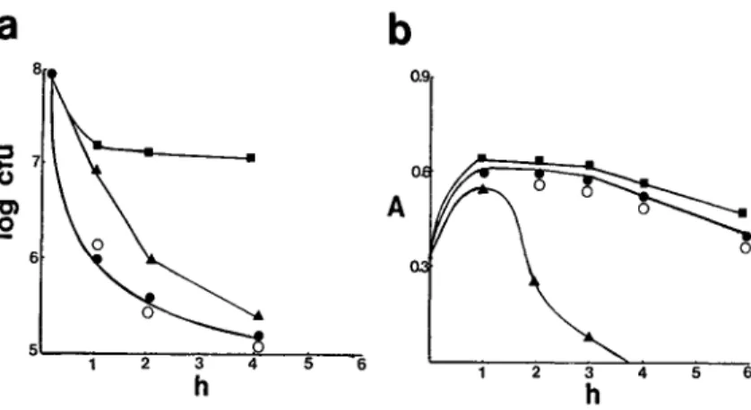
Figure 1. Comparison of the killing
(a)and lysis(b)rates of representative strains from each of the three groups of pneumococci.Triangles, lysis-sen-sitivestrain R6;circles,lysis-defective, kill-sensitive strain Br;squares, kill-resistant(tolerant)strainlyt4-4;closed symbols,penicillin concentration of 10 X MIC; andopen symbols,penicillin concentration of 100 x MIC. 5 6 3 4 h 2
a
b
8 0.9.a
7 o C) A .2 6 5 2 5 6to penicillin (figure 1). Penicillin lysis was minimal, even at 100 x MIC (figure 1, open circles). When rates of cell wall degradation between the penicillin-sensitive, lysis-sensitive/fast-kill (R6) strain and strain Br were compared, cell wall degradation of strain Br was markedly reduced at both 10 x and 100 x MIC (table 1); this response was also found with am-picillin. Rapid lysis analogous to the R6 standard strain was achieved, however, by two other inhibi-tors of cell wall synthesis - imipenem and vancomy-cin. The profile of penicillin-binding proteins for strain Br (determined at saturating concentrations of the radioactive penicillin) was the same as that for the standard laboratory strain R6 (data not shown).
Crude extracts of strain Br contained autolytic ac-tivity capable of solubilizing >90070 of [3H]lysine la-bel from the cell wall of strain R6 in vitro, a result indicating the presence of active autolysin. This re-sult was consistent with the fact that deoxycholate also caused lysis and cell wall degradation of strain
Table1. Percent loss of ['H]choline label from cellwall 4 h after treating strains R6 and Br with various lytic stimuli.
Lytic stimulus
Penicillin 10x MIC (x MIC) of penicillin +
I ug of ami- 0.1010 Strain None 10 100 dase/mL* deoxycholate
R6 9 78 92 92 94
Br 7 17t 34t 87 96
* Purified autolysin of strainR6;results with amidase alone were not different from results with no lytic stimulus.
tp<.01, as compared with strainR6by using the Student's
t test.
Br (table 1) and of all the other lysis-defective clini-cal isolates of pneumococci that we studied (deoxy-cholate-induced lysis is an autolysin-dependent phenomenon [2]). The presence of deoxycholate-in-duced lysis and extensive stationary-phase lysis, al-beit delayed in onset, also indicated that the cell wall of strain Br could be hydrolyzed by autologous en-zyme. Adding exogenous purified autolysin to cul-tures of strain Br, then adding 10 x MIC of penicil-lin, resulted in lysis and cell wall degradation comparable to that in the lysis-sensitive strain R6 (table 1).
Lysis-defective pneumococci in clinical isolates. Figure 2 compares the lytic and killing responses af-ter exposure to 10 x MIC of penicillin for the 51 clinical isolates that were studied. The three general types of responses shown in figure 1 were observed: group a, lysis sensitive and fast killed (16 strains, 31%); group b, lysis defective and fast killed (29 strains, 57%); and group c, lysis defective and slowly killed (6 strains, 12%). The classic lysis-sensitive re-sponse, defined as those isolates showing >50% de-crease in turbidity after 4 h, was characteristic of the standard laboratory strain R6 and 15 clinical isolates (including strain Sin, two pre-penicillin isolates, and 12 Alaskan strains). The remaining 35 strains were lysis defective «50% decrease in turbidity after 4 h), with a spectrum of slow-lysis rates. This group also demonstrated a wide range of rates and degrees of penicillin-induced killing. For the purposes of com-parison, an arbitrary dividing line was established to definefastvs.slowkilling, as follows. The well-characterized laboratory standard (strain R6) was ac-tually the least kill-sensitive strain in group a (mean, 3.0 ± 0.6 log cfu/mL). This value minus one stan-dard deviation (i.e., 2.4) was chosen as the cutoff value for "lysis-sensitive and fast-killed"
pneu-Lysis-Defective Pneumococci
100
I Ia
IR[!]
•
80
I•
••
I 0••
50
• •
I•
I I•
•
60
I 0•
I I<t
-
- --
,----0C
I•
b
(I)40
I•
••
.2
I•
• •
~ I•
•
I•
•
I•
•••
2
I•
•
~B[!]
•
0L
. 0 I•
-
0'"
.g
0
5
Figure 2. Scattergram comparing percent lysis and log killing of pneumococci 4 h after administration of 10 x MIC of penicillin.Dashedlinesrepresent borderlinevalues
betweengroups, derived as explained in the text.A, lysis-sensitive, kill-sensitive;B,lysis-defective, kill-sensitive; C, lysis-defective, kill-resistant (tolerant); .., laboratory strains, including R6(R)and lyt 4-4(L); . ,clinical iso-lates from Alaska; . , isoiso-lates from HIV-infected patients including Br(B); b..,previously well-characterized clini-cal isolates, includingSIll (S); and 0, isolates from the pre-penicillin era.
mococci. On this basis, six strains were classified as "lysis defective" and "slowly killed" (group c): the tolerant laboratory standard strain lyt 4-4; the pre-viously described [1] "tolerant" clinical isolates 8249,
39
Va2, Va3, and Va7; and one Alaskan isolate. Group b, the group with the largest number of strains (29 strains, 571Jfo of the total), was characterized by the novel phenotype "lysis defective" but "fast killed" in response to penicillin. The majority of these strains were as highly kill sensitive (>4 log kill after 4 h) as were the "normal," lysis-sensitive, fast-kill strains in group a. Four of six isolates from the pre-penicillin era, all five isolates from HIV-infected patients (in-cluding strain Br), and 20 of the 33 Alaskan isolates were in this lysis-defective group.
Table 2 describes the relation between lytic and killing properties and the MIC and serotype of the 49 clinical isolates. Lysis-sensitive, kill-sensitive strains were always penicillin susceptible (MIC <0.1 ug/ml.), and most strains were type 14. Tolerant (lysis-defective, kill-resistant) strains were always in-termediatelyor highly resistant to penicillin (MIC >O.1llg/mL), and all but one (6B) were type 19. The mean MIC of the lysis-defective group was some-what elevated, but the strains were nearly evenly dis-tributed above and below an MIC of 0.1ug/rnl.,
The presence of stationary-phase lysis was highly variable in these strains and was often markedly delayed in onset. No relation between the lytic re-sponse to penicillin and stationary-phase lysis was found.
Experimental meningitis due to lysis-sensitive and lysis-defective pneumococci. The lysis-sensitive strainSurhas been studied extensively in the rabbit model of meningitis [9,10].We compared the growth rate, the onset of CSF abnormalities, and the re-sponse to ampicillin of strainSuiwith that of the lysis-defective strain Br. Both strains were penicillin-susceptible (MIC, 0.02 ug/ml.) isolates from chil-dren with meningitis, and both strains had a dou-bling time of "'60 min in vivo in CSF. For strain
Sin,the appearance of >100 leukocytes/ul, of CSF occurred 9 h after infection at a density of 7 x 10⁵
cfu/mL; in contrast, strain Br induced leukocytosis only at 14 h after infection at a density of 7 x 10⁷
cfu/mL. At 24 h after infection the bacterial
den-Table 2. Relation of lytic and killing properties to MIC and serotype of clinical strains. MIC of penicillin(ug/rnl.) No. of strains
Group Mean ± SD Range Total Serotype14 Serotype19
Lysis sensitive/kill sensitive 0.026 ± 0.01 0.015-0.06 15 11 1
Lysis defective/kill sensitive 0.095 ± 0.08 0.02-0.25 29 2 8
Table3. Comparison of CSF cytochemistry and bacterial titers, 4 h after ampicillin therapy, of lysis-sensitive (SIll) and lysis-defective (Br) pneumococci in a rabbit model of meningitis.
CSF cytochemistry"
No. (mean ± SD) of leukocytes at Change in bacterial titer
Strain (no. Percent (mean ± SD
Treatment group of animals) 18 h 22 h change log cfu/mL)
Control SIll (6) 2647 ± 2606 5860 ± 4025 +221 + 1.5 ± 0.8
Br (8) 2775 ± 3616 1630± 1527 -59 +2.5 ± 1.2
Ampicillin Sill (7) 2651 ± 1678 19202 ± 8511 +724t -3.1 ± 0.9
Br (8) 3147 ± 2838 7370 ± 6929 +234 - 1.9 ± 1.1
" Treatment was initiated at 18 h of infection.
tp<.01 when compared with control.
sity of strain Br exceeded the density of Sm by 2 logs. At the same time, however, the leukocyte den-sity and protein concentrations for strain Br re-mained approximately one-third that of SIll.
Table 3 compares the course of meningitis due to strains Sm and Br after treatment with ampicillin. Without treatment, bacterial counts increased 2 logs between 18 and 22 h for both strains, yet the leuko-cyte density for strain Sm doubled and that for strain Br decreased to approximately one-half. In ampicillin-treated animals, both strains were killed, and the change in bacterial titer was 2-3 logs (P
>
0.1). The number of leukocytes appearing in the CSF 4 h after the onset of lysis of strain Siu was, however, a mean of six times greater than the values before ampicillin, whereas the corresponding value for the lysis-defective strain Br was only doubled(P<
.(01). Twenty-four hours after ampicillin, bacterial titers in animals infected with strain Br all reboundedto >3 logs, and all the animals died within 72 h. Three-fourths of the animals infected with strain SIll showed an increase in bacterial titers, but the maximum value was only NIlog, and50010 of these animals survived (i.e., alive at seven days).
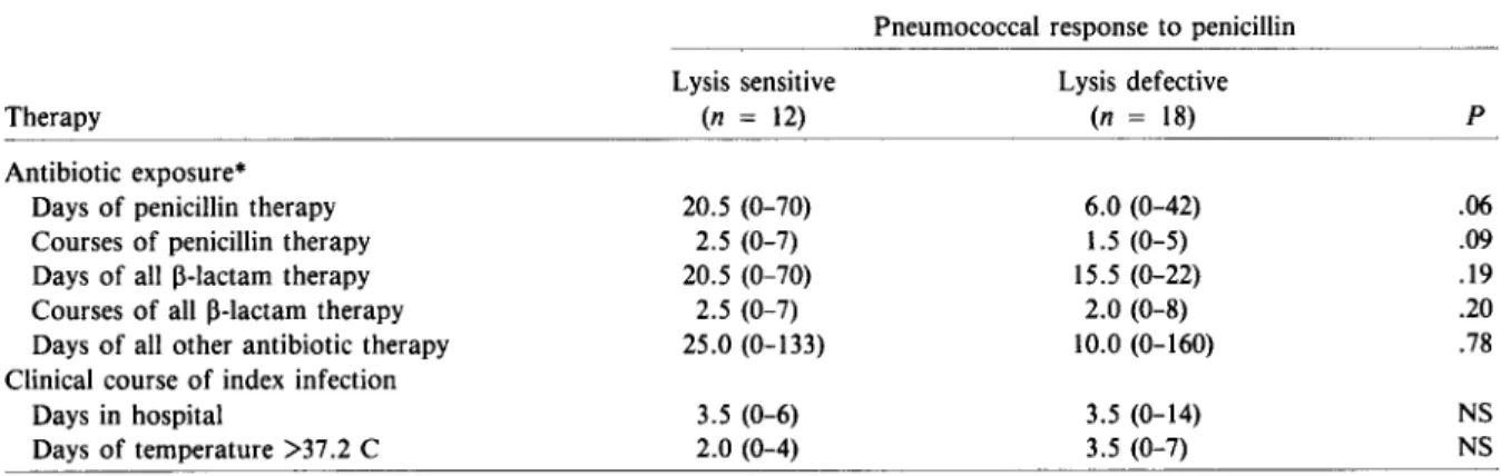
Previous antibioticexposure and clinical course ofpatients withpneumococcal bacteremia. Table 4 compares the antibiotic exposure, before the in-dex infection, of 30 Alaskan patients who had pneu-mococcal bacteremia; 12 patients had lysis-sensitive strains, and 18had lysis-defective strains. No signifi-cant difference could be detected between the me-dian number of days of antibiotic usage by the two groups for three to six months before hospitaliza-tion, a result that eliminated any possible antibiotic-related selection for the lysis defect. In fact, more patients with lysis-defective than lysis-sensitive in-fections did not receive any penicillin (8 of 18 vs. 1 of 12;P
<
.05). A review of the medical records forTable4. Previous antibiotic exposure and clinical course of 30 patients with pneumococcal bacteremia.
Pneumococcal response to penicillin
Therapy
Antibiotic exposure" Days of penicillin therapy Courses of penicillin therapy Days of all 13-lactam therapy Courses of all 13-lactam therapy Days of all other antibiotic therapy Clinical course of index infection
Days in hospital Days of temperature >37.2 C Lysis sensitive (n = 12) 20.5 (0-70) 2.5 (0-7) 20.5 (0-70) 2.5 (0-7) 25.0 (0-133) 3.5 (0-6) 2.0 (0-4) Lysis defective (n = 18) 6.0 (0-42) 1.5 (0-5) 15.5 (0-22) 2.0 (0-8) 10.0 (0-160) 3.5 (0-14) 3.5 (0-7) p .06 .09 .19 .20 .78 NS NS NOTE. Data are median no. (range); NS = not significant,n = no. of patients.
Lysis-Defective Pneumococci
all other members of households in which there was a case of invasive pneumococcal infection showed no difference in previous antibiotic usage.
The data on the clinical courses of the HIV-infected patients and the Alaskan patients with bac-teremia were available for retrospective study. Two of the four HIV-infected patients and one immuno-globulin-deficient child experienced relapsing or per-sistent disease; all of the causative pneumococcal strains were lysis defective. In particular, strain Br, which caused relapsing meningitis in one of the HIV-infected patients, was eradicated only after tandem, 14-day courses of penicillin and ampicillin. Menin-gitis persisted in the immunoglobulin-deficient child after 18 d of penicillin. In contrast, in normal, im-munocompetent hosts, there were no differences de-tected between the patients with lysis-sensitive and lysis-defective strains after comparing days of hos-pitalization or days with fever (>37.2 C), an obser-vation suggesting that the lytic phenotype did not influence the course of bacteremia (table 4).
Discussion
Our study shows that the response to penicillin in clinical isolates of pneumococci varies considerably with respect to bacterial lysis. Recently [1, 11], it has become evident that decreased susceptibility to kill-ing, i.e., tolerance, is increasing in frequency among clinical isolates of pneumococci. We now report that susceptibility to lysis is also changing in pneu-mococci, and these properties may be independent variables. Thus,itis possible to divide pneumococci into three general response patterns to penicillin: group a, both lysis and kill sensitive; group b, lysis defective but kill sensitive; and group c, both rela-tively lysis and kill resistant. The response to peni-cillin of pneumococci in group a typifies the nor-mal response that has been observed in the laboratory strains extensively used over the past de-cades, and the response of pneumococci in group c characterizes a tolerant response. The existence of group b pneumococci, in which lysis and killing are dissociated, suggests that bacterial death and lysis occur by different mechanisms in clinical isolates of pneumococci. Killing without lysis and cell wall degradation are properties that characterize most strains of Lancefield group A streptococci [12, 13]. Penicillin-resistant pneumococci have previously been shown [1] to be lysis defective, but they also show a defect in killing, a result that is much more
41
extensive than that described in figure 2 (compare open triangles in group c and closed circles in group b). Our data indicate that the combination of defec-tive lysis and kill sensitivity is not rare among clini-cal strains, because more than one-half of the strains we tested demonstrated this trait. The actual preva-lence of the lysis defect, with or without alteration of sensitivity to killing, cannot be determined from our data.
The lysis-defective pneumococci cannot be easily recognized in the microbiology laboratory unless they are characterized by using lysis-and-kill curves. Itis important to understand that a lytic defect can-not be used as evidence for a kill defect in the clini-cal microbiology laboratory. The lysis-defectivetrait is clearly compatible with MICs characteristic of highly sensitive(~O.02ug/rnl.)bacteria. The selec-tion pressure that is operative in generating lytic defects in clinical isolates is not known. Recent lab-oratory experiments [14] have demonstrated that when penicillin is used in a cyclic (greater than the MIC to less than the MIC) manner it selects for lysis-defective pneumococci. This type of antibiotic pres-sure characterizes antibiotic usage in the clinical set-ting. Among the isolates collected from bacteremic patients, however, the occurrence of antibiotic ther-apy in both individuals and households before in-fection was not more common in the patients with lysis-defective than with lysis-sensitive strains. The fact that isolates from the pre-penicillin era also dem-onstrate this property suggests that other non-antibiotic selective pressures, e.g., survival in stor-age, also exist. Itshould be noted, however, that a storage artifact does not adequately explain the lysis-defective phenotype, because strain Br and six other defective strains demonstrated the lysis-defective phenotype after only one passage from the clinical specimen. Additional studies [15] have shown the trait to be genetically transformable.
The mechanism of the lytic defect in strain Br is similar to that described for the tolerant South Afri-can pneumococci [1] and in laboratory constructs of lysis-defective strains [14]. The lytic defect mech-anism may involve some aspect of autolysin control, because strain Br lysed and degraded cell wall in re-sponse to deoxycholate, imipenem, and vancomy-cin, a result indicating that active autolysin and hydrolysis-sensitive cell wall substrate were present. Exogenous wild-type autolysin could depolymerize the cell wall of penicillin-treated, lysis-defectivecells; conversely, autolysin extracts of strain Br could
de-grade wild-type cell wall in vitro. Thus, qualitatively, the autolytic capacity of the lysis-defective strain Br appeared to be intact but not triggered by penicillin. Using a rabbit model of meningitis, weinvestigated the autolytic defect to determine the clinical context in which tests to detect the phenotype should be done. A rigorous comparison of the course of in-fection in lysis-defective vs. -sensitive strains would require using isogenic strains of pneumococci, but constructing such strains is difficult at the present time, because no selection technique exists for com-bining defective lysis with kill sensitivity. Despite this limitation, however, several obvious differences in the course of infection were found. Clearly, infec-tion progressed much further before leukocytes were recruited into the CSF when rabbits were challenged with the lysis-defectivestrain as opposed to the highly lytic strain. Continued growth of the lysis-defective strain did not result in increasing CSF leukocytosis. The onset of leukocytosis in CSF has occurred be-tween 0.7 and 1 x 10⁶
cfu/mL for all pneumococci tested thus far, regardless of capsular type [10]. Thus, a delay of 5 h and an increase in the bacterial den-sity to >107cfu/mL before the onset of leukocytosis
for the lysis-defective strain is an important result. The cryptic nature of infection with the lysis-defective strain would contribute to higher morbidity and mortality from this type of infection.Itis known that antibiotic-induced lysis and subsequent release of cell wall-degradation products contribute to generating inflammation in the CNS and the lung [10, 16, 17]. By this criterion, the lysis-defective strains would be expected to generate less inflam-mation, particularly during antibiotic-induced cell death in vivo. This result was indeed what was found; and this result would be expected to improve the out-come of disease once the cryptic infection was dis-covered [16]. The rebound in bacterial titers 24 h af-ter ampicillin was greaaf-ter in the lysis-defectivestrains than in the sensitive strains. Because both lysis-defective and -sensitive strains were killed equally well, this increase may be due to an insufficient num-ber of leukocytes to control a cryptic infection of high bacterial density.
Our results suggest that autolysis plays a role in shaping the course of pneumococcal meningitis in vivo, independent of the sensitivity of the bacterium to antibiotic-induced death. Autolysins can be con-sidered dual-edged swords because when they remain cryptic (lysis defective), recruitment of host defenses to sites of infection outside the blood stream is slower
and the rebound in bacterial growth after antibiot-ics is greater. On the other hand, without lysis, in-flammation associated with antibiotic-induced bac-terial death is less prominent. There appears to be a negative balance between these effects in ex-perimental meningitis, a system that is highly sensi-tive to the complications of lysis. Defecsensi-tive lysis, however, may not require longer therapy when bac-teremia occurs in normal children. A retrospective comparison of the clinical course of 29 im-munocompetent patients with invasive disease caused by lysis-defective strains showed that this property did not promote a relapsing or complicated course for bacteremia. In four HIV-infected patients and one immunoglobulin-deficient patient, however, all six invasive infections were caused by lysis-defective strains; three of the five patients experi-enced relapsing or persistent disease. This result sug-gests that a prospective analysis of the prevalence and the impact on the clinical course of lysis-defective pneumococci is warranted in immunocom-promised patients and in severe invasive disease such as meningitis in the normal host.
References
I. Liu HH, Tomasz A. Penicillin tolerance in multiply drug-resistant natural isolates of Streptococcus pneumoniae. J Infect Dis1985;152:365-72
2. Tomasz A, Albino A, Zanati E. Multiple antibiotic resistance in a bacterium with suppressed autolytic system. Nature 1970;227:138-40
3. Handwerger S, TomaszA.Alterations in penicillin-binding proteins of clinical and laboratory isolates of pathogenic
Streptococcus pneumoniae with low levels of penicillin
re-sistance. J Infect Dis1986;153:83-9
4. Kauffmann F, Morch E, Schmith K. On the serology of the pneumococcus-group. J Immunol1940;39:397-426 5. Morch E. Further studies on the serologyof the pneumococcus
group. J Immunol 1942;43:177-202
6. Tomasz A. Cellular metabolism in genetic transformation of pneumococci: requirement for protein synthesis during in-duction of competence. J Bacteriol 1970;101:860-71 7. Park JT, Hancock R. A fractionation procedure for studies
of the synthesis of cell-wallmuropeptide and of other poly-mers in cells of Staphylococcus aureus. J Gen Microbiol 1960;22:249-58
8. Ronda C, Garcia JL, Garcia E, Sanchez-Puelles JM, Lopez R. Biological role of the pneumococcal amidase. Cloning of the lytA gene in Streptococcus pneumoniae. Eur J Bio-chern1987;164:621-4
9. Sande MA, Korzeniowski OM, Allegro GM, Brennan RO, Zak 0, Scheid WM. Intermittent or continuous therapy of experimental meningitis due to Streptococcus
pneumo-niae in rabbits: preliminary observations on the
Lysis-Defective Pneumococci
10. Tuomanen E, Tomasz A, Hengstler B, Zak0. The relative role of bacterial cell wall and capsule in the induction of inflammation in pneumococcal meningitis. J Infect Dis
1985;151:535-40
ll. Handwerger S, Tomasz A. Antibiotic tolerance among clini-cal isolates of bacteria. Rev Infect Dis 1985;7:368-86 12. Gutmann L, Tomasz A. Pennicillin-resistant and
penicillin-tolerant mutants of group A streptococci. Antimicrob Agents Chemother 1982;22:128-36
13. Horne D, Tomasz A. Tolerant responses ofStreptococcus san-guisto beta-Iactams and other cell wall inhibitors. An-timicrob Agents Chemother 1977;11:888-96
14. Moreillon P, Tomasz A. Penicillin resistance and defective lysis in clinical isolates of pneumococci: evidence for two kinds of antibiotic pressures operating in the clinical en-vironment. J Infect Dis 1988;157:1150-7
43
15. Moreillon P, Tomasz A. Tolerance to beta lactam antibiotics (AB) induced by cyclictreatments(ttt)of pneumococci with high concentrations of penicillin (Pen) [abstract no. 1316). In: Program and abstracts of the 27th Interscience Con-ference on Antimicrobial Agents and Chemotherapy. Washington, DC: American Society for Microbiology, 1987 16. Tuomanen E, Hengstler B, Rich R, Bray MA, zak0,Tomasz A. Nonsteroidal anti-inflammatory agents in the therapy for experimental pneumococcal meningitis. J Infect Dis 1987;155:985-90
17. Tuomanen E, Rich R, Zak0. Induction of pulmonary in-flammation by components of the pneumococcal cell sur-face. Am Rev Respir Dis 1987;135:869-74