REVIEW PAPER
Quo vadis, Pep? Plant elicitor peptides at the crossroads of
immunity, stress, and development
Sebastian Bartels* and Thomas Boller
Zürich-Basel Plant Science Center, University of Basel, Department of Environmental Sciences, Botany, Hebelstrasse 1, CH-4056 Basel, Switzerland
*To whom correspondence should be addressed. E-mail: [email protected] Received 16 February 2015; Revised 14 March 2015; Accepted 17 March 2015
Abstract
The first line of inducible plant defence, pattern-triggered immunity (PTI), is activated by the recognition of exogenous as well as endogenous elicitors. Exogenous elicitors, also called microbe-associated molecular patterns, signal the presence of microbes. In contrast, endogenous elicitors seem to be generated and recognized under more diverse circumstances, making the evaluation of their biological relevance much more complex. Plant elicitor peptides (Peps) are one class of such endogenous elicitors, which contribute to immunity against attack by bacteria, fungi, as well as herbivores. Recent studies indicate that the Pep-triggered signalling pathways also operate during the response to a more diverse set of stresses including starvation stress. In addition, in silico data point to an involvement in the regulation of plant development, and a study on Pep-mediated inhibition of root growth supports this indication. Importantly, Peps are neither limited to the model plant Arabidopsis nor to a specific plant family like the previously intensively studied systemin peptides. On the contrary, they are present and active in angiosperms all across the phylogenetic tree, including many important crop plants. Here we summarize the progress made in research on Peps from their discovery in 2006 until now. We discuss the two main models which describe their likely function in plant immunity, highlight the studies supporting additional roles of Pep-triggered signalling and identify urgent research tasks to further uncover their biological relevance.
Key words: DAMP, danger, Pep, PEPR, plant elicitor peptide, PTI.
Plant immunity triggered by endogenous
elicitors: Peps emerge as the new
paradigms
Plant innate immunity is triggered by the perception of mol-ecules of diverse chemical composition originating from organisms as disparate as bacteria, fungi and herbivores. These molecules are generally called elicitors since they have the capacity to elicit an immune response. Depending on their origin they can be subdivided into MAMPs (microbe-associ-ated molecular patterns; also known as pathogen-associ(microbe-associ-ated
molecular patterns; PAMPs), HAMPs (herbivore-associated molecular patterns) or VAMPs (virus-associated molecular patterns). Plants evolved the ability to perceive these patterns by using pattern recognition receptors (PRRs), which are transmembrane receptors of various classes but all are induc-ing, nevertheless, an astonishingly similar collection of physi-ological responses. This set of defence-associated responses has been termed ‘PAMP-triggered immunity’ (Jones and Dangl, 2006) or, more fittingly, ‘pattern-triggered immunity’ (PTI) (Boller and Felix, 2009). It comprises quick and tran-sient as well as long-lasting physiological reactions, including
© The Author 2015. Published by Oxford University Press on behalf of the Society for Experimental Biology. All rights reserved. For permissions, please email: [email protected]
Abbreviations: BAK1, BRI1-ASSOCIATED KINASE 1; DAMP, damage-associated molecular pattern; ET, ethylene; GDU, GLUTAMINE DUMPER; HAMP, herbivore-associated molecular pattern; JA, jasmonic acid; LRR, leucine-rich repeat; MAMP, microbe-herbivore-associated molecular pattern; MAPK, mitogen-activated protein kinase; NO, nitric oxide; Pep, plant elicitor peptide; PEPR, Pep receptor; Pst, Pseudomonas syringae pv tomato; PTI, pattern-triggered immunity; SA, salicylic acid; VAMP, virus-associated molecular pattern.
for example the production of reactive oxygen species, the induction of defence-related genes or the fortification of the cell wall.
In recent years it has become evident that endogenous pat-terns of the plant host also trigger PTI when perceived by the host itself. These patterns have been assigned in the literature as damage- as well as danger-associated molecular patterns (DAMPs) (Boller and Felix, 2009). The parallel use of damage and danger in the context of DAMPs points already to mech-anistic as well as functional differences among DAMPs which starts with their formation. In brief, oligogalacturonides as well as cutin monomers are related to damage. They are pas-sively released as a result of the activity of fungal enzymes try-ing to make way for the hyphae to enter the plant body (Boller and Felix, 2009; Ferrari et al., 2013). In contrast, the produc-tion and maybe also the release of peptidic DAMPs like sys-temin or plant elicitor peptides (Peps) appear to be under tight control by the host (Ryan and Pearce, 2003; Yamaguchi and Huffaker, 2011). The former, especially oligogalacturonides, have been intensively studied and considerable progress has been made in understanding their generation, perception and subsequent signalling events (Ferrari et al., 2013).
In case of peptidic DAMPs, to date a number of plant peptides have been described which have the ability to trig-ger PTI-like defence responses (reviewed in Albert, 2013). For many years systemin was the paradigm for peptidic DAMPs but due to the controversy about its potential receptor and a limitation to family Solanaceae few recent systemin stud-ies have been published (Ryan and Pearce, 2003; Malinowski et al., 2009). In 2006 a family of plant elicitor peptides from
Arabidopsis, called AtPeps, and their receptor PEPR1
(PEP-RECEPTOR1) were reported to activate components of PTI. After identification of the second receptor for AtPeps, called PEPR2, the Pep research intensified (Huffaker et al., 2006;
Yamaguchi et al., 2006, 2010; Krol et al., 2010). One year later the first homologue of AtPeps in maize (Zea mays), ZmPep1, was characterized and in 2013 it became evident that there are several active Pep homologues present in diverse plant species (Huffaker et al., 2011, 2013). In the meantime perception of Peps was shown to improve the resistance of Arabidopsis and maize plants against bacterial or fungal infections as well as feeding herbivores (Huffaker et al., 2011, 2013; Tintor et al., 2013; Klauser et al., 2015). These studies substantiated the initial hypothesis that Peps act as amplifiers of innate immu-nity. At the same time, an analysis of microarray data indi-cated that Peps might play an additional role in the response to stresses beside biotic stress and may even take part in the regulation of plant development (Bartels et al., 2013). In this regard two studies have recently presented the first experi-mental evidence. Ma et al. reported that Pep perception might inhibit root growth via regulation of GLUTAMINE
DUMPER (GDUs) genes encoding amino acid exporters
(Ma et al., 2014), and work from our lab uncovered an accel-eration of starvation-induced senescence upon Pep percep-tion (Gully et al., 2015). While Pep research has thus far been covered only by broader reviews highlighting advances in plant immunity or the role of signalling peptides in general (Yamaguchi and Huffaker, 2011; Albert, 2013; Ferrari et al.,
2013), we dedicate this review exclusively to the Pep-PEPR system to give a comprehensive overview including Pep-PEPR specific features.
The molecular machinery: genesis of Peps
The first Pep to be described was AtPep1, a peptide isolated from an extract of wounded Arabidopsis leaves, consisting of the last 23 C-terminal amino acids of its precursor protein, called PROPEP1 (Huffaker et al., 2006). PROPEPs are small proteins of ~100 amino acids and are usually encoded by small gene families. Eight PROPEP genes have been identi-fied in Arabidopsis and seven in maize, of which at least five show activity (Huffaker and Ryan, 2007; Bartels et al., 2013;
Huffaker et al., 2013). Despite their low sequence homol-ogy even within the PROPEP gene family of one species, a large number of PROPEPs has been found in numerous spe-cies within the angiosperms including important crop plants (Huffaker et al., 2013; Lori et al., 2015).
In terms of the transcriptional regulation of PROPEPs in Arabidopsis and maize, there are two common principles. First, Pep perception triggers the transcription of at least the corresponding PROPEP in a positive feedback loop. Second, most PROPEPs are induced by wounding and jasmonic acid (JA) (Huffaker and Ryan, 2007; Huffaker et al., 2011, 2013;
Bartels et al., 2013; Ross et al., 2014). In contrast, challenge with pathogens specifically induces individual PROPEPs.
AtPROPEP1 and ZmPROPEP1 have been shown to respond
to infection with fungal pathogens whereas transcription of
AtPROPEP3 and ZmPROPEP3 rises upon detection of
her-bivores (Huffaker et al., 2011, 2013; Liu et al., 2013; Klauser et al., 2015).
The PROPEP gene family of Arabidopsis has been most intensively characterized (e.g. in comparison to the PROPEP gene family of maize) and displays best the complex regula-tion of the individual PROPEPs within one family. Research has focused here on the first three AtPROPEPs due to their apparent connections to plant immunity; thus, little is known about the regulation of AtPROPEP4 to AtPROPEP8. Regarding the latter, currently only wounding seems to induce the transcription of AtPROPEP5 and AtPROPEP8, and this induction is restricted to the midrib of adult leaves, whereas AtPROPEP4 and AtPROPEP7 are not induced at all (Bartels et al., 2013). Moreover, neither treatment with JA, salicylic acid (SA) nor with AtPep1 to AtPep6 led to elevated transcription of AtPROPEP4, AtPROPEP5 and
AtPROPEP6 (Huffaker and Ryan, 2007). Accordingly, a biclustering analysis based on biotic stress-related microar-ray data did not show a clustering of these genes with genes related to defence but rather with genes involved in processes like terpenoid (gibberellin) biosynthesis, chromatin organiza-tion and reproducorganiza-tion. Thus, despite a PTI-inducing activity of AtPep4 to AtPep8, their precursors might be additionally involved in cellular processes unrelated to defence (Bartels et al., 2013).
In contrast, regulation of AtPROPEP1, AtPROPEP2 and
AtPROPEP3 has been studied in more detail. The
three genes with genes linked to plant defence processes, but only AtPROPEP2 and AtPROPEP3 appeared to be regu-lated similarly whereas AtPROPEP1 was found in a different cluster of genes (Bartels et al., 2013).
AtPROPEP1 transcription in leaves was shown to be
induced by danger-related treatments like bacterial elicitors, wounding, fungal infection, methyl jasmonate, ethephon (which releases ethylene), and some AtPeps but not by methyl salicylate (Huffaker et al., 2006; Huffaker and Ryan, 2007;
Yamaguchi et al., 2010; Bartels et al., 2013; Liu et al., 2013). Induction of AtPROPEP1 transcription by AtPep1 was impaired in the ethylene signalling mutant ein2-1 and the JA synthesis triple mutant fad3,7,8, as well as by co-appli-cation of diphenyleneiodonium chloride, an inhibitor of the NADPH oxidases involved in the formation of reactive oxy-gen species (Huffaker et al., 2006).
Microarray data and other recent studies have shown that the transcription of AtPROPEP2 and AtPROPEP3 is induced upon treatment with AtPeps, bacterial elicitors, as well as fungal and bacterial pathogens (Huffaker et al., 2006;
Huffaker and Ryan, 2007; Tintor et al., 2013; Ross et al., 2014). Transcription of both genes is also induced upon wounding but, like the transcription of AtPROPEP1, induc-tion is restricted to the midrib of the leaf (Bartels et al., 2013). Interestingly, treatment with Spodoptera littoralis oral secre-tions or continuous darkness only induced the transcription of AtPROPEP3 and not AtPROPEP1 (Gully et al., 2015;
Klauser et al., 2015). Similarly, induction of AtPROPEP2 transcription by elf18 (the active epitope of bacterial elon-gation factor Tu; EF-Tu) perception was impaired in ein2 mutants whereas AtPROPEP3 transcription was independ-ent of functional ethylene signalling (Tintor et al., 2013). Notably, in their follow-up study the authors showed that elevated transcription of both genes based on treatments with Pseudomonas syringae pv tomato (Pst) ∆hrpS and Pst
avrRpm1 was not impaired by mutations in ein2 as well as dde2 or sid2, affecting ET, JA and SA signalling, respectively.
The authors concluded that induction of both genes is espe-cially robust to perturbations in defence hormone pathways (Ross et al., 2014).
The promoters of AtPROPEP2 and AtPROPEP3 have been analysed in more detail than other PROPEP promot-ers. They share W boxes, cis-regulatory modules bound by WRKY transcription factors. Accordingly, the authors found
in vivo association of WRKY33 with both promoters, and
induction of AtPROPEP2 and AtPROPEP3 transcription by treatment with flg22 (the active epitope of bacterial flagellin) treatment was reduced in wrky33 mutant plants (Logemann et al., 2013).
Comparably little is known about AtPROPEP expres-sion in the different plant tissues. Analysis of trans-genic Arabidopsis promoter::GUS lines indicated that all
AtPROPEPs are expressed in the root, although AtPROPEP4
and AtPROPEP7 are restricted to the root tips of primary and lateral roots. In leaves only the promoter activity of
AtPROPEP5 was found to be relatively strong, whereas the
promoter of AtPROPEP3 led to weak staining and the oth-ers did not produce any detectable GUS staining. Similarly, in addition to AtPROPEP8, AtPROPEP3 and AtPROPEP5 are expressed in flowers (Bartels et al., 2013). To highlight the complexity of the transcriptional data, the current knowl-edge is summarized in Table 1.
As mentioned previously, PROPEPs are believed to be only the precursors of the active Peps since AtPep1 and AtPep5 have been isolated from Arabidopsis leaf extracts as PTI-inducing peptides and not the respective AtPROPEPs (Huffaker et al., 2006; Yamaguchi and Huffaker, 2011). Thus PROPEPs are supposed to be cleaved or somehow processed to release their Peps. Currently, very little is known about processing or cleav-age of signalling peptide precursors in plants (Tabata and Sawa, 2014). Systemin has been shown to be cleaved by treat-ment with intercellular wash fluid from tomato leaves or cell culture medium from tomato cell cultures but the responsible enzyme has not been determined (Dombrowski et al., 1999).
Table 1. The transcriptional landscape of the Arabidopsis PEPR and PROPEP genes
Tissue Treatments Refs
Root Leaf Stem Flower Wounding MAMPs Peps Hormones OS Pathogens Darkness
PEPR1 veins flg22, elf18 1–6 MeJA nd nd 3, 4
PEPR2 stele veins elf18 1, 2, 4 MeJa nd nd 3, 4
PROPEP1 nd midrib flg22, elf18 1, 2, 4, 5 MeJa, ET Bc, Pi 1, 3, 4, 8
PROPEP2 nd midrib flg22, elf18 1–6 nd Pst, Bc, Pi nd 2, 4, 5, 6, 7
PROPEP3 veins nd midrib flg22, elf18 1–6 nd Pst, Bc, Pi 2, 4, 5, 6, 7, 8
PROPEP4 tips nd flg22 1–6 MeJA, MeSA Bc, Pi nd 2, 4
PROPEP5 stele veins nd midrib flg22 1–6 MeJA, MeSA nd Bc, Pi nd 2, 4
PROPEP6 nd nd nd nd nd flg22 1–6 MeJA, MeSA nd Bc, Pi nd 2, 4
PROPEP7 tips nd nd nd nd nd nd nd 4
PROPEP8 stele nd midrib nd nd nd nd nd nd 4
Green represents detected promoter activity (Tissue) or induction (Treatments) whereas red marks tissues without detectable promoter activity or lack of induction after the indicated treatment.
Abbreviations: nd, not determined; OS, oral secretions of Spodoptera littoralis; Pst, Pseudomonas syringae pv. tomato; Bc, Botrytis cinerea; Pi, Phytophthora infestans.
References: 1, Huffaker et al., 2006; 2, Huffaker et al., 2007; 3, Yamaguchi et al., 2010; 4, Bartels et al., 2013; 5, Logemann et al., 2013; 6,
Similarly, Ni and Clark (2006), by treatment with a cauliflower extract, observed the processing of recombinantly produced CLAVATA3 protein, the precursor for CLAVATA3 peptide that interacts with the CLAVATA1/CLAVATA2 receptor complex to regulate the stem cell number in the shoot apical meristem, but again no processing enzyme was identified. Only recently
Arabidopsis type-II metacaspase METACASPASE-9 was
identified to cleave the extracellular protein GRIM REAPER into the GRIM REAPER peptide that triggers cell death via binding to the extracellular domain of POLLEN‐SPECIFIC RECEPTOR‐LIKE KINASE 5 (PRK5) (Wrzaczek et al., 2015). Since METACASPASE-9 as well as other plant meta-caspases are lysine and arginine-specific proteases (Vercammen et al., 2006; Tsiatsiani et al., 2011) and AtPROPEP1 contains an arginine in front of the AtPep1 sequence, which appears to be conserved, it will be intriguing to investigate if metacaspases might process PROPEPs. If METACASPASE-9 would be the processing enzyme an export or release of PROPEPs into the apoplast prior to cleavage would be required. Currently PROPEPs have only been shown to localize to the cytosol with or without association with the tonoplast; thus intracellular metacaspases might be more likely targets for PROPEP pro-cessing (Tsiatsiani et al., 2011; Bartels et al., 2013).
Similar to METACASPASE-9 the extracellular aspartic protease CDR1 has been proposed to be a good candidate for PROPEP cleavage since CDR1 is assumed to create a mobile peptidic PTI-inducing signal which might comprise one or several Peps (Xia et al., 2004; Vlot et al., 2008). But also in this case, PROPEPs would first need to enter the apoplastic space.
The presence of AtPep1 and AtPep5 in the leaf protein extract might also have been an artefact of protein extrac-tion and as a consequence uncleaved PROPEPs could be the active compounds in planta. The structurally and function-ally closely related systemin peptide from tomato (Solanum
lycopersicum) does not need cleavage. It has been shown that
its precursor, prosystemin, is as active as the systemin peptide (Dombrowski et al., 1999).
Cleavage of precursors to release active signalling peptides is a common principle in plant and animal defence and devel-opment (Khimji and Rockey, 2010; Goyette and Geczy, 2011;
van de Veerdonk et al., 2011; Albert, 2013; Czyzewicz et al., 2013). In animals examples for both exist. Prointerleukin-1α, the precursor of interleukin-1α (IL-1α), was similarly active in inducing IL-6 release compared to its mature form IL-1α. In contrast, the proIL-1β was inactive. ProIL-1β needs to be processed e.g. by caspase-1 into the active form IL-1β (Kim et al., 2013).
Taken together, PROPEPs might or might not be cleaved to be active. Detection and localization of cleavage products
in vivo together with the identification of processing enzymes
is one of the most important research tasks at the moment, since it will help to uncover the circumstances of Pep release and perception.
Perception of Peps by PEPRs
PEPRs, the receptors for Peps (and maybe PROPEPs), are transmembrane receptors belonging to the large class of
leucine-rich repeat (LRR) receptor-like kinases (RLKs) (Yamaguchi et al., 2010). In Arabidopsis promoter::GUS analysis showed that both AtPEPR genes are constitutively expressed, mainly in the root (except for the root tip), but also in the leaf veins and the stem (Table 1). Despite a restriction of AtPEPR2 transcription to the stele of the root both show a great overlap in their tissue expression pattern (Bartels et al., 2013; Ma et al., 2014). Transcriptional regulation is similarly uniform. Wounding as well as treatment with methyl jas-monate led to a rapid (30 min to 1 h) but transient induction of AtPEPR1 and AtPEPR2 transcription (Yamaguchi et al., 2010). Moreover, feeding of a range of herbivores triggered a strong induction of both promoters (Klauser et al., 2015). But there are also slight differences between the transcriptional regulation of both AtPEPRs. AtPEPR1 transcript levels rise after treatment with AtPep1 to AtPep6 and the bacterial elicitor derived peptides flg22 and elf18 whereas AtPEPR2 transcription was significantly induced only by perception of AtPep1, AtPep2, AtPep4 and elf18 (Yamaguchi et al., 2010). In summary, both AtPEPRs are transcribed in most plant organs, and they are induced by treatments linked to plant defence. Thus, they show a similar behaviour to the defence-related AtPROPEPs, but intriguingly, they do not overlap with the transcription and regulation of AtPROPEP4 and
AtPROPEP7.
Peps are detected by binding to the extracellular LRR-domain of a PEPR. In Arabidopsis, AtPEPR1 is able to detect all eight AtPeps, whereas AtPEPR2 detects only AtPep1 and AtPep2 (Bartels et al., 2013). Recently, the crystal structure of the AtPEPR1-LRR domain in complex with AtPep1 was solved, revealing that especially the C-terminal ten residues of AtPep1 interact intensively with the AtPEPR1-LRR (Tang et al., 2015). Previously an alanine-substitution approach led to the identification of three crucial and conserved amino acids within these C-terminal ten amino acids. Substitution of either serine15 or glycine17 to alanine or deletion of the terminal asparagine23 resulted in a dramatically decreased sensitivity of cell cultures to these modified AtPep1 peptides (Pearce et al., 2008). The importance of these amino acids was confirmed by the AtPep1/AtPEPR1-LRR crystal structure but additional amino acids also contribute to a stable Pep-PEPR interaction. Moreover, interaction of AtPEPR1 with the co-receptor BAK1 (BRI1-ASSOCIATED KINASE1) was reported to be crucial for mounting full strength defence responses upon AtPep1 per-ception (Roux et al., 2011). Modelling of the AtPEPR1-LRR/ AtPep1/AtBAK1-LRR complex revealed that proline19 as well as glutamine21 and histidine22 seem to support the AtPEPR1 AtBAK1 interaction (Tang et al., 2015).
However, a study on the interspecies compatibility of Peps and PEPRs suggested a high plasticity of Pep and PEPR-LRR sequences with impact on the Pep/PEPR-LRR interaction efficiency (Lori et al., 2015). Generally, Peps from one plant species are not perceived by plants from distantly related fam-ilies. For example AtPep1 is not recognized by maize plants and likewise ZmPep1 is not detected by Arabidopsis. A closer look at the amino acid sequence of these Peps revealed sub-stantial differences and indicated that there is no common and strictly conserved Pep-motif like the aforementioned
ser15, gly17 and asp23, but each plant family evolved its own rather distinct Pep-motif. This hypothesis was supported by a demonstration that Peps from distantly related plant species were recognized if the family-specific motif was introduced into the Pep amino acid sequence (Lori et al., 2015).
Data mining within the growing number of sequenced plant genomes revealed that homologues of AtPEPRs are present in a large number of species throughout the angiosperms. Similar to the situation in Arabidopsis, most plant species con-tain either one or two PEPRs but very few of these have been characterized yet. Beside the two AtPEPRs from Arabidopsis ZmPEPR1 and SlPEPR1 were recently cloned, and their abil-ity to perceive ZmPep1 as well as SlPep1 and subsequently activate PTI was shown by transient expression in Nicotiana
benthamiana (Lori et al., 2015). Based on the insensitivity of the Arabidopsis pepr1 pepr2 double mutant to all AtPeps in all usual bioassays (Krol et al., 2010; Yamaguchi et al., 2010; Flury et al., 2013), we can assume with confidence that these are the only receptors able to perceive Peps. Interestingly, comparison of the conservation of the LRR and the kinase domain of diverse PEPRs has revealed that the LRRs have a much lower level of conservation compared to the kinase domains (Lori et al., 2015). This is another indication for a rapid evolution of the Pep-PEPR interaction, whereas the downstream sig-nalling pathways starting from the kinase domain are highly conserved. In line with this idea is the observation that PEPRs can be transferred between plant families and still operate defence signalling pathways (Lori et al., 2015). This behaviour has been noted before for the EF-Tu receptor (EFR), which is present only in Brassicaceae and triggers PTI upon detec-tion of the bacterial protein EF-Tu. EFR was successfully transferred into plants from the Solanaceae where it improved plant resistance against bacterial pathogens (Lacombe et al., 2010). Since both receptors share BAK1 as their co-receptor, it seems that BAK1-dependent defence signalling pathways are strictly conserved (Lacombe et al., 2010; Schulze et al., 2010; Roux et al., 2011).
PEPR-triggered downstream events
The molecular events following PEPR activation have been rather well studied and are summarized in Fig. 1. Apparently PEPRs operate signalling pathways that are in part similar or even identical to the ones activated by the receptors EFR and FLS2 (FLAGELLIN SENSING2) that detect the bac-terial MAMPs EF-Tu or flg22, respectively. Thus, next we chronologically list these events and highlight the similarities between Pep- and mainly flg22-triggered responses as well as the specialities of the former.
Receptor complex dynamics and phosphorylation events
Similar to FLS2, upon ligand binding AtPEPRs interact with their co-receptor BAK1 followed by the phosphorylation of both BAK1 and AtPEPRs (Schulze et al., 2010). As previ-ously mentioned this interaction is likely to be stabilized by
binding of the Pep peptide (Tang et al., 2015). BOTRYTIS-INDUCED KINASE 1 (BIK1) and its closest homologue PBS1-LIKE 1 (PBL1) constitutively interact with AtPEPR1 and likely AtPEPR2 (Liu et al., 2013). BIK1 also gets phos-phorylated at least by AtPEPR1 upon Pep perception, and might subsequently leave the complex in a similar fashion to how it leaves the FLS2 receptor complex upon flg22 percep-tion (Zhang et al., 2010). Lack of BIK1 and PBL1 compro-mises Pep-induced responses (Liu et al., 2013; Ranf et al., 2014).
Production of cyclic GMP
In contrast to FLS2, AtPEPR1 and maybe also AtPEPR2 contain a cytosolic guanylyl cyclase (GC) domain capable of producing cyclic GMP (cGMP) (Kwezi et al., 2007; Qi et al., 2010; Ma et al., 2012). Although cGMP levels pro-duced by recombinant AtPEPR1 in vitro are extraordinarily low compared to GCs from animals (Ashton, 2011), it has nevertheless been proposed that the GC activity of AtPEPR1 may form locally enough cGMP to activate the plasma mem-brane located CYCLIC NUCLEOTIDE GATED CATION CHANNEL 2 (CNGC2) to promote influx of extracellular Ca2+ and subsequent Ca2+-dependent signalling (Qi et al., 2010; Ma et al., 2012).
Ca2+-influx and signalling
Like flg22, AtPep perception leads to a rapid elevation of cyto-solic Ca2+ levels, which is partially dependent on functional BIK1 and PBL1 (Krol et al., 2010; Ranf et al., 2011, 2014;
Flury et al., 2013). Increase of Ca2+ levels upon AtPep treat-ment (but not flg22) is also significantly reduced in the defence
no death mutant (dnd1), which lacks a functional CNGC2
coding sequence (Qi et al., 2010; Ma et al., 2012). Thus it has been proposed that Pep-triggered signalling involves extracel-lular Ca2+ whereas flg22 signalling rather triggers the release of Ca2+ from intracellular Ca2+-stores (Ma et al., 2012). Ca2+ -dependent signalling triggered upon AtPep1 or flg22 treat-ment requires functional CA2+-DEPENDENT PROTEIN KINASES (CDPKs) since the cpk5 cpk6 cpk11 triple mutant showed reduced ROS production, defence gene expression as well as lowered sensitivity to AtPep- or flg22-triggered resist-ance against infection with the virulent pathogen Pst DC3000 (Boudsocq et al., 2010; Ma et al., 2013).
Production of nitric oxide (NO) and ROS
Addition of flg22 and AtPep to leaf tissue triggers the pro-duction of NO as well as ROS (Krol et al., 2010; Flury et al., 2013; Ma et al., 2013). Both are involved in many signalling pathways including pathogen defence signalling (Moreau et al., 2010; Baxter et al., 2013). Block of NO as well as ROS signalling due to the addition of specific inhibitors impairs Pep-triggered induction of defence gene expression (Huffaker et al., 2006; Ma et al., 2013). Whereas AtPep-triggered NO production appears to be only slightly lower compared to flg22-triggered NO, AtPep-application leads to only minor
amounts of ROS compared to the strong burst triggered by flg22 (Flury et al., 2013; Ma et al., 2013). However, a pretreat-ment of leaf tissue with flg22 led to a specific enhancepretreat-ment of AtPep-triggered ROS reaching ROS levels comparable to flg22 treatments (Flury et al., 2013; Klauser et al., 2013). This was not observed in a similar setup where the pretreatment was done with AtPeps and flg22 was used for eliciting ROS.
Phosphorylation of MAP kinases (MAPKs)
Biotic stress triggers the phosphorylation and therewith the activation of MAPKs. Perception of flg22 as well as AtPeps led to the phosphorylation of at least MPK6 and MPK3 in Arabidopsis (Nühse et al., 2000; Ranf et al., 2011;
Bartels et al., 2013). Activated MAPKs work in parallel and in synergy with CDPKs to induce defence genes upon flg22 perception (Boudsocq et al., 2010). Since AtPep-perception induces MAPK- as well as CDPK-dependent genes it seems that this mode of action is similar for both (Flury et al., 2013).
Receptor endocytosis and degradation
Minutes after flg22 treatment, FLS2-GFP fusion proteins disappear from the plasma membrane and reappear in endo-somal vesicles (Robatzek et al., 2006; Beck et al., 2012). FLS2 degradation is facilitated by ubiquitination via two closely related PLANT U-BOX-TYPE E3 UBIQUITIN LIGASES
(PUBs), PUB12 and PUB13, which are recruited to the FLS2 receptor complex after flg22 detection (Lu et al., 2011). Whether similar endocytosis and degradation routes exist for PEPRs has not been determined, yet. However, other PUBs play a role in either PEPR degradation or downstream signal-ling. PUB22 and its close homologues PUB23 and PUB24 have been shown to act as negative regulators of PTI by tar-geting Exo70B2 (a subunit of the exocyst complex) for deg-radation. Accordingly, the pub22 pub23 pub24 triple mutant showed increased responses to treatments with flg22, elf18, chitin and AtPep1 indicating that AtPEPRs are also regu-lated via PUB-mediated degradation (Stegmann et al., 2012).
Production of defence-related hormones
One of the most striking differences between flg22 and Peps is in the interplay with defence-related hormones. Although both trigger the synthesis of ET in Arabidopsis, flg22 percep-tion leads to elevated SA levels whereas applicapercep-tion of Peps triggers a slight increase in JA levels (Mishina and Zeier, 2007;
Flury et al., 2013). Similarly, in maize perception of ZmPep1 triggers the production of ethylene as well as JA (Huffaker et al., 2011). JA and PEPR-mediated signalling is particularly tightly connected. Pep-triggered responses are reduced in JA-synthesis or JA-perception mutants (Huffaker and Ryan, 2007; Flury et al., 2013), and JA synthesis upon recognition of herbivore oral secretions is reduced in pepr1 pepr2 mutant plants (Klauser et al., 2015).
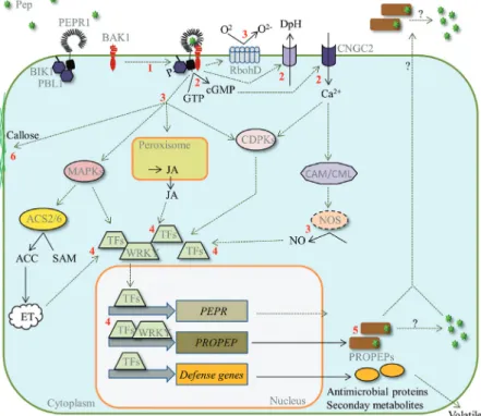
Fig. 1. Overview of the events following Pep perception. Pep perception by PEPRs leads to heteromerization with BAK1, mutual kinase phosphorylation
and further to the phosphorylation and the release of BIK1 (1). Next, ion channels are opened, leading to the alkalinization of the extracellular medium and likely to influx of Ca2+. In addition, PEPRs may produce cGMP, which may activate CNGC2 thereby leading to further influx of extracellular Ca2+ (2). The increase of Ca2+ plays a triple role: it supports RbohD activation leading to an oxidative burst (formation of O
2-), it triggers NO synthesis, likely via CaM and CML Ca2+ sensors, and it activates CDPKs (3). In parallel MAP kinase cascades are activated and levels of the defence hormones ET and JA rise (3). All these together modulate the activity of a multitude of transcription factors (TFs) including WRKYs, which in turn induce defence gene expression as well as the transcription of PEPRs and PROPEPs (4). PROPEPs might then either accumulate or are further processed into Peps and released (5). In the long term Pep perception also leads to the formation of callose (6) and the inhibition of seedling growth.
Changes in gene expression
As mentioned above, Peps as well as flg22 induced simi-lar sets of defence-related genes via MAPK- and CDPK-dependent signalling pathways (Boudsocq et al., 2010;
Flury et al., 2013). A recent study, which analysed transcrip-tomic changes after treatment with AtPep2 or the MAMP elf18, revealed that SA, ET and JA-inducible genes were upregulated by AtPep2 treatment whereas elf18 treatment led to an accumulation of mainly SA-responsive gene tran-scripts (Ross et al., 2014). In addition, even if both treat-ments induce the same gene like PR1 (a SA marker gene) the underlying signalling network is different since upregu-lation of PR1 transcription by elf18 but not by AtPep2 was impaired in the ethylene insensitive mutant ein2 (Tintor et al., 2013).
AtPep perception was reported to induce PDF1.2 and repress VSP2 transcription, both marker genes for JA (Huffaker et al., 2006; Tintor et al., 2013). Accordingly, AtPeps seem to specifically induce the so-called ERF-branch and repress the MYC2-dependent ERF-branch of JA-responsive genes. Furthermore, pepr1 pepr2 mutants showed reduced expression of ethylene responsive genes upon treatment with the ethylene precursor ACC indicat-ing that AtPep-perception contributes to the transcrip-tional upregulation of ethylene-responsive genes (Liu et al., 2013). Thus there is some support for the surpris-ing parallel induction of SA, ET and JA responsive genes upon AtPep2 treatment.
Beside the induction of defence-related genes two stud-ies showed an effect of AtPep perception on genes not directly linked to defence. First, AtPep1 perception led to the repression of GLUTAMINE DUMPER genes (GDUs), which encode amino acid exporters and are supposed to play a role in root development (Ma et al., 2014). And second, genes related to autophagy (APG7 and APG8a) and chlo-rophyll breakdown (PAO) were induced upon treatment of
Arabidopsis leaf tissue with AtPep1 (Gully et al., 2015).
Callose deposition and seedling growth inhibition
Callose deposition and seedling growth inhibition are mark-ers of late PTI responses. AtPep as well as flg22 trigger both responses although here subtle differences exist as well (Bartels et al., 2013; Liu et al., 2013). Flg22 perception appar-ently affects the whole seedling in its development whereas the inhibitory effect of AtPep perception impairs mainly root growth (Krol et al., 2010). The repression of the aforemen-tioned GUD genes might explain why AtPep perception has a special impact on root growth (Ma et al., 2014). Notably, the rise in cytosolic Ca2+ levels was reported to be equal in shoots and roots treated with AtPep1 whereas flg22 treatment trig-gered only a small rise in root Ca2+ levels (Ranf et al., 2011). Thus roots might just be much less sensitive to flg22 than to AtPeps. In contrast to the AtPEPRs, which have shown to be well expressed in roots, FLS2 expression is limited in roots to the stele and lateral root formation sites (Bartels et al., 2013;
Beck et al., 2014).
Production of secondary metabolites
PEPR-mediated induction of secondary metabolite synthesis has currently only been investigated in maize. ZmPep1 treat-ment of maize plants triggered the production of anthranilate and indole, both precursors for benzoxazinoid hydroxamic acid-related defences. Accordingly also the amount of the derived 2,4-dihydroxy-7-methoxy-1,4-benzoxazin-3-one glu-coside (DIMBOA-Glc), which is a strong antibiotic compound against bacterial and fungal pathogens as well as insect pests, increased significantly upon perception of ZmPep1 (Huffaker et al., 2011). In the follow-up study analysing the induction of anti-herbivore defences upon ZmPep3 treatment an increase of indole as well as the highly reactive benzoxazinoid precur-sor 2-hydroxy-4,7-dimethoxy-1,4-benzoxazin-3-one glucoside (HDMBOA-Glc) was reported (Huffaker et al., 2013).
Plants also release volatile secondary compounds in response to herbivores; this is considered to be an anti-herbivore response (by attracting predators) as well as a conserved instrument to communicate with neighbouring plants or tissues. Perception of ZmPep3 in maize was shown to trigger the release of ses-quiterpenes. The amount released was comparable to the one released upon detection of N-linolenoyl-L-glutamine (Gln-18:3), a strong elicitor present in the oral secretions of many lepidopteran species (Huffaker et al., 2013).
The Pep-PEPR system contributes to local
and systemic immunity
There is a growing body of evidence that the Pep-PEPR sys-tem is involved in local as well as syssys-temic immunity, and that it contributes to plant resistance against diverse patho-gens including bacteria, fungi and herbivores. In Arabidopsis, AtPep pretreatment or overexpression of AtPROPEP1 or
AtPROPEP2 has been reported to increase resistance to the
bacterial pathogen Pst DC3000 and the oomycete root path-ogen Pythium irregulare, respectively (Huffaker et al., 2006;
Yamaguchi et al., 2010). But pretreatment approaches are likely to create a rather artificial response, which might not be present under natural conditions. However, further pathogen studies were performed with the pepr1 pepr2 double mutant, which is insensitive to all AtPeps and better suited to uncover the contribution of the Pep-PEPR system to plant immunity.
Spray inoculation of Arabidopsis pepr1 pepr2 plants with
Pst DC3000 revealed a slightly increased susceptibility
towards this pathogen (Tintor et al., 2013). Notably, infiltra-tion of Pst DC3000 and other less virulent P. syringae strains did not show any increased susceptibility indicating that the Pep-PEPR system might play a role in stomatal immunity although neither AtPEPRs nor AtPROPEPs seem to be sig-nificantly expressed in guard cells (Bartels et al., 2013; Tintor et al., 2013; Ross et al., 2014).
The involvement of the Pep-PEPR system in fungal resist-ance also was confirmed. JA and ethylene are key hormones to orchestrate fungal resistance. Treatment of Arabidopsis
pepr1 pepr2 plants with the ethylene precursor ACC revealed
effect of an ACC pretreatment against infection with the fungal pathogen Botrytis cinerea was also impaired in pepr1
pepr2 plants (Liu et al., 2013).
Recently, the contribution to resistance against herbivores, first noted in ZmPep-pretreated maize plants (Huffaker et al., 2013), was confirmed in Arabidopsis by a challenge of pepr1
pepr2 plants with Spodoptera littoralis. Larvae of this generalist
herbivore performed much better on pepr1 pepr2 plants com-pared to wild-type Arabidopsis plants (Klauser et al., 2015).
In maize, resistance against fungi as well as herbivores has been studied with respect to the Pep-PEPR system (Huffaker et al., 2011, 2013). Due to the lack of receptor mutants in maize, current data are based on ZmPep-treatment studies only. The response patterns triggered by ZmPep1 and ZmPep3 show great similarity with those in Arabidopsis triggered by the perception of AtPeps. Both induce the production of JA and ET and acti-vate the transcription of defence-related genes (Huffaker et al., 2011, 2013). Pretreatment of maize plants with ZmPep1 leads to increased resistance against the fungal pathogens Cochliobolis
heterostrophus and Colletotrichum graminicola (Huffaker et al., 2011) whereas ZmPep3 pretreatment strengthens the resistance to the herbivore Spodoptera exigua including the release of anti-herbivore volatiles (Huffaker et al., 2013).
Recently, the first Pep-related study in tomato was per-formed. Silencing of a putative tomato SlPROPEP1 by virus-induced gene silencing led to a reduced expression of defence-related genes compared to the expression of these genes in control-treated plants. Moreover, silenced plants showed a reduced resistance towards the necrotrophic fungus
Pythium dissotocum (Trivilin et al., 2014).
Taken together there are numerous studies supporting the contribution of the Pep-PEPR system to plant resist-ance against a surprising diversity of pathogens. Notably, the induction of JA, SA as well as ethylene-specific genes, revealed by microarray-based determination of the AtPep2-triggered transcriptional changes (Ross et al., 2014), appears to be one special feature of the Pep-PEPR system that enables this broad contribution to the plant’s defence system.
Intriguingly the Pep-PEPR system takes part in systemic immunity as well. Similar to flg22, local AtPep2 application is sufficient to induce systemic immunity (Ross et al., 2014;
Mishina and Zeier, 2007). Also induction of systemic immu-nity by local infection with Pst DC3000 avrRpm1 is impaired in pepr1 pepr2 double mutants (Ross et al., 2014). Although it has been hypothesized that Peps might travel over long dis-tances and contribute to systemic immunity, this seems not to be the case since Pep-responsive genes are not induced in systemic leaves of AtPep2-treated plants. Thus the Pep-PEPR system rather contributes to or amplifies the generation of an unknown systemic signal.
Peps are regarded as damage- or
danger-associated molecular patterns: the
two models
Researchers have long wondered about the role of the Pep-PEPR system in plant biology but due to the lack of
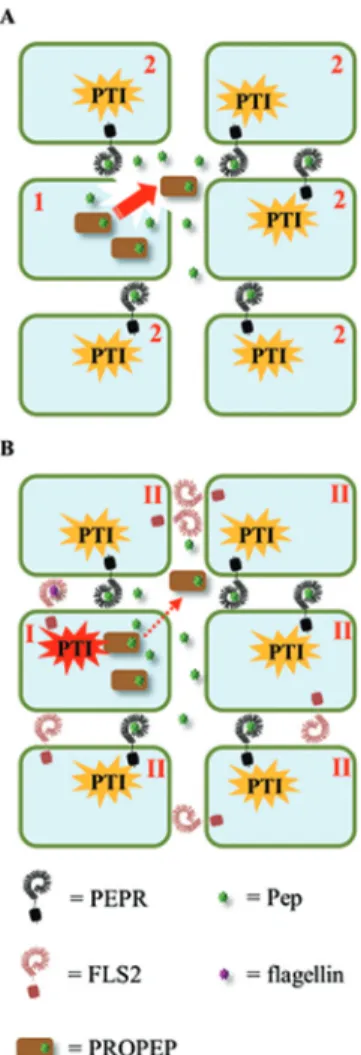
experimental data analysing the molecular circumstances that enable and promote a release of Peps into the extracellular space (and therewith to the potential activation of PEPRs), a clear picture has not yet emerged. Currently, two models are debated (Fig. 2).
(i) The damage model is based on the idea that PROPEPs and Peps reside in the cytosol and are released upon loss of cellular integrity due to damage. Detection of Peps by cells close to the site of damage induces their defence program and thus forms a barrier for pathogens to enter the plant body via the wounded tissue (Fig. 2A). This model would require a constitutive presence of PROPEPs in most cells of the plant body to develop a broad protective effect but sufficient protein data for PROPEPs is lacking. Furthermore, a rapid process-ing of PROPEPs into Peps would be crucial unless PROPEPs
Fig. 2. The damage and the danger model for activation of the Pep-PEPR
system. (A) The damage model: upon cellular damage (1), PROPEPs and Peps are passively released into the extracellular space (red arrow) and diffuse to neighbouring cells. Subsequently surrounding cells (2) detect the presence of PROPEPs and Peps in the extracellular space and induce a PTI-like response (orange). (B) The danger (or amplifier) model: after detection of a MAMP (here flagellin) the cell (I) triggers PTI (red). This cell then produces and actively releases PROPEPs and Peps into the extracellular space (red dotted arrow). As in model A, neighbouring cells (II) will subsequently detect the presence of PROPEPs and Peps in the extracellular space and induce a PTI-like response (orange) and therewith amplify the original danger signal.
are as ‘active’ as Peps. Here experimental data is needed to clarify if and how PROPEPs are processed.
(ii) The amplification model postulates a release of the pep-tides into the extracellular space in the situation of danger. Thereby the Peps might either prolong the immune response in the active cell (autocrine pathway) or spread the informa-tion locally to neighbouring cells to addiinforma-tionally induce their defence response (paracrine pathway) (Fig. 2B).
PROPEPs seem to lack a classical signal sequence to enter the secretory pathway and PROPEP-YFP fusion proteins did not localize to the secretory pathway (Huffaker et al., 2006; Bartels et al., 2013). Thus, PROPEPs or Peps would need to be exported as leaderless secretory proteins (LSPs) via unconventional routes similar to animal interleukin-1β or the yeast mating factor Matα (Ding et al., 2012; Piccioli and Rubartelli, 2013). In brief, release of LSPs can either work via non-vesicular direct crossing of proteins through the plasma membrane or via fusion of membrane-bound structures with the plasma membrane (Ding et al., 2012). Intriguingly two studies showed that after pathogen attack or treatment with SA a large number of LSPs are released into the apoplast but as yet the release of PROPEPs has not been shown (Cheng et al., 2009; Agrawal et al., 2010).
Ultimately both models might be correct, depending on the specific PROPEP. In Arabidopsis, the expression patterns differ strongly between the PROPEPs and their overall amino acid sequence shows little similarity. Moreover they also differ in their subcellular localization; thus it is possible that some are constitutively expressed and released upon damage, whereas others are induced upon danger detection and released in a strictly controlled manner. We should keep in mind that both models are based on the assumption that PROPEPs or Peps enter the extracellular space to bind to the PEPR-LRR domain and activate the PEPRs. If only one of the many PROPEPs is secreted via the secretory pathway it could bind already within the cell to PEPRs and trigger PEPR signalling.
Emerging roles of the Pep-PEPR system
in the regulation of plant stress and
development
Compared to the amount of data connecting PROPEPs and PEPRs to plant immunity there is still only a small number of studies supporting their roles in abiotic stress and plant development. This seems rather surprising since the authors who identified AtPep1 in 2006 already noted that overexpres-sion of AtPROPEP1 or AtPROPEP2 led to increased root biomass production (Huffaker et al., 2006). Remarkably, this observation is counterintuitive since perception of MAMPs and DAMPs often inhibit plant growth. Indeed, addition of AtPeps to Arabidopsis seedlings strongly inhibits root growth (Krol et al., 2010). However, since the roots of Arabidopsis
pepr1 and pepr2 single mutant plants were found to be
signifi-cantly shorter than wild-type roots (Qi et al., 2010; Ma et al., 2014) it has been hypothesized that cell-type-specific expres-sion of PROPEPs and PEPRs might be responsible for a coordinated regulation of root growth (Krol et al., 2010; Ma
et al., 2014). Beside development a study on 69 root-expressed LRR-RLKs reported Arabidopsis pepr1 to be more resistant to osmotic stress and auxin but more sensitive to darkness. Similarly, Arabidopsis pepr2 mutants were found to be more resistant to elevated NaCl concentrations and again more sensitive to darkness (ten Hove et al., 2011). Intriguingly, in
Arabidopsis continuous darkness induced AtPROPEP3
tran-scription (Gully et al., 2015). In the same study we showed that a combination of continuous darkness and treatment with AtPeps accelerated dark/starvation-induced senescence. Due to the observation that AtPep perception triggered the transcription of genes encoding central enzymes of the autophagy machinery we tend to speculate that the Pep-PEPR system might be involved in the regulation of nutrient remobilization. Whether an enhanced nutrient remobiliza-tion is meant to be part of the Pep-induced defence response or if the Pep-PEPR system plays a role in starvation resist-ance needs to be investigated in more detail. However, it is not a side-effect of PTI activation upon Pep perception since the bacterial elicitors flg22 and elf18 had no effect on the dark/ starvation-induced senescence response (Gully et al., 2015).
Further support for roles of the Pep-PEPR system beside plant immunity comes from in silico analyses. First, based on a phylogenetic approach, both AtPEPRs cluster together in the leucine-rich repeat receptor-like kinase subfamily XI, which comprises receptors involved in plant development and differ-entiation, and not in subfamily XII with pattern recognition receptors like FLS2 or EFR (Yamaguchi et al., 2010). This could be an indication of their evolutionary background and thus they might still operate in signalling pathways involved in plant development in addition to the PTI-inducing pathway. Second, an evaluation of microarray data revealed a co-expres-sion of some AtPROPEPs with genes linked to reproduction (Bartels et al., 2013). Also experimental data showed that only some of the AtPROPEP promoters are responsive to biotic stress whereas others are insensitive to this type of stress sug-gesting that they might respond to abiotic stress or develop-mental signals (Huffaker et al., 2006; Bartels et al., 2013).
Important targets for Pep research
Without doubt Peps and PEPRs contribute to plant immunity. Compared to flg22 and elf18, Peps induce a distinct defence response pattern, despite large commonalities of their signal-ling pathways. One of their hallmarks is the simultaneous induction of JA, ET and SA-dependent defence responses and the respective full spectrum resistance against bacterial, fun-gal and herbivorous pathogens. Understanding the likely pro-cessing and release mechanism will reveal if Peps are damage signals or if they amplify signals of danger or even both. The identification of Metacaspase-9 as the processing enzyme for GRIM REAPER points here to a new direction (Wrzaczek et al., 2015). A closer look at the PROPEP sequences reveals a conserved arginine in front of the Pep sequences. Since metacaspases tend to cleave their substrates after arginine and lysine (Vercammen et al., 2004), they appear to be inter-esting candidates for PROPEP cleavage. For the investiga-tion of the release of PROPEPs and Peps two approaches
might be fruitful. First, the ongoing proteomics approaches investigating the Arabidopsis secretome could be combined with immunity-inducing treatments to promote the possible (unconventional) release of PROPEPs or Peps. Alternatively, PROPEPs could be fused to fluorescent proteins known to be detectable in the extracellular milieu like mCherry. Therewith the real-time behaviour of PROPEPs upon damage or danger could be monitored.
Small signalling peptides are widely used by the plant to coordinate its development. Clustering of AtPEPRs with LRR-RLKs involved in plant development, and coregula-tion of some AtPROPEPs with genes linked to develop-mental processes, fosters the idea that the PROPEP-PEPR system is derived from systems regulating plant development (Yamaguchi et al., 2010; Bartels et al., 2013). The aberrant root development of Arabidopsis pepr1 and pepr2 noted by
Ma et al. (2014) may provide a first hint in this direction. In this regard, the exclusive expression of AtPROPEP4 and
AtPROPEP7 in root tips might also be an indication for an
involvement in root development (Bartels et al., 2013). In the future, the Arabidopsis pepr1 pepr2 double mutant should also be carefully investigated with respect to plant develop-ment. This mutant certainly has no obvious phenotype, since it has been studied intensively already by many scientists. However, experts in plant development may have the trained eye and the suitable tools to discover more subtle phenotypes. Thus it is important not to ignore these first fine connections between the Pep-PEPR system and the regulation of plant development.
Acknowledgements
This work was supported by the Swiss National Science Foundation (grant 31003A_127563).
References
Agrawal GK, Jwa NS, Lebrun MH, Job D, Rakwal R. 2010. Plant
secretome: unlocking secrets of the secreted proteins. Proteomics 10,
799–827.
Albert M. 2013. Peptides as triggers of plant defence. Journal of
Experimental Botany 64, 5269–5279.
Ashton AR. 2011. Guanylyl cyclase activity in plants? Proceedings of the
National Academy of Sciences of the United States of America 108, E96;
author reply E97–98.
Bartels S, Lori M, Mbengue M, van Verk M, Klauser D, Hander T, Boni R, Robatzek S, Boller T. 2013. The family of Peps and their
precursors in Arabidopsis: differential expression and localization but similar induction of pattern-triggered immune responses. Journal of Experimental Botany 64, 5309–5321.
Baxter A, Mittler R, Suzuki N. 2013. ROS as key players in plant stress
signalling. Journal of Experimental Botany doi:10.1093/jxb/ert375
Beck M, Wyrsch I, Strutt J, Wimalasekera R, Webb A, Boller T, Robatzek S. 2014. Expression patterns of flagellin sensing 2 map to
bacterial entry sites in plant shoots and roots. Journal of Experimental Botany 65, 6487–6498.
Beck M, Zhou J, Faulkner C, MacLean D, Robatzek S. 2012.
Spatio-temporal cellular dynamics of the Arabidopsis flagellin receptor reveal activation status-dependent endosomal sorting. The Plant Cell 24,
4205–4219.
Boller T, Felix G. 2009. A renaissance of elicitors: perception of
microbe-associated molecular patterns and danger signals by pattern-recognition receptors. Annual Review of Plant Biology 60, 379–406.
Boudsocq M, Willmann MR, McCormack M, Lee H, Shan LB, He P, Bush J, Cheng SH, Sheen J. 2010. Differential innate immune signalling
via Ca2+ sensor protein kinases. Nature 464, 418.
Cheng FY, Blackburn K, Lin YM, Goshe MB, Williamson JD. 2009.
Absolute protein quantification by LC/MS(E) for global analysis of salicylic acid-induced plant protein secretion responses. Journal of Proteome Research 8, 82–93.
Czyzewicz N, Yue K, Beeckman T, De Smet I. 2013. Message in a
bottle: small signalling peptide outputs during growth and development. Journal of Experimental Botany 64, 5281–5296.
Ding Y, Wang J, Wang J, Stierhof Y-D, Robinson DG, Jiang L. 2012.
Unconventional protein secretion. Trends in Plant Science 17, 606–615. Dombrowski JE, Pearce G, Ryan CA. 1999. Proteinase
inhibitor-inducing activity of the prohormone prosystemin resides exclusively in the C-terminal systemin domain. Proceedings of the National Academy of Sciences of the United States of America 96, 12947–12952.
Ferrari S, Savatin DV, Sicilia F, Gramegna G, Cervone F, Lorenzo GD. 2013. Oligogalacturonides: plant damage-associated molecular
patterns and regulators of growth and development. Frontiers in Plant Science 4, 49.
Flury P, Klauser D, Schulze B, Boller T, Bartels S. 2013. The
anticipation of danger: MAMP perception enhances AtPep-triggered oxidative burst. Plant Physiology 161, 2023–2035.
Goyette J, Geczy CL. 2011. Inflammation-associated S100 proteins:
new mechanisms that regulate function. Amino Acids 41, 821–842. Gully K, Hander T, Boller T, Bartels S. 2015. Perception of Arabidopsis
AtPep peptides, but not bacterial elicitors, accelerates starvation-induced senescence. Frontiers in Plant Science 6.
Huffaker A, Dafoe NJ, Schmelz EA. 2011. ZmPep1, an ortholog
of Arabidopsis elicitor Peptide 1, regulates maize innate immunity and enhances disease resistance. Plant Physiology 155, 1325–1338. Huffaker A, Pearce G, Ryan CA. 2006. An endogenous peptide signal
in Arabidopsis activates components of the innate immune response. Proceedings of the National Academy of Sciences of the United States of America 103, 10098–10103.
Huffaker A, Pearce G, Veyrat N, et al. 2013. Plant elicitor peptides are
conserved signals regulating direct and indirect antiherbivore defense. Proceedings of the National Academy of Sciences of the United States of America 110, 5707–5712.
Huffaker A, Ryan CA. 2007. Endogenous peptide defense signals in
Arabidopsis differentially amplify signaling for the innate immune response. Proceedings of the National Academy of Sciences of the United States of America 104, 10732–10736.
Jones JD, Dangl JL. 2006. The plant immune system. Nature 444,
323–329.
Khimji AK, Rockey DC. 2010. Endothelin—biology and disease. Cellular
signalling 22, 1615–1625.
Kim B, Lee Y, Kim E, Kwak A, Ryoo S, Bae SH, Azam T, Kim S, Dinarello CA. 2013. The Interleukin-1alpha precursor is biologically
active and is likely a key alarmin in the IL-1 family of cytokines. Frontiers in Immunology 4, 391.
Klauser D, Desurmont GA, Glauser G, Vallat A, Flury P, Boller T, Turlings TC, Bartels S. 2015. The AtPep-PEPR system is induced by
herbivore feeding and contributes to JA-mediated plant defence against herbivory. Journal of Experimental Botany 66, 5327–5336.
Klauser D, Flury P, Boller T, Bartels S. 2013. Several MAMPs, including
chitin fragments, enhance AtPep-triggered oxidative burst independently of wounding. Plant Signaling & Behavior 8, doi: 10.4161/psb.25346
Krol E, Mentzel T, Chinchilla D, et al. 2010. Perception of the
Arabidopsis danger signal peptide 1 involves the pattern recognition receptor AtPEPR1 and its close homologue AtPEPR2. Journal of Biological Chemistry 285, 13471–13479.
Kwezi L, Meier S, Mungur L, Ruzvidzo O, Irving H, Gehring C.
2007. The Arabidopsis thaliana brassinosteroid receptor (AtBRI1) contains a domain that functions as a guanylyl cyclase in vitro. PLoS ONE 2, e449.
Lacombe S, Rougon-Cardoso A, Sherwood E, et al. 2010. Interfamily
transfer of a plant pattern-recognition receptor confers broad-spectrum bacterial resistance. Nature Biotechnology 28, 365–369.
Liu Z, Wu Y, Yang F, Zhang Y, Chen S, Xie Q, Tian X, Zhou J-M.
Proceedings of the National Academy of Sciences of the United States of America 110, 6205–6210.
Logemann E, Birkenbihl RP, Rawat V, Schneeberger K, Schmelzer E, Somssich IE. 2013. Functional dissection of the PROPEP2 and
PROPEP3 promoters reveals the importance of WRKY factors in
mediating microbe-associated molecular pattern-induced expression. New Phytologist 198, 1165–1177.
Lori M, van Verk M, Hander T, Schatowitz H, Klauser D, Flury P, Gehring C, Boller T, Bartels S. 2015. Evolutionary divergence of the
plant elicitor peptides Peps produced interfamily incompatibility. Journal of Experimental Botany 66, 5315–5325.
Lu D, Lin W, Gao X, Wu S, Cheng C, Avila J, Heese A, Devarenne TP, He P, Shan L. 2011. Direct ubiquitination of pattern recognition receptor
FLS2 attenuates plant innate immunity. Science 332, 1439–1442. Ma C, Guo J, Kang Y, Doman K, Bryan AC, Tax FE, Yamaguchi Y, Qi Z. 2014. AtPEPTIDE RECEPTOR2 mediates the AtPEPTIDE1 induced
cytosolic Ca rise which is required for the suppression of Glutamate Dumper gene expression in Arabidopsis roots. Journal of Integrative Plant Biology 56, 684–694.
Ma Y, Walker RK, Zhao Y, Berkowitz GA. 2012. Linking ligand
perception by PEPR pattern recognition receptors to cytosolic Ca2+ elevation and downstream immune signaling in plants. Proceedings of the National Academy of Sciences of the United States of America 109,
19852–19857.
Ma Y, Zhao Y, Walker RK, Berkowitz GA. 2013. Molecular steps in
the immune signaling pathway evoked by plant elicitor peptides: Ca2+-dependent protein kinases, nitric oxide, and reactive oxygen species are downstream from the early Ca2+ signal. Plant Physiology 163,
1459–1471.
Malinowski R, Higgins R, Luo Y, Piper L, Nazir A, Bajwa VS, Clouse SD, Thompson PR, Stratmann JW. 2009. The tomato brassinosteroid
receptor BRI1 increases binding of systemin to tobacco plasma membranes, but is not involved in systemin signaling. Plant Molecular Biology 70, 603–616.
Mishina TE, Zeier J. 2007. Pathogen-associated molecular pattern
recognition rather than development of tissue necrosis contributes to bacterial induction of systemic acquired resistance in Arabidopsis. Plant Journal 50, 500–513.
Moreau M, Lindermayr C, Durner J, Klessig DF. 2010. NO synthesis
and signaling in plants--where do we stand? Physiologia Plantarum 138,
372–383.
Ni J, Clark SE. 2006. Evidence for functional conservation, sufficiency,
and proteolytic processing of the CLAVATA3 CLE domain. Plant Physiology
140, 726–733.
Nühse TS, Peck SC, Hirt H, Boller T. 2000. Microbial elicitors induce
activation and dual phosphorylation of the Arabidopsis thaliana MAPK 6. Journal of Biological Chemistry 275, 7521–7526.
Pearce G, Yamaguchi Y, Munske G, Ryan CA. 2008. Structure-activity
studies of AtPep1, a plant peptide signal involved in the innate immune response. Peptides 29, 2083–2089.
Piccioli P, Rubartelli A. 2013. The secretion of IL-1beta and options for
release. Seminars in immunology 25, 425–429.
Qi Z, Verma R, Gehring C, Yamaguchi Y, Zhao YC, Ryan CA, Berkowitz GA. 2010. Ca2+ signaling by plant Arabidopsis thaliana Pep peptides depends on AtPepR1, a receptor with guanylyl cyclase activity, and cGMP-activated Ca2+ channels. Proceedings of the National Academy of Sciences of the United States of America 107, 21193–21198.
Ranf S, Eschen-Lippold L, Frohlich K, Westphal L, Scheel D, Lee J.
2014. Microbe-associated molecular pattern-induced calcium signaling requires the receptor-like cytoplasmic kinases, PBL1 and BIK1. BMC Plant Biology 14, 374.
Ranf S, Eschen-Lippold L, Pecher P, Lee J, Scheel D. 2011. Interplay
between calcium signalling and early signalling elements during defence responses to microbe- or damage-associated molecular patterns. The Plant Journal 68, 100–113.
Robatzek S, Chinchilla D, Boller T. 2006. Ligand-induced endocytosis
of the pattern recognition receptor FLS2 in Arabidopsis. Genes & Development 20, 537–542.
Ross A, Yamada K, Hiruma K, Yamashita-Yamada M, Lu X, Takano Y, Tsuda K, Saijo Y. 2014. The Arabidopsis PEPR pathway couples local
and systemic plant immunity. The EMBO Journal 33, 62–75.
Roux M, Schwessinger B, Albrecht C, Chinchilla D, Jones A, Holton N, Malinovsky FG, Tor M, de Vries S, Zipfel C. 2011. The Arabidopsis
leucine-rich repeat receptor-like kinases BAK1/SERK3 and BKK1/ SERK4 are required for innate immunity to hemibiotrophic and biotrophic pathogens. The Plant Cell 23, 2440–2455.
Ryan CA, Pearce G. 2003. Systemins: A functionally defined family
of peptide signal that regulate defensive genes in Solanaceae species. Proceedings of the National Academy of Sciences of the United States of America 100, 14577–14580.
Stegmann M, Anderson RG, Ichimura K, Pecenkova T, Reuter P, Zarsky V, McDowell JM, Shirasu K, Trujillo M. 2012. The ubiquitin
ligase PUB22 targets a subunit of the exocyst complex required for PAMP-triggered responses in Arabidopsis. The Plant Cell 24, 4703–4716. Schulze B, Mentzel T, Jehle A, Mueller K, Beeler S, Boller T, Felix G, Chinchilla D. 2010. Rapid heteromerization and phosphorylation of
ligand-activated plant transmembrane receptors and their associated kinase BAK1. Journal of Biological Chemistry 285, 9444–9451. Tabata R, Sawa S. 2014. Maturation processes and structures of small
secreted peptides in plants. Frontiers in Plant Science 5, 311.
Tang J, Han Z, Sun Y, Zhang H, Gong X, Chai J. 2015. Structural basis
for recognition of an endogenous peptide by the plant receptor kinase PEPR1. Cell Research 25, 110–120.
ten Hove CA, Bochdanovits Z, Jansweijer VM, Koning FG, Berke L, Sanchez-Perez GF, Scheres B, Heidstra R. 2011. Probing the roles
of LRR RLK genes in Arabidopsis thaliana roots using a custom T-DNA insertion set. Plant Molecular Biology 76, 69–83.
Tintor N, Ross A, Kanehara K, Yamada K, Fan L, Kemmerling B, Nurnberger T, Tsuda K, Saijo Y. 2013. Layered pattern receptor
signaling via ethylene and endogenous elicitor peptides during Arabidopsis immunity to bacterial infection. Proceedings of the National Academy of Sciences of the United States of America 110, 6211–6216.
Trivilin AP, Hartke S, Moraes MG. 2014. Components of different
signalling pathways regulated by a new orthologue of AtPROPEP1 in tomato following infection by pathogens. Plant Pathology 63, 1110–118. Tsiatsiani L, Van Breusegem F, Gallois P, Zavialov A, Lam E, Bozhkov PV. 2011. Metacaspases. Cell Death and Differentiation 18,
1279–1288.
van de Veerdonk FL, Netea MG, Dinarello CA, Joosten LAB. 2011.
Inflammasome activation and IL-1 and IL-18 processing during infection. Trends in Immunology 32, 110–116.
Vercammen D, Belenghi B, van de Cotte B, Beunens T, Gavigan JA, De Rycke R, Brackenier A, Inze D, Harris JL, Van Breusegem F. 2006. Serpin1 of Arabidopsis thaliana is a suicide inhibitor for
metacaspase 9. Journal of Molecular Biology 364, 625–636.
Vercammen D, van de Cotte B, De Jaeger G, Eeckhout D, Casteels P, Vandepoele K, Vandenberghe I, Van Beeumen J, Inze D, Van Breusegem F. 2004. Type II metacaspases Atmc4 and Atmc9 of
Arabidopsis thaliana cleave substrates after arginine and lysine. The Journal of Biological Chemistry 279, 45329–45336.
Vlot AC, Klessig DF, Park SW. 2008. Systemic acquired resistance: the
elusive signal(s). Current Opinion in Plant Biology 11, 436–442.
Wrzaczek M, Vainonen JP, Stael S, et al. 2015. GRIM REAPER peptide
binds to receptor kinase PRK5 to trigger cell death in Arabidopsis. The EMBO Journal 34, 55–66.
Xia YJ, Suzuki H, Borevitz J, Blount J, Guo ZJ, Patel K, Dixon RA, Lamb C. 2004. An extracellular aspartic protease functions in Arabidopsis
disease resistance signaling. The EMBO Journal 23, 980–988. Yamaguchi Y, Pearce G, Ryan CA. 2006. The cell surface
leucine-rich repeat receptor for AtPep1, an endogenous peptide elicitor in Arabidopsis, is functional in transgenic tobacco cells. Proceedings of the National Academy of Sciences of the United States of America 103,
10104–10109.
Yamaguchi Y, Huffaker A. 2011. Endogenous peptide elicitors in higher
plants. Current Opinion in Plant Biology 14, 351–357.
Yamaguchi Y, Huffaker A, Bryan AC, Tax FE, Ryan CA. 2010. PEPR2
is a second receptor for the Pep1 and Pep2 peptides and contributes to defense responses in Arabidopsis. The Plant Cell 22, 508–522.
Zhang J, Li W, Xiang T, et al. 2010. Receptor-like cytoplasmic kinases
integrate signaling from multiple plant immune receptors and are targeted by a Pseudomonas syringae effector. Cell Host & Microbe 7,