Chromosome sizes and phylogenetic relationships between
serotypes of Actinobacillus pleuropneumoniae
Bruno Chevallier
a, Dominique Dugourd
b, Kazimirez Tarasiuk
c, Joseèe Harel
b,
Marcelo Gottschalk
b, Maryleéne Kobisch
a, Joachim Frey
d;*
aCNEVA-Ploufragan, Les Croix, P.O. Box 53, F-22440 Ploufragan, France
b Groupe de Recherche sur les Maladies Infectieuses du Porc, Faculteè de Meèdecine Veèteèrinaire, Universiteè de Montreèal,
Saint-Hyacinthe, Queè., Canada
cNational Veterinary Research Institute, 24-100 Pulawy, Poland
dInstitute for Veterinary Bacteriology, University of Berne, Laenggasstrasse 122, CH-3012 Berne, Switzerland
Received 5 January 1998; accepted 22 January 1998
Abstract
The genome size of Actinobacillus pleuropneumoniae was determined by pulsed field gel electrophoresis of AscI and ApaI
digested chromosomal DNA. The genome size of the type strain 4074T (serotype 1) was determined to be 2404 þ 40 kb. The
chromosome sizes for the reference strains of the other serotypes range between 2.3 and 2.4 Mb. The restriction pattern profiles of AscI, ApaI and NheI digested chromosomes showed a high degree of polymorphism among the different serotype reference strains and allowed their discrimination. The analysis of the macrorestriction pattern polymorphism revealed phylogenetic relationships between the different serotype reference strains which reflect to some extent groups of serotypes known to cross-react serologically. In addition, different pulsed fields gel electrophoresis patterns also revealed heterogeneity in the chromosomal structure among different field strains of serotypes 1, 5a, and 5b, while strains of serotype 9 originating from most distant geographical places showed homogeneous ApaI patterns in pulsed field gel electrophoresis. z 1998 Federation of European Microbiological Societies. Published by Elsevier Science B.V.
Keywords: Actinobacillus pleuropneumoniae; Chromosome size; Pulsed ¢eld gel electrophoresis
1. Introduction
During the last 20 years, porcine pleuropneumonia has been recognized as one of the major diseases in swine production world-wide [1]. The etiological
agent of this contagious pulmonary disease is Actino-bacillus pleuropneumoniae [2], a Gram-negative bac-terium of the Pasteurellaceae family [3]. Twelve se-rotypes and two subtypes of A. pleuropneumoniae biotype 1 (NAD-dependent) and two serotypes of biotype 2 (NAD-independent) have been identi¢ed on the basis of capsular and lipopolysaccharide anti-gens [4^6]. Several reports have indicated that the di¡erences in virulence among the various A. pleuro-pneumoniae serotypes are mainly related to the
pro-* Corresponding author. Tel.: +41 (31) 631 2484; Fax: +41 (31) 631 2634; E-mail: jfrey@vbi.unibe.ch
duction of three APX exotoxins [7^9]. Serotypes 1, 2, 5a, 5b, 9 and 11 are frequently involved in severe outbreaks with high mortality and severe pulmonary lesions [2,9]. The other serotypes are less virulent but can be found in outbreaks with lower levels of mortality [2,9]. Serotyping of A. pleuropneumoniae strains is a valuable tool for epidemiological stud-ies and provides important information for the decision taking in control programs aiming at the eradication of the virulent types of the pathogen. Epidemiological analyses showed that the preval-ence of speci¢c serotypes varies with geographic lo-cation [10,11]. However, cross-reactivity, between A. pleuropneumoniae serotypes 1, 9 and 11, between serotypes 3, 6 and 8, and between serotypes 4 and 7 has been described [12]. These cross-reactions were shown to be associated with common epitopes and common components of the cell wall antigens [12,13].
Molecular methodologies applied to the whole ge-nome are becoming more and more relevant in pro-viding means to estimate the genetic relationship be-tween di¡erent biotypes and serotypes of a given bacterial species, and also for accurate bacterial typ-ing and subtyptyp-ing systems [14,15]. Restriction pat-terns of the whole bacterial chromosome, which could be resolved by pulsed ¢eld gel electrophoresis (PFGE) [16], o¡er a good method to understand the degree of genetic relatedness or variability among the di¡erent serotypes. In addition, PFGE of chromoso-mal DNA that was digested by restriction enzymes with recognition sites that occur infrequently in bac-terial genomes presents a reliable and e¤cient meth-od for estimating genome sizes and constructing macro-restriction maps of bacterial chromosomes [17]. The purpose of this study was to determine the chromosome sizes of the A. pleuropneumoniae type strain and all serotype reference strains and to study the genomic relationship between the di¡erent serotype reference strains and between strains of giv-en serotypes.
2. Materials and methods 2.1. Bacterial strains
Reference strains of A. pleuropneumoniae
repre-senting serotypes 1^12 were S4074, S1536, S1421, M62, K17, L20, FemÖ, WF83, 405, CVJ13261, 13039, 56153, and 8329, and came from R. Nielsen (National Veterinary Laboratory, Copenhagen, Den-mark). Field strains of serotypes 1 (21 strains), 5a (11 strains), 5b (10 strains), and 9 (9 strains) for biotype 1, and serotypes 2 (4 strains) and 9 (6 strains) for biotype 2 were isolated in France, Po-land, The Netherlands, Canada, Czech Republic, and Italy.
2.2. Preparation of genomic DNA
Each bacterial strain was grown for 6 h at 37³C in 10 ml PPLO broth (Difco Laboratories, Detroit, MI, USA) containing 12.5 g l31 yeast extract. After cen-trifugation at 1500Ug for 15 min at 4³C, the bacte-rial pellet was washed with 10 ml of cold TE bu¡er (10 mM Tris-(hydroxymethyl)-aminomethane, 1 mM EDTA, pH 8.0). After centrifugation at 1500Ug for 15 min at 4³C, the cells were resuspended in 1 ml of cold TE bu¡er supplemented with 3 mg ml31 lyso-zyme. 1 ml of 1% agarose (Bio-Rad, Ivry sur Seine, France) in TE bu¡er, prewarmed at 60³C, was added and immediately poured into a block former (Bio-Rad). After solidi¢cation, agarose blocks were incubated for 6 h at 37³C in 10 ml TE bu¡er supplemented with 3 mg ml31 lysozyme (Sigma-Al-drich, St Quentin Fallavier, France). Blocks were then transferred to 10 ml fresh TE bu¡er supple-mented with 1% SDS and 0.25 mg ml31 proteinase K (Eurobio, Les Ulis, France) and incubated overnight at 37³C. Blocks were transferred to 10 ml fresh TE bu¡er supplemented with 1% SDS and 0.25 mg ml31 proteinase K and incubated for another 4 h at 37³C. They were then washed three times with TE bu¡er for 1 h at 37³C. The gel blocks were run for 1 h at 6.0 V cm31 in 0.5UTBE bu¡er (45 mM Tris, 45 mM boric acid, 10 mM EDTA). This pre-migration step removed degraded or extrachromosomal DNA from the gel and thus strongly reduced the smear background in PFGE. Blocks were conserved at 4³C in 0.5UTBE for several months. It is important to note that it was important to use PPLO broth complemented with yeast extract as medium for growth of A. pleuro-pneumoniae for PFGE analysis in order to obtain clean and clearly interpretable results.
2.3. Restriction digests and electrophoresis
Restriction enzyme digestion of genomic DNA embedded in agarose blocks was undertaken by a modi¢cation of procedures described by McClelland et al. [16]. Each restriction digest was done with half
of a genomic DNA block. After 30 min in 1 ml of the restriction bu¡er, 800 Wl was removed and the blocks were ¢rst incubated overnight with 30 U of restriction endonuclease at the temperature recom-mended by the manufacturer, then another 10 U was added for 1 h.
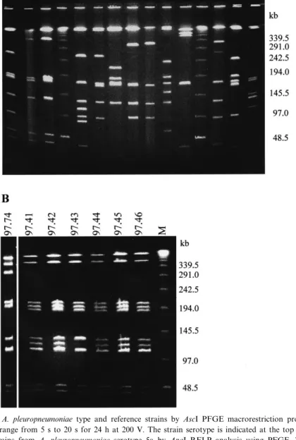
Fig. 1. A: Comparison of A. pleuropneumoniae type and reference strains by AscI PFGE macrorestriction pro¢les. Electrophoresis was performed with pulse time range from 5 s to 20 s for 24 h at 200 V. The strain serotype is indicated at the top of each lane. B: Di¡eren-tiation of di¡erent ¢eld strains from A. pleuropneumoniae serotype 5a by ApaI RFLP analysis using PFGE. The gel shown was made with a pulse time range from 5 s to 20 s for 24 h at 200 V. Panels A and B: Molecular masses of the size standard (lane M) made from polymers of bacteriophage V genomes are indicated on the right of the gel.
PFGE was performed at 14³C with a DRII or DRIII CHEF system (Bio-Rad) in 0.5UTBE. Blocks were then cut in 6 1-mm slices and placed in 1.2 or 1.5% agarose (Bio-Rad Pulsed Field Certi¢ed Agar-ose) gel wells. Pulsed ¢eld gels were then run at var-ious pulse ramps ranging from 0.1 to 4 s (5^100 kb), 5 to 20 s (20^300 kb), 10 to 40 s (200^800 kb) and 40 to 100 s (500^1300 kb), for 24^48 h at 200 V in order to obtain the best resolution of restriction fragments in di¡erent size ranges. Gels were stained for 1 h after electrophoresis with TBE bu¡er containing 0.5 Wg ml31 ethidium bromide, destained overnight with TBE bu¡er, examined over UV transillumina-tion and photographed.
2.4. Data analysis
Gel images were scanned and analyzed using the BIO-GENE Software (Vilber Lourmat, Inc., Marne la Valleèe, France). Sizes of restriction fragments were determined by comparison with standard lambda DNA concatemers (Pharmacia, Orsay, France). Dendrograms were created from a matrix of band matching using the Jaccard coe¤cient [18] and the unweighted pair group method of arith-metic averages (UPGMA) clustering fusion strategy [19].
3. Results 3.1. Genome size
In order to generate chromosomal digests with relatively few, clearly resolvable fragments, genomic DNA A. pleuropneumoniae which contains 42% G+C [20] was digested with restriction endonucleases with GC-rich recognition sequences such as AscI (5P-GGCGCGCC-3P), ApaI (5P-GGGCCC-3P), NgoMI (5P-GCCGGC-3P), SmaI (5P-CCCGGG-3P), SacII (5P-CCGCGG-3P), S¢I (5P-GGCCNNNNNGGCC-3P) and NotI (5P-GCGGCCGC-(5P-GGCCNNNNNGGCC-3P). Among these re-striction enzymes, AscI and ApaI were the most suit-able since they cut the chromosomes of the A. pleu-ropneumoniae serotype reference strains into 6^12 fragments ranging from 11 kb to 1217 kb. Figs. 1A and 2A show photographs from PFGE runs with pulse parameters separating medium-sized (20^400-kb) fragments. For the determination of the chromo-some sizes of the A. pleuropneumoniae type strain and the serotype reference strains, the sizes of the fragments from AscI digested chromosomes were de-termined from PFGE using bacteriophage V multi-mers as molecular mass standards. In each experi-ment three di¡erent gels with di¡erent pulse parameters were used in order to discriminate the
Fig. 2. PFGE of restriction enzyme digests of A. pleuropneumoniae DNA reference strains serotype 1, 9 and 11. The restriction enzymes used were ApaI in A and NheI in B. Molecular sizes (lane M) are indicated on the right of the gel. Pulse time conditions were 5 s to 20 s for 24 h at 200 V in A and 0.1 s to 4 s for 24 h at 200 V in B.
Table 1 Sizes of chromosomal Asc I restriction fragments from A. pleuropneumoniae reference strains Fragment Serotype/Strain 1/S4074 2/S1536 3/S1421 4/M62 5a/K17 5b/L20 6/Fem x 7/WF83 8/405 9/CVJ13261 10/13039 11/56153 12/8329 A 840.5 þ 5.5 832.6 þ 4.2 866.7 þ 0.3 911.8 þ 1.6 854.3 þ 0.6 1217 þ 14 830.3 þ 2.4 614.6 þ 4 852.2 þ 0.7 827.6 þ 1.1 878.7 þ 5.7 831.7 þ 3.1 841.5 þ 0.7 B 644.4 þ 8 665.5 þ 5.2 602.9 þ 6.4 599 þ 8.5 783.4 þ 5.6 333.9 þ 14 628 þ 21 593 þ 4 784.8 þ 3.3 649.3 þ 15 607.9 þ 11 640.5 þ 6.4 600.2 þ 7.6 C 266.4 þ 2.7 263.5 þ 5.1 271.3 þ 14 223.2 þ 4 316.4 þ 10 322 þ 6.7 235.8 þ 2.5 428.7 þ 1.6 329.7 þ 4.9 351.2 þ 9.2 249.9 þ 3.4 349.2 þ 7.8 181.3 þ 5.1 D 182.1 þ 3.8 176.6 þ 1.7 174.7 þ 1.8 188.6 þ 3.5 172.7 þ 3.2 178 þ 2.5 188.3 þ 1.1 390.7 þ 5.2 180 þ 4 186.9 þ 7.2 189.6 þ 3 181.7 þ 3.4 174.7 þ 3.7 E 175.1 þ 3.2 130.7 þ 2.1 170 þ 1.2 175.2 þ 2.3 127.9 þ 3.4 135.4 þ 3.7 173.3 þ 1.4 177.8 þ 4.8 135.8 þ 5.1 180 þ 7 176.1 þ 4.2 175.3 þ 2.4 141.3 þ 4.2 F 121.7 þ 11 114 þ 2.7 130.7 þ 2.1 175.2 þ 2.3 97.3 þ 3.8 97 þ 3.5 151.7 þ 1.1 177.8 þ 4.8 94.5 þ 0.4 137.9 þ 8.9 168.3 þ 2.6 130.9 þ 2.5 133.3 þ 4.2 G 86.9 þ 4.3 98.8 þ 3 96.1 þ 3.7 128.1 þ 2.4 129.2 þ 0.6 25.5 þ 2.3 31.6 þ 1 50.3 þ 1.2 97.7 þ 2.8 50.3 þ 1.2 133.1 þ 4 H 51.2 þ 0.3 86 þ 1.8 70.7 þ 3.1 32.8 þ 2.1 24.4 þ 2.8 32.8 þ 2.1 111.9 þ 4 I 35.8 þ 0.9 29.3 þ 0.9 39.7 þ 0.6 Total (kb) 2404 þ 40 2397 þ 27 2312 þ 30 2401 þ 25 2352 þ 17 2283 þ 44 2407 þ 33 2408 þ 27 2409 þ 19 2416 þ 52 2392 þ 36 2392 þ 29 2357 þ 34 Values are expressed as means þ standard deviation of three independent determinations from di¡erent gels.
medium fragments (20^300 kb) (parameters: 5^20 s for 24 h), the large fragments (200^800 kb) (param-eters: 10^40 s for 48 h) and the very large fragments (500^1300 kb) (parameters 40^100 s for 36 h). The chromosome sizes were calculated by addition of the sizes of the individual fragments from three inde-pendent experiments and are given in Table 1. The size of the chromosome of the A. pleuropneumoniae type strain 4074Twas determined to be 2404 þ 40 kb. The sizes of the di¡erent serotype reference strains di¡ered only slightly from that of the type strain and ranged between 2283 kb and 2416 kb.
3.2. RFLP analysis
A. pleuropneumoniae reference strains gave clear di¡erences in the restriction patterns after digestion with AscI (Fig. 1) and with ApaI (data not shown), showing a high degree of polymorphism with 12 dif-ferent pro¢les for 13 reference strains. Reference strains of serotypes 9 and 11 could not be distin-guished using either ApaI or AscI (Figs. 1A and 2A). However, these two serotypes could be di¡er-entiated using NheI GCTAGC-3P) and XbaI (5P-TCTAGA-3P), which had a higher resolution poten-tial since they generated more than 30 genomic frag-ments ranging from 180 kb to less than 5 kb and produced complex banding patterns (Fig. 2B).
ApaI patterns allowed a clear distinction between biotype 1 reference strains and biotype 2 strains from serotype 2 and from serotype 9. In addition analysis of di¡erent ¢eld strains from serotypes 1, 5a and 5b resulted in di¡erent, but related ApaI patterns as shown for strains of serotype 5a which generally
dif-fer in one or two ApaI fragments (Fig. 1B). In con-trast, no di¡erences in ApaI patterns at all could be detected in serotype 9 ¢eld strains isolated from most distant countries such as Australia, Italy, France, Poland, Czech Republic and Switzerland (results not shown).
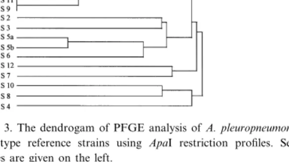
The genetic relatedness between the di¡erent A. pleuropneumoniae serotype reference strains was eval-uated using PFGE pro¢les produced by ApaI (Fig. 3). This analysis reveals the close relationship be-tween serotypes 1, 9 and 11 (85% similarity) and between 5a and 5b (80% similarity) in particular, grouping also serotypes 2 and 3, serotypes 6, 5a and 5b, as well as 4, 8 and 10, at a similarity in the 40^50% range. When the patterns created with AscI were used, slightly di¡erent groupings were ob-tained, due to the lower number of fragments pro-duced by this enzyme.
4. Discussion
The sizes of the chromosomes of the A. pleuro-pneumoniae type and serotype reference strains were determined to range between 2.3 and 2.4 Mb using PFGE analysis of AscI and ApaI digested chromosomal DNA. The size of the A. pleuropneu-moniae chromosome is comparable to that of other species of the family Pasteurellaceae which includes Actinobacillus actinomycetemcomitans (2.3 Mb) [21], Haemophilus parain£uenzae (2.34 Mb), Haemophilus in£uenzae (1.83 Mb) [22] and Haemophilus ducreyi (1.76 Mb) [23]. The sizes of the chromosomes of the di¡erent A. pleuropneumoniae strains analyzed vary relatively little compared to other bacterial spe-cies. In contrast to the well conserved chromosome sizes, the di¡erent A. pleuropneumoniae serotypes and biotypes show polymorphism in their restriction enzyme patterns as revealed by PFGE ¢ngerprinting, re£ecting distinct genetic di¡erences. Such di¡erences were seen between the di¡erent serotypes and be-tween biotypes of the same serotype, but also among strains of the same sero- and biotype. In particular serotypes 1, 5a and 5b showed several di¡erent, but serotype-related restriction patterns. The genomic di¡erences between the two di¡erent biotypes of the same serotype, as deduced by PFGE analyses, must be more abundant than what can be expected
Fig. 3. The dendrogam of PFGE analysis of A. pleuropneumoniae serotype reference strains using ApaI restriction pro¢les. Sero-types are given on the left.
from a single genetic locus involved in NAD metab-olism. Interestingly no di¡erences were encountered in serotype 9 ¢eld strains isolated from most distant geographic areas. Serotype 9 hence seems to present a particularly stable widespread clonal line.
Analysis of the restriction enzyme polymorphisms by using the Jaccard coe¤cient and the UPGMA strategy for clustering showed the close genetic rela-tionship of the serologically and toxigenetically re-lated serotypes 1, 9 and 11 and of serotypes 5a and 5b. However, it clearly di¡erentiates between sero-types 3, 6, and 8 and between 4 and 7 which are known to show serological cross-reactions which are known to interfere in serological subtyping. The genetic relationships between the di¡erent A. pleuropneumoniae serotype reference strains as determined by PFGE show a signi¢cant direct rela-tionship with DNA:DNA homology values of the same strains as determined by free solution hybrid-ization experiments [24]. From our experiments we estimate that PFGE has the same, or even a higher discriminatory potential than other molecular tools such as multilocus enzyme electrophoresis, restric-tion fragment length polymorphism using frequently cutting enzymes together with high resolution gels, or arbitrarily primed polymerase chain reaction [24^ 26]. PFGE, however, seems to be a method that can be well standardized in order to give highly reprodu-cible results which allow comparative studies be-tween di¡erent laboratories. PFGE therefore repre-sents a powerful tool for studies on taxonomy and epidemiology of A. pleuropneumoniae in particular, as well as for many other bacteria where other, mostly phenotypic methods do not allow su¤cient discrimination between di¡erent subtypes of a given species.
Acknowledgments
This work was supported by l'Association Region-ale de l'Interprofession Porcine de Bretagne, l'O¤ce National Interprofessionel de la Viande, de l'Elevage et de l'Aviculture de France, a grant from the Fonds pour la Formation de Chercheurs et l'Aide aé la Re-cherche (FCAR-MEQ-279) Queèbec and from the Swiss National Science foundation (Grant 3100.39123-93). We thank R. Grumdey from
Diag-nostic Nouveau Alimentaire, Montpellier, France for his contribution.
References
[1] Fenwick, B. and Henry, S. (1994) Porcine pleuropneumonia. J. Am. Vet. Med. Assoc. 204, 1334^1340.
[2] Nicolet, J. (1992) Actinobacillus pleuropneumoniae. In: Dis-eases of Swine (Leman, A.D., Straw, B.E., Mengeling, W.L., D'Allaire, S. and Taylor, D.J., Eds.), pp. 401^408. Iowa State University Press, Ames, IA.
[3] Pohl, S., Bertschinger, H.U., Frederiksen, W. and Manheim, W. (1983) Transfer of Haemophilus pleuropneumoniae and the Pasteurella haemolytica-like organism causing porcine necrotic pleuropneumonia to the genus Actinobacillus (Actinobacillus pleuropneumoniae comb. nov.) on the basis of phenotypic and deoxyribonucleic acid relatedness. Int. J. Syst. Bacteriol. 33, 510^514.
[4] Nielsen, R. (1987) Serological characterization of Actinobacil-lus pleuropneumoniae strains and proposal of a new serotype: serotype 12. Acta Vet. Scand. 27, 453^455.
[5] Mittal, K.R., Higgins, R. and Larivieére, S. (1983) Identi¢ca-tion and serotyping of Haemophilus pleuropneumoniae by coagglutination test. J. Clin. Microbiol. 18, 1351^1354. [6] Perry, M.B., Altman, E., Brisson, J.R., Baynon, L.M. and
Richards, J.C. (1990) Structural characteristics of the antigen-ic capsular polysaccharides and lipopolysaccharides involved in the serological classi¢cation of Actinobacillus (Haemophi-lus) pleuropneumoniae strains. Serodiagn. Immunother. Infect. Dis. 4, 299^308.
[7] Beck, M., Van Den Bosch, F., Jongenelen, I.M.C.A., Loe¡en, P.L.W., Nielsen, R., Nicolet, J. and Frey, J. (1994) RTX toxin genotypes and phenotypes in Actinobacillus pleuropneumoniae ¢eld strains. 32, 2749^2754.
[8] Jasen, R., Briaire, J., Smith, H.E., Dom, P., Haesebrouck, F., Kamp, E.M., Gielkens, A.L.J. and Smits, M.A. (1995) Knock-out mutants of Actinobacillus pleuropneumoniae serotype 1 that are devoid of RTX toxins do not activate or kill porcine neutrophils. Infect. Immun. 63, 27^37.
[9] Frey, J. (1995) Virulence in Actinobacillus pleuropneumoniae and RTX toxins. Trends Microbiol. 3, 257^261.
[10] Mittal, K.R., Higgins, R., Larivieére, S. and Nadeau, M. (1992) Serological characterization of Actinobacillus pleuro-pneumoniae strains isolated from pigs in Quebec. Vet. Micro-biol. 32, 135^148.
[11] Chevallier. B., Morvan, H., Guzylack, S. and Kobisch, M. (1997) L'isolement d'Actinobacillus pleuropneumoniae en France. J. Rech. Porcine 29, 23^30.
[12] Mittal, K.R. and Bourdon, S. (1991) Cross-reactivity and antigenic heterogeneity among Actinobacillus pleuropneumo-niae strains of serotypes 4 and 7. J. Clin. Microbiol. 29, 1344^1347.
[13] Beynon, L.M., Gri¤th, D.W., Richards, J.C. and Perry, M.B. (1992) Characterization of the lipopolysaccharide O antigens of Actinobacillus pleuropneumoniae serotypes 9 and 11:
anti-genic relationships among serotypes 9, 11, and 1. J. Bacteriol. 174, 5324^5331.
[14] Bert, F., Branger, C. and Lambert-Zechovsky, N. (1997) Pulsed-¢eld gel electrophoresis is more discriminating than multilocus enzyme electrophoresis and random ampli¢ed pol-ymorphic DNA analysis for typing pyogenic Streptococci. Curr. Microbiol. 34, 226^229.
[15] Grothues, D. and Tuëmmler, B. (1991) New approaches in genome analysis by PFGE: application to the analysis of Pseudomonas species. Mol. Microbiol. 5, 2763^2776. [16] McClelland, M., Jones, R., Patel, Y. and Nelson, M. (1987)
Restriction endonucleases for pulsed ¢eld mapping of bacte-rial genomes. Nucleic Acids Res. 15, 5985^6005.
[17] Fonstein, M. and Haselkorn, R. (1995) Physical mapping of bacterial genomes. J. Bacteriol. 177, 3361^3369.
[18] Colwell, R.R. and Austin, B. (1981) Numerical taxonomy. In: Manual of Methods for General Bacteriology (Gerhardt, P., Murray, R.G.E., Costilow, R.N., Nester, E.W., Wood, W.A., Krieg, N.R. and Phillips, G.B., Eds.), pp. 444^449. American Society of Microbiology, Washington, DC.
[19] Sneath, P.H.A. and Sakal, R.R. (1973) Numerical Taxonomy. W.H. Freeman, San Francisco, CA.
[20] Mannheim, W. (1984) Pasteurellaceae. In: Bergey's Manual of Systematic Bacteriology (Krieg, N.R. and Holt, J.G., Eds.), Vol. 1, pp. 550^5575. Williams and Wilkins, Baltimore, MD.
[21] Valcaret, J., Allardet-Servent, A., Bourg, G., O'Callaghan, D., Michailesco, P. and Ramuz, M. (1997) Investigation of the Actinobacillus actinomycetemcomitans genome by pulsed ¢eld gel electrophoresis. Oral Microbiol. Immunol. 12, 33^ 39.
[22] Fleischmann, R.D., Adams, M.D., White, O., Clayton, R.A., Kirkness, E.-F., Kerlavage, A.R., Bult, C.J., Tomb, J.F., Dougherty, B.A., Merrick, J.M. et al. (1995) Whole-genome random sequencing and assembly of Haemophilus in£uenzae Rd. Science 269, 496^512.
[23] Hobbs, M.M., Leonardi, M.J., Zaretzky, F.R., Wang, T.-H. and Kawula, T.H. (1996) Organization of the Haemophilus ducreyi 35000 chromosome. Microbiology 142, 2587^2594. [24] Borr, J.D., Ryan, D.A.J. and MacInnes, J. (1991) Analysis of
Actinobacillus pleuropneumoniae and related organisms by DNA-DNA hybridization and restriction endonucleases ¢n-gerprinting. Int. J. Syst. Bacteriol. 41, 121^129.
[25] Moller, K., Nielsen, R., Andersen, L.V. and Kilian, M. (1992) Clonal analysis of the Actinobacillus pleuropneumoniae popu-lation in a geographically restricted area by multilocus enzyme electrophoresis. J. Clin. Microbiol. 30, 623^627.
[26] Hennessy, K.J., Iandolo, J.J. and Fenwick, B.W. (1993) Sero-type identi¢cation of Actinobacillus pleuropneumoniae by arbi-trarily primed polymerase chain reaction. J. Clin. Microbiol. 31, 1155^1159.