Morbidity and validity of the hemiclamshell approach for
thoracic surgery
q
Didier Lardinois
a, Martin Sippel
a, Matthias Gugger
b, Michael Dusmet
c, Hans-Beat Ris
a,*
aDepartment of Thoracic and Cardiovascular Surgery, University Hospital, Berne, Switzerland bDivision of Pneumonology, University Hospital, Berne, Switzerland
cDepartment of Surgery, University Hospital, Lausanne, Switzerland
Received 28 September 1998; received in revised form 27 April 1999; accepted 27 April 1999
Abstract
Objective: This is a prospective study to evaluate the indications and outcome of the hemiclamshell incision (longitudinal partial sternotomy combined with an antero-lateral thoracotomy) as used for a consecutive series of patients requiring surgery for various thoracic pathologies not ideally approached by postero-lateral thoracotomy, sternotomy or thoracoscopy. Methods: All patients with a hemiclamshell incision performed between 1994 and 1998 were prospectively analyzed regarding indications, postoperative morbidity and outcome (clinical examination and pulmonary function testing) in order to validate this incision for thoracic surgery. Results: 25 patients (15 men, 10 women) with an age ranging from 16 to 73 years (mean 43 years) underwent a hemiclamshell incision. The indications for the hemiclamshell approach were (1) chest trauma with massive hemorrhage requiring urgent access to the mediastinum and the ipsilateral pleural space (40%), (2) tumors of the anterior cervico-thoracic junction with suspicion of vascular involvement (28%) and (3) lesions involving both one chest cavity and the mediastinum (32%). The 30-day mortality was 8%. One patient suffered a sternal wound infection, mediastinitis and pleural empyema after a gun shot wound, whereas wound healing was uneventful in all other patients. Analgesic require-ments for postoperative pain relief were not increased as compared to those following a standard thoracotomy. At 3 months normal sensitivity of the entire chest wall and intact shoulder girdle function was noted in 90% of the patients. Pulmonary function testing showed no restriction due to the hemiclamshell incision. Conclusions: The hemiclamshell incision is a useful approach in selected patients and does not cause more morbidity or long-term sequelae than a standard thoracotomy. q 1999 Elsevier Science B.V. All rights reserved.
Keywords: Hemiclamshell; Trap door incision; Shoulder girdle function; Pulmonary function testing; Postoperative pain
1. Introduction
Tumors arising from the cervico-thoracic junction or in the anterior mediastinum with extension to one chest cavity are usually dif®cult to access by classical incisions such as a postero-lateral thoracotomy or median sternotomy. These incisions do not provide optimal exposure of the operative ®eld [1±3]. Alternative incisions such as the anterior cervico-thoracic approach as described by Dartevelle [4], or the manubrial cervico-thoracic approach have been promoted in order to improve the exposure in these situa-tions. However, these incisions may cause impaired shoulder girdle function, irritation of the brachial plexus and cosmetic problems, due to the resection of the clavicle,
especially if an additional thoracotomy is required. The hemiclamshell approach was therefore proposed as a possi-ble alternative in patients suffering from anterior cervico-thoracic tumors or injuries to the subclavian vessels [5]. However, this approach has not gained wide spread accep-tance since it is believed that this incision causes more morbidity and long-term sequelae than standard approaches. We have therefore analyzed the indications and the post-operative morbidity of the hemiclamshell incision in a consecutive series of patients in order to validate this approach for thoracic pathologies which are not easy to access through standard incisions.
2. Materials and methods
This prospective study included all patients who under-went a hemiclamshell incision in our institution between 1994 and 1998. The indications for a hemiclamshell access
European Journal of Cardio-thoracic Surgery 16 (1999) 194±199
1010-7940/99/$ - see front matter q 1999 Elsevier Science B.V. All rights reserved. PII: S1010-7940(99)00156-6
qPresented at the 12th Annual Meeting of the European Association for
Cardio-thoracic Surgery, Brussels, Belgium, September 20±23, 1998. * Corresponding author. Thoracic Surgery, Centre Hospitalier Universi-taire Vaudois, Lausanne, Switzerland. Tel.:3141111; fax: 141-21-3142360.
were classi®ed as follows: (1) severe chest trauma with massive hemorrhage requiring urgent access to the medias-tinum and the chest cavity; (2) large tumors of the anterior cervico-thoracic junction; (3) lesions with involvement of both the upper mediastinum and one lung.
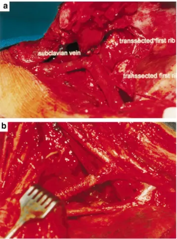
The hemiclamshell access consists of an antero-lateral thoracotomy usually in the fourth or ®fth intercostal space, combined with a partial longitudinal median sternot-omy [1], with the possible extension of the incision along the anterior edge of the sternocleidomastoid muscle (Fig. 1). The patient is positioned in the supine position with the arm slightly abducted. The skin is incised from the jugular notch to the mid portion of the sternum and then follows the submammary fold to the mid-axillary line. The pectoralis major muscle is divided at its origin and the pectoralis minor muscle is split along its ®bers without being divided. The chest cavity is entered through the 4th or 5th intercostal space (depending on the size and location of the lesion). The internal mammary vessels are ligated and divided and a median sternotomy is performed from this level up to the jugular notch. The intercostal muscles are bluntly dissected off of the underlying rib back to the costo-transversal junc-tion of the involved interspace without dividing the over-lying chest wall muscles. This enables good exposure of the involved chest cavity, the mediastinum and the anterior cervico-thoracic junction. If greater access to the anterior cervico-thoracic junction is required, the ®rst rib may be transsected laterally to the internal mammary vessels using a gygli saw after identi®cation and preservation of the subclavian vein. The division of the ®rst rib at this point will further improve the exposure of the anterior cervico-thoracic junction (Fig. 2a,b) while preserving the integrity of the shoulder girdle.
At the end of the procedure, the sternum is closed with wires and the thoracotomy with resorbable pericostal vicryl sutures. The chest wall muscles are readapted in layers.
The postoperative course of all patients who underwent a
hemiclamshell incision was prospectively assessed. A clin-ical control as well as pulmonary function testing were performed 3 months after the operation on each patient. The clinical evaluation included a subjective assessment of chest wall and shoulder girdle complaints, of physical and professional activities, followed by clinical examination with assessment of the chest wall integrity and the shoulder girdle function.
3. Results
Ten patients (40%) underwent operation for severe chest trauma with life-threatening hemorrhage requiring urgent access to the mediastinum and the injured chest cavity via a hemiclamshell incision. Five lesions were related to pene-trating chest wounds, three to blunt chest trauma following motor vehicle accidents and two to iatrogenic lesions of respectively the innominate and the azygos veins during mediastinoscopy. Pulmonary procedures included three lobectomies, four segmentectomies or wedge resections and two pulmonary tractotomies of the involved lung [6]. One patient also required celiotomy for simultaneous thor-acic and abdominal injury. Intrapericardial control of the pulmonary vessels was required in four patients, with repair
Fig. 1. The hemiclamshell incision consists of a median sternotomy combined with an anterolateral thoracotomy and gives excellent exposure of the mediastinum and the involved chest cavity, as shown in this patient suffering from a large cyst of the thoracic duct situated in the upper cervi-cothoracic junction.
Fig. 2. Further access to the cervicothoracic junction can be obtained by dividing the ®rst rib just lateral to the internal mammary vessels (a); after resection and reconstruction of the subclavian vessels for an anteriorly situated Pancoast tumor (b).
of the pulmonary veins. In the two patients with non small cell lung carcinoma (NSCLC) who suffered iatrogenic lesions during mediastinoscopy, therapy required repair of the azygos and innominate vein, respectively, followed by lobectomy and mediastinal lymph node dissection.
Seven patients (28%) operated through a hemiclamshell incision had large tumors of the anterior cervico-thoracic junction, comprising neurogenic tumors in three patients, a cervico-thoracic adult teratoma. Three of these patients had a tracheal sequestration, a necrotizing infection of left upper lobe with erosion of the left subclavian artery in an immunocompromised patient, and an aneurysm of the left subclavian artery. In the last patient a hemiclamshell inci-sion was used because simultaneous aorto-coronary bypass surgery was required in addition to the exclusion of a large (5 cm) aneurysm of the left subclavian artery. In this group of seven patients, resection and reconstruction of the subcla-vian vessels was required in three patients.
Eight patients (32%) required a hemiclamshell incision to resect lesions with involvement of the anterior mediastinum and one chest cavity including three patients with upper lobe
NSCLC who underwent resection after induction radioche-motherapy, two patients with a thymoma and additional lesions in one lung and three patients with a lymphangioma extending into the anterior mediastinum. Three patients required en bloc resection of adjacent structures such as the innominate vein, the phrenic nerve and parts of the anterior chest wall; the chest wall was reconstructed by the sandwich technique [7].
The overall 30-day mortality was 8% (2/25). One patient died as a result of multiorgan failure after a severe traf®c accident and one due to acute mesenteric infarction follow-ing aorto-coronary bypass and left subclavian artery aneur-ysm repair.
Complications were observed in six patients (24%). These included atelectasis and pneumonia of the involved lung in three, a chylothorax in two, and mediastinitis with pleural empyema in one patient. All three patients with pneumonia were treated by bronchoscopy, minitracheotomy and aggressive bronchial toilette and antibiotics and recov-ered without sequelae. The two patients with chylothorax required reoperation. One patient underwent ligation of the thoracic duct supradiaphragmatically through a small right thoracotomy and one patient direct thoracic duct ligation at the level of the left subclavian vein where it was injured during operation for a large cervico-thoracic teratoma. The chylothorax resolved and further recovery was uneventful. The patient who developed mediastinitis and pleura empyema suffered from gun shot injury requiring urgent left upper lobectomy and had an initially uneventful recov-ery. Suppuration was then observed in the sternal wound and empyema developed once mediastinitis was diagnosed. The hemiclamshell incision was re-opened, the mediasti-num was debrided and the left lung was decorticed with an additional intrathoracic muscle transposition. This covered the debrided mediastinum and the bronchial stump of the left lower lobe. The patient recovered after a prolonged period of hospitalization but with uneventful local wound healing.
Fig. 3. The cosmetical result observed 2 weeks after a hemiclamshell incision required to access an aspergilloma invading the left subclavian artery.
Table 1
Hemiclamshell incision: pulmonary function at 3 months following an elective procedurea
Preop FEV1 (l) VR (% pred) Postop FEV1 (l) VR (% pred)
Lobectomy 3.1 139 3.1 139 1.1 36 1.8 59 2.5 80 1.6 52 4.7 125 3.6 99 2.5 87 2.2 82 3.1 70 2.4 66 Lesser/no resection 3.5 83 3.4 80 3.2 90 3.4 95 1.9 72 2.7 102 3.9 97 3.6 92 3.9 115 3.7 109 3.1 99 2.9 93
Three month follow-up was performed in 23 of 25 patients (92%) and revealed uneventful healing in all patients at that time point, including the patient with mediastinitis and empyema (Fig. 3). Sternal stability was observed in all patients. A loss of sensitivity of the anterior chest wall was noted in 2/23 patients (9%) and was persis-tent in one. Nineteen of 23 patients (83%) had no chest wall complaints. Two patients had constant pain in the operated chest wall and two others had intermittent discomfort. One of the two patients complaining of constant pain had under-gone an en bloc resection of the chest wall. Shoulder girdle function was symmetrical and normal in 92% of the patients. In two patients, the abduction of the involved upper extremity was limited to 908 and 1208, respectively. One of those patients had simultaneous chest wall resection. Pulmonary function testing 3 months post surgery suggested that the hemiclamshell incision per se did not lead to restriction. In 6/7 patients who survived an emer-gency procedure without assessment of their preoperative lung function, pulmonary function tests 3 months after the operation were normal. The patient with empyema and mediastinitis demonstrated mild restriction at pulmonary function testing. All 12 patients who survived an elective procedure had preoperative pulmonary function testing. The six patients who underwent a lobectomy had a slight decrease in postoperative FEV1. The six patients without formal lung resection had a pulmonary function at follow-up which was unchanged (Table 1). Preoperative pulmonary function was not measured in four elective cases.
At 3 months 74% of the patients had returned to work, 17.3% were working at a rate of 50% and 8.7% did not return to work, one due to tumor progression and the patient with empyema and mediastinitis for psychiatric reasons. 4. Discussion
Although the classical incisions in thoracic surgery have stood the test of time and have provided indubitable merits for the approach to a variety of intrathoracic pathologies, some inconveniences and drawbacks have been observed in selected patients. The postero-lateral thoracotomy initially described by Crafoord gives a superb view of all aspects of the lung and its hilum, the oesophagus, the aorta and the diaphragm [8]. This incision can be enlarged by means of the interscapulo-vertebral approach to resect the upper part of the chest cavity in Pancoast tumours, as described by Paulson [9]. The main disadvantage of the standard postero-lateral thoracotomy is the poor exposure of the anterior cervico-thoracic junction. In addition, the visualization and access to the mediastinum is limited, and the lateral position required for thoracotomy may not be tolerated by hemody-namically unstable patients in emergency situations. Median sternotomy offers a suitable alternative for a variety of indi-cations, especially for the treatment of cardiac injuries and mediastinal tumours. It has shown some advantages compared to thoracotomy, with less pain and no need to
dissect the chest wall muscles. However, the inferior and posterior regions of the chest cavity, mediastinum and lungs are less well exposed than by thoracotomy, and the distal part of the subclavian vessels is not easily accessible [10]. The drawbacks of these classical incisions have led to the development of other incisions for selected indications, especially to improve the accessibility to diseases situated in the anterior aspect of the cervico-thoracic junction or to injured subclavian vessels. The approach described by Darte-velle [4], which is an anterior cervico-thoracic incision (consisting in an L-shaped anterior cervical incision with resection of the medial part of the clavicle) allows good exposure of the thoracic inlet. However, irritation of the brachial plexus and impairment of shoulder girdle function have been observed after this approach, especially if this incision is combined with a thoracotomy [4]. Furthermore, exposure of the hilar region and the proximal part of the subclavian vessels is limited and a thoracotomy has to be added if a resection of the chest wall below the second rib is indicated. The hemiclamshell incision (trap door incision or book thoracotomy) was described to access injured subcla-vian arteries [1,5]. It consists of a median sternotomy combined with an antero-lateral thoracotomy and provides an excellent view of the thoracic inlet including the subcla-vian vessels, and of the mediastinum and the involved chest cavity. Exposure can be improved by dividing the ®rst rib laterally to the internal mammary vessels. This incision is also well suited for emergency thoracotomies since it can be performed with the patient in the supine position and less manipulation of the heart is required to reach the infero-posterior aspect of the left chest cavity as compared to ster-notomy [11,12].
Chest trauma represented approximately half of the patient population. These patients were haemodynamically unstable due to life-threatening hemorrhage within one chest cavity arising from parenchymal or hilar injuries. In these situations, the hemiclamshell incision gave good exposure to the entire mediastinum and the chest cavity. In addition, several patients needed urgent intrapericardial access to the pulmonary vessels in order to control the massive hemorrhage which was due to a proximal rupture of the pulmonary vessels. This was very easily to perform through the hemiclamshell incision. All the other intrathor-acic structures within one hemithorax are well exposed by the hemiclamshell incision [2,10,13]. In two patients with NSCLC, access to vessels injured during mediastinoscopy was urgently required, and lobectomy with a formal mediastinal lymph node dissection was then performed without any additional dif®culty as compared to standard thoracotomy. Injuries of the upper branches of the aorta are also easily approached with this incision, with safe prox-imal control [14±16].
The second group of patients suffered from large tumours of the anterior cervico-thoracic junction. In this situation, the classical incisions do not give good exposure of the entire operative ®eld [17]. The hemiclamshell incision offers a
good alternative in this situation and it can be extended into the neck to become the trap door thoracotomy described by Nazzaro [18]. The visualization of the thoracic inlet can be further improved by transection of the ®rst rib at its medial end [19]. In addition, this incision can be helpful to reach the anterior aspect of the cervico-thoracic spine (C4-T3) [20± 22].
The third group of patients had lesions involving one hemithorax and the mediastinum.
The 30-day mortality in our series was 8% and the complication rate was 24%. These were mainly related to the nature of the underlying disease or injury. One major complication which might have been related to the hemi-clamshell incision consisted in mediastinitis and empyema which occurred in a patient suffering from a gun shot wound. The reported incidence of wound infection follow-ing elective thoracotomy or sternotomy is low (0.4±0.8%) [23,24]. This also seems to be the case for the hemiclam-shell approach since no infections were observed in any of our elective cases.
At 3 months following surgery 17.4% of the patients complained of chest wall discomfort. This is similar to the results obtained in a prospective study comparing chest wall complaints after thoracotomy and thoracoscopy, respec-tively, in which 35% of the patients in each group presented with chest wall complaints during follow-up [25]. The shoulder girdle function in our series of patients assessed at 3 months can also be compared favourably to that observed after thoracotomy [25]. It has been suggested that impaired shoulder girdle function might be related to postoperative pain control and physiotherapy rather than to the transection of chest wall muscles [26,27]. Our results also suggest that the hemiclamshell incision per se does not lead to impaired postoperative pulmonary function. In our series, in patients with elective operations requiring lung resection lesser than lobectomy, the pulmonary function tests at 3 months after the operation showed results very similar to the preoperative values.
In conclusion, our results suggested that the hemiclam-shell incision offers improved exposure of the operative ®eld in selected patients without causing more postoperative morbidity, chest wall complaints, shoulder girdle dysfunc-tion or impairment of pulmonary funcdysfunc-tion than standard approaches.
References
[1] Schaff HV, Brawley RK. Operative management of penetrating vascular injuries of the thoracic outlet. Surgery 1977;82:182±191. [2] Mitchell ME, Muakkassa FF, Poole GV, Rhodes RS, Griswold JA.
Surgical approach of choice for penetrating cardiac wounds. J Trauma 1993;34:17±20.
[3] Tavares S, Hankins JR, Moulton AL, Sequeira A, McLaughlin JS. Management of penetrating cardiac injuries: the role of emergency room thoracotomy. Ann Thorac Surg 1984;38:183±187.
[4] Dartevelle PG, Chapelier AR, Macchiarini P, Lenot B, Cerrina J,
Lafont D. Anterior transcervical-thoracic approach for radical resec-tion of lung tumors invading the thoracic inlet. J Thorac Cardiovasc Surg 1994;108:389±392.
[5] Hood RM. Techniques in general thoracic surgery, Philadelphia: Saunders, 1985 pp. 64±67.
[6] Wall MI, Hirshberg A, Mattox KL. Pulmonary tractotomy with selec-tive vascular ligation for penetrating injuries of the lung. Am J Surg 1989;168:665±669.
[7] Arnold PG, Pairolero PC. Chest-wall reconstruction: an account of 500 consecutive patients. Plast Reconstr Surg 1996;98:804±810. [8] Destable MD, Azorin JF, Branscheid D. La thoracotomie
posteÂro-lateÂrale. La paroi thoracique, voies d'abord, reÂparation, approches multidisciplinaires. Int. Thorax 1998;:23±25.
[9] Azorin JF, Lancelin C. La thoracotomie posteÂro-lateÂrale haute. La paroi thoracique, voies d'abord, reÂparation, approches multidiscipli-naires. Int. Thorax 1998;98±100.
[10] Martinez Ibanez V, Marques Gubern A, Sanchez de Toledo J, Perez A, Tarroch J. Surgical treatment in tumors of the anterior mediasti-num. Cir Pediatr 1990;3:154±159.
[11] Graham JM, Feliciano DV, Mattox KL, Beall Jr AC, De Bakey ME. Management of subclavian vascular injuries. J Trauma 1980;20:537± 544.
[12] Mattox KL. Thoracic vascular trauma. J Vasc Surg 1988;7:725±729. [13] Bergeron P, Guennaoui T, Henric A, Rudondy P, Alessandri C. Primary surgical resection of tumors of the anterior mediastinum. Ann Chir 1993;47:167±169.
[14] Steenburg RW, Rawitch MM. Cervico-thoracic approach for subcla-vian vessel injury from compound fracture of the clavicle. Ann Surg 1963;157:839±846.
[15] George Jr SM, Croce MA, Fabian TC, Mangiante EC, Pate JW. Cervicothoracic arterial injuries: recommendations for diagnosis and management. World J Surg 1991;15:134±139.
[16] Wall Jr MJ, Granchi T, Liscum K, Mattox KL. Penetrating thoracic vascular injuries. Surg Clin North Am 1996;76:749±761.
[17] Kraus DH, Huo J, Burt M. Surgical access to tumors of the cervi-cothoracic junction. Head Neck 1995;17:131±136.
[18] Nazzaro JM, Arbit E, Burt M. `Trap-door' exposure of the cervi-cothoracic junction (Technical note). J Neurosurg 1994;80:338±341. [19] Nomori H, Nara S, Horio H. Modi®ed trap-door thoracotomy for malignancies invading the subclavian and innominate vessels. Thorac Cardiovasc Surg 1995;43:204±207.
[20] Zimmermann JM, De Graeve B, Cador L, Colonna MA, Coblence JF, Lemesle L. Rare tumor of the postero-superior mediastinum. Thera-peutic approach. Apropos of a case. Review of the literature. Ann Chir 1991;45:695±698.
[21] Gieger M, Roth PA, Wu JK. The anterior cervical approach to the cervicothoracic junction. Neurosurgery 1995;37:704±709.
[22] Haniuda M, Morimoto M, Nishimura H, Kobayashi O, Fujioka F. A case of chondrosarcoma arising from the left ®rst rib. Kyobu Geka 1990;43:835±838.
[23] Hazelrigg SR, Wellons Jr HA, Schneider JA, Kolm P. Wound compli-cations after median sternotomy. Relationship to internal mammary grafting. J Thorac Cardiovasc Surg 1989;98:1096±1099.
[24] Grmoljez PF, Barner HH, Willman VL, Kaiser GC. Major complica-tions of median sternotomy. Am J Surg 1975;130:679±681. [25] Furrer M, Rechsteiner R, Eigenmann V. Signer. Ch., Althaus, U., Ris,
H.B. Thoracotomy versus thoracoscopy: differences in postoperative pulmonary function, pain and chest wall complaints. Eur J Cardi-othorac Surg 1997;12:82±87.
[26] Landreneau RJ, Mack MJ, Hazelrigg SR, Keenan RJ, Ferson PF. Prevalence of chronic pain after pulmonary resection by thoracotomy or video-assisted thoracic surgery. J Thorac Cardiovasc Surg 1995;109:1255±1256.
[27] Hazelrigg SR, Landreneau RJ, Boley TM, Walls JT, Curtis JJ. The effect of muscle-sparing versus standard posterolateral thoracotomy on pulmonary function, muscle strength, and postoperative pain. J Thorac Cardiovasc Surg 1991;101:394±400.
Appendix A. Conference discussion
Dr T. Dosios (Athens, Greece): Do you always divide the sternocleido-mastoid muscle? And, if you divide it, do you repair the muscle after that? Also do you have any experience of excising the clavicle or dividing the clavicle?
Dr Lardinois: We divided the sternocleidomastoid muscle only in patients with large tumors of the anterior cervicothoracic junction and those requiring distal control of the subclavian artery. We never resect the clavicle. If we need greater access to the anterior cervicothoracic junc-tion, we perform a transection of the ®rst rib. This preserves in an optimal manner the cosmetic and functional integrity of the chest wall while provid-ing a large access to the cervicothoracic region. The hemiclamshell access affords a quick and ef®cient exposure of the mediastinum and the involved chest cavity without the need for repositioning of the patient in emergency situations. Bleeding arising from lung lacerations, hilar lesions, or great vessels of the involved chest cavity is ef®ciently controlled by this access. Dr Dosios: When you excise the ®rst rib, have you ever had any bleed-ing from the subclavian vein?
Dr Lardinois: There was no laceration of the subclavian vein after division of the ®rst rib in our series. However, careful identi®cation of the vein and dissection of the ®rst rib are mandatory in this respect. The ®rst rib was always transected with a Gigli saw, we think it is safer than with a rib cutter.
Dr P. Macchiarini (Les Plessis-Robinson, France): Just to say that I don't believe that with the Dartevelle approach you cannot control the area of the subclavian artery. I think that if you do it in the right way, you can control whatever branches over the aortic arch. All supraaortic arch branches can be controlled by this approach. And again, the clavicle or not, I think it's a minor problem. The fact is that if you have excellent results, as you've shown, it doesn't matter if you do the Dartevelle, the hemiclamshell, or the clamshell incision.
Dr Lardinois: I don't want to debate about that polemic again, I think it is a question of preference. However, our results suggest that in selected situations, access to the subclavian artery and particularly to the hilus can be ef®ciently performed with the hemiclamshell approach, with excellent cosmetical and functional outcome.