This paper is available online free of all access charges (see http://jxb.oxfordjournals.org/open_access.html for further details)
RESEARCH PAPER
Antagonistic peptide technology for functional dissection of
CLE peptides revisited
Nathan Czyzewicz1, Mari Wildhagen2, Pietro Cattaneo3, Yvonne Stahl4, Karine Gustavo Pinto4, Reidunn B. Aalen2, Melinka A. Butenko2, Rüdiger Simon4, Christian S. Hardtke3, Ive De Smet1,5,6,7,* 1 Division of Plant and Crop Sciences, School of Biosciences, University of Nottingham, Loughborough LE12 5RD, UK 2 Department of Biosciences, Section for Genetics and Evolutionary Biology, University of Oslo, N-0316 Oslo, Norway 3 Department of Plant Molecular Biology, University of Lausanne, CH-1015, Lausanne, Switzerland
4 Institute for Developmental Genetics, Heinrich-Heine University, D-40225 Düsseldorf, Germany 5 Centre for Plant Integrative Biology, University of Nottingham, Loughborough LE12 5RD, UK 6 Department of Plant Systems Biology, VIB, B-9052 Ghent, Belgium
7 Department of Plant Biotechnology and Genetics, Ghent University, B-9052 Ghent, Belgium * To whom correspondence should be addressed. E-mail: [email protected] Received 23 February 2015; Revised 10 April 2015; Accepted 20 April 2015
Editor: Thomas Dresselhaus
Abstract
In the Arabidopsis thaliana genome, over 1000 putative genes encoding small, presumably secreted, signalling tides can be recognized. However, a major obstacle in identifying the function of genes encoding small signalling pep-tides is the limited number of available loss-of-function mutants. To overcome this, a promising new tool, antagonistic peptide technology, was recently developed. Here, this antagonistic peptide technology was tested on selected CLE peptides and the related IDA peptide and its usefulness in the context of studies of peptide function discussed. Based on the analyses, it was concluded that the antagonistic peptide approach is not the ultimate means to overcome redundancy or lack of loss-of-function lines. However, information collected using antagonistic peptide approaches (in the broad sense) can be very useful, but these approaches do not work in all cases and require a deep insight on the interaction between the ligand and its receptor to be successful. This, as well as peptide ligand structure consid-erations, should be taken into account before ordering a wide range of synthetic peptide variants and/or generating transgenic plants.
Key words: CLE, IDA, peptide structure, peptide variants, root, small signalling peptides.
Introduction
Small signalling peptides are able to elicit a vast array of biologi-cal and physiologibiologi-cal responses, allowing the plant to develop and adapt to changes in the surrounding environment (Czyzewicz et al., 2013; Murphy et al., 2012). In the Arabidopsis thaliana genome, over 1000 putative genes encoding small, presumably secreted, signalling peptides can be recognized (Lease and Walker, 2006). These small signalling peptides are mainly perceived
by receptors, such as receptor kinases, and in the A. thaliana genome, over 600 genes encoding putative receptor-like kinase (RLK) proteins have been detected (Shiu and Bleecker, 2001a,
b). However, to date, only a small portion of these putative small signalling peptides have been functionally characterized and few have been linked to a receptor (Butenko et al., 2009; Czyzewicz et al., 2013; Lee and Torii, 2012; Murphy et al., 2012).
This is an Open Access article distributed under the terms of the Creative Commons Attribution License (http://creativecommons.org/licenses/by/3.0/), which permits unrestricted reuse, distribution, and reproduction in any medium, provided the original work is properly cited.
Small signalling peptides consist of usually <20 amino acids in their mature form and rarely >120 amino acids as a full-length precursor. Although there are hardly any data for most small signalling peptides, they are likely often present at very low (nanomolar range) physiological concentrations. Forward and reverse genetic approaches have been employed to study the biological function of genes encoding small sig-nalling peptides. For example, CLAVATA3 (CLV3), a peptide regulating maintenance of plant stem cells, was identified in a forward genetic screen (Clark et al., 1995; Fletcher et al., 1999). The clv3 mutants have an enlarged shoot apical mer-istem (SAM) and floral mermer-istems, which generate supernu-merary floral organs, suggesting a general role in regulating above-ground meristematic growth (Clark et al., 1996). CLV3 belongs to the family of CLV3/EMBRYO SURROUNDING REGION-related (CLE) peptides, which consists of 31 mem-bers in A. thaliana. These peptides share a conserved 12–14 amino acid C-terminal domain that is proteolytically released and has been shown to function in various contexts, including shoot and root meristem development, nodulation, embryo and endosperm development, regulation of root architecture in response to nutrients, and vascular development (Araya et al., 2014; Cock and McCormick, 2001; Fiers et al., 2005;
Fiume and Fletcher, 2012; Hirakawa et al., 2008; Hobe et al., 2003; Jun et al., 2010; Lim et al., 2011; Mortier et al., 2010;
Okamoto et al., 2013; Reid et al., 2011; Stahl et al., 2009). Genetic interaction studies suggested CLV3 to act as a small signalling peptide since mutations in the RLK encoding gene, CLV1, had a similar phenotype to clv3 mutants and the overexpression phenotype of CLV3 was lost in the clv1 mutant background (Brand et al., 2000). Indeed, the identi-fication of the mature active CLV3 peptide and biochemical evidence for its interaction with CLV1 was confirmed almost a decade later (Ogawa et al., 2008; Ohyama et al., 2009). This example illustrates some of the difficulties in identifying the mature active form of small signalling peptides in plants and thereafter finding their receptors and/or interacting signalling partners.
One major obstacle in identifying the function of genes encoding small signalling peptides is the limited number of available loss-of-function mutants, since most have no use-ful T-DNA insertions, partly because small genes are less likely to be targeted by a T-DNA insertion. To complicate matters further, the functional redundancy of some small signalling peptides and RLKs can mask phenotypes when only one family member is successfully disrupted. Although some small signalling peptides have been discovered through screening of T-DNA or transposon insertion mutants—such as INFLORESECENCE DEFICIENT IN ABSCISSION (IDA), TAPETUM DETERMINANT1 (TPD1), CLV3, and CLE40 (Butenko et al., 2003; Fletcher et al., 1999; Hobe et al., 2003; Yang et al., 2003), new approaches and technolo-gies are required to facilitate the functional analyses of genes encoding small signalling peptides and their putative corre-sponding receptor partners (Butenko et al., 2014; Stes et al., 2015).
To interfere with and unravel endogenous peptide func-tion, antagonistic peptides—such as mutant peptide variants,
chemically modified peptides or peptide-like molecules that can affect peptide ligand–receptor (kinase) pathways are an important tool. In this context, structure-function/activity analyses can provide useful information on peptide residues critical for function. With respect to CLE peptides, such analy-ses were used to test, for example, suppression of nodulation capability in soybean (Glycine max) roots of the nodulation-controlling RHIZOBIA-INDUCED CLE1 (GmRIC1) (Reid et al., 2013) or regulation of primary and lateral root growth of various CLE peptides (Czyzewicz et al., 2015; Kondo et al., 2008). Recently, this approach was used to develop a promis-ing new tool, referred to as antagonistic peptide technology, for functional dissection of CLE peptides (Song et al., 2013). Based on transgenic plants carrying CLV3 variants where each of the 12 residues in the core CLE motif were one by one replaced by alanine (Ala), it was shown that the glycine (Gly) to Ala substitution at position six gave a weak clv3 phe-notype. Subsequently, replacing this highly conserved Gly resi-due with other amino acids revealed that a Gly to threonine (Thr) produced a phenotype most similar to clv3 mutants. This was further tested using synthetic CLV3 peptide with the Gly to Thr substitution (CLV3p6Thr), which was also able
to produce—although less effective—the clv3 mutant pheno-type, and which could compete with wild-type synthetic CLV3 peptide (CLV3p). These exciting observations suggested that the CLV3p6Thr variant could act as an antagonistic peptide.
Specifically, a loss-of-function phenotype is suggested to be obtained through competitive inhibition, namely the peptide is proposed to be able to bind to the native receptor, but unable to activate it, since a functionally critical amino acid is mutated. Probably the CLV3p6Thr variant has compromised peptide
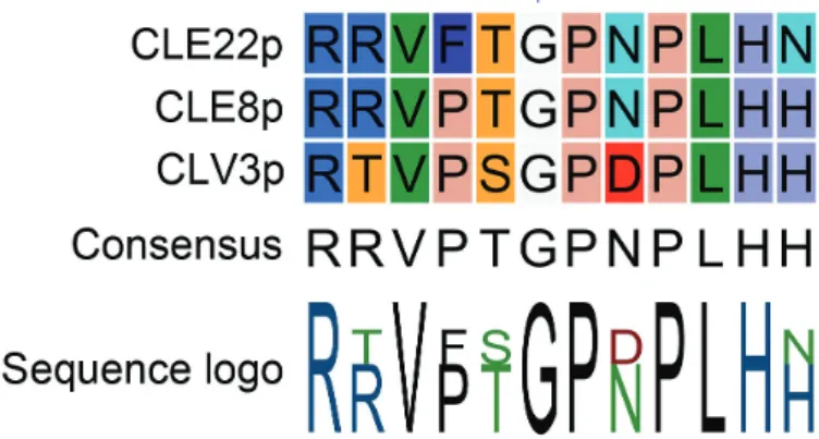
flexibility leading to stronger interaction with corresponding receptors and to disrupted downstream signal transduction. Taken together, such antagonistic peptides would provide a powerful tool for the functional dissection of CLEs in plants, and might also have the potential to be used for other plant peptides. Based on this assumption and the conserved nature of the Gly at position six (Fig. 1), this technology was applied to CLE8 (giving rise to embryo-lethal phenotype) and CLE22 (giving rise to short root phenotype) (Song et al., 2013).
Here, this antagonistic peptide technology was tested, spe-cifically Gly6-to-Ala or Gly6-to-Thr, as used by Song et al.
(2013), on selected CLE peptides and the related IDA pep-tide, and its usefulness discussed in the context of studies of peptide function.
Materials and methods
Plant growth conditions
For the work on CLE40 and CLV3, seeds were surface sterilized with chlorine gas and imbibed in 0.1% (w/v) agarose for 2 d at 4 °C before being plated onto 0.5× Murashige and Skoog (MS) medium with Gamborgs No. 5 vitamins (Duchefa), 0.5 g/l 2-(N-morpholino) ethanesulfonic acid (MES), 1% (w/v) sucrose, and 1.2% (w/v) plant agar. Plates were incubated vertically in a growth chamber with con-stant light at 21 °C for 5 d. For peptide-containing plates, synthetic dodecapeptides were added to a final concentration of 1 µM. For the work on CLE1/4, CLE7, CLE26, and CLE27, seeds were sur-face sterilized by immersion in 70% ethanol for 30 s, and incubated
in 20% bleach at room temperature for 20 min. Sterile seeds were vernalized in water at 4 °C for 2 d, before being plated onto 0.5× MS medium supplemented with 0.1 g/l Myo-inositol (Sigma Aldrich), 0.5 g/l MES (Sigma Aldrich), and 1% (w/v) bacteriological agar. Plants were incubated vertically under constant light at 21 °C until 12 d after germination. Synthetic CLE was added to a final con-centration of 10 µM or 10 nM. The work on CLE45 was essentially performed as previously described (Rodriguez-Villalon et al., 2014).
Starch staining
Starch granules and cell walls in root tips were stained with the mPSPI method and imaged with a confocal microscope as previ-ously described (Truernit et al., 2008).
Oxidative burst experiments
For transient expression, Agrobacterium tumefaciens carrying HAESA-LIKE 2 (HSL2) in frame with eGFP in an estradiol-inducible expression vector described previously (Bleckmann et al., 2010), was infiltrated into Nicotiana benthamiana leaves according to (Mueller et al., 2012). The oxidative burst experiment was per-formed as previously described by (Butenko et al., 2014), with the exception that 3 d after infiltration with A. tumefaciens, leaf pieces of N. benthamiana were induced with 20 μM estradiol before cut. Light emission was measured in a Wallac 1420 VICTOR2™ micro-plate luminometer (PerkinElmer).
Peptide structure predictions
The recently published solution structure of CLE10p, solved using nuclear magnetic resonance (NMR) (MMDB ID: 125940; PMBID: 2MD), depicts the backbone of the PXGP core (position 4–7) as a smooth curve protruding from the rest of the peptide. To inves-tigate the effect of mutations in this core of the peptides investi-gated, amino acid sequences with the structure AAA[core]AAA with the core PGGP, PGAP, PGTP, PRGP, PRTP, PSAP, or PSTP were submitted for analysis in PEP-FOLD ( http://mobyle.rpbs.univ- paris-diderot.fr/cgi-bin/portal.py?form=PEP-FOLD#forms::PEP-FOLD) using standard settings.
Results and discussion
‘Antagonistic’ CLE peptides
Among many processes (Cock and McCormick, 2001; Fiume and Fletcher, 2012; Hirakawa et al., 2008; Okamoto et al., 2013), various CLE peptides affect primary and lateral root
growth and development (Czyzewicz et al., 2015; Depuydt et al., 2013; Fiers et al., 2005; Hobe et al., 2003; Jun et al., 2010;
Rodriguez-Villalon et al., 2014; Rodriguez-Villalon et al., 2015; Stahl et al., 2009). To build on previous work investigat-ing CLE peptides in the context of lateral root development, primary root growth, root apical stem cell maintenance, and vascular development, putative antagonistic versions of CLV3, CLE1/4, CLE7, CLE26, CLE27, CLE40, and CLE45 peptides were designed—based on the findings by Song et al. (2013)—to further unravel CLE peptide function (Figs 2A,
3A, 4A). To assess the function of these mutated chemically synthesized CLE peptides with Gly/cysteine (Cys) to Ala or Gly/Cys to Thr substitutions (referred to as mCLEpAla6
or mCLEp6Thr, respectively), a number of biological assays
were used.
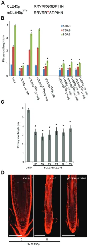
The antagonistic peptide technology was first applied to CLE45 peptide (CLE45p), which, when applied exogenously, leads to shorter primary roots because it suppresses pro-tophloem differentiation (Depuydt et al., 2013; Rodriguez-Villalon et al., 2014). To explore a potential loss-of-function phenotype, synthetic mCLE45p6Thr peptide was applied and
its effect on primary root development upon external appli-cation evaluated (Fig. 2A, B). This revealed that at the low nanomolar range mCLE45p6Thr does not have an effect on
primary root length as compared with the wild-type CLE45p (Fig. 2B), again confirming that position six is important for peptide activity. However, a higher concentration of 1 μM mCLE45p6Thr had the same effect as the unmodified wild-type
CLE45p (Fig. 2B). In addition, this peptide was not able to out-compete the effects of simultaneous CLE45p application (Fig. 2B). Thus, while the mCLE45p6Thr peptide does not act
as an antagonistic peptide, a CLE45p variant was obtained, which has identical effects as the wild-type version but required application of higher peptide concentrations. The notion that mCLE45p6Thr is a weak CLE45p, rather than an
antago-nistic version, was confirmed in planta (Rodriguez-Villalon et al., 2014). Plants that express a wild-type pCLE45::CLE45 transgene are notoriously difficult to create, presumably because of the detrimental effects of increased CLE45 dosage. However, the few lines that were eventually obtained recapit-ulated the root phenotype observed upon external CLE45p application (Fig. 2C). Specifically, in pCLE45::CLE45 lines, root growth was impaired, the periclinal division of the sieve element precursor cell was frequently abolished, and pro-tophloem differentiation was often suppressed (Fig. 2D). This phenotype is similar to plants that express a correspond-ing pCLE45::CLE456Thr transgene, which are much easier to obtain (Rodriguez-Villalon et al., 2014). Thus, the data for both tissue culture assay and in planta are consistent with the interpretation that mCLE45p6Thr is a weak rather than an
antagonistic version of the CLE45 peptide.
Next, the antagonistic peptide approach for CLE40 was explored (Fig. 3A). It was previously shown that an increas-ing concentration of synthetic CLE40 peptide reduces stemness and causes differentiation of columella stem cells (CSCs), quiescent centre (QC) cells, and proximal initial (P1) cells in wild-type roots (Fig. 3B, D, H, Supplementary Table S1 available at JXB online) (Stahl et al., 2013). Also Fig. 1. Alignment of CLE peptides used in Song et al. (2013). Conserved
glycine (G) at position six is indicated with a blue arrowhead. (This figure is available in colour at JXB online.)
synthetic CLV3 peptide acts similarly on the stemness in the root tip (Fig. 3E, G, Supplementary Table S1 available at
JXB online). In contrast, in the shorter cle40 mutant roots,
differentiation of CSC daughters into CCs was significantly delayed (Fig. 3B, F, H, Supplementary Table S1 available at
JXB online) (Hobe et al., 2003; Stahl et al., 2009). Wild-type roots carry mostly one (at D1 position) or, after a recent cell division, two layers of CSCs distal to the QC (at D1 and D2 positions), which lack stainable starch granules (Fig. 3B, H,
Supplementary Table S1). In cle40 root tips, additional CSCs in more distal positions (D2) were found (Fig. 3B, C, H,
Supplementary Table S1). To assess if synthetic mCLE40p6Thr
and mCLV3p6Thr variants could be used as antagonistic
pep-tides to obtain a loss-of-function phenotype, their impact on the distal root stemness was evaluated (Fig. 3A). This revealed a response comparable with the wild-type CLE40p or CLV3p treatments (Fig. 3E, G, H, Supplementary Table S1 available at JXB online). Taken together, this suggests that the Gly to Thr substitution in CLE40 and CLV3 does not give rise to an antagonistic peptide.
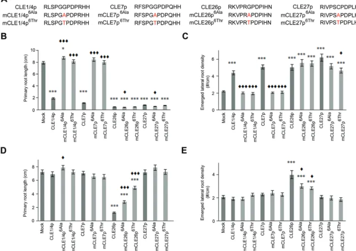
Finally, while treatment of A. thaliana seedlings with 10 µM wild-type CLE1/4p, CLE7p, CLE26p, and CLE27p resulted in a short primary root and more lateral roots (Fig. 4B, C) (Czyzewicz et al., 2015; Depuydt et al., 2013; Kinoshita et al., 2007; Rodriguez-Villalon et al., 2014), this does not neces-sarily reflect their natural function. However, based on the
CLE1/4, CLE7, CLE26, and CLE27 expression patterns, a
role in lateral root development might be expected (Czyzewicz et al., 2015; Jun et al., 2010). In this context, only CLE26p gave rise to a short primary root and increased lateral root density at a concentration of 10 nM (Fig. 4B) (Czyzewicz et al., 2015; Rodriguez-Villalon et al., 2015), further support-ing caution when interpretsupport-ing exogenous peptide application results, especially at higher concentrations. To assess if the above-mentioned CLE peptides have a role in primary and lateral root development, the antagonistic peptide technology was attempted (Fig. 4A). However, analysis of mutated chem-ically synthesized CLE peptides (mCLEp) at 10 μM revealed that, although mCLE1/4p6Ala/Thr and mCLE7p6Ala/Thr did not
induce a primary root shortening or a lateral root density increase—unlike the non-mutated forms of these peptides, mCLE1/4p6Thr and mCLE7p6Ala/Thr were also unable to
pro-duce an obvious dominant negative root phenotype, namely an expected longer primary root and/or decrease in lateral root density (Fig. 4B, C). However, for mCLE1/4p6Ala, a
sub-tle increase in primary root length, but no effect on lateral root density was observed (Fig. 1C). It should be pointed out that since the receptor, and the associated loss-of-function pheno-type, for these peptides is not known, the expected dominant negative root phenotype remains speculative. Nevertheless, this outcome suggested that for CLE1p, CLE4p, and CLE7p activity, the Gly at position six is essential, but that this mutant form did not appear to act as an antagonistic peptide. In con-trast, mCLE26p6Ala/Thr and mCLE27p6Ala/Thr displayed a
simi-lar phenotype to the non-mutated forms, namely a significant reduction in primary root length (92–95%) and increased lat-eral root density (110–151%) (Fig. 4B, C), suggesting that the sixth amino acid in their respective sequences is not critical Fig. 2. CLE45 peptide treatment and pCLE45::CLE45 transgenic lines.
(A) Sequence of synthetic CLE45 and mCLE45 peptides used. (B) Primary root length following treatment of wild-type seedlings with indicated concentrations of CLE45p or mCLE45p6Thr. The bar graph indicates the mean ± standard error. Statistical significance (Student’s t-test) compared with mock is indicated for each time point (DAG, days after germination): * P <0.01. (C) Primary root length of pCLE45::CLE45 lines. The bar graph indicates the mean ± standard error. Statistical significance (Student’s t-test) compared with Col-0 is indicated: * P <0.01. (D) Confocal images of primary root meristems of 7-d-old seedlings (propidium iodide-stained; composite images). The asterisks highlight the two protophloem strands that can be distinguished in wild-type (Col-0) grown on mock (left), but that do not develop when grown on 10 nM CLE45p (middle). Protophloem strands also do not develop in wild-type seedlings that express a pCLE45::CLE45 transgene (right). Scale bar, 100 µm.
to their function, and also, did not appear to give rise to an antagonistic peptide when mutated. Intriguingly, at 10 nM, mCLE26p6Ala/Thr retained activity, but was less potent than
CLE26p. This suggests that mCLE26p6Ala/Thr is a weak rather
than an antagonistic version of the CLE26 peptide, which is in agreement with the results on CLE45. In contrast, most Fig. 3. Distal root phenotypes after antagonistic peptide treatments. (A) Sequence of synthetic CLE and mCLE peptides used. (B–H) Distal root cell fates were analysed by mPSPI staining 5 d after germination in wild-type (Col-0) and cle40-2 mutant roots (C, D). Representative examples of Col-0 roots grown on media with 1 µM CLE40p (E), mCLE40p6Thr (F), CLV3p (G), and mCLV3p6Thr (H) are shown. Frequency of roots carrying starch granules in the designated domains is shown in (B). Arrowheads: blue, QC position; yellow, CSC position (D1); red, CC position (D2). Double yellow arrowheads indicate CSC fate in D2, whereas the lack of a yellow arrowhead indicates CC fate in D1 position. QC, quiescent centre position; D1, distal layer position one; D2, distal layer position two; CC, columella cell position. Scale bars represent 15 µm.
Fig. 4. CLE1/4, CLE7, CLE26, and CLE27 peptide treatment. (A) Sequence of synthetic CLE and mCLE peptides used. (B–E) Treatment of wild-type seedlings with 10 µM (B, C) or 10 nM of CLE or mCLE peptide (D, E). Quantification of primary root length (B, D) and emerged lateral root density (C, E) for CLEp and mCLEp-treated wild-type seedlings. The bar graph indicates the mean ± standard error. Statistical significance (Student’s t-test) compared with no peptide (*) and to CLEp treatment (♦) is indicated: ***/♦♦♦, P <0.001, */♦, P <0.05. (This figure is available in colour at JXB online.)
mCLE1/4p, mCLE7p, and mCLE27p variants had no altered activity compared with the wild-type variant at 10 nM, except mCLE1/4p6Ala (Fig. 4D, E). In general, it appears that also
for CLE1/4p, CLE7p, CLE26p, and CLE27p, the antagonis-tic peptide technology is not easily applicable.
In conclusion, other amino acid mutations are likely required to give rise to (strong) antagonistic CLE1p, CLE4p, CLE7p, CLE26p, CLE27p, CLE40p, and CLE45p peptides, or alternatively, this approach cannot be universally applied with respect to synthetic CLE peptides. A poor effect of syn-thetic antagonistic peptides could be due to delivery to rel-evant tissues and/or instability. However, synthetic peptide stability issues were not observed in these assays or with respect to synthetic control peptides, nor was a lack of phe-notypes observed when synthetic (antagonistic) peptides were exogenously applied to the root. While the latter can be a non-specific effect in some cases, specific and local pheno-types were also observed.
‘Antagonistic’ IDA peptides
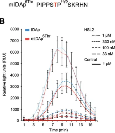
In addition, the extent the antagonistic peptide technology can be applied to other small signalling peptides was assessed. For this, the IDA and IDA-LIKE (IDL) family were cho-sen, given their sequence similarity to CLEs (Stenvik et al., 2006). The IDA and IDL1 peptides of 12 amino acids share a common core at positions four to seven [PS(G/A)P] and the C-terminal end [H(N/H)] with CLV3 and some CLE peptides (Figs 5A, 6A). Like CLV3, hydroxylation of the Pro at posi-tion seven of the IDA dodecapeptide (IDAp, also referred to as PIPPo) increases the activity of the peptide (Butenko et al., 2014). An oxidative burst response in Nicotiana benthamiana can be employed as readout for the RLK HAESA-LIKE2 (HSL2) activation by exogenously applied synthetic IDA pep-tides (Butenko et al., 2014). Previous results indicated that IDAp binds to HSL2 with a Kd of 20 nM (Butenko et al., 2014). As the wild-type IDA peptide has an Ala at position six corresponding to the Gly at that position in CLV3, and the
ida mutant phenotype can be fully rescued by IDL1, which
has a Gly at this position (Stenvik et al., 2008) (Fig. 6A); both of these small amino acids are evidently compatible with high signalling activity. It was, however, conceivable that substi-tution to the larger Thr (mIDAp6Thr) (Fig. 5A) could have
an effect on receptor binding and/or activation. Therefore, the activity of mIDAp6Thr in comparison with the activity
of synthetic IDAp was assessed in an oxidative burst assay. For all peptide concentrations tested, mIDAp6Thr gave the
same response as IDAp in the presence of its receptor HSL2 (Fig. 5B), indicating that the mutated peptide was just as active as its wild-type counterpart. In conclusion, this muta-tion neither produced a ligand with weaker activity, nor a peptide with antagonistic effect.
Conclusion
Information collected using antagonistic peptide approaches (in the broad sense) can be very useful, but these approaches do not work in all cases and require a deep insight on the
interaction between the ligand and its receptor to be success-ful. While the antagonistic peptide approach might work in a number of cases, as described by Song et al. (2013) and Xu et al. (2015), its universal applicability remains to be deter-mined. Initial data were presented for CLV3, CLE8, and CLE22, and recently for CLE19 but in the absence of the pertinent wild-type control transgenes and genetic knock-out lines, it remains difficult to judge whether the pheno-types triggered by mCLE86Thr, mCLE196Thr, and mCLE226Thr transgenes are antagonistic or not. Importantly, in view of the results presented here, and in agreement with the results of Song et al. (2013), it appears that the antagonistic peptide technology cannot be easily applied to synthetic CLE peptides and—at least—requires expressing mutant variants to deliver dominant peptides to their endogenous locations. However, as was shown with the CLE456Thr transgene, the latter also does not always work. Nevertheless, it can provide novel insight that can lead to other tools to dissect peptide activ-ity, as— for example—the weakened activity of mCLE456Thr
could be used to functionally characterize CLE45. In addi-tion, it also does not appear to be straightforward to trans-late this approach to other peptide families, as exemplified Fig. 5. IDA peptide treatment. (A) Sequence of synthetic IDA peptides used. (B) N. benthamiana leaf pieces expressing HSL2 were exposed to various concentrations of peptides as indicated. Oxidative burst by the luminol-based assay was monitored over time as relative light units (RLU). Leaf pieces infiltrated with Agrobacterium without HSL2 were exposed to 1 μM of both peptides and used as control. Error bars indicate standard error of n=3 or 4 replicates.
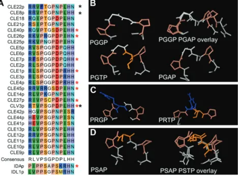
through analyses on IDA. In general, it was observed that whether the mutations have an effect or not, seems dependent on the context, with differential sensitivity to conformational changes (Fig. 6A and Supplementary Table S2 available at
JXB online). CLE1/4p and CLE7p are highly similar
pep-tides with the same PGGP core at position four to seven, and both lose activity when the Gly at position six is mutated to Ala or Thr. Structure prediction for the peptides may suggest that a mutation in this context, with the small Gly at posi-tion five, easily changes the peptide conformaposi-tion (Fig. 6B). Alternatively, all size increases in the side chain of the amino acid at position six could interfere with binding of the puta-tive receptor(s) of CLE1, CLE4, and CLE7. CLE26p and CLE45p both have an Arg in the core sequence (PRGP and RRGS, respectively) and react similarly to the introduced mutations, namely weaker activity when the Gly at position six is mutated. The long side chain of Arg might change direc-tion in the mutant peptides, which might reduce its binding affinity for a receptor (Fig. 6C). In contrast, mutation of Ala at position six to Thr did not reduce the activity of the IDA peptide, which has a PSAP core, suggesting that the serine (Ser) residue might stabilize the peptide structure (Fig. 6D).
In conclusion, the antagonistic peptide approach can be a useful tool to study the function of some CLE genes (Song et al., 2013; Xu et al., 2015), but not the ultimate means to overcome redundancy or lack of loss-of-function lines (Rodriguez-Villalon et al., 2014; this study). However, while the approach described by Song et al. (2013), when applied to synthetic CLE peptide variants, did not work—for the pep-tides selected in this study and with respect to the phenotypes investigated, it does not preclude there being any other substi-tution, modification, or combination thereof or a transgene that may induce the desired effects. This, as well as structure
considerations, should be taken into account before ordering a wide range of synthetic peptide variants and/or generating transgenic plants.
Supplementary data
Supplementary data are available at JXB online.
Supplementary Table S1. Quantification of distal root phe-notypes after antagonistic peptide treatments.
Supplementary Table S2. Summary of mutations and phenotypes.
Acknowledgements
This work was supported by a BBSRC David Phillips Fellowship (BB_ BB/H022457/1) and a Marie Curie European Reintegration Grant (PERG06-GA-2009–256354) to IDS, a BBSRC CASE Studentship co-funded by Bayer CropScience to NC, and a Swiss National Science Foundation grant (310030B_147088) to CSH. This work was supported by Grants 13785/F20, 218735, and 204756 to MAB, MW, and RBA, and 230849/F20 and 225299 to MAB from the Research Council of Norway. Work by RS, YS, and KGP was supported by the German excellence initiative (CEPLAS, EXC1028).
References
Araya T, Miyamoto M, Wibowo J, et al. 2014. CLE-CLAVATA1 peptide-receptor signaling module regulates the expansion of plant root systems in a nitrogen-dependent manner. Proceedings of the National Academy of Sciences USA 111, 2029–2034.
Bleckmann A, Weidtkamp-Peters S, Seidel CAM, Simon R. 2010. Stem Cell Signaling in Arabidopsis Requires CRN to Localize CLV2 to the Plasma Membrane. Plant Physiology 152, 166–176.
Brand U, Fletcher JC, Hobe M, Meyerowitz EM, Simon R. 2000. Dependence of stem cell fate in Arabidopsis on a feedback loop regulated by CLV3 activity. Science 289, 617–619.
Fig. 6. Peptide structure. (A) Manually adjusted alignment of the 12 amino acids of CLE peptides, with IDA and IDL1 for comparison. Red stars mark peptides used in the present study, black stars those tested by Song et al. (2013). (B–D) Examples of structures of the PXXP core predicted by PEP-FOLD. (B) PGGP. The larger Thr might interfere with receptor binding, and substitution of Gly6 with Ala6 may change the angles between the Pro residues. (C) PRGP. The side chain of Arg may change direction when the Gly6 is exchanged with a Thr. (D) PSAP. A change from Ala to Thr in position six may not result in major conformational changes when Ser is present in the core. (This figure is available in colour at JXB online.)
Butenko MA, Patterson SE, Grini PE, Stenvik GE, Amundsen SS, Mandal A, Aalen RB. 2003. Inflorescence deficient in abscission controls floral organ abscission in Arabidopsis and identifies a novel family of putative ligands in plants. The Plant Cell 15, 2296–2307.
Butenko MA, Vie AK, Brembu T, Aalen RB, Bones AM. 2009. Plant peptides in signalling: looking for new partners. Trends in Plant Science 14, 255–263.
Butenko MA, Wildhagen M, Albert M, Jehle A, Kalbacher H, Aalen RB, Felix G. 2014. Tools and Strategies to Match Peptide-Ligand Receptor Pairs. The Plant Cell 26, 1838–1847.
Clark SE, Running MP, Meyerowitz EM. 1995. CLAVATA3 is a specific regulator of shoot and floral meristem development affecting the same processes as CLAVATA1 Development 121, 2057–2067.
Clark SE, Jacobsen SE, Levin JZ, Meyerowitz EM. 1996. The CLAVATA and SHOOT MERISTEMLESS loci competitively regulate meristem activity in Arabidopsis. Development 122, 1567–1575. Cock JM, McCormick S. 2001. A large family of genes that share homology with CLAVATA3. Plant Physiology 126, 939–942.
Czyzewicz N, Yue K, Beeckman T, De Smet I. 2013. Message in a bottle: small signalling peptide outputs during growth and development. Journal of Experimental Botany 64, 5281–5296.
Czyzewicz N, Shi C, Vu LD, Van De Cotte B, Butenko MA, De Smet I. 2015. Modulation of Arabidopsis and monocot root architecture by CLAVATA3/EMBRYO SURROUNDING REGION 26 peptide. Journal of Experimental Botany 66, 5229–5243.
Depuydt S, Rodriguez-Villalon A, Santuari L, Wyser-Rmili C, Ragni L, Hardtke CS. 2013. Suppression of Arabidopsis protophloem differentiation and root meristem growth by CLE45 requires the receptor-like kinase BAM3. Proceedings of the National Academy of Sciences USA 110, 7074–7079.
Fiers M, Golemiec E, Xu J, van der Geest L, Heidstra R, Stiekema W, Liu CM. 2005. The 14-amino acid CLV3, CLE19, and CLE40 peptides trigger consumption of the root meristem in Arabidopsis through a CLAVATA2-dependent pathway. The Plant Cell 17, 2542–2553. Fiume E, Fletcher JC. 2012. Regulation of Arabidopsis embryo and endosperm development by the polypeptide signaling molecule CLE8. The Plant Cell 24, 1000–1012.
Fletcher JC, Brand U, Running MP, Simon R, Meyerowitz EM. 1999. Signaling of cell fate decisions by CLAVATA3 in Arabidopsis shoot meristems. Science 283, 1911–1914.
Hirakawa Y, Shinohara H, Kondo Y, et al. 2008.
Non-cell-autonomous control of vascular stem cell fate by a CLE peptide/receptor system. Proceedings of the National Academy of Sciences USA 105, 15208–15213.
Hobe M, Muller R, Grunewald M, Brand U, Simon R. 2003. Loss of CLE40, a protein functionally equivalent to the stem cell restricting signal CLV3, enhances root waving in Arabidopsis. Development Genes and Evolution 213, 371–381.
Jun J, Fiume E, Roeder AH, et al. 2010. Comprehensive analysis of CLE polypeptide signaling gene expression and overexpression activity in Arabidopsis. Plant Physiology 154, 1721–1736.
Kinoshita A, Nakamura Y, Sasaki E, Kyozuka J, Fukuda H, Sawa S. 2007. Gain-of-function phenotypes of chemically synthetic CLAVATA3/ ESR-related (CLE) peptides in Arabidopsis thaliana and Oryza sativa. Plant and Cell Physiology 48, 1821–1825.
Kondo T, Nakamura T, Yokomine K, Sakagami Y. 2008. Dual assay for MCLV3 activity reveals structure-activity relationship of CLE peptides. Biochemical and Biophysical Research Communications 377, 312–316.
Lease KA, Walker JC. 2006. The Arabidopsis unannotated secreted peptide database, a resource for plant peptidomics. Plant Physiology 142, 831–838.
Lee JS, Torii KU. 2012. A tale of two systems: peptide ligand-receptor pairs in plant development. Cold Spring Harbor Symposia on Quantitative Biology 77, 83–89.
Lim CW, Lee YW, Hwang CH. 2011. Soybean nodule-enhanced CLE peptides in roots act as signals in GmNARK-mediated nodulation suppression. Plant and Cell Physiology 52, 1613–1627.
Mortier V, Den Herder G, Whitford R, Van de Velde W, Rombauts S, D’Haeseleer K, Holsters M, Goormachtig S. 2010. CLE peptides
control Medicago truncatula nodulation locally and systemically. Plant Physiology 153, 222–237.
Mueller K, Bittel P, Chinchilla D, Jehle AK, Albert M, Boller T, Felix G. 2012. Chimeric FLS2 receptors reveal the basis for differential flagellin perception in Arabidopsis and tomato. The Plant Cell 24, 2213–2224. Murphy E, Smith S, De Smet I. 2012. Small signaling peptides in Arabidopsis development: how cells communicate over a short distance. The Plant Cell 24, 3198–3217.
Ogawa M, Shinohara H, Sakagami Y, Matsubayashi Y. 2008. Arabidopsis CLV3 peptide directly binds CLV1 ectodomain. Science 319, 294.
Ohyama K, Shinohara H, Ogawa-Ohnishi M, Matsubayashi Y. 2009. A glycopeptide regulating stem cell fate in Arabidopsis thaliana. Nature Chemical Biology 5, 578–580.
Okamoto S, Shinohara H, Mori T, Matsubayashi Y, Kawaguchi M. 2013. Root-derived CLE glycopeptides control nodulation by direct binding to HAR1 receptor kinase. Nature Communications 4, 2191.
Reid DE, Ferguson BJ, Gresshoff PM. 2011. Inoculation- and nitrate-induced CLE peptides of soybean control NARK-dependent nodule formation. Molecular Plant-Microbe Interactions 24, 606–618.
Reid DE, Li D, Ferguson BJ, Gresshoff PM. 2013. Structure-function analysis of the GmRIC1 signal peptide and CLE domain required for nodulation control in soybean. Journal of Experimental Botany 64, 1575–1585.
Rodriguez-Villalon A, Gujas B, Kang YH, Breda AS, Cattaneo P, Depuydt S, Hardtke CS. 2014. Molecular genetic framework for protophloem formation. Proceedings of the National Academy of Sciences USA 111, 11551–11556.
Rodriguez-Villalon A, Gujas B, van Wijk R, Munnik T, Hardtke CS. 2015. Primary root protophloem differentiation requires balanced phosphatidylinositol-4,5-biphosphate levels and systemically affects root branching. Development 142, 1437–1446.
Shiu SH, Bleecker AB. 2001a. Plant receptor-like kinase gene family: diversity, function, and signaling. Science Signaling 2001 , re22. Shiu SH, Bleecker AB. 2001b. Receptor-like kinases from Arabidopsis form a monophyletic gene family related to animal receptor kinases. Proceedings of the National Academy of Sciences USA 98, 10763–10768. Song XF, Guo P, Ren SC, Xu TT, Liu CM. 2013. Antagonistic peptide technology for functional dissection of CLV3/ESR genes in Arabidopsis. Plant Physiology 161, 1076–1085.
Stahl Y, Wink RH, Ingram GC, Simon R. 2009. A signaling module controlling the stem cell niche in Arabidopsis root meristems. Current Biology 19, 909–914.
Stahl Y, Grabowski S, Bleckmann A, et al. 2013. Moderation of Arabidopsis root stemness by CLAVATA1 and ARABIDOPSIS CRINKLY4 receptor kinase complexes. Current Biology 23, 362–371.
Stenvik GE, Butenko MA, Urbanowicz BR, Rose JK, Aalen RB. 2006. Overexpression of INFLORESCENCE DEFICIENT IN ABSCISSION activates cell separation in vestigial abscission zones in Arabidopsis. The Plant Cell 18, 1467–1476.
Stenvik GE, Tandstad NM, Guo Y, Shi CL, Kristiansen W, Holmgren A, Clark SE, Aalen RB, Butenko MA. 2008. The EPIP peptide of INFLORESCENCE DEFICIENT IN ABSCISSION is sufficient to induce abscission in Arabidopsis through the receptor-like kinases HAESA and HAESA-LIKE2. The Plant Cell 20, 1805–1817.
Stes E, Gevaert K, De Smet I. 2015. Phosphoproteomics-based peptide ligand-receptor kinase pairing. Commentary on: “A peptide hormone and its receptor protein kinase regulate plant cell expansion”. Frontiers in Plant Science 6, 224.
Truernit E, Bauby H, Dubreucq B, Grandjean O, Runions J,
Barthelemy J, Palauqui JC. 2008. High-resolution whole-mount imaging of three-dimensional tissue organization and gene expression enables the study of phloem development and structure in Arabidopsis. The Plant Cell 20, 1494–1503.
Xu T-T, Ren S-C, Song X-F, Liu C-H. 2015. CLE19 expressed in the embryo regulates both cotyledon establishment and endosperm development in Arabidopsis. Journal of Experimental Botany 66, 5217–5227. Yang SL, Xie LF, Mao HZ, Puah CS, Yang WC, Jiang L, Sundaresan V, Ye D. 2003. Tapetum determinant1 is required for cell specialization in the Arabidopsis anther. The Plant Cell 15, 2792–2804.