
Inhibition of matrix metalloproteinases and tumour necrosis factor α converting enzyme as adjuvant therapy in pneumococcal meningitis
9
0
0
Texte intégral
(2) Metalloproteinases in bacterial meningitis membrane-bound form of TNF-α is converted to its active soluble form by TNF-α converting enzyme (TACE or ADAM-17), a metalloproteinase closely related to the MMP family (Maskos et al., 1998). TNF-α administered intracisternally results in CSF leucocytosis, whereas intracerebral injection of TNF-α leads to a dose-dependent response in MMP activation and the opening of the blood–brain barrier (Ramilo et al., 1990; Rosenberg et al., 1995). In addition to its proinflammatory properties, TNF-α induces loss of hippocampal neurones in experimental meningitis, an effect that is antagonized by administration of monoclonal antibodies directed against TNF-α (Bogdan et al., 1997). MMPs are a family of zinc-dependent endopeptidases with substrate affinity for different components of the extracellular matrix (Alexander and Werb, 1989). Furthermore, MMPs sustain the inflammatory host response by activating cytokines and cleaving cytokine receptors (Gearing et al., 1994; Clements et al., 1997; Wallace et al., 1999). In bacterial meningitis, MMP-8 and -9 are specifically upregulated and act as effectors of subarachnoid space inflammation, brain oedema, blood–brain barrier opening and neuronal injury (Rosenberg et al., 1995; Azeh et al., 1998; Paul et al., 1998; Kieseier et al., 1999; Leib et al., 2000; Leppert et al., 2000). However, the precise array of MMPs induced in CSF and brain parenchyma during bacterial meningitis has not been evaluated so far. Here, time-course studies in a rat model of experimental pneumococcal meningitis were performed to investigate the spectrum and kinetics of MMPs induced at the mRNA level both in CSF cells and in brain tissue. At the protein level, the activity profile of MMPs and TACE during disease development was delineated in the CSF. TNF-α and MMPs both contribute to the development of neuronal injury in experimental bacterial meningitis (Bogdan et al., 1997; Leib et al., 2000). Therefore, this study evaluated the effect of combined inhibition of MMPs and TACE by BB-1101, a hydroxamic acid type inhibitor, on clinical disease parameters, the histopathological outcome and learning function (Clements et al., 1997; Wallace et al., 1999).. Material and methods Model of meningitis An established model of pneumococcal meningitis in infant rats was used (Auer et al., 2000; Leib et al., 2000; Loeffler et al., 2000; Pfister et al., 2000). The animal studies were approved by the Animal Care and Experimentation Committee of the Canton of Bern, Switzerland, and followed National Institutes of Health guidelines for the performance of animal experiments. Sprague–Dawley rats were infected on postnatal day 11 by direct intracisternal injection of 10 µl saline solution containing a defined inoculum of Streptococcus pneumoniae (serogroup 3) with a 32-gauge needle. Uninfected control animals were injected with 10 µl of sterile saline solution. Eighteen hours later, animals were. 1735. weighed and assessed clinically using the following scores: 1 ⫽ coma; 2 ⫽ does not turn upright when positioned on the back; 3 ⫽ turns upright within 30 s; 4 ⫽ minimal ambulatory activity, turns upright in ⬍5 s; 5 ⫽ normal. CSF (10–30 µl) was obtained by puncture of the cisterna magna and 5 µl was cultured quantitatively to document meningitis. All infected animals then received antibiotic therapy (ceftriaxone 100 mg/kg subcutaneously, twice daily; Roche Pharma, Reinach, Switzerland). Seizures, defined as tonic convulsions for ⬎15 s, were monitored for 2 h starting 18 h after infection. Animals were sacrificed and perfused via the left cardiac ventricle with 30 ml of either ice-cold PBS (phosphate-buffered saline) for assessment of mRNA in brain homogenates or with 4% paraformaldehyde in PBS for histopathological evaluation. Animals were sacrificed when they became terminally ill (cyanosis, difficulty breathing, protracted seizures). Animals dying unobserved were excluded from the histopathological evaluation. For timecourse studies, animals (n ⫽ 21) were infected with log10 7.0 c.f.u. (colony-forming units)/ml S. pneumoniae as described above and at each time point, i.e. 0, 0.25, 4, 8, 12, 16 and 20 h after infection, the animals (n ⫽ 3) were killed immediately after CSF sampling.. Analysis of mRNA expression Transcriptional expression of MMPs was analysed with the real-time polymerase chain reaction (PCR) based on a 5⬘-nuclease assay (TaqMan; Applied Biosystems, Foster City, Calif., USA) with the ABI PRISM 7700 Sequence Detection System (Applied Biosystems) in two independent laboratories (CSF cells were analysed in Basel and brain tissue samples in Oxford). Primers and probes for all target genes (Table 1) were designed with Primer Express software (Applied Biosystems). CSF samples were centrifuged and cells were suspended in lysis buffer (QiaGen, Valencia, Calif., USA) and stored at –80°C until RNA isolation. RNA was isolated according to the manufacturer’s instructions (RNeasy; QiaGen). Total RNA (final concentration 5–10 ng/µl) was first incubated with 0.5 µg oligo-dT at 70°C for 2 min and then reversetranscribed at 37°C for 1 h in a reaction mix containing a final concentration of 1⫻ first strand buffer (Promega Corporation, Madison, Wis., USA), 10 mM dithiothreitol, 500 µM of each deoxynucleotide triphosphate, 1 U/µl of Moloney murine leukaemia virus reverse transcriptase (Promega) and 1 U/µl of ribonuclease inhibitor (RNAsin, Promega). cDNA corresponding to 5–10 ng RNA was used as a template for PCR analysis. PCR conditions were: 2.5 µl of 10⫻ buffer A, 200 µM dATP, dCTT, dGTP, 400 µM dUTP, 3 mM MgCl2, 0.625 U AmpliTaqGold and 0.25 U AmpErase Uracil N-glycosylase (all reagents were from Applied Biosystems). Primer and probe concentrations were 300–900 and 100–200 nM, respectively, depending on the target gene. Whole brains were dissected in ice-cold PBS from animals.
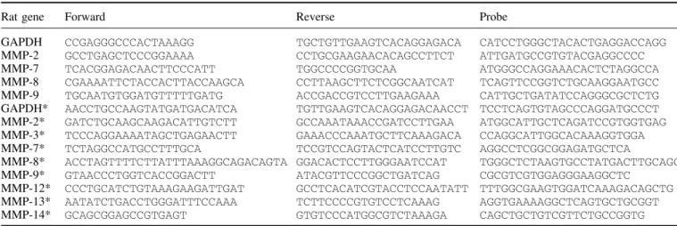
(3) 1736. S. L. Leib et al.. Table 1 Primers and probes used for transcriptional expression analysis in CSF cells and brain tissue (*) by real-time PCR Rat gene. Forward. Reverse. Probe. GAPDH MMP-2 MMP-7 MMP-8 MMP-9 GAPDH* MMP-2* MMP-3* MMP-7* MMP-8* MMP-9* MMP-12* MMP-13* MMP-14*. CCGAGGGCCCACTAAAGG GCCTGAGCTCCCGGAAAA TCACGGAGACAACTTCCCATT CGAAAATTCTACCACTTACCAAGCA TGCAATGTGGATGTTTTTGATG AACCTGCCAAGTATGATGACATCA GATCTGCAAGCAAGACATTGTCTT TCCCAGGAAAATAGCTGAGAACTT TCTAGGCCATGCCTTTGCA ACCTAGTTTTCTTATTTAAAGGCAGACAGTA GTAACCCTGGTCACCGGACTT CCCTGCATCTGTAAAGAAGATTGAT AATATCTGACCTGGGATTTCCAAA GCAGCGGAGCCGTGAGT. TGCTGTTGAAGTCACAGGAGACA CCTGCGAAGAACACAGCCTTCT TGGCCCCGGTGCAA CCTTAAGCTTCTCGGCAATCAT ACCGACCGTCCTTGAAGAAA TGTTGAAGTCACAGGAGACAACCT GCCAAATAAACCGATCCTTGAA GAAACCCAAATGCTTCAAAGACA TCCGTCCAGTACTCATCCTTGTC GGACACTCCTTGGGAATCCAT ATACGTTCCCGGCTGATCAG GCCTCACATCGTACCTCCAATATT TCTTCCCCGTGTCCTCAAAG GTGTCCCATGGCGTCTAAAGA. CATCCTGGGCTACACTGAGGACCAGG ATTGATGCCGTGTACGAGGCCCC ATGGGCCAGGAAACACTCTAGGCCA TCAGTTCCGGTCTGCAAGGAATGCC CATTGCTGATATCCAGGGCGCTCTG TCCTCAGTGTAGCCCAGGATGCCCT ATGGCATTGCTCAGATCCGTGGTGAG CCAGGCATTGGCACAAAGGTGGA AGGCCTCGGCGGAGATGCTCA TGGGCTCTAAGTGCCTATGACTTGCAGC CGCGTCGTGGAGGGAAGGCTC TTTGGCGAAGTGGATCAAAGACAGCTG AGGTGAAAAGGCTCAGTGCTGCGGT CAGCTGCTGTCGTTCTGCCGGTG. with bacterial meningitis at predetermined time points (n 艌 2 for each time point). The brains were rolled individually on filter paper to remove the meninges and brain vessels. Total RNA was isolated from brain homogenates using the SV total RNA preparation kit (Promega) and further treated with 10 U of RQ1 DNase (Promega). RNA was reverse-transcribed to cDNA using Universal RiboClone (Promega). For every RNA sample, a reaction was also performed without reverse transcriptase (RT–) to provide a negative control in subsequent reactions. To account for variations in input RNA and reverse transcriptase efficiency, the MMP levels were normalized to glyceraldehyde phosphate dehydrogenase (GAPDH) expression in each sample. To account for PCR amplification of contaminating genomic DNA, a RT– control was included. In all cases, contaminating genomic DNA accounted for ⬍0.001% of the cDNA sample. For each primer set, the assay was linear over a range of at least 5 log units. PCR reactions were performed in a total volume of 25 µl, containing 0.5 µl cDNA sample, 50 mM KCl, 10 mM Tris– HCl (pH 8.3), 10 mM EDTA, 60 nM Passive Reference 1, 200 µM dATP, dCTP, dGTP and 400 µM dUTP, 5.5 mM MgCl2, 400 nM of each primer, 0.625 U AmpliTaqGold and 0.25 U AmpErase Uracil N-glycosylase. Each reaction contained 100–175 nM of the corresponding detection probe. Each PCR reaction was performed in duplicate wells using the following conditions: 2 min at 50°C and 10 min at 94°C, followed by a total of 40 cycles of 15 s at 94°C and 1 min at 60°C.. Quantitation of MMP-9 in CSF The amount of MMP-9 in CSF was assessed by zymography as described previously (Leib et al., 2000). In brief, CSF samples (3 µl) were diluted with 2 µl of 4⫻ sample buffer (Novex, San Diego, Calif., USA) to a loading volume of 10 µl and electrophoresed under non-reducing conditions in 10% polyacrylamide–sodium dodecyl sulphate gels. containing type A gelatin from porcine skin (1% v/v; Sigma, Buchs, Switzerland) as proteinase substrate. After electrophoresis for 2 h at 95 V, gels were incubated for 1 h in SDSremoving buffer (1% Triton X-100, three changes), followed by incubation buffer (10 mMCaCl2, 50 mM Tris, 50 mM NaCl, pH 7.65) for 18 h at 37°C. The gelatinolytic activity of MMP-9 was determined by densitometric quantitation (public domain NIH Image program; National Institutes of Health, Bethesda, Md., USA) of the substrate lysis zones around 92 (MMP-9) and 72 (MMP-2) kDa in gels stained with Coomassie blue (Fig. 1C). The amount of MMP-9 protein induced was expressed as a percentage of that of the constitutively expressed MMP-2 for each sample.. Quantitation of TNF-α in CSF The concentration of rat TNF-α in CSF was measured with a high-sensitivity sandwich ELISA (enzyme-linked immunosorbent assay) kit (Cytoscreen, Rat Tumour Necrosis Factor-Alpha Ultra Sensitive ELISA, KRC3014, BioSource International, Camarillo, Calif., USA). CSF supernatant (2 µl) was diluted 1 : 50 and assayed in duplicate according to the manufacturer’s instructions. The detection limit of the assay was ⬍35 pg/ml.. Treatment studies For the pre-treatment studies, animals were infected with a large inoculum (log10 7 ⫾ 0.4 c.f.u./ml) to produce severe disease and randomized for treatment with BB-1101 (British Biotech, Oxford, UK) (15 mg/kg subcutaneously, twice daily) or vehicle (200 µl PBS, 1% Tween 80) and sacrificed 24 h after infection (Clements et al., 1997; Barlaam et al., 1999; Wallace et al., 1999). To evaluate adjuvant therapy with BB1101, animals were infected (log10 6.4 ⫾ 0.4 c.f.u./ml) and delayed administration of BB-1101 or vehicle was started 18 h after infection, simultaneously with the first dose of.
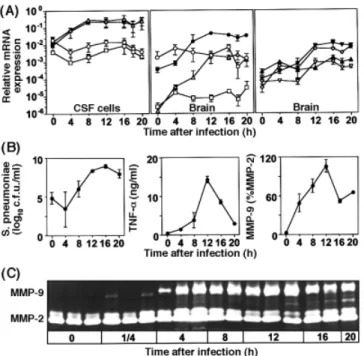
(4) Metalloproteinases in bacterial meningitis. 1737. sections per animal were evaluated for neuronal injury of the cortex and hippocampus, as described in detail previously (Leib et al., 2000; Pfister et al., 2000). The area of cortical brain damage was expressed as a percentage of the total area of cortex in each section and expressed as the mean value per animal. Apoptotic death of neurones in the dentate granule cell layer of the hippocampus was scored and an averaged score per animal was calculated from all sections evaluated. All histopathological evaluations were performed by an investigator blinded to the clinical, microbiological and treatment data of the respective animal.. Assessment of learning function. Fig. 1 Time-course studies in experimental pneumococcal meningitis. (A) Transcriptional expression of MMPs was standardized by expression of the housekeeping gene GAPDH separately in CSF cells and brain tissue (data are mean ⫾ standard deviation). In CSF cells, MMP-8 (open triangles) and MMP-9 (closed circles) mRNAs showed a rapid increase, of 10to 100-fold, while MMP-2 (open circles) and MMP-7 (open squares) mRNAs remained virtually unchanged. In brain tissue, transcription of MMP-2 and MMP-7 remained at basal level, while MMP-3 (inverted closed triangles), MMP-8, MMP-9, MMP-12 (inverted open triangles), MMP-13 (open diamonds) and MMP-14 (closed triangles) mRNAs were significantly upregulated by a factor of 100–1000. (B) Time course of CSF parameters. The amount (c.f.u.) of S. pneumoniae in CSF and protein expression of TNF-α and MMP-9 showed rapid, synchronous increases, starting 4 h after infection. (C) Zymography of CSF samples. MMP-2 was present constitutively in all samples, without significant change during disease. In contrast, MMP-9 was not present at the time of infection but appeared as early as 15 min after infection, reached maximal intensity at 12 h and was present 20 h after infection.. ceftriaxone. Animals were killed 48 h after infection. Animals to be tested for learning capacity by the water maze procedure were infected (log10 6.5 ⫾ 0.6 c.f.u./ml) and randomized for treatment with BB-1101 (10 mg/kg subcutaneously; n ⫽ 23), vehicle (200 µl PBS, 1% Tween 80, n ⫽ 26, or 300 µl saline, n ⫽ 31) every 8 h intraperitoneally starting 18 h after infection, for a duration of 4 days. Uninfected animals from the same litters were sham infected with 10 µl of sterile saline and treated with vehicle (200 µl PBS, 1% Tween 80; n ⫽ 26 or 300 µl saline; n ⫽ 22) every 8 h intraperitoneally starting 18 h after infection, for a duration of 4 days.. Histology and morphometry For histopathological examination, animals were perfused with 4% paraformaldehyde in PBS and 12 coronal brain. For the water maze procedure, we used a round grey tank of 1.8 m diameter (surface 2.54 m2) and of 0.7 m height, filled with water at a temperature of 25 ⫾ 1°C to a depth of 48 cm (Riedel et al., 1999; Iida et al., 2000; Loeffler et al., 2000). The water was darkened by the addition of non-toxic food colouring. A video camera was fastened to the ceiling above the centre of the pool. Before the test, gross vestibulomotor dysfunction of animals to be assessed in the water maze was excluded by the use of a rotating rod. Rats were placed on a foam cylinder with a circumference of 22 cm, fastened to a motor with adjustable speed, 20 cm above the table surface. Animals had to stay on the cylinder at different speeds (4, 8, 12, 16 r.p.m.) for a minimum of 10 s to qualify for the water maze test. For the assessment of learning function, swimming patterns of the rats were registered with the video tracking system Ethovision (Noldus Information Technology, Wageningen, The Netherlands). The water surface was virtually divided into four inner quadrants and a periphery with a width of 18 cm. An adjustable platform measuring 16 ⫻ 13 cm, covered with a rough black mat, was placed in the centre of the first quadrant 0.5 cm below the water surface. Four entry zones, each situated between two quadrants, were marked outside the pool. Three posters measuring 0.6 ⫻ 0.3 m with different black-and-white patterns (horizontal and diagonal stripes, circles) were placed on three walls 0.5 m from the edge of the pool to serve as visual cues. The room was illuminated by indirect light from the ground. Thirty-two-day-old survivors of meningitis and uninfected littermate controls were transferred to the experiment room, where they were given 24 h to acclimatize in a light cycle of 12 h light–12 h darkness, the light being switched on at 08.00 hours. Animals were provided with water and food ad libitum. From Day 1 to Day 4, animals performed five training trials per day with the invisible platform in a fixed position throughout the test. The rats were put into the water with their head directed towards the wall of the tank. If an animal found the platform within 90 s, it was allowed to stay on it for 15 s before it was put back to the cage. If the rat did not find the platform within 90 s, it was guided there by hand and was allowed to stay on it for 15 s. Between trials, animals rested for 45 min. Entry zones were randomized.
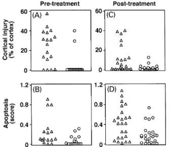
(5) 1738. S. L. Leib et al.. with a dice for each trial. Tracks were recorded by Ethovision with five samples per second, and the total distance moved to reach the platform was evaluated for each trial.. analysis of the broth culture of S. pneumoniae drawn every 4 h over 18 h did not show MMP-9-specific zymographic activity (data not shown).. Statistical analysis. Effect of MMP/TACE inhibition on clinical and CSF parameters. Normally distributed variables are presented as mean ⫾ standard deviation. Variables that were not normally distributed were compared by the Kruskal–Wallis test. When this test yielded a statistically significant value (P ⬍ 0.05), pair-wise comparison was done using the two-tailed nonparametric Mann–Whitney U-test. The association between continuous variables was assessed with the Pearson correlation coefficient. Proportions in different groups were compared by Fisher’s exact test. Survival curves were analysed by Kaplan–Meier analysis. A professional statistician blinded to the treatment and infection status of the animals performed the statistical analysis of data from time-course studies on mRNA expression and data from the water maze studies. Levels of mRNA expression were analysed for trends over time by multivariant regression analysis, and significant increases in the slopes were analysed with the t-test. Distances moved in the water maze task were compared with repeated measures analysis of variance, and pairwise comparison was done with the Tukey–Kramer adjustment. SAS version 8.0 (SAS Institute, Cary, NC, USA) software was used.. Results mRNA for MMPs in CSF cells and brain tissue during meningitis Transcriptional regulation of specific MMPs was assessed by quantitative real-time PCR separately in CSF cells and brain tissue (Fig. 1A). In CSF cells, mRNA encoding MMP-8 and -9 was upregulated rapidly and significantly, reaching maximum levels 8 h after infection, while expression of MMP-2 and -7 remained at basal levels (P ⬍ 0.005 for MMP-8 and -9; P not significant for MMP-2 and -7). In brain tissue, transcription of MMP-2 and -7 remained unchanged (P not significant) during the course of the study, whereas the numbers of transcripts for MMP-3, -8, -9, -12, -13 and -14 showed significant (P ⬍ 0.01) increases of 100to 1000-fold over baseline expression.. Protein expression of MMP-9 and TNF-α in CSF during meningitis The concentrations of MMP-9 and TNF-α in CSF were significantly correlated (P ⬍ 0.0001; Pearson r ⫽ 0.83) and exhibited synchronous kinetic changes in the course of the disease. There was a marked increase as early as 4 h and peak concentrations were reached at 12 h, persisting until 20 h after infection (Fig. 1B and C). The bacteria in CSF samples did not contribute to the amount of MMP-9, as. To maximize the effect of treatment and to evaluate the therapeutic intervention early in the course of meningitis, animals were infected and were treated with BB-1101 (15 mg/kg subcutaneously twice daily, n ⫽ 34) or vehicle (n ⫽ 36) at the time of infection (pretreatment). Eighteen hours after infection, all infected animals had meningitis, as indicated by positive CSF cultures and disease symptoms (weight loss and clinical score ⬍5). Pretreatment with BB1101, compared with vehicle, led to a reduction in the incidence of seizures (67 versus 22%, P ⬍ 0.02) and reduced disease severity, as evidenced by a higher activity score (3.8 ⫾ 0.5 for BB-1101 versus 3.3 ⫾ 1.1 for controls, P ⬍ 0.03), whereas bacterial titres of CSF (8.3 ⫾ 0.9 log10 c.f.u./ml for BB-1101 versus 8.8 ⫾ 0.6 log10 c.f.u./ml for controls, P not significant) and weight loss (0.6 ⫾ 0.5 g for BB-1101 versus 0.9 ⫾ 0.4 g for controls, P not significant) were not affected. CSF analysis showed a marked reduction in TNF-α concentrations (256 ⫾ 270 pg/ml for BB-1101 versus 3347 ⫾ 1513 pg/ml for controls, P ⬍ 0.001), indicating efficient TACE inhibition, but showed no effect on the amount of MMP-9. Mortality associated with the disease was significantly reduced (P ⬍ 0.001, Kaplan–Meier analysis) in infected animals treated with BB-1101 versus vehicle.. Effect of MMP/TACE inhibition on brain injury Two forms of neuronal injury can be identified in this model: cortical necrosis and apoptotic neuronal injury in the dentate gyrus of the hippocampus (Fig. 2).. Pre-treatment study In control animals treated only with antibiotics, bacterial meningitis led to substantial cortical damage, defined as ⬎5% of cortex injured, in 14 out of 18 (78%) animals. The proportion of animals with cortical injury was significantly (P ⬍ 0.001) reduced to two out of 18 (11%) when BB1101 was given at the time of infection (Fig. 3A). The neuroprotective effect of BB-1101 was also evident in the hippocampus, where apoptosis was significantly (P ⬍ 0.01) reduced by pretreatment (Fig. 3B). Uninfected control animals treated with BB-1101 did not show histopathological changes.. Post-treatment study We evaluated the effect of MMP/TACE inhibition in an experimental setting more closely reflecting the clinical situation, in which therapy is started when patients have.
(6) Metalloproteinases in bacterial meningitis. Fig. 2 Brain histology of infant rats with pneumococcal meningitis 24 h after infection. (A) In the cortex, neuronal injury consisted of areas with markedly reduced neuronal density with a wedge-shaped distribution (arrows) suggestive of ischaemic damage. Magnification ⫻10. (B) Focus of cortical neuronal loss (right side), containing neurones with morphological features of necrosis (including cell swelling and fading of cytoarchitecture), is sharply demarcated from preserved neurones (left side). Magnification ⫻200. (C) Dentate gyrus of the hippocampus, containing neurones with dense and shrunken nuclei (arrows). Magnification ⫻200. (D) Neurones with condensed, fragmented nuclei (arrows) forming apoptotic bodies, a morphological feature characteristic of programmed cell death. Magnification ⫻400.. 1739. Fig. 3 Effect of vehicle (triangles) and inhibition of MMP/TACE by BB-1101 (circles) on neuronal injury in the cortex and in the hippocampus. Treatment was started at the time of infection as a pre-treatment (A and B) and in established pneumococcal meningitis as a post-treatment (C and D). In the pretreatment study, injury in the infected control animals was [median (range)] 28.3% (0–57.3%) in the cortex and 0.1 (0–0.91) in the hippocampus. BB-1101 significantly reduced injury to 0% (0–39.9%) in the cortex (P ⬍ 0.0004) and to 0 (0–0.31) in the hippocampus (P ⬍ 0.01). When BB-1101 was first administered 18 h after infection, together with antibiotics, injury was reduced from 2.12% (0–39.77%) to 0.2% (0–12.63%) (P ⬍ 0.04) in the cortex and from 0.42 (0–1.08) to 0.16 (0–0.73) (P ⬍ 0.02) in the hippocampus.. disease symptoms. Rats were infected and BB-1101 (n ⫽ 25) or vehicle (n ⫽ 25) was given together with antibiotics 18 h later, when meningitis was fully developed. Brain injury was assessed 31 ⫾ 12 h after infection. Again, injury of both cortical and hippocampal neurones was attenuated significantly (P ⬍ 0.04 for cortex and P ⬍ 0.02 for hippocampus) by adjuvant therapy with BB-1101 (Fig. 3C and D).. Effect of MMP/TACE inhibition on learning Learning ability, assessed by the decreasing distance needed to reach a submersed platform in repetitive trials over 4 days, was significantly (P ⬍ 0.0001) impaired in animals 3 weeks after pneumococcal meningitis (n ⫽ 57) in comparison with uninfected controls (n ⫽ 48) (Fig. 4). In contrast, posttreatment with BB-1101 (n ⫽ 23) reduced (P ⬍ 0.03) learning deficits as a consequence of bacterial meningitis, compared with animals treated with antibiotics alone (Fig. 4). The learning curve of BB-1101-treated animals after meningitis was not different (P ⫽ 0.96) from that of uninfected control animals.. Fig. 4 Assessment of learning function of rats by the Morris water maze 3 weeks after pneumococcal meningitis. Analysis of the distance moved to reach the platform showed a decrease over time in all tested groups, indicating that all animals had the ability to learn. However, uninfected control animals (squares) learned significantly faster than survivors of meningitis (P ⬍ 0.0001). Adjuvant therapy of meningitis with BB-1101 (circles) led to improved performance (P ⬍ 0.03) compared with animals treated with antibiotics alone (triangles)..
(7) 1740. S. L. Leib et al.. Discussion The present time-course studies in a rat model of pneumococcal meningitis document a 100- to 1000-fold transcriptional induction of MMP-3, -8, -9, -12, -13 and -14, but not of MMP-2 and -7, in brain parenchymal tissue. In CSF cells, mRNAs of MMP-8 and -9 were increased 10- to 100-fold, while MMP-2 and -7 mRNAs remained at basal levels. These findings extend those of an earlier report in a model of CSF inflammation caused by intracisternal application of heat-killed Neisseria meningitidis in rats, in which mRNA encoding MMP-9 was upregulated and no change was found in the mRNA expression pattern of MMP2 and -7 (Kieseier et al., 1999). The strong transcriptional upregulation of MMPs in brain parenchyma and the appearance of MMP-9 in CSF as early as 15 min after infection indicate that, in early phases of bacterial meningitis, MMPs are produced primarily by resident brain parenchymal cells, as blood-derived neutrophils are virtually absent at this stage. The close correlation of TNF-α with MMP-9 and the lack of MMP-2 induction in CSF cells is fully congruent with previous findings in experimental models and in patients with bacterial meningitis, where MMP-2 was unaffected by the disease while MMP-9 and TNF-α were increased in parallel (Leib et al., 2000; Leppert et al., 2000). During inflammation, specific metalloproteinases (i.e. TACE) may contribute to the pathophysiology of bacterial meningitis, not only by their proteolytic activity, but also by their ability to release TNF-α, thus increasing the stimulus that initiates MMP upregulation via a positive feedback loop (Chandler et al., 1997). The lack of an effect of MMP/TACE inhibition on CSF concentrations of MMP-9 in the acute disease (18 h after infection in the present study) may reflect the fact that BB-1101 inhibits the activity but not the release of preformed MMPs, whereas a potential effect of TNF-α on MMP expression is likely to occur at later disease stages. In patients with bacterial meningitis, TNF-α levels in CSF are elevated and correlate positively with morbidity and mortality (Mustafa et al., 1989). Similarly, children who developed neurological sequelae of bacterial meningitis had higher CSF levels of MMP-9 than those who recovered fully (Leppert et al., 2000). TNF-α injected intracerebrally produces a dose-dependent increase in the permeability of the blood–brain barrier and MMP activity, whereas administration of TNF-α into the CSF results in pathophysiological changes characteristic of bacterial meningitis, including the breakdown of the blood– brain barrier, the recruitment of neutrophils and the generation of meningeal inflammation (Rosenberg et al., 1995). Thus, in bacterial meningitis, TNF-α mediates the inflammatory response in the subarachnoid space and is likely to perpetuate its own release via the induction of MMPs. The marked beneficial effect of BB-1101 on clinical parameters, mortality, neuropathology and learning abilities suggests that the inhibition of both the signalling (shedding of cytokines including TNF-α and its receptors) and effector mechanisms (proteolytic MMP activity) of meningitis is a particularly promising strategy for adjuvant therapy.. Histopathologically, the disease model is characterized by the development of extensive neuronal damage (Auer et al., 2000; Leib et al., 2000; Loeffler et al., 2000; Pfister et al., 2000). In the cortex, areas of reduced neuronal density exhibit features of ischaemic injury, with wedge-shaped lesions and morphological evidence of acute neuronal necrosis (Fig. 2A and C). In the dentate gyrus of the hippocampus, the nature of neuronal death is predominantly apoptotic, as evidenced by morphological studies and increased DNA fragmentation (Fig. 2B and D) (Leib et al., 1996). Both forms of injury in the meningitis model (necrotic cortical injury and apoptotic hippocampal injury) were attenuated by treatment with BB-1101. This suggests that metalloproteinase activity is critical for both forms of neuronal injury, even though the detailed mechanisms of cellular injury may well differ. Previous studies of the present meningitis model found an association between cerebral blood flow reduction and the development of cortical brain injury (Leib et al., 1996; Pfister et al., 2000). Cerebral blood flow reduction during meningitis can be global, as a result of the blood–brain barrier opening with subsequent brain oedema and reduced cerebral perfusion pressure, or it can be focal, resulting from vasculitis of cerebral blood vessels caused by inflammation of the surrounding subarachnoid space (Leib and Ta¨ uber, 1999). Processes that are modulated by MMP inhibition include both leakage through the blood–brain barrier and the inflammatory reaction of the CSF space. Accordingly, GM6001, a different hydroxamic acid MMP inhibitor with only minor TACE inhibitory activity, attenuated cortical injury efficiently but had no effect on hippocampal apoptosis compared with BB-1101 (Leib et al., 2000). In contrast, results from another study in the same model argue for TACE inhibitory activity as a possible cause of the observed effect of BB-1101 on hippocampal injury. Administration of a monoclonal antibody against TNF-α significantly reduced neuronal loss in the dentate gyrus of the hippocampus, but had no effect on damage to the cortex (Bogdan et al., 1997). Furthermore, apart from the proteolytic cleavage of TNF-α and its receptors (p75 and p55), TACE has been found to contribute to the shedding of several other transmembrane proteins, including transforming growth factor-α, L-selectin, the amyloid precursor protein and the non-signalling type II interleukin 1 receptor (Reddy et al., 2000). Investigations into the contribution of TACE substrates to the development of neuronal injury in bacterial meningitis are likely to reveal further explanations for the effects observed in the present study. Attenuation of hippocampal apoptosis by adjuvant therapy with BB-1101 prevented the learning disturbances observed in animals that had recovered from pneumococcal meningitis. Damage to the hippocampus results in impaired learning, both in humans and in animals, and the extent of hippocampal apoptosis as a consequence of bacterial meningitis is correlated positively with the reduction in learning capacity (Dusek and Eichenbaum, 1997; Loeffler et al., 2000). In patients, neuropsychological evidence supports the clinical.
(8) Metalloproteinases in bacterial meningitis importance of impaired function of the hippocampal formation after bacterial meningitis. In a retrospective survey of patients surviving bacterial meningitis, 31% reported disturbances of attention and 24% memory deficits, which affected their ability to work (Bohr and Rasmussen, 1988). Memory, the development of speech and verbal performance were significantly affected in children after bacterial meningitis (Letson et al., 1992; Thomas, 1992; Anderson et al., 1997). Histopathological studies in patients who died from bacterial meningitis showed apoptosis exclusively in neurones of the granular cell layer of the hippocampal dentate gyrus (Nau et al., 1999). On the basis of the results of the present study, we envisage that the consequences of bacterial meningitis for learning and other integrative neuronal functions can be prevented by the inhibition of MMP/ TACE activity. In summary, time-course studies in a pneumococcal meningitis model documented transcriptional upregulation of MMP-9 and -8 in CSF cells and brain parenchyma, whereas four additional MMPs, whose roles in the pathogenesis of bacterial meningitis are not yet defined, are upregulated in the latter. TNF-α and MMP-9 increased synchronously in CSF, peaking 12 h after infection. Combined MMP and TACE inhibition by BB-1101 downregulated the protein concentration of TNF-α in CSF, decreased the incidence of seizures and mortality and attenuated neuronal necrosis in the cortex and apoptosis in the hippocampus. A marked neuroprotective effect was seen even when therapy with BB1101 and antibiotics was started simultaneously 18 h after infection, reflecting the clinical situation in human disease. The attenuation of brain damage by BB-1101 preserved learning performance of animals that had recovered from pneumococcal meningitis. These results suggest that adjunctive therapy with MMP/TACE inhibitors could prevent neurological deficits in bacterial meningitis and provide the scientific basis for the evaluation of MMP/TACE inhibitors in clinical studies.. Acknowledgements We thank Graham Wells (British Biotech Pharmaceuticals, Oxford, UK), Caroline Grygar (Department of Research, University Hospitals Basel, Switzerland), Oliver Schu¨ tz and Philipp Joss (Institute for Infectious Diseases, University of Bern, Switzerland) for excellent technical support. This work was supported by grants from the Swiss National Science Foundation (NRP 4038-52841, 32-61654), the The´ odore Ott Foundation, The Meningitis Research Foundation and by the NIH (NS-35902).. References Alexander CM, Werb Z. Proteinases and extracellular matrix remodeling. [Review]. Curr Opin Cell Biol 1989; 1: 974–82.. 1741. Anderson V, Bond L, Catroppa C, Grimwood K, Keir E, Nolan T. Childhood bacterial meningitis: impact of age at illness and acute medical complications on long term outcome. J Int Neuropsychol Soc 1997; 3: 147–58. Auer M, Pfister LA, Leppert D, Ta¨ uber MG, Leib SL. Effects of clinically used antioxidants in experimental pneumococcal meningitis. J Infect Dis 2000; 182: 347–50. Azeh I, Mader M, Smirnov A, Beuche W, Nau R, Weber F. Experimental pneumococcal meningitis in rabbits: the increase of matrix metalloproteinase-9 in cerebrospinal fluid correlates with leucocyte invasion. Neurosci Lett 1998; 256: 127–30. Barlaam B, Bird TG, Lambert-Van Der Brempt C, Campbell D, Foster SJ, Maciewicz R. New alpha-substituted succinate-based hydroxamic acids as TNFalpha convertase inhibitors. J Med Chem 1999; 42: 4890–908. Bogdan I, Leib SL, Bergeron M, Chow L, Ta¨ uber MG. Tumor necrosis factor-alpha contributes to apoptosis in hippocampal neurons during experimental group B streptococcal meningitis. J Infect Dis 1997; 176: 693–7. Bohr VA, Rasmussen N. Neurological sequelae and fatality as prognostic measures in 875 cases of bacterial meningitis. Dan Med Bull 1988; 35: 92–5. Chandler S, Miller KM, Clements JM, Lury J, Corkill D, Anthony DC, et al. Matrix metalloproteinases, tumor necrosis factor and multiple sclerosis: an overview. [Review]. J Neuroimmunol 1997; 72: 155–61. Clements JM, Cossins JA, Wells GM, Corkill DJ, Helfrich K, Wood LM, et al. Matrix metalloproteinase expression during experimental autoimmune encephalomyelitis and effects of a combined matrix metalloproteinase and tumour necrosis factor-alpha inhibitor. J Neuroimmunol 1997; 74: 85–94. Dusek JA, Eichenbaum H. The hippocampus and memory for orderly stimulus relations. Proc Natl Acad Sci USA 1997; 94: 7109–14. Gearing AJ, Beckett P, Christodoulou M, Churchill M, Clements J, Davidson AH, et al. Processing of tumour necrosis factor-alpha precursor by metalloproteinases. Nature 1994; 370: 555–7. Grimwood K, Nolan TM, Bond L, Anderson VA, Catroppa C, Keir EH. Risk factors for adverse outcomes of bacterial meningitis. J Paediatr Child Health 1996; 32: 457–62. Iida R, Saito K, Yamada K, Basile AS, Sekikawa K, Takemura M, et al. Suppression of neurocognitive damage in LP-BM5-infected mice with a targeted deletion of the TNF-alpha gene. FASEB J 2000; 14: 1023–31. Kieseier BC, Paul R, Koedel U, Seifert T, Clements JM, Gearing AJ, et al. Differential expression of matrix metalloproteinases in bacterial meningitis. Brain 1999; 122: 1579–87. Leib SL, Ta¨ uber MG. Pathogenesis of bacterial meningitis. Infect Dis Clin North Am 1999; 13: 527–48, v–vi. Leib SL, Kim YS, Chow LL, Sheldon RA, Ta¨ uber MG. Reactive oxygen intermediates contribute to necrotic and apoptotic neuronal injury in an infant rat model of bacterial meningitis due to group B streptococci. J Clin Invest 1996; 98: 2632–9..
(9) 1742. S. L. Leib et al.. Leib SL, Leppert D, Clements J, Ta¨ uber MG. Matrix metalloproteinases contribute to brain damage in experimental pneumococcal meningitis. Infect Immun 2000; 68: 615–20.. Pfister L-A, Tureen JH, Shaw S, Christen S, Ferriero DM, Ta¨ uber MG, et al. Endothelin inhibition improves cerebral blood flow and is neuroprotective in pneumococcal meningitis. Ann Neurol 2000; 47: 329–35.. Leppert D, Leib SL, Grygar C, Miller KM, Schaad UB, Hollander GA. Matrix metalloproteinase (MMP)-8 and MMP-9 in cerebrospinal fluid during bacterial meningitis: association with blood–brain barrier damage and neurological sequelae. Clin Infect Dis 2000; 31: 80–4.. Ramilo O, Saez-Llorens X, Mertsola J, Jafari H, Olsen KD, Hansen EJ, et al. Tumor necrosis factor alpha/cachectin and interleukin 1 beta initiate meningeal inflammation. J Exp Med 1990; 172: 497–507.. Letson GW, Gellin BG, Bulkow LR, Parks DJ, Ward JI. Severity and frequency of sequelae of bacterial meningitis in Alaska Native infants. Correlation with a scoring system for severity of sequelae. Am J Dis Child 1992; 146: 560–6.. Reddy P, Slack JL, Davis R, Cerretti DP, Kozlosky CJ, Blanton RA, et al. Functional analysis of the domain structure of tumor necrosis factor-alpha converting enzyme. J Biol Chem 2000; 275: 14608–14.. Loeffler JM, Ringer R, Hablu¨ tzel M, Ta¨ uber MG, Leib SL. The free radical scavenger alpha-phenyl-tert-butyl-nitrone aggravates hippocampal apoptosis and learning deficits in experimental pneumococcal meningitis. J Infect Dis 2001; 183: 247–52.. Riedel G, Micheau J, Lam AG, Roloff E, Martin SJ, Bridge H, et al. Reversible neural inactivation reveals hippocampal participation in several memory processes. Nat Neurosci 1999; 2: 898–905.. Maskos K, Fernandez-Catalan C, Huber R, Bourenkov GP, Bartunik H, Ellestad GA, et al. Crystal structure of the catalytic domain of human tumor necrosis factor-alpha-converting enzyme. Proc Natl Acad Sci USA 1998; 95: 3408–12. Mustafa MM, Ramilo O, Saez-Llorens X, Mertsola J, Magness RR, McCracken GH Jr. Prostaglandins E2 and I2, interleukin 1-beta, and tumor necrosis factor in cerebrospinal fluid in infants and children with bacterial meningitis. Pediatr Infect Dis J 1989; 8: 921–2. Nau R, Soto A, Bruck W. Apoptosis of neurons in the dentate gyrus in humans suffering from bacterial meningitis. J Neuropathol Exp Neurol 1999; 58: 265–74. Paul R, Lorenzl S, Koedel U, Sporer B, Vogel U, Frosch M, et al. Matrix metalloproteinases contribute to the blood–brain barrier disruption during bacterial meningitis. Ann Neurol 1998; 44: 592– 600.. Rosenberg GA, Estrada EY, Dencoff JE, Stetler-Stevenson WG. Tumor necrosis factor-alpha-induced gelatinase B causes delayed opening of the blood–brain barrier: an expanded therapeutic window. Brain Res 1995; 703: 151–5. Saukkonen K, Sande S, Cioffe C, Wolpe S, Sherry B, Cerami A, et al. The role of cytokines in the generation of inflammation and tissue damage in experimental gram-positive meningitis. J Exp Med 1990; 171: 439–48. Thomas DG. Outcome of paediatric bacterial meningitis 1979– 1989. Med J Aust 1992; 157: 519–20. Wallace GR, Whiston RA, Stanford MR, Wells GM, Gearing AJ, Clements JM. The matrix metalloproteinase inhibitor BB-1101 prevents experimental autoimmune uveoretinitis (EAU). Clin Exp Immunol 1999; 118: 364–70.. Received November 21, 2000. Revised April 9, 2001. Accepted May 4, 2001.
(10)
Figure
Documents relatifs