ECTOPIC OSSIFICATION FOLLOWING TOTAL HIP
ARTHROPLASTY: IS DIFFUSE IDIOPATHIC SKELETAL
HYPEROSTOSIS A RISK FACTOR?
BY H. FAHRER1, P. KOCH2, P. BALLMER2, P. ENZLER2 AND N. GERBER1
Departments of 'Rheumatology and 2Orthopaedic Surgery, University of Bern, Inselspital,
CH-3010 Bern, Switzerland
SUMMARY
Total hip arthroplasty may be followed by ectopic bone formation. An increased frequency has been suspected in patients with diffuse idiopathic skeletal hyperostosis (DISH). In 204 patients we found that, of the 38 subjects with pre-existing DISH, 29% developed postoperative ossification compared with only 10% in those without DISH (p < 0.01). DISH is therefore a risk factor for postoperative ectopic bone formation. In a separate study of 1325 patients (not analysed for spinal DISH), we looked for correlations between the severity of postoperative ectopic bone and clinical measurements. Even for the more severe ossification grades (n = 112), only 10% reported serious pain and only 26% had reduced hip flexion (<70°). Thus, periprosthetic ectopic bone is not sufficiently important to justify the routine use of preventative drugs such as bisphosphonates in patients with DISH undergoing total hip replacement.
KEY WORDS: Ectopic bone, Hip arthroplasty, Diffuse idiopathic skeletal hyperostosis.
THE formation of periprosthetic ectopic cal-cification and ossification in the first months fol-lowing total hip replacement arthroplasty is well known. (In this paper we will use only the term 'ossification'. Persistent calcification usually becomes trabecular with time.) Reports of its frequency vary. A notable degree of periarticu-lar postoperative ossification was seen in 5% of Charnley's patients [1]. Others have reported frequencies of 8-90% [2-6].
No consensus is found in the literature about the clinical significance of this abnormality in terms of pain or reduced motion (ROM). How-ever, symptoms seem to occur in 1-5%, especi-ally loss of movement [7].
Predisposing factors which have been exam-ined include operative technique, haemorrhage and infection [ 1,8,9]. DeLee et al. [3] stated that the degree of preoperative reduced ROM influ-enced the amount of postoperative ossification. Male sex, osteoarthritis, hyperglycaemia and obesity are other factors which have been incriminated [2, 10-12]. Several authors have suggested that DISH (formerly called For-estier's disease) might be a risk factor (Fig. 1) [2, 13-16], others have denied any influence of DISH [17].
The objective of this retrospective radiologi-Submitted 17 August; revised version accepted 10 November 1987.
Address correspondence to Heinz Fahrer, MD.
cal and clinical study was to elucidate two ques-tions. First, do patients with pre-existing spinal hyperostosis develop ectopic bone more fre-quently around their hip arthroplasty than con-trols without DISH? Second, are such bone formations associated with serious pain or restricted hip function?
PATIENTS AND METHODS There were two groups of patients:
Group I consisted of 204 consecutive patients with an original M.E. Muller straight-stem pros-thesis, operated on in the years 1977/1978 (sub-group la, n = 91) and 1979/1980 (sub(sub-group Ib, n = 113), respectively. They all underwent
sur-FtG. 1.—Typical flowing ossification in a 67-year-old male with DISH on the spine. Within 2 years (left to right), a new lateral bridge developed between L2 and L3. 187
188 BRITISH JOURNAL OF RHEUMATOLOGY VOL. XXVII NO. 3
TABLE I
CLASSIFICATION FOR ECTOPIC PERI PROSTHETIC BONE FORMATION*
Class I: Islands of bone within the soft tissues about the hip.
Class II: Bone spurs from the pelvis or proximal end of the femur, leaving at least 1 cm between opposing surfaces.
Class III: Bone spurs from the pelvis or proximal end of the femur, reducing the space between oppos-ing bone surfaces to less than 1 cm. Class IV: Apparent bone ankylosis of the hip. * After Brooker el al. [4].
gery in the University Ginic for Orthopaedic Surgery in Bern. These patients were followed up after 5 years in 1983 and 1985, respectively. Of the 204 patients 117 were men with a mean age of 67±8 (range 43-84) years at follow-up, and 87 were women, mean age 70±10 (range 35-S9).
Group II consisted of 1325 patients with a Muller prosthesis, operated on in different Swiss clinics for orthopaedic surgery and examined again after 1 year (mean age: men 64±9; women 66±11).
Radiological investigation
At the 5-year follow-up, all the patients of group / had an anteroposterior radiograph of the pelvis. In addition, patients in subgroup la (n = 91) had anteroposterior (AP) and lateral radio-gTaphs of the thoracic and/or lumbar spine. Those of subgroup Ib (n = 113) had AP and lateral radiographs of the chest and 49 had addi-tional spinal views.
A rheumatologist (H.F.) and two orthopaedic surgeons (P.B. and P.E.) first examined all radiographs of the spine and chest of group I independently, looking for DISH. The criteria used were those of Resnick [18], and required flowing calcification and ossification along the anterolateral aspect of at least four contiguous vertebral bodies and absence of extensive 'degenerative' disc disease. Radiographs which did not completely fulfil these criteria were clas-sified as 'probable DISH'. The individual assess-ments were compared and where discordant, the films were regraded by both observers together. The pelvic radiographs of group I were evalu-ated similarly for any ectopic bone formation • about the prosthesis. Radiographs of spine and
pelvis were not seen simultaneously, thus mak-ing the gradmak-ing 'blind'.
The degree of ossification was defined accord-ing to the scale of Brooker (Table I) [4, 7]. This
refers to ossification situated between the greater trochanter and the upper border of the acetabulum, and was adequate since changes develop laterally in 94% [3] (Fig. 2).
All group //-patients had an AP radiograph of the pelvis 1 year after operation but views of the spine were not routinely obtained.
Clinical investigation
In group II, we graded patients' hip pain as none, slight, moderate or severe 1 year post-operatively and measured their maximal hip flexion as >90°, 70-90°, 30-70° or <30°.
RESULTS
The radiological prevalence of DISH was 38/204 (19%) (Table II). In subgroup la the prevalence of DISH was 25%, higher than in the subgroup Ib (13%). In more than half of the patients in subgroup Ib the diagnosis of DISH had to be made from a chest radiograph. DISH was more frequent in men than in women.
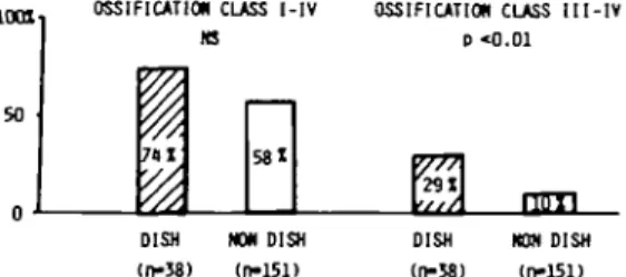
Table III shows the frequency of the different degrees of postoperative ossification (0-FV). The main findings are illustrated in Fig. 3 and 74% of 38 DISH probands formed postoperative grade I-IV ossification, compared with only 58% of the 151 non-DISH probands. The differ-ence was not significant (x2 = 2.65, p = 0.1). When only the more severe grades III and IV were compared, there were 29% in the DISH-probands and 10% in non-DISH-DISH-probands (x2 = 7.72, p < 0.01).
The correlations between postoperative ossification, pain and reduced ROM are shown for group II in Fig. 4. Of patients with no or
Fio. 2.—Formation of ectopic ossification (grade III—IV, after Brooker) following total hip arthroplasty.
TABLE II
PREVALENCE OF DISH (SUBGROUPS IA, IB, TOTAL GROUP I) IN 204 PATIENTS UNDERGOING TOTAL HIP
ARTHROPLASTY
No. (%) Group Ia Group Ib Total Total Men DISH Men Non-DISH DISH probable 91 51 23 18 61 7 (100) (56) (25) (78) (67) (8) 113 70 15 11 90 8 (100) (62) (13) (73) (80) (7) 204 121 38 29 151 15 (100) (59) (19) (76) (74) (7) TABLE III
FREQUENCY OF ECTOPIC BONE FORMATION FOLLOWING HIP ARTHROPLASTY IN PATIENTS WITH DISH AND WITHOUT
DISH (CLASSIFICATION I-IV AFTER BROOKER)
Class 0 I II III IV Total DISH no 10 13 4 6 5 38 . (%) (26) (34) (11) (16) (13) (100) Non-DISH no 64 52 20 11 4 151 (42) (35) (13) (7) (3) (100) DISH prob. no 3 7 0 5 0 15 • (%) (20) (47) (33) (100) Total no 77 72 24 22 9 204 . (%) (38) (35) (12) (11) (4) (100)
slight ossifications (classes 0-11), 4% experi-enced moderate or severe pain. In patients with marked ossification (classes III-IV) the fre-quency was 10%. Postoperative flexion of less than 70° was found in 6% of gTades 0-11 and in 26% of grades III-IV. Both findings were statis-tically significant (x2 = 9.8, p < 0.01; x2 = 54.19, p < 0.001, respectively).
DISCUSSION
The prevalence of spinal DISH in 19% of our population agrees with previous published studies of similar ages [7, 11, 12], as does the greater prevalence of ossification in men [2,3, 10, 11].
Severe postoperative ectopic bone formation around the hip arthroplasty was three times more frequent in those with spinal DISH. Irrespective of DISH, other authors have noted postoperative ossification in 10% of patients [3,8].
This threefold increased frequency in DISH-probands supports the hypothesis that DISH predisposes to ectopic bone formation as part of a generalized 'ossifying diathesis'. Blasingame et al. [7] suggested a correlation between vertebral hyperostosis and ectopic bone formation based on a small series of patients. Jacqueline [16]
found ossification in 57 of 67 subjects with DISH, but in only 10 of 33 controls. The severity of these ossifications and the criteria used for the diagnosis of DISH were not given. Pilet et al. [11] in a similar study, used less rigorous criteria for DISH and found 22.4% postoperative ossification in subjects with DISH and 9.6% in controls (grades III-IV), and this is in accord with our results.
Our second question concerned the clinical significance of ectopic ossification. Others have suggested a relationship with pain or impaired hip function [6-8,10,19, 20] and Pilet et al. [11] found a favourable postoperative ROM in 66% of males with DISH compared with 83 % in those without. They concluded that DISH does not contra-indicate total hip replacement.
Our results indicate that ectopic bone forma-tion about the hip is associated less with pain than with functional impairment. We noted moderate and severe pain in our group II of 1325 arthroplasties and this was significantly more frequent in marked ossification (10% versus 4% in controls). However, this is not a striking prev-alence. Restricted flexion of less than 70° was found in 26% of 'ossifying' patients versus 6% in 'non-ossifying'.
We may conclude that approximately 30% of patients with DISH develop serious ectopic bone formation following hip replacement and
so OSSIFICATION CLASS I - I V M 581 OSSIFICATION CLASS I I I - I V p < 0 . 0 1 i in 11 DISH (n-38) NOH DISH ( n - 1 5 1 ) DISH (n-58) WW DISH (n-151) FIG. 3.—Frequency (percentage) of postoperative ectopic bone formation in DISH and non-DISH probands.
1001 1 1 OSSIFICATION V777X OSSIFICATION PAIN WDERATE/SEVERE CLASS CLASS O-II I I I - I V 6 1 i (n-1213) (n- 112) FLEXION <7O-2 6 1 p<0.01 CXO.001 FIG. 4.—Correlation of degree of ossification with pain and reduced ROM 1 year after hip arthroplasty (group II, n = 1325).
190 BRITISH JOURNAL OF RHEUMATOLOGY VOL. XXVII NO. 3
70% will remain free. Only 25% of those with ectopic bone show important loss of ROM and only 6% develop serious pain. This means that only 7 . 5 % of all patients with DISH suffer from restricted ROM and 1.8% from pain. This low risk does not justify the use of drugs in an attempt to prevent ossification. Bisphospho-nates have proved relatively effective in this situ-ation, but the recommended 4 months of treatment is both expensive and may cause side-effects [21-23]. Prevention should be confined to patients with a history of severe ossification of the other hip or other articulations.
ACKNOWLEDGEMENT
We wish to thank particularly Professor A. St. J. Dixon, Bath, for assistance in the preparation of the manuscript.
REFERENCES
1. Charnley J. The long-term results of low-fric-tion arthroplasty of the hip performed as a primary intervention. J Bone Joint Surg [Br] 1972;54:61-76.
2. Lazansky M. Complications revisited: the debit side of total hip replacement. Clin
Orthop 1973;95:96-103.
3. DeLeeJ, Ferrari A, Charnley J. Ectopic bone formation following low friction arthroplasty of the hip. Clin Orthop 1976;121:53-9. 4. Brooker AF, Bowerman JW, Robinson RA,
Riley Jr LH. Ectopic ossification following total hip replacement. J Bone Joint Surg
[Am] 1973;55:1626-32.
5. Nollen AJG, Sloof TJ. Para-articular ossifica-tion after total hip replacement. Acta Orthop
Scand 1973;44:230-41.
6. Rosendahl S, Krogh Christoffersen J, Ntfrgaard M. Para-articular ossification fol-lowing hip replacement. Acta Orthop Scand 1977;48:400-4.
7. Blasingame JP, Resnick D, Courts RD, Danzig LA. Extensive spinal osteophytosis as a risk factor for heterotopic bone formation after total hip arthroplasty. Clin Orthop Rel Res
9 8 1 1 6 1 1 9 7 1981;161:1917.
8. Holz U, Kraner F, Weller S. Periartikulare Verknocherungen nach Hufttotalen-doprothesen. Z Orthop 1977;115:146-58. 9. Rigler HF, Harries CM. Heterotopic bone
for-mation after total hip arthroplasty. Clin
Orthop 1976;117:2O9-16.
10. Ritter MA, Vaughan RB. Ectopic ossification
following total hip replacement. J Bone Joint
Surg [Am] 1977 £9:345-51.
11. Pilet F, Waldburger M, Livio JJ. Periarticular ossification following tota| hip prosthesis in cases of diffuse idiopathic skeletal hyper-ostosis. Rev Chir Orthop 1983;69:455-63. 12. Julkunen H, Heinonen OP, Pyorala K.
Hyper-ostosis of the spine in an adult population, its relationship to hyperglycemia and obesity.
Ann Rheum Dis 1971;30:605-12.
13. Forestier J, Rotes-Querol J. Senile ankylosing hyperostosis of the spine. Ann Rheum Dis 1950;9:321-30.
14. Forestier J, Lagier R. Ankylosing hyperostosis of the spine. Clin Orthop Rel Res 1971 ;74: 65-83.
15. Resnick D, Limovita RJ, Feingold ML. Post-operative heterotopic ossification in patients with ankylosing hyperostosis of the spine (Forestier's disease). / Rheumatol 1976^3: 313-20.
16. Jacqueline F. Ankylose osseuse de coxopathies hyperostosiques (4 observations).
Rhum-atologie 1983^5:231-5.
17. Bundrick TJ, Cook DE, Resnik CS. Hetero-topic bone formation in patients with DISH following total hip replacement. Radiology 1985;155:595-7.
18. Resnick D, Niwayama G. Diffuse idiopathic skeletal hyperostosis (DISH): ankylosing hyperostosis of Forestier and Rotes-Querol. In: Resnick D, Niwayama G, eds. Diagnosis
of bone and joint disorders. Philadelphia:
WB Saunders, 1981;2:1416-52.
19. Kromann-Andersen C, Scherff Sarensen T, Hougaard K, Zdravkovic D, Frigaard E. Ectopic bone formation following Charnley hip arthroplasty. Acta Orthop Scand 1980; 51:633-8.
20. SlStis P, Kiviluoto O, Santavirta S. Ectopic ossification after hip arthroplasty. Ann Chir
Gynaecol 1978;67:89-93.
21. Finerman GAM, Stover SL. Heterotopic ossification following hip replacement or spi-nal cord injury: two clinical studies with EHDP. Metab Bone Dis Relat Res 1981 ;4/5: 337-42.
22. Sloof TJJH, Feith R, Bijvoet OLM, Nollen AJG. The use of a disphosphonate in para-articular ossifications after total hip replace-ment. Acta Orthop Belg 1974;40:820-8. 23. Thomas BJ, Amstutz HC. Results of the
administration of diphosphonate for the pre-vention of heterotopic ossification after total hip arthroplasty. J Bone Joint Surg [Am]
BEFORE IT'S TOO LATE
Salazopyrin EN-tabs has a favourable benefit: risk ratio
for early intervention when NSAIDs alone prove inadequate^ and because
it works promptly, patients improve early - often within one month?
13Salazopyrin
enteric coated sulphasalazine
Positive action in rheumatoid arthritis
»ha|Mfi .f M »
hi^.-r»a**Orra™»«3>CLJgaej(a»tttea)lCTcm«tt,t
torn mdat I r»«i <* v . Pnctdarc bod dado ad IFTt rtouM
X caw Gtma, «m Imt priorKM
N A P R O S Y N
ABBREVIATED
PRESCRIBING I N F O R M A T I O N NAPROSYN Tablets (naproxen 250mg per tablet)
NAPROSYN 500 Tablets (naproxen 500mg per tablet)
NAPROSYN Suspension (naproxen 25mg/ml) NAPROSYN Suppositories (naproxen 500mg per suppository)
NAPROSYN Granules (naproxen 500mg per sachetl
Uses:
Rheumatoid arthritis, osteoarthritis, ankylosing spondylitis, acute gout and acute musculo-skeletal disorders.
Dosage:
For rheumatoid arthritis, osteoarthritis and ankylosing spondylitis: usually 500mg to 1g daily taken in t w o doses at 12-hour intervals. Use lowest effective dose in the elderly. For acute gout: 750mg at once, then 2S0mg every eight hours until the attack has passed. For juvenile arthritis in children over 5 years: Smg/kg body weight twice daily. For acute musculoskeletal dis-orders: 500mg initially, then 25Omgat 6-8 hour intervals as needed with a maximum daily dose after the first day of 1250mg.
C o n t r a i n d i c a t i o n s :
Active peptic ulceration. Hypersensitivity to naproxen or naproxen sodium formulations. Aspirin/anti-inflammatory-induced allergy.
Warnings, Precautions, etc:
Episodes of Gl bleeding have been reported. Use with care in patients with a history of Gl disease. Use lowest effective dose in impaired hepatic function. Use with caution in patients with impaired renal function. Monitor renal function and consider reducing dosage in patients where renal blood flow is compromised (e.g. extracellular volume depletion, cirrhosis of the liver, sodium restriction, congestive heart failure, pre-existing renal disease) - some elderly patients may fall in this category. Use with caution in patients with asthma or allergic disease. Caution is required if any of the following are administered concurrently: hydantoins, anti-coagulants or highly protein-bound sulphonamides; frusemide; propranolol or other beta-blockers; lithium; probenecid; methotrexate. NAPROSYN decreases platelet aggregation and prolongs bleeding time. Use in pregnant or breast-feeding women should be avoided if possible.
Side-effects:
Gl - nausea, vomiting, pain; occasionally bleeding
and ulceration. Dermatologicol/hypersensitMty -skin rashes, urticaria, angio-oedema; rarely anaphylactic reactions, eosinophilic pneumonitis, alopecia, erythema multiforme, Stevens Johnson syndrome, epidermal necrolysis and photo-sensitive dermatitis. CNS - headache, insomnia, inability to concentrate, cognitive dysfunction.
Haematological - thrombocytopenra,
granulo-cytopenia, aplastic anaemia, haemolytic anaemia. Other - tinnitus, hearing impairment, vertigo, mild peripheral oedema (patients with compro-mised cardiac function m«y be at a greater risk on NAPROSYN); rarely jaundice, fatal hepatitis, nephropathy, haematuria, visual disturbances, vasculitis and ulcerative stomatitis. NAPROSYN
Suppositories (loco!) - rectal discomfort, soreness,
burning, itching, rectal bleeding, tenesmus, proctitis.
Basic N H S Cost:
Tablets 25Omg E7.60 for 60 tablets, £30.35 for 250 tablets. Tablets 500mg £14.57 for 60 tablets original pack (OP), £24.28 for 100 tablets. Suspension £8.24 for 500ml. Suppositories £2.96 for 10 suppositories. Granules £19.47 for 60 sachets. Product Licence N o . : PL 0286/0031 -Tablets (250mg). PL 0286/0061 - Tablets (500mg). PL 0286/0047 - Suspension. PL 0286/0053 - Suppositories. PL0286/0098-Granules. Further information is available from: SYNTEX Pharmaceuticals Limited, St. ives Road. Maidenhead, Berkshire SL6 1RD. 'NAPROSYN is a trade mark.
^ SYNTEX
Original Pack O P containing 60 tablets.