Gene flow and cross-mating in Plasmodium falciparum in
households in a Tanzanian village
H. A. BABIKER
1, J. D. CHARLWOOD
2, T. SMITH
3W D . W A L L I K E R
1 1Institute of Cell, Animal and Population Biology, University of Edinburgh, West Mains Road, Edinburgh
EH9 3JT, Scotland, UK
2
National Institute for Medical Research, Ifakara Centre, P.O. Box 53, lfakara, Tanzania
3
Department of Public Health and Epidemiology, Swiss Tropical Institute, Postfach, CH-4002, Basel, Switzerland
(Received 15 November 1994; revised 1 May 1995; accepted 1 May 1995)SUMMARY
The diversity of the genes encoding 2 merozoite surface proteins (MSP-1 and MSP-2) of Plasmodium falciparum has been
examined in parasites infecting members of 4 households in a village in Tanzania. The polymerase chain reaction (PCR)
was used to characterize allelic variants of these genes by the sizes and sequences of regions of tandemly repeated bases
in each gene. In each household extensive polymorphism was detected among parasites in the inhabitants and in infected
mosquitoes caught in their houses. Similar frequencies of the alleles of these genes were observed in all households.
Capture-recapture data indicated that both Anopheles gambiae and A. funestus freely dispersed among households in the
hamlet. The results confirm that cross-mating and gene flow occur extensively among the parasites, and are discussed
within the context of spatial clustering of natural populations of P. falciparum.
Key words: Plasmodium falciparum, merozoite surface proteins, genetic polymorphisms, spatial clustering.
INTRODUCTION
The idea that malaria parasites are 'clustered' in
nature has been proposed by several workers. For
example, clustering of Plasmodium malariae
infec-tions in certain houses has been reported in a village in
Guinea-Bissau (Snounouei al. 1993). Local variations
in the intensity and duration of infection and of
clinical symptoms among infected individuals in a
given community (Molineaux & Gramiccia, 1980)
provide circumstantial evidence that such clustering
of parasites may occur. Clustering of mosquitoes
occurs in nature, and variations in levels of
trans-mission have been observed between different
house-holds in a single community (Gamage-Mendis et al.
1991). Such variations in transmission could result
in local variations in the genetic structure of the
parasite populations. Space-time clustering of
mini-epidemics of severe P. falciparum malaria in a coastal
region of Kenya has been attributed to antigenically
distinct parasite 'strains' (Snow et al. 1993).
Few studies have been carried out to determine
whether parasite genotypes are clustered at
house-hold levels. It is now possible to examine this subject
by studying the distribution of the numerous
polymorphic genes which exist in malaria parasite
populations, especially in P. falciparum and P. vivax
(Kemp, Cowman & Walliker, 1990; Cheng et al.
1993). Isolates of P. falciparum from different
countries have been found to possess similar alleles
of these genes, but often at different frequencies,
suggesting that there is a closer relatedness of
parasites within rather than between different areas
(Creasey et al. 1990). Forsyth et al. (1989) have
produced evidence for variations in the frequency of
an S-antigen allele at the community level in
different villages in Papua New Guinea.
Never-theless, the diversity of parasites occurring in a
single community may be very great; for example,
each of 29 isolates of P. falciparum examined from a
Sudanese village possessed unique genotypes
(Babi-ker et al. 1991). In a peri-urban area in The Gambia,
the gene pool of P. falciparum was smaller in small
communities than in larger ones, although there was
no evidence of differences in allele frequencies
among the communities studied (Conway &
McBride, 1991 a).
In this paper, we examine whether parasite
clustering occurs in P. falciparum in houses in a
village in Tanzania. We make use of the polymerase
chain reaction (PCR) to examine polymorphic alleles
of 2 antigen genes among these parasites in
finger-prick blood samples of the inhabitants, in mosquito
bloodmeals, and in oocysts in mosquitoes caught in
the houses. We show that there is considerable
movement of infected mosquitoes between different
households, and that frequent crossing between
different clones occurs during mosquito
trans-mission. As a result, extensive genetic diversity of
parasites occurs among the parasites in all the
households studied.
MATERIALS AND METHODS
Study area
T a b l e 1. Oocyst rate in mosquitoes in households of Kining'ina, collected by resting catch
Uninfected Household mosquitoes Infected mosquitoes Total 8001/1-2 8003/1-3 8004/1-4 8006/1-4 Total 323 96 347 73 839 60(17) 11 (10) 31 (8) 10(12) 112(12) 383 107 378 83 951
hamlet named Kining'ina, which is 1 km north of Michenga village, near Ifakara, south-east Tanzania. T h e area is holo-endemic for malaria, P. falciparum constituting u p to 9 5 % of all malaria cases. T r a n s -mission by Anopheles gambiae and A.funestus occurs t h r o u g h o u t the year, with peaks following rains in A p r i l / M a y and N o v e m b e r / D e c e m b e r (Kilombero Malaria Project, 1992; Smith et al. 1993).
Kining'ina lies in an area which is farmed. T h e hamlet consists of 4 families, each of which inhabits a group of 1-4 small huts. T h e groups of huts are approximately 500 m apart from each other. Each h u t contains 1 or 2 beds. At the start of the study, no mosquito nets or other anti-mosquito devices were in use in any of the huts. Households were allocated serial n u m b e r s 8001, 8003, 8004 and 8006, and each h u t was allocated a s u b n u m b e r . T h u s , for example, 8 0 0 4 / 1 - 4 designates a group of 4 huts, inhabited by m e m b e r s of 1 family.
Isolates of P. falciparum
Fingerprick blood samples (500—700^1) were col-lected from all m e m b e r s of the 4 families on 1 day, 21 J u n e 1991. T h e r e were 30 inhabitants altogether, of whom 17 were positive for P. falciparum by thick blood smear. T h e s e 17 blood samples were cryo-preserved (Aley et al. 1984), stored at - 8 0 °C, and transported to Edinburgh. P. falciparum genomic D N A was isolated from the blood samples by the method of Foley, Ranford-Cartwright & Babiker (1992) for subsequent P C R work.
Collection and capture—recapture of mosquitoes Daily collections of A. gambiae s.l. and A. funestus were made in each of the huts in the early morning by resting collections (Molineaux et al. 1988) between 10 and 23 June 1991. On 15 June, some of the mosquitoes caught in households 8001 and 8004 were dusted with different coloured fluorescent powder (Charlwood, Graves & Birley, 1986) and released from the sites of collection. From 17 June, mosquito nets with four 200 mm diameter holes cut into their sides were placed over 12 of the 15 beds in the hamlet. Mosquitoes were collected from these nets every morning for the next 5 days, and taken
immediately to an insectary at the Ifakara Centre for processing. At the end of the experiment new intact nets were given to each family.
Isolation of parasites from mosquitoes
Mosquitoes collected from the huts were maintained in the insectary for 5-7 days to allow oocysts to develop. After dissection and careful microscopical examination, midguts containing only single oocysts were washed twice in drops of fresh RPMI medium on a glass slide, placed individually into Eppen-dorf tubes containing a PCR lysis buffer/proteinase K mixture, and incubated for 1 h at 55 °C (Ranford-Cartwright et al. 1991). After incubation, all preparations were stored at —20 °C, before being transported to Edinburgh. DNA was subsequently isolated from oocysts as described by Ranford-Cartwright et al. (1991).
Blood was also prepared from 12 mosquitoes from household 8001. These mosquitoes were dissected on the morning of their collection. Their abdomens with midguts containing blood from the previous night's feeds were placed individually into Eppen-dorf tubes containing PCR lysis buffer/proteinase K, and then treated in a similar manner to the oocysts, as described above.
Polymerase chain reaction (PCR) of MSP-1 and MSP-2 genes
PCR primers were designed to amplify regions of genes encoding 2 antigen genes denoted MSP-1 and MSP-2. These genes occur as single copies in the genome, MSP-1 being located on chromosome 9 and MSP-2 on chromosome 2 (Triglia, Wellems & Kemp, 1992). Both genes contain regions of tan-demly repeated bases. In MSP-1, these repeats occur in block 2 of the gene near its 5' end (Tanabe et al. 1987), while in MSP-2 they occur in a central portion of the gene, also denoted block 2 (Smythe et al. 1991). Details of the primers and PCR conditions are given by Ranford-Cartwright et al. (1993) and Babiker et al. (1994a).
Amplified PCR fragments were subjected to electrophoresis on a 1-6% agarose gel. Variations in the number of repeats in different alleles of MSP-1 and MSP-2 could be recognized by differences in size of the amplified products (Kimura et al. 1990). Fragments were then Southern blotted on to nylon membranes (Sambrook, Fritsch & Maniatis, 1989). Hybridization of the PCR-amplified fragments with sequence-specific oligonucleotides
Amplified PCR fragments were classified on the basis of their sequence by hybridization of Southern blots with allele-specific probes. All MSP-1 alleles so far examined contain 1 of 3 sequences in block 2, denoted K l , MAD20 and RO33 after the isolates
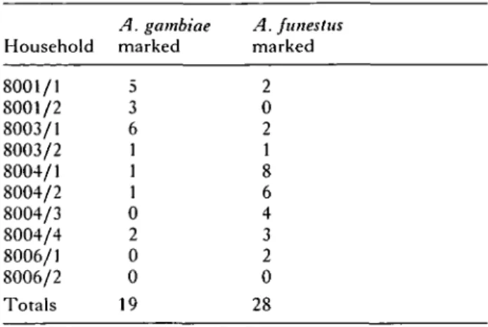
Table 2. Recapture of marked mosquitoes originally released in houses 8001/1 and 8004/4
Household 8001/1 8001/2 8003/1 8003/2 8004/1 8004/2 8004/3 8004/4 8006/1 8006/2 Totals A. gambiae marked 5 3 6 1 1 1 0 2 0 0 19 A. funestus marked 2 0 2 1 8 6 4 3 2 0 28
from which they were originally described (Kimura et al. 1990). Similarly, block 3 of the MSP-2 gene has been classified into 2 families distinguishable by specific DNA sequence (Fenton et al. 1991; Smythe et al. 1991); two oligonucleotide probes denoted IC1 and FC27 were used to identify the two types (Babiker et al. 1994a). Oligonucleotides for each of these sequences were made, then labelled at their 3' ends with fluorescein-11-dUTP, using the Amer-sham 3' oligolabelling kit. These probes were then hybridized to Southern blots of the PCR-amplified products of each respective gene, and detected using the Enhanced Chemiluminescence (ECL) detection kit (Amersham) (Babiker et al. 1994a).
RESULTS
Mosquito collections
(a) Resting collections. I n d o o r resting catch col-lections of mosquitoes were m a d e in all h o u s e h o l d s t h r o u g h o u t the study period. T h e r e w e r e c o n s i d e r -able variations between t h e h o u s e h o l d s in t h e n u m b e r s collected, as well as in t h e oocyst rates (Table 1).
(b) Collections from nets with holes. Collections of mosquitoes from nets with holes were m a d e daily between 17 and 22 J u n e . A total of 913 s p e c i m e n s of A. funestus and 432 of A. gambiae was c a u g h t . D u r i n g the period of s t u d y , t h e p o p u l a t i o n s of b o t h species were relatively s t a b l e a n d old (mostly parous). M e a n n u m b e r s of A. gambiae decreased from 24 to 1 5 / n e t / n i g h t , w h e r e a s m e a n n u m b e r s of A. funestus increased from 28 to 4 2 . T h e r e was n o significant difference in t h e p a r o u s rate of t h e t w o species, 31 of 38 ( 8 2 % ) A. funestus e x a m i n e d and 20 of 26 (77 % ) A. gambiae being p a r o u s (%2 = 0 2 1 , P = 0-65). Despite these similarities, t h e oocyst rates were significantly higher in A. gambiae t h a n in A. funestus; 60 out of 276 (21 %) A. gambiae dissected were infected, compared to 57 o u t of 514 (11 % ) A. funestus (X2 = 1 6 1 , P < 0 0 0 0 1 ) .
(c) Mosquito movement and cross-infection of P . falciparum between different households. M o s q u i t o e s collected from nets with holes were e x a m i n e d for
T a b l e 3. M S P - 1 and M S P - 2 alleles of isolates collected on 21 J u n e 1991 from infected i n d i v i d u a l s in 4 households in Kining'ina, T a n z a n i a
(Alleles are designated by sequence type and by size of their amplified PCR fragment. For example, for M S P - 1 , allele Kl5 2 0 contains the Kl sequence and has a fragment size of 520 base pairs (bp); —, allele not detected, N.D. Not done.) House number 8001/1-2 8003/1-3 8004/1-4 8006/1-4 Isolstc number IfB2 IfB3 IfB4 IfBlO IfBll IfB13 IfB14 IfB16 IfB17 IfB18 IfB20 IfB22 IfB23 IfB26 IfB27 IfB28 IfB29 MSP-1 Kl5 2 0 Kl5 2 0 Kl5 4 0 Kl4 1 0 Kl4 8 0 Kl4 8 0 — N.D. Kl4 8 0 Kl4 8 0 K 1i s o Kl5 6 0 Kl4 8 0 Kl5 4 0 Kl4 7 0 Kl5 8 0 — alleles MAD20470 MAD20520 — MAD20470 MAD20560 MAD20520 N.D. — — MAD20'8 0 — — MAD20500 — RO33470 — RO33470 RO33470 — RO33470 RO33470 RO33470 N.D. — — RO33470 RO33470 — — RO33470 RO33470 MSP-2 alleles IC1520 IC1650 IC1600 I C 15 2 0 I C 15 2 0 I C 16 5 0 I C 15 2 0 — I C 15 2 0 I C 15 2 0 I C 15 2 0 I C 16 5 0 I C 16 5 0 I C 16 5 0 I C 15 2 0 N.D. FC276 0 0 FC274 8 0 FC274 8 0 FC275 2 0 FC275 2 0 FC275 8 0 FC275 2 0 FC275 2 0 FC275 2 0 FC274 6 0 — FC276 0 0 FC275 2 0 FC275 2 0 FC274 8 0 FC275 2 0 N.D.
1 2 3 4 5 6 7 8
B
D
FC27 IC1
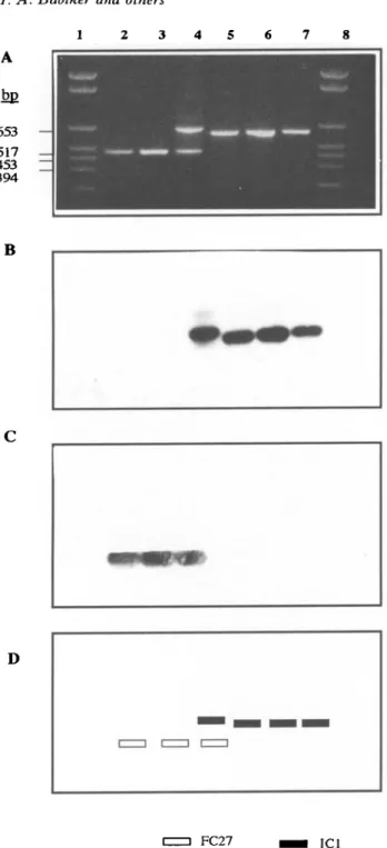
Fig. 1. Alleles of the MSP-2 gene of Plasmodium falciparum extracted from parasites from bloodmeals
acquired by fed mosquitoes {Anopheles gambiae and A. funestus). Following electrophoresis on 1-6% agarose gel
and ethidium bromide staining of PCR-amplified DNA fragments (A), blots were hybridized with allele-specific probes IC1 (B) and FC27 (C). (D) Schematic
interpretation of the results. Lanes 1 and 8: size markers, lanes 2-7: bloodmeals from individual mosquitoes. Note that in the mosquito in lane 4, two alleles were detected, indicating that the bloodmeal contained parasites of mixed genotypes.
fluorescent markings 2 days, i.e. 1 gonotrophic cycle,
after release in houses 8001/1 and 8004/1. All b u t 1
net, from house 8006/2, contained at least 1 marked
mosquito (Table 2). It is clear that many of these
mosquitoes had moved from the house of release
(either 8001/1 or 8004/4) to different houses.
Household-specific markings provided some limited
evidence of a difference in the recapture pattern
between the two species. For example, the majority
of the recaptured A. funestus originally released in
house 8004/4 (21/28) were found in huts of the same
household (8004); conversely, 11 out of 19
recap-tured A. gambiae originally released in household
8001 had moved between different households.
T h e data obtained in this study can be used to
estimate the proportion of infected mosquitoes
caught in the nets with holes which carry oocysts
from infections in other households. This proportion
is estimated by EPI, where E is the proportion of
mosquitoes caught in different houses from those of
their release, P is the parous rate of these mosquitoes,
and / is the chance of a mosquito taking up
gametocytes giving rise to oocysts. Details of the
calculations involved are given in the Appendix. EPI
is calculated from the data as 0-056 for A. gambiae
and 0-O132 for A. funestus.
Genotypes of parasites in infected people
Allele typing of MSP-1 and MSP-2 was carried out
on parasite DNA prepared from fingerprick samples
of the 17 individuals found to be positive for P.
falciparum by blood smear examination (Table 3).
Twelve alleles of M S P - 1 , and 8 of MSP-2,
dis-tinguishable by size and/or sequence were detected.
Many samples contained mixed infections, in which
2 or more alleles of 1 or both genes could be detected.
Each infected individual in households 8001 and
8003, 6 of the 8 in household 8004 and 3 of the 4 in
household 8006 had such mixed infections. In our
previous study, mixed infections were found in
approximately 85 % of the inhabitants of Michenga
village (Babiker et al. 19946).
T h e majority of isolates contained parasites with
different combinations of alleles of each gene.
Exceptions were IfB18 and IfB20 in household 8004
which both contained genotypes possessing the
K l / 4 8 0 and IC1/52O alleles of MSP-1 and MSP-2
respectively (Table 3). This combination could also
have been present in IfBll and IfB14; however,
since these isolates contained more than 1 allele at
both loci, it was not possible to define the precise
genotypes present without cloning. For the same
reason, it is possible but not certain that other mixed
isolates could have contained parasites with the same
genotypes.
Genotypes of parasites in blood samples from
mosquito midguts
Bloodmeals from 10 of the 12 fed mosquitoes
collected in house 8001/1 gave positive PCR
pro-ducts for MSP-1 and/or MSP-2. Results are shown
in Fig. 1 and Table 4.
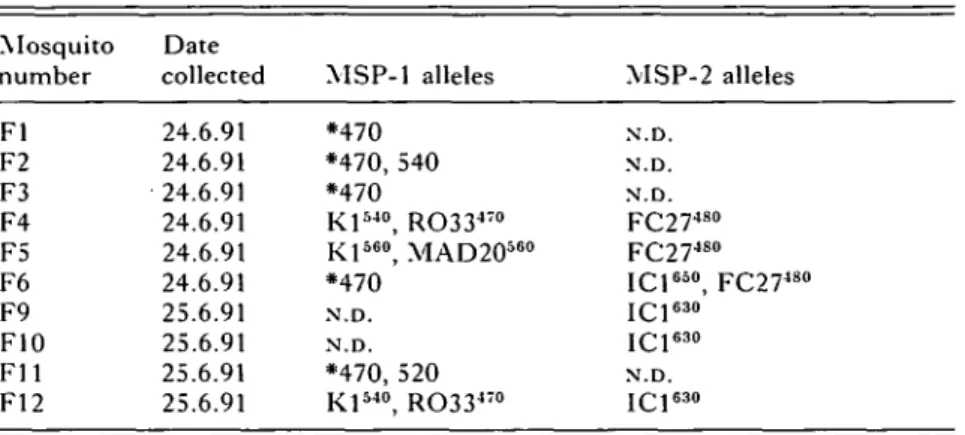
Table 4. MSP-1 and MSP-2 alleles of parasites extracted from bloodmeals of mosquitoes collected in house 8001/1 in Kining'ina (See Table 3 legend for explanation of symbols. *, Sequence not determined. N.D. Not done.) Mosquito number Fl F2 F3 F4 F5 F6 F9 F10 F l l F12 Date collected 24.6.91 24.6.91 24.6.91 24.6.91 24.6.91 24.6.91 25.6.91 25.6.91 25.6.91 25.6.91 MSP-1 alleles *470 *470, 540 *470 Kl540, RO33470 Kl560, MAD20560 *470 N.D. N.D. *470, 520 Kl5 4 0, RO33470 MSP-2 alleles N.D. N.D. N.D. F C 2 74 8 0 F C 2 74 8 0 I C 16 5 0, F C 2 74 8 0 I C 16 3 0 I C 16 3 0 N.D. I C 16 3 0
T a b l e 5. T w o - l o c u s genotypes of oocysts collected from h o u s e h o l d s in K i n i n g ' i n a
(For household 8004, oocysts nos. If22, 23, 32, 39 and 40 were from a pooled collection of mosquitoes caught in the 4 huts of this household. See Table 3 legend for explanation of symbols. N.D., Not determined.)
Hut number 8001/1 8003/1 8004/1-4 8004/1-4 8004/1-4 8004/1-4 8004/1-4 8004/1-3 8004/1-4 8004/1-3 8004/1-2 8004/1-2 8004/1-2 8004/1-4 8006/1 Oocyst number If 14 If 15 If25 If34 If42 If44 IfSO If57 If58 If59 If68 If9 IflO If 11 If31 If73 If22 If23 If32 If39 If40 If48 If66 If70 If71 If72 If 74 If77 If47 If51 If61 If62 If63 If69 If75 If 76 Date collected 11.6.91 11.6.91 11.6.91 12.6.91 13.6.91 12.6.91 18.6.91 19.6.91 19.6.91 19.6.91 20.6.91 11.6.91 11.6.91 11.6.91 13.6.91 20.6.91 12.6.91 12.6.91 13.6.91 12.6.91 12.6.91 17.6.91 20.6.91 20.6.91 20.6.91 20.6.91 21.6.91 21.6.91 17.6.91 18.6.91 19.6.91 19.6.91 19.6.91 20.6.91 21.6.91 21.6.91 MSP-1 Kl4 6 0 — — — Kl5 2 0 Kl5 4 0 — Kl5 2 0 Kl4 8 0 Kl5 2 0 Kl5 4 0 — Kl5 2 0 Kl5 4 0 — Kl5 6 0 Kl5 4 0 Kl5 4 0 Kl4 8 0 Kl5 2 0 Kl5 0 0 Kl5 0 0 Kl5 2 0 Kl5 4 0 — Kl4 7 0 Kl5 4 0 Kl4 8 0 Kl5 2 0 Kl4 8 0 Kl5 2 0 Kl5 2 0 Kl5 4 0 Kl5 6 0 Kl5 2 0 — alleles MAD20500 MAD20520 MAD20520 — — MAD20500 — MAD20480 MAD20520 — — — MAD20520 MAD20520 — — — MAD20520 — — MAD20520 MAD20520 MAD20520 — — — — — — — — — — — — — — — RO33470 — — — RO33470 RO33470 — — — RO33470 — — —-— — — — RO33470 RO33470 RO33470 — RO33470 RO33470 RO33470 — RO33470 RO33470 RO33470 RO33470 MSP-2 alleles JQ1600.520 N.D. N.D. I C 14 8 0 I C 16 2 0 I C 15 6 0 I C 15 8 0 JC 15 6 0 — I C 14 6 0 N.D. — N.D. I C 16 8 0 I C 16 0 0 N.D. I C 16 8 0 N.D. — I C 14 8 0 | p i 580;540 I CJ54O I / s i 65O;54O |Qj62O;56O I C 16 0 0 — J £ j600;J40 JQJ580;540 I C 1o 8 0 N.D. JQJ620;500 I C 16 0 0 I CJ600 F C 2 76 0 0 N.D. N.D. F C 2 76 2 0 F C 2 74 8 0 — — F C 2 75 2 0 F C 2 74 8 o — N.D. F C 2 74 8 0 N.D. — N.D. N.D. F C 2 75 0 0 F C 2 76 2 0 — F C 2 75 4 0 F C 2 75 4 0 — — F C 2 74 5 0 F C 2 75 0 0 — — F C 2 75 2 0 N.D. F C 2 75 2 0 — —
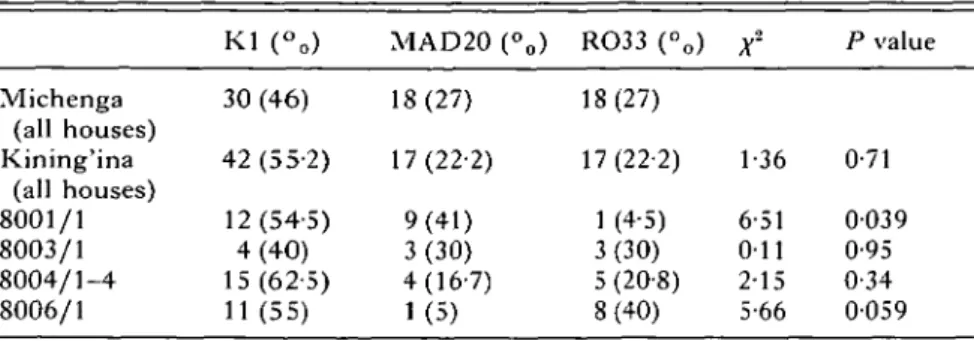
Table 6. Comparison of allele frequencies of the MSP-1 gene between Kining'ina hamlet and Michenga village, and between individual households in Kining'ina and Michenga village
(Data for Michenga village are from Babiker et al. (19946).)
Michenga (all houses) Kining'ina (all houses) 8001/1 8003/1 8004/1-4 8006/1 K l (-„) 30 (46) 42 (55-2) 12(54-5) 4(40) 15(62-5) 11(55) MAD20 (°0) 18(27) 17(22-2) 9(41) 3(30) 4(16-7) 1(5) RO33 (°0) 18(27) 17(22-2) 1 (4-5) 3(30) 5 (20-8) 8(40) X2 1-36 6-51 O i l 2-15 5-66 P value 0-71 0039 0-95 0-34 0059 * Likelihood ratio chi-squared (2 D.F.), to test whether the distribution of alleles in households in Kining'ina differs from that in Michenga.
T a b l e 7. Comparison of allele frequencies of M S P - 2 within households of Kining'ina
(Likelihood ratio chi-squared (3 D.F.), to test whether the distribution of alleles differs between households in Kining'ina. x~, I '4 7; p = 069.) House number Michenga (all houses) Kining'ina (all houses) House 8001/1 House 8003/1-3 House 8004/1-4 House 8006/1-4 ICl (%) 33(53) 36 (60) 11 (61) 4(67) 10(50) 11 (69) FC27 (%) 29 (47) 24 (40) 7(39) 2(33) 10(50) 5(31) Total 62 60 18 6 20 16
T h e parasites in these mosquitoes were most probably derived from the inhabitants of the hut in which they were caught. However, it cannot be entirely excluded that they might have come into the huts with a bloodmeal taken elsewhere, or that they might have contained oocysts from previous feeds. Only parous mosquitoes could have had developing oocysts from previous feeds, and only immigrant mosquitoes would have been infected elsewhere.
Oocyst genotypes
A total of 951 m o s q u i t o e s w e r e e x a m i n e d for oocysts 5—7 days after collection ( T a b l e 1). Of these, 110 c o n t a i n e d d e v e l o p i n g oocysts, 36 of w h i c h contained only single oocysts. T h e s e w e r e e x a m i n e d for M S P - 1 a n d M S P - 2 alleles ( T a b l e 5). T h e r e m a i n i n g 74 infected m o s q u i t o e s c o n t i n u e d 2 o r m o r e oocysts, a n d these were n o t s t u d i e d further.
F o u r t e e n alleles of M S P - 1 a n d 20 of M S P - 2 were d e t e c t e d . I n 7 oocysts ( I f l 5 , If23, If.31, If57, If59, If77 and If76), only 1 t y p e of allele of each gene was d e t e c t e d . T h e s e oocysts h a d m o s t p r o b a b l y derived
from homozygous zygotes, resulting from self-fertilization events between identical gametes, al-though it cannot be entirely excluded that they were heterozygous at other loci not examined here. Oocysts with different alleles of one or both genes (e.g. If42, If44, etc.) had clearly originated from heterozygotes, derived from crossing between unlike gametes (Ranford-Cartwright et al. 1993; Babiker et al. 19946). T w e n t y - t h r e e oocysts were in this category. Six oocysts which produced positive results only for M S P - 1 were homozygous for this gene, but in the absence of M S P - 2 data it is not known whether they were derived from zygotes homozygous at both loci.
T h e oocysts found in the mosquitoes kept in the insectary 5 days had almost certainly derived from the inhabitants of the h u t in which they were caught, although it cannot be entirely ruled out that they had developed from gametocytes in a blood meal taken elsewhere. It is also possible that sporozoites re-sulting from earlier feeds by the mosquitoes could have been present a r o u n d t h e m i d g u t s which could have been the source of some P C R p r o d u c t s ; how-ever, we consider this to be unlikely because of the washing of each m i d g u t sample before preparation for P C R .
Comparisons of MSP-1 and MSP-2 allele frequencies of oocysts from different households
T o investigate the possibility of spatial clustering of the parasites, frequencies of the alleles of each gene in oocysts found in Kining'ina were calculated and compared to those of Michenga village as a whole, using t h e likelihood chi-squared test. In view of the small n u m b e r of samples involved, the alleles were ' b i n n e d ' into g r o u p s classified by sequence only (Babiker et al. 19946). T a b l e 6 shows the results for M S P - 1 . N o significant differences were seen in these frequencies (x2 = 1"36; P = 0-71). T h e frequencies of the M S P - 1 alleles found in each separate
house-hold were also compared to those of the whole
village. Those of households 8003, 8004 and 8006
were not significantly different; however, in
house-hold 8001 the x
2 t e s tindicated a border value
(X
2= 5-66; P = 0059) (Table 6).
The frequencies of MSP-2 alleles showed no
significant differences between the households
(Table 7) (
A-
2= l-47; P = 069), or between
Kin-ing'ina as a whole and Michenga (P > 0-5).
DISCUSSION
The most striking finding of this study is the
remarkable degree of genetic diversity which occurs
in P. falciparum in individual households in the
hamlet of Kining'ina. The fingerprick blood samples
and the mosquito bloodmeals illustrate clearly the
numbers of infections in the inhabitants containing
mixtures of clones. The oocysts show that an
extensive degree of crossing between clones occurs
in this small community. The mosquito
capture-recapture data illustrate that considerable gene flow
is likely to occur among the parasites of the
households. Taken together, the findings illustrate
emphatically the inappropriateness of the idea that
P. falciparum in such communities consists of a
collection of distinct 'strains'.
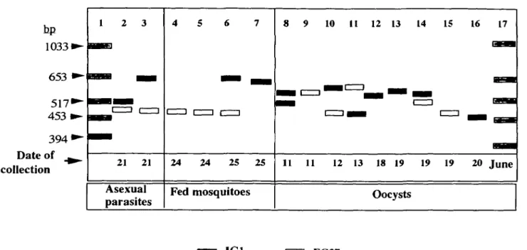
The information obtained from the single hut
8001/1 provides a good example of the complexity of
infection in just a single family. Fig. 2 illustrates the
MSP-2 alleles of parasites of fingerprick samples of
2 inhabitants of this hut, and in the bloodmeals and
oocysts of mosquitoes caught there. The oocysts
possessed very diverse alleles of both MSP-1 and
MSP-2, in homozygous and heterozygous
combin-ations. These alleles can only have derived from the
gametocytes taken up by the mosquitoes, but most
were not present in the blood samples examined.
While it is possible that some of the oocysts were the
result of feeds on inhabitants of other houses in the
area, it is more likely that the inhabitants of hut
8001/1 did indeed contain parasites with these alleles
at the times when the mosquitoes fed. Studies in
Papua New Guinea have shown that marked changes
in P. falciparum genotypes occur in a single patient
over periods as short as 3 days (M. C. Bruce and
M. Walmsley, unpublished observations).
Three of the 5 members of household 8001 were
positive for P. falciparum on the day of sampling.
Each of the 3 contained mixed clones, as shown by
the presence of 2 alleles of 1 or both genes. Their
parasites contained a total of 5 alleles of MSP-1 and
4 of MSP-2. Certain alleles were found in 2 of the 3
people, e.g. FC27/480 of MSP-2 in IfB3 and IfB4,
while others were present in only one, e.g. IC1/650.
Both these alleles were found in the bloodmeals of
mosquitoes caught in this hut 3 and 4 days later,
implying that parasites with these alleles were still
present in the inhabitants at the time of the feed. The
FC27/480 allele was also found in the oocyst in
mosquito If59 caught on 19 June, as well as in If44
caught 1 week earlier.
A similar degree of parasite diversity was observed
in each of the other Kining'ina households. This was
not unexpected in such a highly endemic situation,
where each unprotected individual can receive an
annual average of 329 infective bites (Smith et al.
1993) and where there is free movement by vectors
between households. The mosquito sampling
showed that 20% of the A. gambiae and 10% of the
A. funestus were infected. Ignoring the mosquitoes
that we did not collect, this means that parasites
were developing in at least 18 A. gambiae and 20
A. funestus in the hamlet every day of the study.
A few instances were found of parasites of similar
genotypes in different people in the same house,
which were most probably due to infection from a
common mosquito; a similar explanation was used
by Conway & McBride (19916) for parasites
con-taining identical alleles of MSP-1 and MSP-2 in
family members of a village in The Gambia.
We have found no evidence of clustering of P.
falciparum genotypes among the Kining'ina
house-holds. The frequencies of alleles found in individual
oocysts are more precise than those estimated from
those of blood forms alone (Babiker et al. 19946).
These data showed no significant difference in allele
frequencies of either gene among the households of
Kining'ina and Michenga village, with the exception
of MSP-1 in household 8001. We consider that this
result was most probably produced by the small
numbers of oocysts examined in the study, rather
than by the spatial isolation of this household from
the others. When the information obtained from
blood samples was taken together with the oocysts,
the numbers of alleles of MSP-1 and MSP-2 found
were comparable to those in Michenga village as a
whole; a total of 17 alleles of MSP-1 and 24 of MSP-2
were detected in 53 blood samples and 71 oocysts
from the whole village (Babiker et al. 19946), while
there were 14 and 18 respectively in the Kining'ina
households.
In summary, while the number of samples studied
here was small, there was no evidence for spatially
structured parasite populations within which mating
could be restricted, as suggested in the Papua New
Guinea studies by Day et al. (1992). The high level
of genetic diversity among the parasites studied here
is a consequence of the intensity of transmission and
sexual reproduction among these parasites. It is
possible that a larger survey involving more remote
houses in this area might produce evidence of spatial
variations. It would also be of considerable interest
to compare the findings obtained in this highly
endemic area with those in an area of much lower
10331
653 <
517
1453 1
394"
Date of ^
collection
1 2 3
21 21
Asexual
parasites
4 5 6 7
24 24 25 25
Fed mosquitoes
8 9 10 11 12 13 14 15 16 17
11 11 12 13 18 19 19 19 20 June
Oocysts
IC1
FC27
Fig. 2. Schematic illustration of the MSP-2 alleles of parasites in fingerprick blood samples (lanes 2 and 3),
bloodmeals from fed mosquitoes (lanes 4—7), and oocysts from mosquitoes (lanes 8-16) collected from hut number
8001/1. Alleles are classified by size and sequence of PCR-amplified fragments. Lanes 1 and 17: size markers.
intensity of transmission, to determine whether such
spatial clustering of parasites does occur in such
places.
We are indebted to the entomological team of the Ifakara
Centre of the Tanzanian National Institute for Medical
Research, Tanzania for their excellent assistance and
expertise. We are particularly grateful for the cooperation
of all the inhabitants of Kining'ina in this study. We thank
Professor W. Kilama, Director-General, NIMR,
Tan-zania, Professor Marcel Tanner, Swiss Tropical Institute,
Basel and Dr Thomas Teuscher of the Ifakara Centre for
their encouragement and support. The work was funded
by the Medical Research Council of Great Britain, the
World Health Organization and The Wellcome Trust.
APPENDIX
Estimate of the proportion of infected mosquitoes
caught in nets with holes which carry oocysts from
infections in different households
To estimate the proportion of infected mosquitoes caught
in the nets with holes in each house in Kining'ina which
carry oocysts from gametocytes acquired elsewhere, the
following assumptions are made, (i) The chances of
mos-quitoes acquiring gametocytes giving rise to any oocysts
(/) and to single oocysts (/') per feed are constant for all
days and households, (ii) Oocysts detected may be derived
from a previous feed, but rarely from an even earlier one.
(iii) Parous mosquitoes move between households with
probability E between feeds, E being the emigration rate
which is constant over houses and time, (iv) The system is
closed, (v) Intensity and risk of infection are independent
of mosquito parity, infection and migration status.
Then, before feeding (a) Proportion of immigrant
mos-quitoes with any oocysts = /. (b) Proportion of immigrant
mosquitoes with single oocysts = /'. (c) Proportion of
im-migrant mosquitoes with more than 1 oocyst = I—I', (d)
Proportion of fed mosquitoes which are parous = P. (e)
Proportion of fed mosquitoes which are immigrants = EP.
(f) Proportion of fed mosquitoes already having a single
oocyst infection = PI'. (g) Proportion of fed mosquitoes
already having any oocyst infection = PI. (h) Proportion
of fed mosquitoes already having an alien single oocyst
infection = EPT. (i) Proportion of fed mosquitoes already
having any alien oocyst infection = EPI.
After feeding, among fed mosquitoes in the
net:-(j) Overall proportion of mosquitoes with an alien single
oocyst infection and no superinfection = EPT (1 —/). (k)
Overall proportion of mosquitoes with a new single oocyst
infection and no previous infection = (1 —PI)I'. (1) Overall
proportion of mosquitoes with a single oocyst (/?') =
EPI\\-I) + (\-PI)r. (m) Overall proportion of
unin-fected mosquitoes = (1 —P/)(l —/). (n) Overall proportion
of infected mosquitoes (R) = 7(1 +P-PI).
Solving for /, we obtain:
To obtain I' we rearrange (1), to give / ' = R'/{EP{\ —/) +
(1— PI). In the current study, the relevant figures are as
follows.
E
P
R
R'
I
I'
EPI
A. gambiae
(11/19) 0-58
0-77
0-21
(36/951) 00379*
0-125
0029
0056
A. funestus
(7/28) 0-25
0-82
011(36/951) 00379*
0062
0033
0013
* Data for A. gambiae and A. funestus aggregated.
R E F E R E N C E S
ALEY, S. B., BARN'WELL, J. W., WENDELL, D. & HOWARD,
R. j . (1984). Identification of parasite proteins in a
membrane preparation enriched for surface membrane
of erythrocytes infected with Plasmodium knowlesi.
Molecular and Biochemical Parasitology 12, 69-84.
BABIKER, H. A., CREASEV, A. M., FENTON, B., BAYOUMI, R.
A. L.,
ARNOT, D. E.&
WALLIKER,D. (1991). Genetic
diversity of Plasmodium falciparum in a village in
eastern Sudan. 1. Diversity of enzymes, 2D-PAGE
proteins and antigens. Transactions of the Royal
Society of Tropical Medicine and Hygiene 85, 572-7.
BABIKER, H. A., RANFORD-CARTWRIGHT, L., SULTAN, A.,
SATTI, G.
&
WALLIKER, D.(1994a). Genetic evidence
that Rl chloroquine resistance of Plasmodium
falciparum is caused by recrudescence of resistant
parasites. Transactions of the Royal Society of Tropical
Medicine and Hygiene 88, 328-31.
BABIKER, H. A., RANFORD-CARTWRIGHT, L. C , CURRIE, D., CHARLWOOD, D., BILLINGSLEY, P., TEUSCHER, T. &
WALLIKER, D.
(19946). Random mating in a natural
population of the malaria parasite Plasmodium
falciparum. Parasitology 109, 413-21.
CHARLWOOD, D. J., GRAVES, P. & BIRLEY, M. H. (1986).
Capture-recapture studies of Anopheles punctulatus
Donitz (Diptera: Culicidae) from Papua New Guinea.
Bulletin of Entomological Research 76, 211-27.
CHENG, Q., STOWERS, A., HUANG, T.-Y., BUSTORS, D., HUANG, Y.-M., RZEPCZYK, C. & SAUL, A. (1993).
Polymorphism in Plasmodium vivax MSA1 gene - the
result of intragenic recombination ? Parasitology 106,
335-45.
CONWAY, D.
j . &
MCBRIDE,j . s. (1991 a). Population
genetics of Plasmodium falciparum within a malaria
hyperendemic area. Parasitology 103, 7-16.
CONWAY, D.
j . &
MCBRIDE,j . s. (1991 b). Genetic evidence
for the importance of interrupted feed by mosquitoes
in the transmission of malaria. Transactions of the
Royal Society of Tropical Medicine and Hygiene 85,
454-6.
CREASEY, A., FENTON, B., WALKER, A., THAITHONG, S., OLIVEIRA, S., MUTAMBU, S. & WALLIKER, D. (1990).
Genetic diversity of Plasmodium falciparum shows
geographical variation. American Journal of Tropical
Medicine and Hygiene 42, 403-13.
DAY, K. P., KOELLA, J. C , NEE, S., GUPTA, S. & READ, A. F.
(1992). Population genetics and dynamics of
Plasmodium falciparum: an ecological view.
Parasitology 104, S35-S52.
FENTON, B., CLARK, J. T., ANJAM KHAN, C. M., ROBINSON, J. V., WALLIKER, D., RIDLEY, R., SCAIFE, J. G. &
MCBRIDE,
j . s. (1991). Structural and antigenic
polymorphism of the 35- to 48-kilodalton merozoite
surface antigen (MSA-2) of the malaria parasite
Plasmodium falciparum. Molecular and Cellular Biology
11, 963-71.
FOLEY, M., RANFORD-CARTWRIGHT, L. C. & BABIKER, H. A.
(1992). Rapid and simple method for isolating malaria
DNA from fingerprick samples of blood. Molecular
and Biochemical Parasitology 53, 241-4.
FORSYTH, K., ANDERS, R. F., CATTANI, J. A. & ALPERS, M. P.
(1989). Small area variation in prevalence of an
S-antigen serotype of Plasmodium falciparum in
villages of Madang, Papua New Guinea. American
Journal of Tropical Medicine and Hygiene 40, 344—50.
GAMAGE-MENDIS, A. C , CARTER, R., MENDIS, C , DEZOYSA, A. P. K., HERATH, P. R. J. & MENDIS, K. (1991).
Clustering of malaria infections within an endemic
population: risk of malaria associated with the type of
housing construction. American Journal of Tropical
Medicine and Hygiene 45, 77-85.
KEMP, D.
j . ,
COWMAN, A.&
WALLIKER, D.(1990). Genetic
diversity in Plasmodium falciparum. Advances in
Parasitology 29, 75-149.
KILOMBERO MALARIA PROJECT
(1992). The level of
anti-sporozoite antibodies in a highly endemic malaria area
and its relationship with exposure to mosquitoes.
Transactions of the Royal Society of Tropical Medicine
and Hygiene 86, 499-504.
KIMURA, E., MATTEI, D., DI SANTI, S. M. & SCHERF, A.
(1990). Genetic diversity in the major merozoite
surface antigen of Plasmodium falciparum: high
prevalence of a third polymorphic form detected in
strains derived from malaria patients. Gene 91, 57-62.
MOLINEAUX, L. & GRAMICCIA, G. (1980). The Garki
Project: Research on Epidemiology and Control of
Malaria in the Sudan Savanna of West Africa.
Geneva: World Health Organization.
MOLINEAUX, L., MUIR, D. A., SPENCER, H. C. &
WERNSDORFER, w. H. (1988). The epidemiology of
malaria and its measurement. In Malaria : Principles
and Practice of Malariology (ed. Wernsdorfer, W. H.
& McGregor, I. A.), pp. 999-1089. Edinburgh:
Churchill Livingstone.
RANFORD-CARTWRIGHT, L. C , BALFE, P., CARTER, R. &
WALLIKER, D.
(1991). Genetic hybrids of Plasmodium
falciparum identified by amplification of genomic
DNA from single oocysts. Molecular and Biochemical
Parasitology 49, 239-44.
RANFORD-CARTWRIGHT, L. C , BALFE, P., CARTER, R. &
WALLIKER, D.
(1993). Frequency of cross-fertilization
in the human malaria parasite Plasmodium falciparum.
Parasitology 107, 11-18.
SAMBROOK, J., FRITSCH, E. F. & MANIATIS, T. ( 1 9 8 9 ) .
Molecular Cloning: a Laboratory Manual. Cold Spring
Harbor: Cold Spring Harbor Laboratory Press.
SMITH, T., CHARLWOOD, J. D., KIHONDA, J., MWANKUSYE, S., BILLINGSLEY, P., MEUWISSEN, J., LYIMO, E., TAKKEN,
w.,
TEUSCHER,T. &
TANNER, M.(1993). Absence of
seasonal variation in malaria parasitaemia in an area
of intense seasonal transmission. Acta Tropica 54,
55-72.
SMYTHE, J. A., COPPEL, R. L., DAY, K. P., MARTIN, R. K., ODUOLA, A. M. J., KEMP, D. J. & ANDERS, R. F. ( 1 9 9 1 ) .
Structural diversity in the Plasmodium falciparum
merozoite surface antigen MSA-2. Proceedings of the
National Academy of Sciences, USA 88, 1751-5.
SNOUNOU, G., PINHEIRO, L., GONCALVES, A., FONSECA, L., DIAS, F., BROWN, K. N. & ROSARIO, V. E. (1993). T h e
importance of sensitive detection of malaria parasites
in the human and insect hosts in epidemiological
studies, as shown by analysis of field samples from
Guinea Bissau. Transactions of the Royal Society of
Tropical Medicine and Hygiene 87, 649-53.
SNOW, R. W., ARMSTRONG SCHELLENBERG, J. R. M., PESHU, N., FORSTER, D., NEWTON, C. R. J. C , WINSTANLEY, P. A.,
MWANGi, I., WARLIRU, c , WARN, p. A., NEWBOLD, c. & (1987). Allelic dimorphism in a surface antigen gene MARSH, K. (1993). Periodicity and space-time of the malaria parasite Plasmodium falciparum. Journal clustering of severe childhood malaria on coast of of Molecular Biology 195, 273-87.
Kenya. Transactions of the Royal Society of Tropical TRIGLIA, T., VVELLEMS, T. E. & KEMP, D. J. (1992). Medicine and Hygiene 87, 386-90. Towards a high resolution map of the Plasmodium