Prevalence of Polyomavirus BK and JC Infection and
Replication in 400 Healthy Blood Donors
Adrian Egli,1Laura Infanti,3Alexis Dumoulin,1,2Andreas Buser,3Jacqueline Samaridis,1Christine Stebler,3 Rainer Gosert,1and Hans H. Hirsch1,2,4
1Transplantation Virology, Institute for Medical Microbiology, Department of Biomedicine, and2Division of Diagnostics, Institute for Medical
Microbiology, University of Basel,3Blutspendezentrum Schweizerisches Rotes Kreuz beider Basel,4Infectious Diseases and Hospital Epidemiology,
University Hospital of Basel, Switzerland
Background. The replication of BK virus (BKV) and JC virus (JCV) is linked to polyomavirus-associated ne-phropathy, hemorrhagic cystitis, and multifocal leukoencephalopathy in immunodeficient patients, but the behavior of these viruses in immunocompetent individuals has hardly been characterized.
Methods. We used EIA to study samples obtained from 400 healthy blood donors aged 20 –59 years for BKV- and JCV-specific antibodies against virus-like particles. We also studied BKV and JCV loads in plasma and urine among these individuals by use of real-time polymerase chain reaction.
Results. IgG seroprevalence was 82% (328 of 400 donors) for BKV and 58% (231 of 400) for JCV. As age increased (age groups were divided by decade), the seroprevalence of BKV decreased from 87% (87 of 100) in the youngest group (aged 20 –29 years) to 71% (71 of 100) in the oldest group (aged 50 –59 years) (P⫽ .006), whereas the seroprevalence of JCV increased from 50% (50 of 100) in the youngest group to 68% (68 of 100) in the oldest group (P⫽ .06). Asymptomatic urinary shedding of BKV and JCV was observed in 28 (7%) and 75 (19%) of 400 subjects, respectively, with median viral loads of 3.51 and 4.64 log copies/mL, respectively (P⬍ .001). Unlike urinary BKV loads, urinary JCV loads were positively correlated with IgG levels. The shedding of JCV was more commonly observed among individuals who were seropositive only for JCV, compared with individuals who were seropositive for both BKV and JCV, suggesting limited cross-protection from BKV immunity. Noncoding control regions were of archetype architecture in all cases, except for 1 rearranged JCV variant. Neither BKV nor JCV DNA was detected in plasma.
Conclusions. Our study provides important data about polyomavirus infection and replication in healthy, im-munocompetent individuals. These data indicate significant differences between BKV and JCV with respect to virus-host interaction and epidemiology.
The human polyomaviruses BK (BKV) and JC (JCV) infect immunocompetent individuals without specific signs or symptoms and remain latent thereafter in the renourinary tract [1]. In seropositive individuals with altered immunity, viral replication can reactivate and progress to serious organ diseases such as
polyomavirus-associated nephropathy, hemorrhagic cystitis, and pro-gressive multifocal leukoencephalopathy [2– 4]. Because no specific antiviral drugs are available to treat polyoma-virus disease, treatment depends on improving immune functions to regain control over viral replication [5].
Polyomavirus-associated nephropathy (PVAN) in in-dividuals who have undergone kidney transplantation is primarily caused by BKV replication , with rates ranging from 1%–10% [6, 7]. The stringent sequence of high-level viruria of⬎7 log genome equivalents (GEq)/mL followed by viremia and histologically manifest disease after transplantation has become a paradigm of poly-omavirus pathogenesis and diagnosis [8, 9]. The dynam-ics of BKV replication are rapid, with a half-life of 2 h–12 h in plasma and urine [10, 11]. The extensive cytopathic wear of 4⫻ 107 cells per day is further accelerated by more pathogenic BKV variants with rearranged non-Received 24 September 2008; accepted 20 October 2008; electronically
pub-lished 27 January 2009.
Potential conflicts of interest: none reported.
Financial support: Swiss National Fund (grant 3200B0 –110040/1 to H.H.H.); “Freie Akademische Gesellschaft Basel” (to H.H.H.); Lichtenstein Foundation (to A.E.).
Reprints or correspondence: Hans H. Hirsch, MD, MSc, Transplantation Virol-ogy, Institute for Medical MicrobiolVirol-ogy, Department of Biomedicine, University of Basel, Petersplatz 10, CH-4003 Basel, Switzerland ([email protected]).
The Journal of Infectious Diseases 2009; 199:837– 46
© 2009 by the Infectious Diseases Society of America. All rights reserved. 0022-1899/2009/19906-0011$15.00
DOI: 10.1086/597126
coding control regions (NCCRs) that increase viral replication capacity [12].
High-level urothelial BKV replication is also observed in ⬎50% of patients after hematopoietic stem-cell transplantation, but only 10% develop severe polyomavirus-associated hemor-rhagic cystitis [13, 14]. The key JCV-mediated disease is progres-sive multifocal leukoencephalopathy, which was diagnosed in 聿7% of patients with HIV or AIDS prior to the advent of com-bined antiretroviral therapy [15–17]. JCV viruria seems not to correlate with histologically confirmed JCV disease, and JCV vi-remia is exceptional and low level [18 –21].
The behavior of BKV and JCV in immunocompetent individ-uals has hardly been investigated, and some results seem contra-dictory. Ling et al. detected JCV but not BKV in urine, and pe-ripheral blood mononuclear cells (PBMCs) were uniformly negative for both viruses [22]. By contrast, Dolei et al. reported detecting BKV and JCV DNA in 7% and 0.9%, respectively, of 231 DNA preparations of PBMCs from healthy blood donors [23]. Zhong et al. detected BKV and JCV DNA in centrifugation-enriched urine pellets from 351 presumably immunocompetent outpatients and 99 healthy volunteers, and the percentage of subjects in whom the viruses were detected increased with age, from 14% to 44% and 10% to 72%, respectively [24]. None of these studies conducted antibody testing to distinguish between reactivation and primary replication. Research that used hemag-glutination inhibition assays (HIA) indicated that the overall seroprevalence rates were 81% for BKV and 35% for JCV, but urinary viral shedding was not investigated [25]. Thus, there is considerable uncertainty about the BKV and JCV detection rates and the viruses’ respective seroprevalence. For the analysis of samples obtained from 400 healthy blood donors at the time of blood donation, we used current real-time polymerase chain re-action (PCR) and EIA for antibodies to virus-like particles (VLP) to identify BKV and JCV DNA in urine and plasma and to de-termine antibody responses in plasma.
SUBJECTS AND METHODS
Blood donor population. We enrolled 400 consecutive healthy blood donors from the blood donation center in Basel, Switzerland (Schweizerisches Rotes Kreuz [Swiss Red Cross]) from February 2007 to January 2008. Participants gave written informed consent to the protocol (267/06) approved by the local institutional review board. Participants were enrolled into 1 of 4 age groups of 100 per-sons each (divided by decade into age 20 –29, 30 –39, 40 – 49, and 50 –59 years); each group consisted of 50 women and 50 men (total in all 4 age groups, 200 men and 200 women). From each partici-pant, citrate-anticoagulated blood (CPT Vacutainer; Becton Dick-inson) and urine were collected at the time of regular blood dona-tion. The donors fulfilled all criteria required for blood donation, including serologic and PCR negativity for hepatitis B virus (HBV), hepatitis C virus (HCV), HIV, and Treponema pallidum.
Polyomavirus molecular diagnostic assays. BKV and JCV loads were quantified after DNA extraction from citrate-anticoagulated plasma or urine using the Magnapure reagents and robotics (Roche). The real-time PCR protocols for detection of BKV and JCV DNA in urine and plasma samples have been described elsewhere [8, 12, 21, 26]; the primers and probes used are listed in table 1. For the purpose of the study, the limit of detection was arbitrarily set to 3 log GEq/mL, which is the limit used routinely in our diagnostic laboratory.
EIA for antibodies to BK and JC VLP. The VP1 coding sequence of BKV and JCV was inserted into a pFastBac vector (Invitrogen) by using the restriction enzymes BamHI-XhoI and BamHI-HindIII (New England Biolabs). Recombinant baculovirus genomes containing the BKV or JCV VP1 gene sequence were generated by using the Bac-to-Bac expression system (Invitrogen) and trans-fected in Sf 9 insect cells, in accordance with the manufacturer’s instructions. The 3-dimensional structure and quality of BK VLP and JC VLP was confirmed by electron microscopy. BK VLP and JC VLP were used as antigens in a standard EIA assay, as described elsewhere [27]. The optical density at 492 nm (OD492) obtained with PBS alone was taken as background and subtracted. The cutoff defining a positive serologic response was OD4920.110 for BKV- and JCV-specific IgG and IgM.
Sequencing of viral NCCR and BK VP1 gene. To deter-mine the architecture of the BKV and JCV NCCRs, we per-formed nested PCR using BKV- and JCV-specific primer sets (table 1). The amplicons were analyzed with 2% Tris, sodium acetate, and EDTA–agarose gel electrophoresis to identify arche-typal structure, as well as deletions and/or insertions. Bands of altered mobility were extracted and sequenced as described else-where [12]. The BKV serotype-defining VP1 sequences [28] were identified by cycle sequencing on an AB3130 Genetic Ana-lyzer, using the primers shown in table 1 .
Statistical analysis. For statistical analysis, we used SPSS (version 13; SPSS). Data were summarized as medians and ranges or as mean (⫾SD), as appropriate. If the Kolmogorov-Smirnov Z Test indicated a nonnormal distribution, we used nonparametric tests such as Mann-Whitney U tests, Kruskal-Wallis tests, Spearman’s correlation analyses, linear regression analysis, and paired Wilcoxon tests. Categorical data were ana-lyzed by using Fisher’s exact test or Pearson’s2test, depending on sample size. All tests were 2 sided and P⬍ .05 was considered statistically significant. Bonferroni correction was used where appropriate.
RESULTS
General characteristics of the study population. More than 95% of blood donors were white. Their blood and rhesus groups corresponded to the distribution found in central Europe; group A was the most common, observed in 194 (49%) of 400 subjects, followed by group 0 in 146 subjects (37%), group B in 23 subjects
(6%), and group AB in 37 subjects (9%). Of 400 subjects, 331 (83%) were positive for rhesus factor.
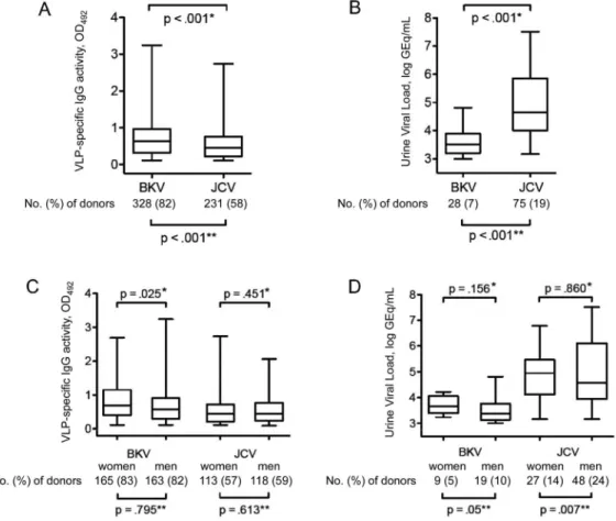
Prevalence of BKV and JCV antibodies, DNAemia, and DNAuria. BKV IgG seroprevalence among healthy blood do-nors was 82% (328 of 400), significantly higher than the corre-sponding JCV IgG seroprevalence of 58% (231 of 400) (P⬍ .001, by2test; figure 1A). The mean IgG level as defined by OD
492was higher for BKV than for JCV (P⬍ .001, by Mann-Whitney U test). No significant difference was detected in IgM seropreva-lence, which was 1% (4 of 400) for BKV and 0% (0 of 400) for JCV. All of the IgM-positive individuals were also IgG positive. BKV was detected in urine samples from 28 (7%) of 400 healthy blood donors, a detection rate lower than that for JCV, which was detected in samples from 75 (19%) of the donors (P⬍ .001, by2test; figure 1B). Median urinary viral loads were also lower for BKV than for JCV (3.5 log vs. 4.8 log GEq/mL;
P⬍ .001, by Mann-Whitney U test). Four (1%) of 400 healthy
blood donors had both BKV and JCV DNA detected in the urine. No difference in urinary BKV or JCV load was detectable when we compared individuals who shed just one of the viruses with individuals who shed both viruses (table 2). There was no asso-ciation between polyomavirus shedding and ABO blood group. However, individuals who shed BKV in the urine were more
likely to be positive for rhesus factor than were those who did not (27 of 331 vs. 1 of 69; P⫽ .047, by2test), whereas no associa-tion between rhesus factor positivity and JCV shedding was found (60 of 331 vs. 15 of 69; P⫽ .484, by2test). Of note, neither BKV nor JCV DNA was detected in any of the 400 plasma samples.
Sex and age dependent prevalence of BKV and JCV anti-bodies and viruria. BKV IgG seroprevalence was not different between female and male blood donors (165 [83%] vs. 163 [82%] of 200; P⫽ .795, by2test) (figure 1C). However, the level of BKV IgG activity was higher for women than for men (P⫽ .025, by Mann-Whitney U test). No difference in JCV IgG seropreva-lence or the level of JCV IgG activity was apparent between women and men (113 [57%] of 200 vs. 118 [59%] of 200;
P⫽ .613, by2test; P⫽ .451, by Mann-Whitney U test) (fig-ure 1C). Both BKV and JCV were more frequently detected in urine samples obtained from men, compared with samples ob-tained from women (P⫽ .05 for BKV and .007 for JCV, by2 test) (figure 1D). However, there were no significant differences between the sexes with respect to urinary BKV or JCV loads (figure 1D).
When we compared the antibody responses in the different age groups, we found that IgG seroprevalence and the level of Table 1. Primers and probes used for detection of BK and JC polyomavirus (BKV and JCV)
in plasma and urine from 400 healthy blood donors.
Analysis Sequence PCR quantification BKV LT-forward 5'-AGCAGGCAAGGGTTCTATTACTAAAT-3' LT-reverse 5'-GAAGCAACAGCAGATTCTCAACA-3' Probe FAM-AAGACCCTAAAGACTTTCCCTCTGATCTACACCAGTTT-TAMRA JCV LT-forward 5'-CTAAACACAGCTTGACTGAGGAATG-3' LT-forward 5'-CATTTAATGAGAAGTGGGATGAAGAC-3' Probe FAM-TAGAGTGTTGGGATCCTGTGTTTTCATCATCACT-TAMRA NCCR rearrangement BKV Outer 1, BKTT5 5'-GAGCTCCATGGATTCTTC-3' Outer 2, BKTT6 5'-CCAGTCCAGGTTTTACCA-3' Inner 1, BKTT7 5'-CCCTGTTAAGAACTTTATCCATTT-3' Inner 2, BKTT8 5'-AACTTTCACTGAAGCTTGTCGT-3' JCV Outer 1 5'-AGGCCTAATAAATCCATAAGCTCCA-3' Outer 2 5'-GTTCCACTCCAGGTTTTACTAACTT-3' Inner 1 5'-TTTTAGCTTTTTGCAGCAAAAAATTA-3' Inner 2 5'-CCTGGCGAAGAACCATGGCCAG-3' BKV VP1 sequencing Outer 1 5'-GTGCAAGTGCCAAAACTACTAATAA-3' Outer 2 5'-TGCATGAAGGTTAAGCATGCTAGT-3' Inner 1 5'-CAACCAAAAGAAAAGGAGAGTGTC-3' Inner 1 5'-TCCTCCACCATGCTCATGCACT-3'
BKV IgG activity decreased significantly with increasing age, from 87% (87 of 100) in the youngest group (aged 20 –29 years) to 71% (71 of 100) in the oldest group (aged 50 –59 years) (P⫽ .006; figure 2A), whereas a strongly opposite trend was found for JCV IgG (P⫽ .06) (figure 2A). BKV shedding was not associated with age group (P⫽ .429, by2test) (figure 2B). By contrast, JCV shedding was more frequent in older age groups, increasing from 9% (9 of 100) in the youngest group to 28% (28 of 100) in the oldest group (P⫽ .02, by2test), but no correla-tion between age and urinary viral load was found (figure 2B). When we compared individuals who shed just one virus with those who shed both viruses, we found that individuals who shed only BKV tended to be younger than those who shed only JCV. All individuals who shed both BKV and JCV belonged to the oldest age group (50-59 years old).
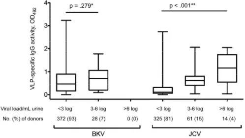
Serologic responses and viral loads. To investigate the re-lationship between antibody response and the level of urinary viral shedding, we divided subjects into the following 3 strata, according to their urinary viral load: undetectable (⬍3 log GEq/ mL), low (3– 6 log GEq/mL), and high (⬎6 log GEq/mL). BKV loads were undetectable in 372 (93%) of the donors and low in
the remaining 28 (7%). JCV loads were undetectable in 325 (81%), low in 61 (15%), and high in 14 (4%) of the donors (figure 3). There was no correlation between BKV antibody level and urinary BKV load. For JCV, the IgG level was significantly higher in individuals in the high-shedding group, compared with those in the low-shedding group (P⬍ .001) (figure 3). Similar results were obtained for JCV by using a Spearman test and a linear regression model (Spearman, 0.284 [P ⫽ .017]; and linear regression coefficient [R2], 0.139 [P⫽ .001]), whereas only a trend was observed for BKV (Spearman, 0.423 [P⫽ .027]; and R2, 0.122 [P⫽ .081). No cross-correlation was found between BKV loads and JCV IgG activity level (data not shown).
Cross-reactivity of BKV and JCV IgG responses. Seropositivity for both BKV IgG and JCV IgG was found in 182 (46%) of 400 indi-viduals. When we compared the IgG antibody level in individuals who were seropositive for only one virus with that in individuals who were seropositive for both viruses, we found that the BKV IgG level was sig-nificantly higher in individuals who were seronegative for JCV than in individuals who were seropositive for both BKV and JCV (P⫽ .012; table 3). Conversely, individuals who were seropositive only for Figure 1. Box-whisker plots showing overall prevalence of BK and JC polyomavirus (BKV and JCV) antibodies and viruria. A, BK and JC virus-like
particle (VLP)–specific IgG seroprevalence (N⫽ 400). B, Urinary BKV and JCV load (N ⫽ 400). C, BK-VLP–specific and JC-VLP–specific IgG
seroprevalence, according to sex (n⫽ 200, for each sex). D, Urinary BKV and JCV load, according to sex (n ⫽ 200, for each sex). Boxes span the interquartile range; the line within each box denotes the median, and whiskers indicate the minimum and maximum values. *P calculated by Mann-Whitney U test; **P calculated by2test. GEq, genome equivalents; OD
JCV showed a higher JCV IgG level than did those who were seropositive for both BKV and JCV (P⫽ .028; table 3). When we compared the rate at which urinary BKV shedding occurred, we found no difference between individuals who were seropos-itive for BKV and those who were seroposseropos-itive for BKV and JCV (P⫽ .169). Interestingly, urinary shedding of JCV was signifi-cantly less common among individuals who were seropositive for both viruses, compared with those who were seropositive only for JCV (P⬍ .001; table 3). When we compared the BKV IgG level and the JCV IgG level, we found no positive correlation (figure 4A). By contrast, some limited correlation suggestive of cross-reactivity was apparent between BKV and JCV IgM activ-ity levels (figure 4B). Taken together, these data indicated that BK-VLP and JC-VLP IgG responses were specific to the respec-tive polyomavirus capsid and that cross-reactivity was not a ma-jor concern.
Types of BKV detected in urine. To investigate the serotype-defining genotype of BKV in the urine of healthy blood donors, we sequenced the relevant VP1 region. Although urinary BKV loads were low, 25 of 28 samples positive for BKV by PCR could be se-quenced. BKV type 1 was most common, occurring in 18 (80%) of 25 samples, followed by type 4 (in 5 samples [20%]), and type 2 (in 2 samples [8%]). BKV type 3 was not found. No differences in dian urinary BKV loads were found between the subtypes (the me-dian [range] BKV load for type 1 was 3.57 log GEq/mL [3.0 – 4.8 GEq/mL], for type 2 it was 3.27 GEq/mL [3.0 – 4.2 GEq/mL] and for type 4, it was 3.27 GEq/mL [3.2– 3.8 GEq/mL]). When we com-pared the BKV IgG level in individuals who shed BKV, we found a significantly higher BKV IgG level in individuals who shed BKV type 1, compared to those who shed non-type 1 BKV (figure 4C). When the BKV IgG level among the 18 individuals who shed BKV type 1 was compared according to viral subtype (i.e., subtypes 1a [n⫽ 5)], 1b-1 [n ⫽ 6], and 1b-2 [n ⫽ 7]), it showed a trend
toward increase among patients who shed BKV subtype 1-b2 (figure 4D).
Architecture of BKV and JCV NCCR in subjects who shed virus. Given the role the polyomavirus NCCR plays in viral replication capacity in transplant recipients, we examined the NCCR architecture of BKV and JCV found in the urine of healthy blood donors. In 20 of 28 BKV-positive urine samples, the NCCR could be amplified and sequenced but no NCCR re-arrangement was identified. In 70 of 75 JCV-positive urine sam-ples, the NCCR could be characterized, and in only 1 case was a rearranged JCV NCCR was identified. The JCV NCCR rear-rangement was confirmed in 2 subsequent urine samples ob-tained 1 and 2 months after the original sample, which revealed identical insertion in the F-block (GeneBank accession numbers FJ233068 –70). In the first sample, the urinary JCV load was 4.78 log GEq/mL; in the second, it was 3.78 log GEq/mL; and in the third, it was 3.25 log GEq/mL.
DISCUSSION
This comprehensive study of samples from 400 consecutive blood donors provides important baseline data on human poly-omavirus infection and replication. First, the overall IgG sero-prevalence for BKV was 82% (328 of 400), which was signifi-cantly higher than the 58% (231 of 400) seroprevalence observed for JCV. Second, urinary shedding of BKV was observed in 28 subjects (7%), which was significantly fewer than the 75 subjects (19%) observed to shed JCV. Third, the median urinary BKV load was 3.51 log GEq/mL, which was significantly lower than the median urinary JCV load of 4.64 log GEq/mL. Fourth, anal-ysis of the NCCR of all urinary BKV and JCV genomes revealed archetype architecture in all but 1 case, which involved a rear-ranged JCV NCCR. Finally, neither BKV nor JCV was detected Table 2. Age and viral status of blood donors evaluated for BK and JC polyomavirus (BKV and JCV), according to virus detected.
BKV and JCV (n⫽ 4) BKV only (n⫽ 24) JCV only (n⫽ 71) None (n⫽ 301) P Demographic characteristic
Age, median (range), years 55 (53–56) 39 (22–58) 46 (20–59) 38 (20–59) ⬍.001a
Viral data BKV
VLP-specific IgG, median (range) 0.4415 (0.0974–0.9066) 0.8079 (0.0858–1.7773) 0.3012 (0.0001–1.876) 0.5353 (0.0035–3.2405) .001a
Urinary viral load, median (range), GEq/mL
2812 (1020–5660) 3270 (1000–64,900) 0 0 .59b
JCV
VLP-specific IgG, median (range), OD492 1.2752 (0.1753–2.062) 0.0774 (0.0205–0.9538) 0.6596 (0.0306–2.0533) 0.1101 (0.0035–2.7329) ⬍.001a
Urinary viral load, median (range), GEq/mL 5,469,450 (31,997–2.1⫻ 107) 0 42,251 (1471–32.7⫻ 106) 0 .132b
NOTE. GEq, genome equivalents; OD492, optical density at 492 nm; VLP, virus-like particles. aBy Kruskal-Wallis test.
in plasma under conditions routinely used to monitor viremia in patients undergoing kidney and/or hematopoietic stem-cell transplants. Unlike earlier studies, we studied healthy donors at the time of regular blood donation that was conducted in accor-dance with international standards and was not confounded by pregnancy, clinical and laboratory abnormalities, or infections with T. pallidum, HIV, HBV, or HCV. Thus, we may conclude that BKV and JCV infection and replication are common in bona fide healthy, immunocompetent adults, but the rates of seropositivity and urinary viral shedding point to significant dif-ferences in the epidemiology of BKV and JCV infection.
We noted that the BKV IgG seroprevalence was highest in the youngest age group (aged 20 –29 years) at 87% (87 of 100) and decreased with older age to 71% (71 of 100) in the oldest group
(aged 50 –59 years), whereas the opposite trend was observed for JCV IgG seroprevalence, which increased from 50% (50 of 100) in the youngest group to 68% (68 of 100) in the oldest group. Knowles et al. [25] used HIA in a large study of 2435 serum samples obtained in England in 1991 and observed a similar age-related decrease in BKV seroprevalence from 91% to 74% (P⬍ .01; overall mean seroprevalence, 81%), whereas JCV se-roprevalence increased with age from 34% to 45% (P⬍ .01; overall mean seroprevalence, 35%). We may conclude that no gross alterations in the epidemiology of BKV infection seem to have occurred in the last 15 years. Similar data have been re-ported for immunocompetent Japanese outpatients [24]. The data indicate that the puzzling increase in the incidence of PVAN is not related to changes in the epidemiology of BKV infection in Figure 2. Box-whisker plots showing age-dependant prevalence of BK and JC polyomavirus (BKV and JCV) antibodies and viruria. A, BK and JC virus-like particle (VLP)–specific IgG levels, according to age group. B, Urinary BKV and JCV load, according to age group. Boxes span the interquartile range; the line within each box denotes the median, and whiskers indicate the minimum and maximum values. *P calculated by Kruskal-Wallis test; **P calculated by2test. GEq, genome equivalents; OD
the general population, but rather to other modulating factors, of which increased immunosuppression following kidney trans-plantation is the key suspect.
The decline in BKV IgG antibody level with age is also of interest, as it suggests that significant (re)exposure to BKV does not seem to occur in most healthy, immunocompetent individ-uals during adult life. This is in line with our data showing low-level urinary shedding of BKV in only 28 individuals (7%). By contrast, significant increases in BKV-specific antibody titers have been observed in patients undergoing kidney transplanta-tion who had high-level viruria and viremia [27, 29 –33]. Unlike BKV, JCV starts with a lower IgG seroprevalence that increases with age. Initial serological studies indicated that BKV infection occurs earlier during childhood, whereas JCV infection follows
later, which is supported by our data as well. This suggests that significant exposure to JCV leads to primary and secondary in-fections during adult life. This observation is of interest for the as yet unresolved pathogenesis of progressive multifocal leukoen-cephalopathy in patients with AIDS and suggests the hypothesis that exogenous infection and replication may be the cause in some profoundly immunosuppressed individuals.
Our data show that urinary shedding of JCV was not only more common but occurred in significantly greater quantities than BKV shedding, with 14 (4%) of 400 participants having JCV loads⬎6 log GEq/mL. By contrast, the urinary BKV load was observed to be⬍5 log GEq/mL for all subjects. Thus, com-pared to immune control of BKV, JCV-directed humoral and/or cellular immunity seems less efficient at controlling viral repli-Figure 3. Box-whisker plots showing virus-like particle (VLP)–specific IgG activity for BK and JC polyomavirus (BKV and JCV), according to urinary viral load. Subjects were stratified as follows:⬍3 log genome equivalents (GEq)/mL (undetectable), 3–6 log GEq/mL (low), and ⬎6 log GEq/mL (high). Boxes span the interquartile range; the line within each box denotes the median, and whiskers indicate the minimum and maximum values. *P calculated by Mann-Whitney U test; **P calculated by Kruskal-Wallis test. OD492, optical density at 492 nm.
Table 3. IgG activity levels and viral shedding in the urine of 400 healthy blood donors evaluated for BK and JC polyomavirus (BKV and JCV), according to serologic results.
Viral data BKV⫺/JCV⫺ (n⫽ 23) BKV⫹/JCV⫺ (n⫽ 146) BKV⫺/JCV⫹ (n⫽ 49) BKV⫹/JCV⫹ (n⫽ 182) P VLP-specific IgG, OD492 BKV 0.0586 (0.0035–0.1002) 0.7709 (0.1141–3.2405) 0.0776 (0.0001–0.1065) 0.5241 (0.1142–2.6862) .012a JCV 0.0398 (0.0035–0.094) 0.0523 (0.0059–0.1085) 0.6096 (0.1144–2.0533) 0.4343 (0.1101–2.7329) .028b
Virus detected in urine, proportion of subjects
BKV 0/23 15/146 2/49 11/182 .169c
JCV 0/23 4/146 19/49 52/182 ⬍.001c
NOTE. OD492, optical density at 492 nm; VLP, virus-like particle. aComparison of BKV⫹/JCV⫺and BKV⫹/JCV⫹, by Mann-Whitney U test. b Comparison of BKV⫺/JCV⫹and BKV⫹/JCV⫹, by Mann-Whitney U test. cComparison of individuals who shed virus, by2test.
cation. The absence of IgM in practically all individuals who shed virus indicates that these events represent persistent or re-activated replication. Given the role of cellular immune control in other viral infections that have a propensity for latency and reactivation, such as cytomegalovirus infection, we suspect that these differences most likely reside in the efficiency of cellular immune surveillance [7, 34]. However, BKV and JCV are closely related to each other with 72%– 81% nucleotide homology in the regions of the 5.1-kb DNA genome, yielding amino acid homol-ogies of 59%– 83% [35]. We observed that JCV shedding was significantly more common among individuals who were posi-tive only for JCV, compared with those who were seroposiposi-tive for both BKV and JCV, suggesting limited cross-protection from a BKV-induced immune response. A precedent for limited cross-protection has been established for herpes simplex type 1 and type 2, which are⬃70% homologous to one another [36]. Moreover, cross-reacting CD8⫹T cells have been described re-cently for some closely related peptide epitopes in the large T antigen and in the VP1 capsid protein [37–39]. Because T cell immunity requires major histocompatibility complex presenta-tion by infected cells or processing by antigen-presenting cells, we suspect that JCV shedding is not necessarily a sign of T cell
dysfunction but may instead reflect differences between BKV-and JCV-infected cells with respect to presentation, anatomic location, and/or accessibility to T cells (e.g., in mucosal sites) [6]. Our data suggest that male sex and older age may be contribu-tory factors. In this respect, Drachenberg et al. noted that reduc-ing immunosuppression in patients undergoreduc-ing kidney trans-plantation who had JCV-mediated PVAN resulted in clearance of allograft involvement but not clearance of urinary JCV loads [21]. Clearly, further studies are needed to address these differ-ences in the respective polyomavirus-host balance.
Our serological analysis indicated that there is little cross-reactivity between BKV and JCV IgG when baculovirus-expressed VLP are used as EIA antigens. This result is in line with VLP studies by Hamilton et al., Viscidi et al., and Stolt et al., who also included preabsorption studies [40 – 42]. We noted a higher BK-VLP IgG level in individuals who shed BKV serotype 1 compared with those who shed virus that was not serotype 1. Among those who shed serotype 1 virus, the individuals who shed subtype 1-b1— which was used to generate the BK VLP—showed higher levels of IgG activity. Of note, (sub)serotypes 1-b1 and 1-b2 only differ by 1 amino acid in the VP1 gene (position 1908). These differ-ences suggest that the IgG response is highly specific for the re-Figure 4. Virus-like particle (VLP)–specific IgG levels in serum samples from 400 healthy blood donors evaluated for BK and JC polyomavirus (BKV and JCV). BK-VLP–specific and JC-VLP–specific IgG levels (A) and IgM levels (B) in samples were plotted against each other, and regression analysis was performed to check for statistical evidence of cross-reactivity. Note that log scales differ in A and B. C, Comparison of BK-VLP–specific IgG levels between donors who had BKV serotype 1 in urine and donors who had non-serotype 1 (i.e., serotypes 2– 4) BKV in urine. D, BK-VLP–specific IgG levels in donors with BKV serotype 1 in urine were further compared among BKV subtypes 1a, 1b-1, and 1b-2. Boxes span the interquartile range; the line within each box denotes the median, and whiskers indicate the minimum and maximum values. *P calculated by Mann-Whitney U test. **P calculated by Kruskal-Wallis test. OD492, optical density at 492 nm; R2, regression coefficient.
spective BKV subtype, leading to underquantification of IgG lev-els in response to other BKV (sub)types. Another explanation could be that secondary exposure to BKV subtype 1-b1 is more prevalent, and therefore the cognate antibody responses are boosted. Although none of the individuals who shed BKV sub-types other than 1-b1 were seronegative, our data indicate that further studies are needed to investigate the diagnostic and pathogenic relevance of BKV subtype-specific IgG. For JCV, only a single capsid serotype has been reported.
Urinary polyomavirus shedding among immunocompetent, healthy blood donors provided a unique opportunity to investi-gate the architecture of the regulatory NCCR of both BKV and JCV. In all but 1 case of JCV infection, the BKV and JCV NCCR were of archetype architecture. Earlier in vitro studies showed that rearrangements of the NCCR (rr-NCCR) arise rapidly dur-ing polyomavirus replication [43]. Typically, partial duplica-tions (inserduplica-tions) and deleduplica-tions are found, which confer in-creased early gene expression and higher replication capacity. JCV and BKV with rr-NCCR have been detected in vivo in pa-thology samples of affected brain and transplant tissue, as well as in the cerebrospinal fluid and blood of patients with PML and PVAN, respectively [44]. In patients undergoing kidney trans-plantation, the emergence of rr-NCCR BKV in plasma is associ-ated with higher plasma BKV loads and more advanced pathol-ogy both in vivo and in vitro [12, 45, 46]. In healthy individuals, little data is available on the prevalence of rearranged viruses. In urine samples from pregnant women, Markowitz et al. found predominantly archetypal BKV in 18 of 127 samples and pre-dominantly archetypal JCV in 5 of 76 samples [47]. Thus, our data support the hypothesis that high-level replication in vitro or in vivo in severely immunocompromised patients seems to be linked to the frequent emergence of polyomavirus variants with rr-NCCR [44].
We could not detect BKV or JCV in plasma. Dolei et al. exam-ined PBMC DNA from 205 blood donors and 26 workers in a hematology unit and reported that 21.6% of PBMCs tested pos-itive for BKV, whereas JCV DNA was found in 0.9% by use of nested PCR [23]. By contrast, Ling et al., who did not use nested PCR, failed to detect BKV or JCV DNA in any of 30 blood sam-ples; no BKV was found in urine samples but JCV was detected in 47% of samples and was more common in samples obtained from individuals⬎40 years old (P ⫽ .03) [22]. Thus, we cannot exclude the possibility that more sensitive PCR protocols might allow the detection of low levels of polyomavirus DNA, but viral genome integrity, the cell types involved, and contamination needs to be addressed to elucidate the potential significance of such results [48].
In summary, our study provides important baseline data on polyomavirus infection and viral replication in healthy, immu-nocompetent individuals. These data indicate significant differ-ences between BKV and JCV with respect to virus-host interac-tion and the epidemiology of infecinterac-tion, which may have an
impact on the pathogenesis of infection in immunocompro-mised patients.
Acknowledgments
We wish to thank Dr. Sohrab Bodaghi for helping with the preparation of BK VLP and the JC VLP, Claudia Mistl for support and sequencing results. Also, we are grateful to Ingrid Ziekau, Sabrina Köhli, and Anna C. Hirsch for preservation of the samples; the team of the Molecular Diagnostics Labora-tory for PCR analysis of blood and urine samples; the team at the Blood Donation Center Basel for motivating the study participants; and all healthy blood donors in Basel who contributed to this work.
References
1. Chesters PM, Heritage J, McCance DJ. Persistence of DNA sequences of BK virus and JC virus in normal human tissues and in diseased tissues. J Infect Dis 1983; 147:676 – 84.
2. Hirsch HH. BK virus: opportunity makes a pathogen. Clin Infect Dis
2005; 41:354 – 60.
3. Arthur RR, Shah KV, Baust SJ, Santos GW, Saral R. Association of BK viruria with hemorrhagic cystitis in recipients of bone marrow trans-plants. N Engl J Med 1986; 315:230 – 4.
4. Cinque P, Koralnik IJ, Clifford DB. The evolving face of human immu-nodeficiency virus-related progressive multifocal leukoencephalopathy: defining a consensus terminology. J Neurovirol 2003; 9(Suppl 1):88 –92. 5. Rinaldo CH, Hirsch HH. Antivirals for the treatment of polyomavirus
BK replication. Expert Rev Anti Infect Ther 2007; 5:105–15. 6. Hirsch HH, Steiger J. Polyomavirus BK. Lancet Infect Dis 2003; 3:611–23. 7. Egli A, Binggeli S, Bodaghi S, et al. Cytomegalovirus and polyomavirus BK posttransplant. Nephrol Dial Transplant 2007; 22(Suppl 8):viii72– viii82.
8. Hirsch HH, Knowles W, Dickenmann M, et al. Prospective study of polyomavirus type BK replication and nephropathy in renal-transplant recipients. N Engl J Med 2002; 347:488 –96.
9. Nickeleit V, Klimkait T, Binet IF, et al. Testing for polyomavirus type BK DNA in plasma to identify renal-allograft recipients with viral nephrop-athy. N Engl J Med 2000; 342:1309 –15.
10. Funk GA, Steiger J, Hirsch HH. Rapid dynamics of polyomavirus type BK in renal transplant recipients. J Infect Dis 2006; 193:80 –7. 11. Funk GA, Gosert, R., Comoli P, Ginevri F, Hirsch HH. Polyomavirus BK
replication dynamics in vivo and in silico to predict cytopathology and viral clearance in kidney transplants. Am J Transplant 2008; 8:2368 –77. 12. Gosert R, Rinaldo CH, Funk GA, et al. Polyomavirus BK with rearranged noncoding control region emerge in vivo in renal transplant patients and increase viral replication and cytopathology. J Exp Med 2008; 205:841–52. 13. Bedi A, Miller CB, Hanson JL, et al. Association of BK virus with failure of prophylaxis against hemorrhagic cystitis following bone marrow transplantation. J Clin Oncol 1995; 13:1103–9.
14. Leung AY, Suen CK, Lie AK, Liang RH, Yuen KY, Kwong YL. Quantifi-cation of polyoma BK viruria in hemorrhagic cystitis complicating bone marrow transplantation. Blood 2001; 98:1971– 8.
15. Dörries K, Johnson RT, ter Meulen V. Detection of polyoma virus DNA in PML-brain tissue by (in situ) hybridization. J Gen Virol 1979; 42:49 –57. 16. Berger JR, Kaszovitz B, Post MJ, Dickinson G. Progressive multifocal
leukoencephalopathy associated with human immunodeficiency virus infection. A review of the literature with a report of sixteen cases. Ann Intern Med 1987; 107:78 – 87.
17. Cinque P, Scarpellini P, Vago L, Linde A, Lazzarin A. Diagnosis of cen-tral nervous system complications in HIV-infected patients: cerebrospi-nal fluid acerebrospi-nalysis by the polymerase chain reaction. AIDS 1997; 11:1–17. 18. Markowitz RB, Thompson HC, Mueller JF, Cohen JA, Dynan WS. Inci-dence of BK virus and JC virus viruria in human immunodeficiency virus-infected and -uninfected subjects. J Infect Dis 1993; 167:13–20. 19. Hirsch HH, Meylan PR, Zimmerli W, Iten A, Battegay M, Erb P.
for Toxoplasma gondii, Epstein-Barr virus, and JC virus. Clin Microbiol Infect 1998; 4:577– 84.
20. Yousry TA, Major EO, Ryschkewitsch C, et al. Evaluation of patients treated with natalizumab for progressive multifocal leukoencephalopa-thy. N Engl J Med 2006; 354:924 –33.
21. Drachenberg CB, Hirsch HH, Papadimitriou JC, et al. Polyomavirus BK versus JC replication and nephropathy in renal transplant recipients: a prospective evaluation. Transplantation 2007; 84:323–30.
22. Ling PD, Lednicky JA, Keitel WA, et al. The dynamics of herpesvirus and polyomavirus reactivation and shedding in healthy adults: a 14-month longitudinal study. J Infect Dis 2003; 187:1571– 80.
23. Dolei A, Pietropaolo V, Gomes E, et al. Polyomavirus persistence in lymphocytes: prevalence in lymphocytes from blood donors and healthy personnel of a blood transfusion centre. J Gen Virol 2000; 81:1967–73. 24. Zhong S, Zheng HY, Suzuki M, et al. Age-related urinary excretion of BK polyomavirus by nonimmunocompromised individuals. J Clin Micro-biol 2007; 45:193– 8.
25. Knowles WA, Pipkin P, Andrews N, et al. Population-based study of antibody to the human polyomaviruses BKV and JCV and the simian polyomavirus SV40. J Med Virol 2003; 71:115–23.
26. Hirsch HH, Mohaupt M, Klimkait T. Prospective monitoring of BK virus load after discontinuing sirolimus treatment in a renal transplant patient with BK virus nephropathy. J Infect Dis 2001; 184:1494 –5. 27. Ginevri F, Azzi A, Hirsch HH, et al. Prospective monitoring of
polyoma-virus BK replication and impact of pre-emptive intervention in pediatric kidney recipients. Am J Transplant 2007; 7:2727–35.
28. Jin L, Gibson PE, Booth JC, Clewley JP. Genomic typing of BK virus in clinical specimens by direct sequencing of polymerase chain reaction products. J Med Virol 1993; 41:11–17.
29. Randhawa PS, Gupta G, Vats A, Shapiro R, Viscidi RP. Immunoglobulin G, A, and M responses to BK virus in renal transplantation. Clin Vaccine Immunol 2006; 13:1057– 63.
30. Hariharan S, Cohen EP, Vasudev B, et al. BK virus-specific antibodies and BKV DNA in renal transplant recipients with BKV nephritis. Am J Transplant 2005; 5:2719 –24.
31. Leuenberger D, Andresen PA, Gosert R, et al. Human polyomavirus type 1 (BK virus) agnoprotein is abundantly expressed but immunologically ignored. Clin Vaccine Immunol 2007; 14:959 – 68.
32. Bohl DL, Brennan DC, Ryschkewitsch C, Gaudreault-Keener M, Major EO, Storch GA. BK virus antibody titers and intensity of infections after renal transplantation. J Clin Virol 2008; 43:184 –9.
33. Randhawa P, Bohl D, Brennan D, et al. longitudinal analysis of levels of immunoglobulins against BK virus capsid proteins in kidney transplant recipients. Clin Vaccine Immunol 2008; 15:1564 –71.
34. Egli A, Binet I, Binggeli S, et al. Cytomegalovirus-specific T-cell re-sponses and viral replication in kidney transplant recipients. J Transl Med 2008; 6:29.
35. Egli A, Dumoulin A, Köhli S, Hirsch HH. Polyomavirus BK after kidney transplantation—role of molecular and immunological markers. Trends in Transplantation 2009 (in press).
36. Langenberg AG, Corey L, Ashley RL, Leong WP, Straus SE. A prospec-tive study of new infections with herpes simplex virus type 1 and type 2. Chiron HSV Vaccine Study Group. N Engl J Med 1999; 341:1432– 8. 37. Provenzano M, Bracci L, Wyler S, et al. Characterization of highly
fre-quent epitope-specific CD45RA⫹/CCR7⫹/- T lymphocyte responses against p53-binding domains of the human polyomavirus BK large tu-mor antigen in HLA-A*0201⫹ BKV-seropositive donors. J Transl Med
2006; 4:47.
38. Randhawa PS, Popescu I, Macedo C, et al. Detection of CD8⫹T cells
sensitized to BK virus large T antigen in healthy volunteers and kidney transplant recipients. Hum Immunol 2006; 67:298 –302.
39. Li J, Melenhorst J, Hensel N, et al. T-cell responses to peptide fragments of the BK virus T antigen: implications for cross-reactivity of immune response to JC virus. J Gen Virol 2006; 87:2951– 60.
40. Hamilton RS, Gravell M, Major EO. Comparison of antibody titers de-termined by hemagglutination inhibition and enzyme immunoassay for JC virus and BK virus. J Clin Microbiol 2000; 38:105–9.
41. Viscidi RP, Rollison DE, Viscidi E, et al. Serological cross-reactivities between antibodies to simian virus 40, BK virus, and JC virus assessed by virus-like-particle-based enzyme immunoassays. Clin Diagn Lab Im-munol 2003; 10:278 – 85.
42. Stolt A, Sasnauskas K, Koskela P, Lehtinen M, Dillner J. Seroepidemiol-ogy of the human polyomaviruses. J Gen Virol 2003; 84:1499 –1504. 43. Sundsfjord A, Flaegstad T, Flo R, et al. BK and JC viruses in human
immunodeficiency virus type 1-infected persons: prevalence, excretion, viremia, and viral regulatory regions. J Infect Dis 1994; 169:485–90. 44. Loeber G, Dörries K. DNA rearrangements in organ-specific variants of
polyomavirus JC strain GS. J Virol 1988; 62:1730 –5.
45. Randhawa P, Zygmunt D, Shapiro R, et al. Viral regulatory region se-quence variations in kidney tissue obtained from patients with BK virus nephropathy. Kidney Int 2003; 64:743–7.
46. Olsen GH, Andresen PA, Hilmarsen HT, et al. Genetic variability in BK Virus regulatory regions in urine and kidney biopsies from renal-transplant patients. J Med Virol 2006; 78:384 –93.
47. Markowitz RB, Eaton BA, Kubik MF, Latorra D, McGregor JA, Dynan WS. BK virus and JC virus shed during pregnancy have predominantly archetypal regulatory regions. J Virol 1991; 65:4515–9.
48. Dörries K, Sbiera S, Drews K, Arendt G, Eggers C, Dörries R. Association of human polyomavirus JC with peripheral blood of immunoimpaired and healthy individuals. J Neurovirol 2003; 9(Suppl 1):81–7.