Glycobiology vol. 5 no. 4 pp. 435-441, 1995
Characterization of a 21 amino acid peptide sequence of the
laminin G2 domain that is involved in HNK-1 carbohydrate
binding and cell adhesion
Heike Hall
2, Thomas Vorherr
1and Melitta Schachner
3 Department of Neurobiology, Swiss Federal Institute of Technology, Hanggerberg, 8093 Zurich and 'Hoffmann La Roche, PRPC 15/4, 4002 Basel, Switzerland2Present address: Department of Experimental Pathology, Guy's Hospital,
London Bridge, London SE1 9RT, UK 'To whom correspondence should be addressed
The N-linked HNK-1 carbohydrate expressed by several
recognition molecules mediates the adhesion of early
post-natal cerebellar neurons to the G2 domain of the terminal
globular domain of the laminin a l chain (H.Hall et al.,
sub-mitted). To define this binding site more precisely, G2-derived
synthetic peptides were used for binding and competition
studies. Peptide 5-G2, comprising the amino add residues
3431-3451 of G2, inhibited the interaction between the
HNK-1-carrying glycolipid and laminin in a
concentration-dependent and saturable manner. Peptides which overlap
only partially with this sequence interfered less. Peptides
comprising other amino acid sequences from G2, and
pep-tides derived from Gl and G3 or a scrambled version of
peptide 5-G2, did not show significant effects. Direct binding
of peptide 5-G2 to the HNK-1 glycolipid was also
demon-strated. Furthermore, peptide 5-G2 interfered in a
concen-tration-dependent and saturable manner with the adhesion
of early postnatal cerebellar neurons to laminin. These
obser-vations indicate that amino acid residues 3431-3451 of the
laminin G2 domain are involved in HNK-1
carbohydrate-mediated cell adhesion.
Key words: cell adhesion/HNK-1 carbohydrate/laminin/nervous
system
Introduction
The extracellular matrix of the nervous system plays important
roles during development and regeneration (for reviews, see
Sanes, 1989; Reichardt and Tomaselli, 1991; Venstrom and
Reichardt, 1993). Extracellular matrix molecules have been
implicated in cell attachment, spreading and migration, and
growth cone guidance. One of the components of the
extra-cellular matrix in the developing and adult central and
peri-pheral nervous system is laminin. Laminin is a self-aggregating,
multifunctional glycoprotein consisting of the three
polypep-tide chains a l , pi, yl (Timpl et al, 1979; Martin and Timpl,
1987; Beck et al, 1990, 1991; Yurchenco et al, 1990, 1992;
Yurchenco and Cheng, 1993; Burgeson et al, 1994). The fil
and yl chains are linked by a disulphide bond and associated
non-covalently with the a l chain forming a coiled-coil structure.
The C-terminus of the a l chain forms a globular domain
consisting of five G domains each comprising -180 amino
acids. These domains share 25-30% internal homology (Sasaki
et al, 1988) and represent independent folding units (Beck
et al, 1990, 1991) capable of exerting different functions
such as binding of various cell surface receptors (for review,
see Edgar, 1989; Mecham, 1991; Hynes and Lander, 1992),
promotion of neurite outgrowth (Edgar et al, 1984; Sephel
et al, 1989; Lander, 1990; Calof et al, 1994) and recognition
of sulphated carbohydrates (Ott et al, 1982; Roberts et al,
1985, 1988; Skubitz et al, 1988, 1991; Kouzi-Koliakos et al,
1989; Taraboletti et al, 1990). One of these carbohydrates
which has been shown to be involved in neural cell adhesion to
laminin is the HNK-1 carbohydrate (Mohan et al, 1990; Hall
etal, 1993).
The HNK-1 carbohydrate expressed by several neural
recog-nition molecules (Kruse etal, 1984; Schachner, 1989) and two
structurally related nervous system-derived glycolipids have
been defined by their interaction with the monoclonal antibody
HNK-1 directed against a subset of human natural killer cells
(Abo and Balch, 1981). The structure was determined as 3'
sulphoglucuronylneolactotetraosyl- or hexaosyl-ceramide (Chou
etal, 1986, 1991; Ariga etal, 1987). In vivo experiments with
the monoclonal HNK-1 antibodies have demonstrated that it is
involved in the migration of neural crest cells (Bronner-Fraser,
1987). In in vitro assays, the HNK-1 carbohydrate has been
im-plicated in migration of neural crest cells and neurons, neurite
outgrowth (Kiinemund et al, 1988; Lallier and Bronner-Fraser,
1991; Lallier et al, 1992) and in short-term cell-to-cell and
cell-to-laminin adhesion (Keilhauer et al, 1985; Kiinemund
et al, 1988; Hall etal, 1993). Cell-to-laminin adhesion is
medi-ated by direct binding of the cell surface-expressed HNK-1
carbohydrate to the G2 domain of the terminal globular domain
of the laminin a l chain (Mohan et al, 1990; H.Hall et al,
submitted).
Since it was found that the binding of HNK-1 carbohydrate
to laminin was not affected when laminin was treated with
urea or reduced and alkylated (H.Hall et al, submitted), we
searched for a peptide sequence responsible for binding to the
HNK-1 carbohydrate. For this purpose we used a variety of G2
domain-derived synthetic peptides and tested their ability to
inhibit the binding. One peptide could be determined which
interfered with the binding, bound directly to the HNK-1
car-bohydrate and inhibited HNK-1-mediated neural cell adhesion
to laminin.
Results
Previous studies defined the HNK-1 carbohydrate binding site
on the G2 domain of the terminal globular domain of the
lam-inin a l chain (see schematic drawing in Figure 1). Since this
binding could not be affected by reduction and alkylation of
disulphide bonds or urea denaturation of the laminin E8
frag-ment (H.Hall et al, submitted), the native conformation of G2
H.Hall, T.Vorherr and MSchachner
pi
a lyl
&S1
2245-2266 2431-2451 2619-2838Fig. 1. Schematic structure of the distal portion of the long arm of EHS tumour laminin ( a l ^ l y l ) and the adjacent G domains which are comprised in the proteolytic fragment E8 (Ott et al., 1982). This structure consists of a coiled-coil rod contributed by the C-terminal portions of the a l , pl and yl chains where the pi and the y\ chains are connected by a disulphide bond (ss) and non-covalently associated to the A chain. The most C-terminal portion of the a l chain is composed of five similar G domains with only Gl, G2 and G3 belonging to the E8 fragment. Positions of peptides 5-G1, 5-G2 and 5-G3 are indicated.
might not be important for binding. We therefore started to
search for a peptide which is able to interfere with the binding
of the HNK-1 carbohydrate to laminin and which also binds
directly to the HNK-1 carbohydrate. Ten different synthetic
peptides derived from the G2 domain were studied with respect
to the binding of laminin to substrate-immobilized
HNK-1-carrying glycolipids (hereafter called HNK-1 glycolipid). The
sequences and positions of the peptides within the G domains
are indicated in Figure 1 and Table I. One out of these peptides
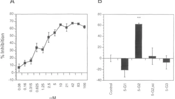
(peptide 5-G2), comprising the amino acid residues 2431-2451
of the laminin a l chain, showed a pronounced inhibition of
binding of 62 ± 2%. Peptide 5-G2 inhibited the binding in a
concentration-dependent manner, reaching saturation at 10 uM
and a maximal inhibition of 65% (Figure 2A). Peptide 4-G2,
which overlaps with the six N-terminal amino acids of peptide
5-G2, showed an inhibition of 41 ± 8% and peptide 6-G2,
com-prising the 12 C-terminal amino acids of peptide 5-G2, showed
a 43 ± 6% inhibition. Peptide 7-G2 corresponds to the C-terminal
heptapeptide of peptide 5-G2 and showed an inhibition of 23 ±
14%. Peptides 1-G2, 2-G2, 3-G2, 8-G2, 9-G2 and 10-G2 did
not show a significant inhibition (Table I). Peptides derived
from Gl (5-G1) and G3 (5-G3), comprising sequences at
posi-tions corresponding to those of peptide 5-G2, did not inhibit the
binding of laminin to the HNK-1 glycolipid at a concentration
of 166 uM (-21 ± 13% and -7 ± 12%, respectively) (Table I
and Figure 2B). The scrambled version of peptide 5-G2
(5-G2,sc) did not show any inhibition of the binding (4 ± 15%).
These results indicate that a G2 domain-derived peptide is
involved in the binding of laminin to the HNK-1 glycolipid.
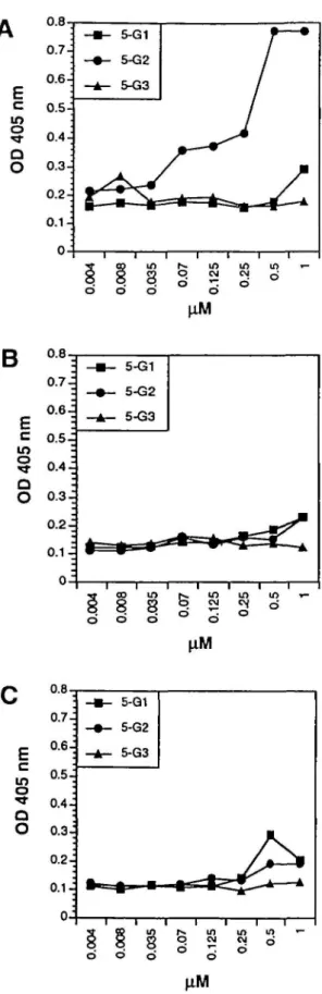
To demonstrate that peptide 5-G2 interacts directly with
the HNK-1 carbohydrate, binding studies with biotinylated
peptides 5-G1, 5-G2 and 5-G3 were performed on
substrate-immobilized HNK-1 glycolipid, sulphatides or cerebrosides.
Peptide 5-G2 showed concentration-dependent binding to the
HNK-1 glycolipid, reaching a saturating concentration at 0.5 uM
(Figure 3 A). Peptides 5-G1 and 5-G3 did not show any binding
to the HNK-1 glycolipid (Figure 3A). None of the peptides
bound to sulphatides (Figure 3B) or cerebrosides (Figure 3C).
These results indicate that peptide 5-G2 binds directly and
specifically to the HNK-1 glycolipid. The experiments
also exclude the possibility that biotinylation of the peptides
alters their functional properties, since the binding specificity
corresponded to that obtained from the studies on laminin
binding to the HNK-1 glycolipid using non-biotinylated
peptides.
Cell adhesion experiments with small cerebellar neurons,
85% of which express the HNK-1 carbohydrate (Wernecke
et al., 1985), were performed to investigate whether peptide
5-G2 also interferes with the adhesion of neural cells to laminin.
In the presence of peptide 5-G2, cell adhesion was inhibited by
63 ± 3% at a concentration of 83 uM (Figure 4). Control
pep-tides did not (-3 ± 8% for 5-G1) or only slightly (14 ± 4% for
Table I. Name 1-G2 2-G2 3-G2 4-G2 5-G2 6-G2 7-G2 8-G2 9-G2 10-G2 5-G1 5-G2,sc 5-G3
List of G2 domain-derived synthetic peptides used to interfere with the binding of HNK-1 glycolipid to laminin
2370- 2394- 2419- 2426- 2431- 2440- 2445- 2451- 2458- 2245- 2619-Sequence1 TWYKIAFQRNRK SDKETKQGETPG YVGGLPHSKAVR SKAVRKGVSSRS KGVSSRSYVGCIKNLEISRST GCIKNLEISRST LEISRST LEISRSTFDLLR TFDLLRNSYGVRK SYGVRKGCALEP PAVKVTHFKGCMGEAFLNG KVILSCKSTGSRNIRVESGYS MLKMRTSFHGCIKNVVLDAQ -2381 -2405 -2430 -2437 -2451 -2451 -2451 -2456 -2463 -2469 -2263 -2638 Inhibition11 -26 ± 15 9 ± 7 - 8 ± 6 41 ±8** 6 2 ± 2 « * 43 ± 6** 23 ± 14* 5 ± 8 - 3 ± 6 2 ± 9 -21 ± 13 4 ± 1 5 - 7 ± 12 T h e sequences of the peptides from 1-G2 to 10-G2 are derived from the G2 domain of the laminin a l chain. Peptide 5-G1 is derived from Gl and peptide 5-G3 from G3 (see also Figure 1). Peptide 5-G2,sc contains a scrambled version of peptide 5-G2. The numbers in the front and at the end of each peptide indicate the positions of the amino acids within the Gl, G2 and G3 domain, respectively, according to the alignment of Sasaki el aL (1988). Bold letters represent sequences which are contained in overlapping peptides.
bValues represent inhibition of binding in * and are normalized setting the control value as 0% inhibition. Values ± SD represent data from at least three independent experiments carried out in duplicates. Asterisks represent values which are significantly different from the control
Binding site for the HNK-1 carbohydrate on laminln
B
T - CJ •rf 00 (O CD C? ir> in 04 O CO9
Fig. 2. Binding of laminin to substrate-coated HNK-1 glycolipid in the absence and presence of laminin-derived synthetic peptides. Bound laminin was detected by ELISA with pab LN, followed by goat anti-rabbit HRP-conjugated secondary antibodies. Per cent inhibition was calculated relative to the binding in the absence of additives (= 0% inhibition, control). (A) Per cent inhibition is shown as a function of increasing concentrations of peptide 5-G2. Mean values ± SDs are from five independent experiments carried out in triplicates. (B) Per cent inhibition in the presence of 166 uM of each of the peptides 5-Gl, 5-G2, the scrambled version of 5-G2 (5-G2,sc) and 5-G3 is shown. Mean values ± SDs are from at least five independent experiments carried out in triplicates. Asterisks represent values which differ significantly from the control (**•/> :£ 0.001).
5-G3) interfered with the binding of neurons to laminin
(Figure 4). The scrambled version of 5-G2 (5-G2,sc) did not
show any inhibition of cell adhesion (-14 ± 6%) (Figure 4).
For control, the same experiments were performed with
L-cells, with none of the peptides showing significant
inhi-bitory effects (not shown).
To demonstrate that peptide 5-G2 specifically interferes with
the HNK-1 carbohydrate-mediated adhesion of small cerebellar
neurons to laminin, adhesion assays were performed in the
presence of 5-G2, HNK-1 glycolipid and Fab fragments of the
monoclonal antibody (mab) 412 (Figure 5). Inhibition of cell
adhesion to substrate-bound laminin in the presence of
differ-ent concdiffer-entrations of peptide 5-G2 (83, 33, 16, 1.6 and 0.3 uM)
was dose dependent, with an inhibition of 63 ± 5% at saturating
concentrations of 33 uM peptide (Figure 5A). Control peptides
5-Gl and 5-G3 were used at a concentration of 83 \xM without
significant inhibition (Figure 4). The adhesion of cells to
lam-inin was also inhibited in a concentration-dependent manner
in the presence of HNK-1 glycolipid at 20, 5, 2.5, 1.25 and
0.6 uM (Figure 5B). Saturation of adhesion was achieved at
5 uM, resulting in 56 ± 10% inhibition of cell adhesion.
Sulphatides and cerebrosides used as control glycolipids
did not show significant inhibition (-8 ± 9% and - 3 ± 10%,
respectively) at a concentration of 50 uM, indicating that
the glycolipid backbone is not involved in binding to laminin.
Fab fragments of mab 412 were used in the same assay at
concentrations of 10, 2, 1, 0.1, 0.06 and 0.02 uM with a
concentration-dependent and saturable inhibition of cell
adhesion (Figure 5C). Fab fragments of mab 412 inhibited by
maximally 81 ± 2% at a concentration of 5 uM. Fab fragments
of a polyclonal antibody (pab) NCAM, even at a concentration
of 10 jiM, did not show inhibition of cell adhesion (-7 ± 6%)
(not shown). Adhesion of L-cells to laminin in identical assays
was not altered by peptide 5-G2, HNK-1 glycolipid micelles
or Fab fragments of mab 412 (not shown). The combined
observations show that residues 2431-3451 of the G2 domain
of the laminin a l chain are involved in the HNK-1
carbohydrate-mediated cell adhesion to laminin.
Discussion
In this study, we define a sequence of 21 amino acids within the
a l chain of laminin which is involved in the binding of laminin
to the HNK-1 carbohydrate. Out of 10 different peptides derived
from the G2 domain, which were distributed over the central
100 out of 180 amino acids of the complete G2 domain, only
the peptide comprising amino acid residues 3431-3451 was
shown to be active in direct binding of the HNK-1 carbohydrate
and in interfering with the binding of the HNK-1 carbohydrate
to laminin. This peptide has a positive net charge of three and
could therefore interact with the negatively charged HNK-1
carbohydrate. This is noteworthy in the context that according
to surface probability predictions (Chou and Fasman, 1978;
Emini et al., 1985) this sequence of the G2 domain is exposed
at the surface of the G2 domain and thus accessible to putative
ligands. Similarly, the sequences of the equivalent peptides
derived from Gl and G3 are likely to be exposed at the surface
of the G domain.
That this peptide is also involved in the adhesion of HNK-1
carbohydrate-carrying neurons was shown by inhibition
experi-ments. For all inhibitors, a concentration dependence and
satura-tion could be demonstrated. However, there are differences in
the molar concentrations and the maximal inhibition achieved.
HNK-1 glycolipid micelles and the Fab fragments of mab 412
reached saturation of inhibition at - 5 uM. For the inhibitory
peptide, a six times higher concentration (-33 uM) was required
to achieve saturation of inhibition. A possible explanation could
be that the peptide has a lower affinity to the carbohydrate
H.Hall, T.Vorherr and MSchacfaner
E
c o QO
oB
m o Q OE
c m o ^-O O 0.7 -j 0.6-j 0.5^ 0.4-j 0.3-i 0.2-j 0.1-: - • - 5-G1 - • - 5-G2 - A - 5-G3 i i 0.8 07 -. 0.6-j 0.5-j 0.4-; 0.3 J 0.2-j0.1-:
0
S S "" o d d d uo in CM oj5 °
in d o d in CM *-; U) CM d dFig. 3. Concentration dependence of the binding of laminin-derivcd peptides to different glycolipids. HNK-1 glycolipid (A), sulphatides (B) and cerebrosides (C) were substrate immobilized at a concentration of 1 uM. Bound biotinylated peptides 5-G1, 5-G2 and 5-G3 were detected by EUSA with HRP-conjugated streptavidin. One representative out of three independent experiments is shown. SDs were <59b.
Fig. 4. Inhibition of neural cell adhesion in the absence and presence of different laminin-derived peptides. After pre-incubation of small cerebellar neurons in the absence (control) or in the presence of peptides 5-G1, 5-G2, the scrambled version of 5-G2 (5-G2,sc) and 5-G3 bound cells were recorded. Per cent inhibition was shown in relation to the control value which was determined in the absence of any peptides (= 0% inhibition). Mean values ± SDs arc from at least three independent experiments carried out in quadruplicates. Asterisks represent values which differ significantly from the control (•••/> £ 0.001).
ligand than the Fab fragments of mab 412. A reduced affinity
would thus have to be compensated for by an increased peptide
concentration. The slight inhibition of cell adhesion observed
in the presence of peptide 5-G3 could be due to the similarity
between the peptides 5-G2 and 5-G3, since both share the five
central amino acids GCIKN (residues 2440-2445 in peptide
5-G2 and 2628-2633 in peptide 5-G3, respectively).
The question is which molecules on cerebellar neurons may
be the carriers of the HNK-1 carbohydrate. Cerebellar neurons
express this carbohydrate as a glycolipid from early embryonal
stages until adulthood (Schwarting et al, 1987; Prasadarao
etal., l990;Chouetal, 1991). The HNK-1 carbohydrate could
also be presented by several neural recognition molecules which
are expressed by cerebellar neurons, such as LI (Kruse et al,
1984, 1985), NCAM (Goridis et al, 1983; Edelman, 1984),
TAG-1 (Dodd et al, 1988), integrins (Pesheva et al, 1987) and
possibly other molecules. Since it is known for mouse brain LI,
the myelin-associated molecule MAG, NCAM and P0 that only
subpopulations carry the HNK-1 carbohydrate (Kruse et al,
1985; Faissner, 1987; Poltorak et al, 1987), the expression of
the HNK-1 carbohydrate is regulated independently of the
protein backbone. Thus, cell interactions may be subject to
independent sets of regulatory mechanisms.
With regard to the functional roles of integrins, it is
noteworthy that the sequence of 5-G2 overlaps with the nine
C-terminal amino acids of another peptide (GD-3, comprising
the residues 2443—2463) described as a cell binding and
spread-ing site for human fibrosarcoma cells (Skubitz et al, 1991).
Antibodies against the fil integrin subunit inhibited cell
adhesion to this peptide (Skubitz et al, 1991), thus attributing
to the GD-3 and possibly also for the 5-G2 peptide sequences
an important functional role in integrin-laminin-mediated
cell-substrate interactions.
Another candidate for the HNK-1-mediated cell interaction
with laminin is Ng-CAM, an LI-like molecule in the chicken,
Binding site for the HNK-1 carbohydrate on lamlnln C
o
B
c
o
p
'E
c
10 80- 70- 60- 50- 40- 30- 20-10-Ik
fi
/
i
• i —a2 8
S
HNK-1-dependent interactions with laminin are coupled with
HNK-1-independent mechanisms through one ligand, although
the binding sites might be localized on distant parts of the
laminin molecule. It will now be interesting to determine which
adhesion cell surface ligands of cerebellar neurons are involved
in binding to laminin and, if there are several, how they
contri-bute separately and possibly even cooperatively to
laminin-mediated cell-to-substrate interactions.
Materials and methods
Glycolipids and glycoprvteins
The SGGL glycolipids (3'-sulphoglucuronylneolactotetraosyl- and hexaosyl-ceramide), designated as HNK-1 glycolipid, were purified as described by Hall
et al. (1993) with the exception that the HNK-1 glycolipid was purified over a
column of silica gel-60 (Merck no. 15101) instead of a DEAE-Sephadex A-25 ion-exchange column. The HNK-1 glycolipid was eluted with chloroform: ethanol:water (60:35:8 v/v). The purity of the glycolipid was tested by TLC in a chloroform:methanol:water (60:35:8 v/v) system. Sulphatides from bovine brain were purchased from Sigma, cerebrosides were obtained from Sigma or Boehringer Mannheim and EHS-tumour laminin from Gibco/BRL or Boehringer Mannheim.
Antibodies
A monoclonal antibody against the HNK-1 carbohydrate (mab 412), a polyclonal antibody against mouse EHS sarcoma laminin (pab LN) and an immuno-affinity-purified polyclonal antibody against the neural cell adhesion molecule NCAM from mouse brain (pab NCAM) were obtained and purified according to Kruse et al. (1984), Pesheva et al. (1989), and Martini and Schachner (1986), respectively. Fab fragments of mab 412 and pab NCAM were prepared by pro-teolytic digestion with papain (Porter, 1959). Horseradish peroxidase (HRP> conjugated secondary antibodies directed against rat immunoglobulin IgG and IgM or rabbit IgG were purchased from Dianova (Hamburg, FRG).
Fig. 5. Concentration dependence of the binding of neural cell adhesion to
laminin in the presence of peptide 5-G2 (A), HNK-1 glycolipid (B) and Fab fragments of mab 412 (C). Small cerebellar neurons were pre-incubated either in the presence of peptide 5-G2, HNK-1 glycolipid or Fab fragments of mab 412 at different concentrations before adding this mixture to the substrate. Inhibition of cell adhesion was calculated relative to the binding in the absence of additives (= 0% inhibition). Mean values ± SDs are from at least three independent experiments carried out in quadruplicates.
which has been reported to bind to laminin (Grumet et al.,
1993). Interestingly, the laminin fragment involved in
Ng-CAM binding is not fragment E8 comprising the G2 domain,
but the PI fragment which consists of the cross-like region and
the truncated short arms of laminin. Thus, it is possible that
Synthetic peptides
Peptides derived from the G domains of the laminin a l chain were obtained by solid-phase peptide synthesis on a Milligen 9050 continuous-flow peptide synthesizer following preparative HPLC purification as described by Knorr et aL (1989). The indicated position and the amino acid sequence (Table I) of the acetylated carboxamide peptides correspond to the alignment by Sasaki et aL (1988). The synthesis of the following peptides was performed: peptide 3-G2 Ac-YVGGLPHSKAVR-NH2, peptide 5-G2
Ac-KGVSSRSYVGCKNLFJSRST-NH2, peptide 6-G2 Ac-GCIKNLEISRST-NH2, peptide 8-G2
Ac-LEISRST-FDLLR-NH2, peptide 10-G2 Ac-SYGVRKGCALEP-NH2, peptide 5-G1
Ac-PAVKVTHFKGCMGEAFLNG-NH2 and 5-G3
Ac-MLKMRTSFHG-CIKNWDAQ-NH2. After purification by preparative HPLC, the peptides were
>95% pure as determined by analytical HPLC. The ion spray mass spectra (API IU, Sciex) showed the expected ion series. Peptide 1-G2 TWYKIA-FQRNRK, peptide 2-G2 SDKETKQGETPG, peptide 4-G2 SKAVRKGVSSR and peptide 9-G2 TFDLLRNSYGVRK were kindly provided by Yoshihiko Yamada, NIH, NIDR, Bethesda, MD, and peptides 7-G2 LEISRST and 5-G2,sc KVILSCKSTGSRNIRVESGYS by Werner GUrr, Department of Immunology, University of Zurich, Switzerland.
Solid-phase binding assays
Inhibition of the binding of laminin to the HNK-1 glycolipid by synthetic peptides. The HNK-1 glycolipid was substrate coated onto 96-well microtitre
plates (Falcon, Dietikon, Switzerland) in elhanolic solution at 3 uM by evapor-ating to dryness. After blocking with 1% fatty acid-free bovine serum albumin (BSA; Sigma catalogue no. A-7030), in phosphate buffered saline (PBS; pH-7.4) for 1 h, the laminin-derived peptides 5-G1, 5-G2, 5-G3 and 5-G2,sc were pre-incubated with the substrates at a concentration of 166 uM for 30 min at room temperature. In another binding assay, peptide 5-G2 was used in a 1:1 dilution series starting at 166 uM in 0.1 M NaHCO3 containing 10 mM EDTA.
Laminin was then added at 12.5 nM in 0.1 N NaHCO3 supplied with 10 mM
EDTA. For control, laminin was incubated with the substrate in the absence of peptides. Bound laminin was detected by incubation with pab LN and HRP-conjugated goat anti-rabbit secondary antibodies. Incubation of antibodies was
RHall, T.Vorberr and M.Schachner
carried out at room temperature for 2 and for 1 h, respectively. The generation and detection of the coloured reaction product were performed as described by Horstkorte et al (1993). Per cent inhibition was calculated with reference to the control value obtained in the absence of additives (= 0% inhibition).
Binding of biotinylated synthetic peptides to different glycolipids. Peptkles 5-G1,
5-G2 and 5-G3 were biotinylated according to Cole et al. (1987) using NHS-LC-biotin (Pierce, Rockford, IL). The efficiency of biotinylation was tested by enzyme-linked immunosorbent assay (ELISA) (Bayer and Wilchek, 1990) and found to be equal for all peptides. HNK-1 glycolipid, sulphatides or cerebro-sides were substrate immobilized at 1 uM in 96-well microtitre plates as described in the previous paragraph. After blocking with 10% BSA in PBS for 1 h at room temperature, biotinylated peptides 5-G1, 5-G2 or 5-G3 were added in a 1:1 dilution series starting at 1 (iM in 0.1 N NaHCOj containing 10 mM EDTA for 1 h at room temperature. Bound peptides were detected by HRP-conjugated streptavidin (Sigma catalogue no. S-5512) and the OD determined as described in the previous paragraph.
Cell adhesion assays
Preparation of single-cell suspensions. Single-cell suspensions of small
cere-bellar neurons (-99% pure) from 6-day-old ICR mice were prepared by mild trypsinization (Schnitzer and Schachner, 1981; Keilhauer et al., 1985). Cells were used for the adhesion assay at a concentration of 1 x 10⁶ cells/ml in basal
medium Eagle's (Gibco/BME from BRL). Cells expressed the HNK-1 carbo-hydrate as tested by indirect immunofluorescence staining (Schnitzer and Schachner, 1981) with mab 412 immediately after preparation.
Mouse fibroblast L-cells (L929; Pantazis and Jensen, 1988) were cultured in BME containing 1 0 * horse serum. Confluent monolayer cells were trypsinized 0.05% trypsin in Hank's balanced salt solution (HBSS) without Ca2* and Mg2+
for 5 min at room temperature and used for the adhesion tests at a final concentration of 0.5 x 10⁶ cells/ml.
Cell adhesion to laminin in the presence of different peptides, glycolipids and Fab fragments of mab 412 and pab NCAM. Laminin was substrate immobilized
overnight at 8°C in 24-well microtitre plates (NUNC, Nunclon, Switzerland). Coating was performed in 3 pi spots at 25 nM in BME. The coating efficiency was determined to be 15—17% by measuring the protein concentration of the coating solution before and after the coating procedure (Bradford, 1976). The substrate was blocked for 1 h at room temperature with 3% heat-inactivated BSA in PBS. Cells were pre-incubated for 30 min at room temperature either with the peptides 5-G1, 5-G2, 5-G3 or 5-G2,sc at 166 |iM or with decreasing concentrations of 5-G2 (83, 33, 16, 1.6 and 0.3 uM) in HBSS without Ca2* and
Mg2+. The cells were viable during the time of the assay according to their
mor-phological appearance by phase-contrast microscopy and exclusion of trypan blue for all peptides used. Cells were also pre-incubated for 30 min at room temperature in the presence of Fab fragments of pab NCAM (5 uM) and Fab fragments of the mab 412 at 10, 2, 1, 0.1, 0.06 and 0.02 uM in HBSS without Ca2+ and Mg2+. Pre-incubation was also performed with the HNK-1 glycolipid
at 20, 5, 2.5, 1.25 and 0.6 uM, and sulphatides or cerebrosides al 50 uM. The mixture of cells and inhibitors was incubated with the substrate for 30 min at room temperature. For the control, cells were incubated with laminin in the absence of any additive. Bound cells were washed in HBSS without Ca2+ and
Mg2+, fixed for 1 h with 2.5% glutaraldehyde and stained with crystal violet
(Aumailley el al., 1989). The number of cells per area (0.1 x 0.06 mm) was determined with the IBAS graphics system (Kontron, Zurich, Switzerland). Data obtained by this procedure were normalized and per cent inhibition was calculated with respect to the control value obtained in the absence of additives (= 0% inhibition).
Statistical analysis
Statistical analysis was performed on non-normalized data by one-way ANOVA and Scheffd's F or Fischer's PLSD comparison among the means (Winer, 1971). All values marked with an asterisk differ significantly from the control value at*/5 s O . 0 5 , **P S 0 . 0 1 , ***P s 0.001.
Acknowledgements
We are most grateful to Enrico Tongiorgi from our department for help with the sequence analysis of the G2 domain using the GCG computer program, and to Hans Voshol and Georg Orberger from our department for helpful discussions and comments on the manuscript
Abbreviations
BME, basal medium Eagle's; BSA, bovine serum albumin; ELISA, enzyme-linked immunosorbent assay; HBSS, Hank's balanced salt solution; HRP, horseradish peroxidase; mab, monoclonal antibody; OD, optical density; pab, polyclonal antibody; PBS, phosphate-buffered saline; SD, standard deviations.
References
Abo.T. and Balch.C.M. (1981) A differentiation antigen of human NK and K cells identified by a monoclonal antibody (HNK-1). J. Immunol., 127,
1024-1029.
Ariga,T, Kohriyama,T., Freddo.L., Latov,N., Saito,M., Kon.K., Ando.S., Suzuki,M., Hemling,M.E., Rinehart,K.L.J., Kusunoki.S. and Yu.R.K. (1987) Characterization of sulfated glucuronic acid containing glycolipids reacting with IgM M-proteins in patients with neuropathy. /. Biol. Chenu, 262, 848-853.
AumailleyAi., Mann.K., von der Mark,H. and Timpl.R. (1989) Cell attachment properties of collagen type VI and Arg-Gly-Asp dependent binding to its a2 (VI) and a 3 (VI) chains. Exp. Cell Res., 181, 463-474.
BayerJEA and Wilchekjvl. (1990) Protein biotinylation. Methods EnzymoL,
184, 138-160.
Beck,K., Hunter.I. and EngelJ. (1990) Structure and function of laminin: anatomy of a multidomain glycoprotein. FASEB J., 4, 148-160.
Bcck,K., SpringJ., Chiquet-Ehrismann,R., EngelJ. and Chiquet,M. (1991) Structure of the basement membrane protein laminin: variations on a theme. In Taylor,W. and Argos.P. (eds), Springer Series in Biophysics. Springer Verlag, Heidelberg, New York , Vol. 7, pp. 231-256.
Bradford,M.M. (1976) A rapid and sensitive method for quantitation of micro-gram quantities of protein utilizing the principle of protein-dye binding.
Anal. Biochem., 72, 248-254.
Bronner-Fraser,M. (1987) Perturbation of neural crest migration by the HNK-1 antibody. Dev. Biol, HNK-123, 32HNK-1-33HNK-1.
Burgeson,R.E., Chiquet,M., Deutzmann,R., Ekblom.P, EngelJ., Kleinman.H., Martin.G.R., Menegussi.G., Paulssonjvl., SanesJ., Timpl,R., Tryggvason.K., Yamada,Y. and Yurchenco.P.D. (1994) A new nomenclature for laminins.
Matrix Biol, 14, 209-211.
Calof.A.L., Campanero,M.R., O'RearJ.J., Yurchenco.P.D. and LanderAD. (1994) Domain-specific activation of neural migration and neurite outgrowth-promoting activities of laminin. Neuron, 13, 117-130.
Chou.P.Y. and Fasman.G.D. (1978) Prediction of the secondary structure of proteins from their amino acid sequence. Adv. Enzymol, 47, 45-148. Chou.D.K.H., IlyasAA., EvansJ.E., Costello.C, Quarles.R.H. and Jungalwala,
F.B. (1986) Structure of sulfated glucuronyl glycolipids in the nervous system reacting with HNK-1 antibody and some IgM paraproteins in neuropathy.
J. Biol Chem., 261, 11717-11725.
Chou.D.K.H., Prasadarao,N., Koul.O. and JungalwalaJ.B. (1991) Develop-mental expression of HNK-1 reactive antigens in rat cerebral cortex and molecular heterogeneity of sulfoglucuronylneolactotetraosyl ceramide in CNS versus PNS. J. Neurochem., 57, 852-859.
Cole.S.R., Ashman.L.K. and EyJ>.L. (1987) Biotinylation: an alternative to radioiodination for the identification of cell surface antigens in immuno-precipitates. Mol. Immunol., 24, 699-705.
DoddJ., Morton.S.B., Karagogeos.D., Yamamoto.M. and Jessell.T.M. (1988) Spatial regulation of axonal glycoprotein expression on subsets of embryonic spinal neurons. Neuron, 1, 105-116.
Edelman.G.M. (1984) Modulation of cell adhesion during induction, histogen-esis and perinatal development of the nervous system. Annu. Rev. Neurosci., 7, 339-377.
Edgar.D. (1989) Neuronal laminin receptors. Trends Neurosci., 12, 248-251. Edgar.D., Timpl,R. and Thoenen,H. (1984) The heparin binding domain of
lam-inin is responsible for its effects on neurite outgrowth and neuronal survival.
EMBOJ., 3, 1463-1468.
Emini.EA., Schleif.WA, Colonno.RJ. and Wimmer.E. (1985) Antigenic con-servation and divergence between the viral-specific proteins of poliovirus type 1 and various picomaviruses. Virology, 140, 13-20.
FaissnerA (1987) Monoclonal antibody detects carbohydrate microheterogen-eity on the munne cell adhesion molecule LI. Neurosci. Lett., 83, 327-332. Goridis.C, Deagostini-Bazin.H., Him,M., HirschJVi.R., Rougon.G., Sadoul.R., Langely.O.K., Gambos.G. and FinrteJ. (1983) Neural surface antigens during neural development. Cold Spring Harbour Symp. Quant. BioL, 48,527-537. Grumet>4., Friedlander.D.R. and Edelman.G.M. (1993) Evidence for the
bind-ing of Ng-CAM to laminin. Cell Adhes. Commun., 1, 177-190.
Hall,H., Liu.L.. Schachnerjvl. and Schmitz^. (1993) The L2/HNK-1 carbo-hydrate mediates adhesion of neural cells to laminin. Eur. J. Neurosci., 5, 34-^3.
Binding site for the HNK-1 carbohydrate on laminin
Hallji., Deutzmann.R., Timpl.R., Vaughan,L., Schmitz3. and Schachner,M. (1995) Localization of the binding site for the L2/HNK-1 carbohydrate medi-ated cell adhesion on the G2 domain of the laminin a l chain. Submitted. Horstkorte,R., Schachner,M., MagyarJ.P., Vorherr.T. and Schmitz,B. (1993)
The fourth immunoglobulin-like domain of NCAM contains a carbohydrate recognition domain for oligomannosidic glycans implicated in association with LI and neurite outgrowth. J. Cell Biol. , 121, 1409-1421.
Hynes,R.O. and Lander,A.D. (1992) Contact and adhesive specificities in the associations, migrations, and targeting of cells and axons. Cell, 68, 303-322.
Keilhauer.G., Faissner,A. and Schachner,M. (1985) Differential inhibition of neurone-neurone, neurone-astrocyte and astrocyte-astrocyte adhesion by LI, L2 and NCAM antibodies. Nature, 316, 728-730.
Knon\R., Trzeciak,A., Bannwarth.W. and Gillessen.D. (1989) New coupling reagents in peptide chemistry. Tetrahedron Lett., 30, 1927-1930.
Kouzi-Koliakos.K., Koliakos.G.G., Tsilibary.E.C, Furcht,L.T. and Charonis, A.S. (1989) Mapping of three major heparin binding sites on laminin and identification of a novel heparin binding site on the B1 chain. J. BioL Chem.,
264, 17911-17978.
KruseJ., Mailhammer,R., Wernecke.H., Faissner.A., Sommer, I., Goridis.C. and Schachner.M. (1984) Neural cell adhesion molecules and myelin-associated glycoprotein share a common carbohydrate moiety recognized by monoclonal antibodies L2 and HNK-1. Nature, 311, 153-155.
KruseJ., Keilhauer.G., FaissnerA, Timpljl. and Schachnerjvi. (1985) The Jl -glycoprotein—a novel nervous system cell adhesion molecule of the L2/HNK-1 family. Nature, 316, 146-148.
KUnemund.V., Jungalwala,F.B., Fischer.G., Chou.D.K.H., Keilhauer.G. and Schachner,M. (1988) The L2/HNK-1 carbohydrate of neural cell adhesion molecules is involved in cell interactions. /. Cell Biol., 106, 213-223. Lallier.T. and Bronner-Fraser,M. (1991) Avian neural crest cell attachment to
laminin: involvement of divalent cation dependent and independent integrins.
Development, 113, 1069-1084.
Lallier.T., Leblanc.G., Artinger.K.B. and Bronner-Fraser,M. (1992) Cranial and trunk neural crest cells use different mechanisms for attachment to extra-cellular matrices. Development, 116, 531-541.
Lander,A.D. (1990) Mechanisms by which molecules guide axons. Curr. Opin.
Cell BioL, 2, 907-913.
Martin.G.R. and Timpl,R. (1987) Laminin and other basement membrane components. Annu. Rev. Cell Biol., 3, 57-85.
Martini,R. and Schachner,M. (1986) Immunoelectron microscopic localization of neural cell adhesion molecules (LI, NCAM and MAG) and their shared carbohydrate epitope and myelin basic protein in developing sciatic nerve.
J. Cell Biol., 103, 2439-2448.
Mecham,R.P. (1991) Receptors for laminin on mammalian cells. FASEB J., 5, 2538-2546.
Mohan.P.S., Chou.D.K.H. and Jungalwala.F.B. (1990) Sulfoglucuronyl glycolipids bind laminin. J. Neuwchcm., 54, 2024-2031.
Ott,U., OdermattE., EngeU., Furthmayr.H. and TimplR. (1982) Protease resistance and conformation of laminin. Eur. J. Biochenu, 123, 63—72. Pantazis,NJ. and Jensen,R. (1988) Nerve growth factor, not laminin, is the major
neurite outgrowth promoting component in medium conditioned by mouse L929 ftbroblast cells. Dev. Brain Res., 40, 123-137.
Pesheva,P., Horwitz,A.F. and Schachner.M. (1987) Integrin, the cell surface receptor for fibronectin and laminin, expresses the L2/HNK-1 and L3 carbo-hydrate structures shared by adhesion molecules. Neuwsci. Lett., 83, 303-306. Pesheva,P., Spiess,E. and Schachner.M. (1989) Jl-160 and Jl-180 are oligodendrocyte-secreted nonpermissive substrates for cell adhesion. / Cell
Biol. 109, 1765-1778.
PoltoralcM., SadouLR., Keilhauer.G., Landa,C, FahrigX and Schachner.M. (1987) Myelin-associated protein, a member of the L2/HNK-1 family of neural cell adhesion molecules is involved in neuron-oligodendrocyte and oligodendrocyte-oligodendrocyte interaction. / Cell Biol., 105, 1893-1899. Porter, K.R. (1959) The hydrolysis of rabbit gamma-globulin and antibodies
with crystalline papain. Biochcm. J., 73, 119-126.
PrasadaracsN., KouLO., Tobet,S.A., Chou,D.K.R and Jungalwala,F.B. (1990) Developmental expression of HNK-1-reactive antigens in the rat cerebellum and localization of sulfoglucuronyl glycolipids in molecular layer and deep cerebellar nuclei. J. Neurochem., 55, 2024-2030.
Reichardt,L.F. and TomaselllKJ. (1991) Extracellular matrix molecules and their receptors: functions in neural development. Annu. Rev. Neuwsci., 14, 531-570.
Roberts.D.D., Rao.C.N., Magnani.J.C, Spitalnik,S.L., Liotta,L.A. and Ginsburg.V. (1985) Laminin binds specifically to sulfated glycolipids. Proc.
NatlAcad. Sci. USA, 82, 1306-1310.
Roberts, D.D., Wener.U.M., Liotta,L.A. and Ginsburg.V. (1988) Laminin-dependent and laminin-inLaminin-dependent adhesion of human melanoma cells to sulfatides. Cancer Res., 48, 3367-3373.
SanesJ. (1989) Extracellular matrix molecules that influence neural develop-ment. Annu. Rev. Neuwsci, 12, 491-516.
SasakiM-, Kleinman.H.K., Huber.H., Deutzmann.R. and Yamada,Y. (1988) Laminin, a multidomain protein. The A chain has a unique globular domain and homology with the basement membrane proteoglycan and the laminin B chains. J. BioL Chem., 263, 16536-16544.
Schachner,M. (1989) Families of neural cell adhesion molecules. In Bock.G. and Hamett,S. (eds.), Carbohydrate Recognition in Cellular Function. Ciba
Foundation Symposium 145. John Wiley & Sons, Chichester, pp. 156-172.
SchnitzerJ. and Schachner,M. (1981) Characterization of isolated mouse cere-bellar cell populations in vitro. J. NeuwimmunoL, 1, 457-470.
Schwarting.G.A., Jungalwala,F.B., Chou.D.K., Boyer.A.M. and Yamamoto.M. (1987) Sulfated glucuronic acid containing glycoconjugates are temporally and spatially regulated antigens in the developing mammalian nervous system. Dev. Biol., 120, 65-76.
Sephel.G.C, Tashiro.K.I., Sasaki,M., Greatorex.D., Martin.G.R., Yamada.Y. and Kleinman.H.K. (1989) Laminin A chain synthetic peptide which supports neurite outgrowth. Biochem. Biophys. Res. Commun., 162, 821—829. Skubirz,A.P.N., McCarthyJ.B., Charonis.A.S. and Furcht.L.T. (1988)
Local-ization of three distinct heparin-binding domains of laminin by monoclonal antibodies. /. BioL Chem.. 263, 4861^868.
Skubitz,A.P., Letoumeau^.C, Wayner.E. and Furcht.L.T. (1991) Synthetic peptides from the carboxy-terminal globular domain of the A chain of lam-inin: their ability to promote cell adhesion and neurite outgrowth, and interact with heparin and the fil integrin subuniL / Cell Biol., 115, 1137-1148. Taraboletti,G., Rao.C.N., Krutzsch.H.C, LiottaXA. and Roberts.D.D. (1990)
Sulfatide-binding domain of the laminin A chain. /. Biol. Chem., 265, 12253-12258.
Timpl.R., Rohde,H., Robey.P.G., Rennard.S.I., FoidartJM., and Martin.G.R. (1979) Laminin-a glycoprotein from basement membranes. J. Biol. Chem.,
254, 9933-9937.
Wernecke,K, LindnerJ. and Schachnerjvl. (1985) Cell type specificity and developmental expression of L2/HNK-1 epitopes in mouse cerebellum.
J. Neuroimmunol., 9, 115-130.
Winer3J- (1971) Statistical Principles in Experimental Design. McGraw-Hill, New York.
Venstrom,K.A. and Reichardt,L.F. (1993) Extracellular matrix 2: Role of extra-cellular matrix molecules and their receptors in the nervous system. FASEB
J., 7, 996-1003.
Yurchenco.P.D. and Cheng, Y.S. (1993) Self assembly and calcium binding sites on laminin. J. BioL Chem., 268, 17286-17299.
Yurchenco.P.D., Cheng,Y.S. and SchittnyJ.C. (1990) Heparin modulation of laminin polymerization. / BioL Chem., 265, 3981-3991.
Yurchenco.P.D., Cheng,Y.S. and Colognato.H. (1992) Laminin forms an inde-pendent network in basement membranes. J. Cell BioL, 117, 1119-1133.
Received on February 7, 1995; revised on March 30, 1995; accepted on March 30, 1995