ATP2A2 Mutations in Darier's Disease: Variant Cutaneous Phenotypes Are Associated with Missense Mutations, But Neuropsychiatry Features Are Independent of Mutation Class
10
0
0
Texte intégral
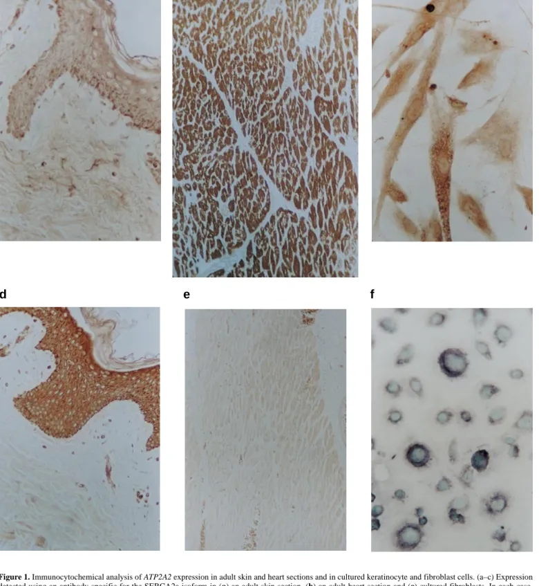
(2) 1622 Human Molecular Genetics, 1999, Vol. 8, No. 9. rheic’ areas of head, neck and upper chest, and there is often a history of photoaggravation. Histologically, these lesions show suprabasal acantholysis of the epidermis, with overlying dyskeratotic cells, so-called corps ronds and grains. Other epithelial features of DD include focal palmar pits and keratoses, linear nail lesions with distal notching, and multiple plane keratoses of the dorsa of the hands (acrokeratosis verruciformis). Mucosae can be involved; narrowed salivary ducts may lead to obstructive sialadenitis, and corneal lesions are also recognized. The cutaneous manifestations vary considerably in severity even within families, and the diagnosis may be overlooked (2). Several variants in the presentation of cutaneous lesions have also been described (1). The most severe of these are bullous, erosive or vegetating forms of disease, in particular in flexures, or confluent cornifying disease of the lower legs (3). Less severe lesions seen in other cases include hypopigmented macules in pigmented skin. In some cases, acantholytic histology has been found in acneiform comedones and epidermoid cysts. A familial haemorrhagic variant is also evident in which red/ black macular and bullous lesions are found in acral sites, especially on the hands, although typical keratoses may be present elsewhere (4,5). There are no consistent extra-epithelial manifestations of DD. An immunological defect has been debated, and in isolated instances other associations have been reported. However, beginning with early reports (6,7), the frequent coexistence of neuropsychiatric disorder in DD has been noted, including mental handicap, schizophrenia and bipolar disorder, or epilepsy. The majority of patients have no such problems, but several reports have documented the familial occurrence of neuropsychiatric features (1,8–10). A survey of 37 patients from Denmark reported that seven were mentally subnormal or ‘deranged’, and nine others ‘destitutes’ (11). Ascertainment bias may have played a role in these findings as several patients were in institutional care. In a British survey, 5% of 163 patients were mentally retarded, and a similar proportion epileptic (1). Less easy to quantify is the impression, common amongst dermatologists, that milder degrees of learning or behavioural problems are over-represented in patients with DD, and that there may be several such cases within a family. Such families usually have poor social circumstances, and it is difficult to distinguish the contribution of other factors to the severity of disease. Social isolation due to severe, often malodorous, disease may in turn play a role in psychological problems (1). A pathogenetic defect of epidermal cell structure or adhesion has been proposed, in particular as the acantholytic cells show anomalous distribution of desmosomal components (12). The DD gene was mapped to 12q24.1 (13–19) but no convincing candidate gene was evident. Recently, a consortium of British groups reported causative mutations in a gene at this locus, ATP2A2, which encodes the sarco/endoplasmic reticulum calcium ATPase type 2 (SERCA2) (20). This enzyme is one of a family of membrane ATPases which pump calcium from the cytoplasm into reservoirs in the endoplasmic reticulum (21). Two isoforms exist, SERCA2a and SERCA2b, which differ only in the C-terminal domains (22), but isoform specificity of SERCA2 expression in skin has not been established. Mutations in ATP2A2 have been suggested to cause DD through haploinsufficiency, resulting in impaired uptake of cytosolic calcium into the endoplasmic reticulum, and conse-. quent disruption of calcium signalling (20). In the epidermis, the presumed result is loss of cellular adhesion, proliferation and disordered keratinization. The control of cytoplasmic calcium signalling is complex (23) and the sequence and details of this pathogenic mechanism remain to be elucidated. Even so, the above model does not explain adequately certain disease features. In particular, if clinical variants are allelic with the classical disease, certain mutations must have effects additional to those arising from haploinsufficiency alone. In the present study, we show that the SERCA2b isoform is the one which is expressed predominantly in skin. Our mutational analyses in a large collection of European Darier pedigrees reveal that clinical variant forms of DD are allelic to classical DD, and that cutaneous variants, most significantly the familial acral haemorrhagic variant, are particularly associated with missense mutations. In contrast, neuropsychiatric features did not appear to be associated with a specific mutation class. RESULTS ATP2A2 expression in the epidermis The finding of ATP2A2 mutations in DD by Sakuntabhai et al. (20) suggested a role for this gene in the epidermis. Several expression surveys on ATP2A2 or vertebrate homologues have been carried out, but expression in skin has not been investigated. We used antibodies specific for the two major isoforms of the protein (24,25) to track expression in skin sections and in cultured keratinocytes and fibroblasts (see Materials and Methods). With an antibody specific for the longer isoform (SERCA2b), adult skin sections showed clear immunopositivity in epidermal structures including interfollicular epidermis, pilosebaceous units and sweat glands (Fig. 1d). Little staining was observed, however, in the dermis. In contrast, the signal in adult skin sections for the shorter isoform (SERCA2a) was difficult to distinguish from background (Fig. 1a) and, although low level expression cannot be excluded, positive staining of pilar muscle provided a good internal control (data not shown) and strong staining was produced in normal adult heart sections (Fig. 1b) whereas, as expected, no signal was detected using the SERCA2b-specific antibody (Fig. 1e). Both cultured keratinocytes and fibroblasts did, however, show clear staining for both antibodies (Fig. 1c and 1f, and data not shown). In both skin and cultured cells, signal was confined to the cytoplasm in a pattern compatible with the putative role of these gene products. Extended mutation analyses of the ATP2A2 gene in DD patients Patient-specific mutations in five pedigrees from our group were included in the consortium report of Sakuntabhai et al. (20). To obtain a firmer grounding for possible genotype– phenotype correlations, we conducted standard single-strand conformation polymorphism (SSCP) and heteroduplex analyses on patients drawn from >100 families from the UK and continental Europe. Patient-specific mutations were identified in a total of 47 families (Table 1). A total of 40 different mutations were found, comprising 13 short intragenic deletions/ insertions (11 of which were frameshifting), seven splice site.
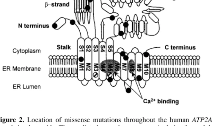
(3) Human Molecular Genetics, 1999, Vol. 8, No. 9 1623. a. d. c. b. e. f. Figure 1. Immunocytochemical analysis of ATP2A2 expression in adult skin and heart sections and in cultured keratinocyte and fibroblast cells. (a–c) Expression detected using an antibody specific for the SERCA2a isoform in (a) an adult skin section, (b) an adult heart section and (c) cultured fibroblasts. In each case, staining was with DAB. (d–f) Expression detected using an antibody specific for the SERCA2b isoform in (d) an adult skin section, staining with DAB, (e) an adult heart section, staining with DAB, and (f) cultured keratinocytes, staining with nickel–DAB and nuclear counterstaining using methyl green.. mutations, three nonsense mutations and 17 missense mutations. The latter were found to be distributed throughout the protein molecule (Fig. 2).. In cases in whom unusual cutaneous manifestations were present, only missense mutations were found. In two such pedigrees, the consistency of the phenotype could not be estab-.
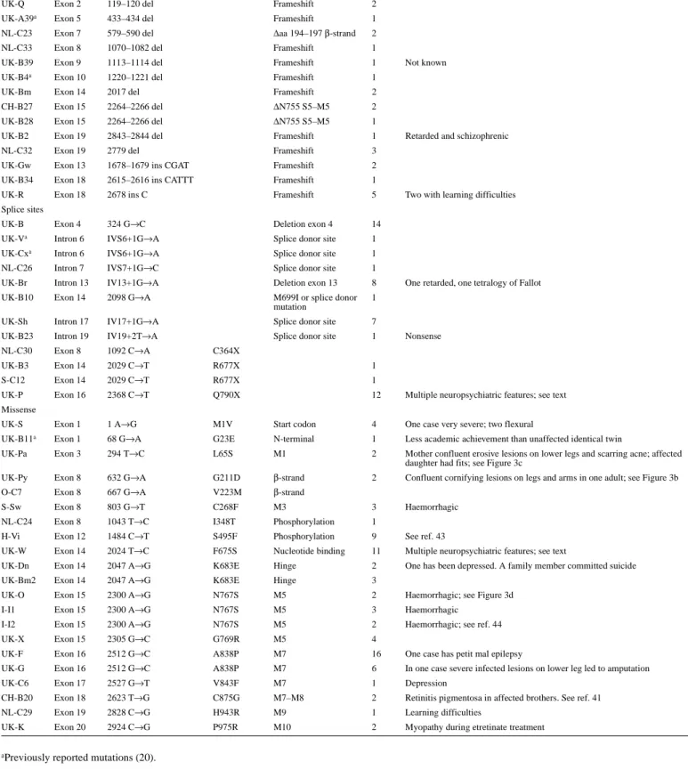
(4) 1624 Human Molecular Genetics, 1999, Vol. 8, No. 9. Table 1. ATP2A2 mutations in 47 pedigrees of Darier’s disease Origin-ID. Site. Mutation. Amino acid. Protein effect, or domain Cases. Exceptional or associated features. Deletion/insertion UK-Q. Exon 2. 119–120 del. Frameshift. 2. UK-A39a. Exon 5. 433–434 del. Frameshift. 1. NL-C23. Exon 7. 579–590 del. ∆aa 194–197 β-strand. 2. NL-C33. Exon 8. 1070–1082 del. Frameshift. 1. UK-B39. Exon 9. 1113–1114 del. Frameshift. 1. UK-B4a. Exon 10. 1220–1221 del. Frameshift. 1. UK-Bm. Exon 14. 2017 del. Frameshift. 2. CH-B27. Exon 15. 2264–2266 del. ∆N755 S5–M5. 2. UK-B28. Exon 15. 2264–2266 del. ∆N755 S5–M5. 1. UK-B2. Exon 19. 2843–2844 del. Frameshift. 1. NL-C32. Exon 19. 2779 del. Frameshift. 3 2. UK-Gw. Exon 13. 1678–1679 ins CGAT. Frameshift. UK-B34. Exon 18. 2615–2616 ins CATTT. Frameshift. 1. UK-R. Exon 18. 2678 ins C. Frameshift. 5. UK-B. Exon 4. 324 G→C. Deletion exon 4. 14. UK-Va. Intron 6. IVS6+1G→A. Splice donor site. 1. Not known. Retarded and schizophrenic. Two with learning difficulties. Splice sites. UK-Cxa. Intron 6. IVS6+1G→A. Splice donor site. 1. NL-C26. Intron 7. IVS7+1G→C. Splice donor site. 1. UK-Br. Intron 13. IV13+1G→A. Deletion exon 13. 8. UK-B10. Exon 14. 2098 G→A. M699I or splice donor mutation. 1. One retarded, one tetralogy of Fallot. UK-Sh. Intron 17. IV17+1G→A. Splice donor site. 7. UK-B23. Intron 19. IV19+2T→A. Splice donor site. 1. NL-C30. Exon 8. 1092 C→A. C364X. UK-B3. Exon 14. 2029 C→T. R677X. 1. S-C12. Exon 14. 2029 C→T. R677X. 1. UK-P. Exon 16. 2368 C→T. Q790X. 12. Multiple neuropsychiatric features; see text One case very severe; two flexural. Nonsense. Missense UK-S. Exon 1. 1 A→G. M1V. Start codon. 4. UK-B11a. Exon 1. 68 G→A. G23E. N-terminal. 1. Less academic achievement than unaffected identical twin. UK-Pa. Exon 3. 294 T→C. L65S. M1. 2. Mother confluent erosive lesions on lower legs and scarring acne; affected daughter had fits; see Figure 3c. UK-Py. Exon 8. 632 G→A. G211D. β-strand. 2. Confluent cornifying lesions on legs and arms in one adult; see Figure 3b. O-C7. Exon 8. 667 G→A. V223M. β-strand Haemorrhagic. S-Sw. Exon 8. 803 G→T. C268F. M3. 3. NL-C24. Exon 8. 1043 T→C. I348T. Phosphorylation. 1. H-Vi. Exon 12. 1484 C→T. S495F. Phosphorylation. 9. See ref. 43. UK-W. Exon 14. 2024 T→C. F675S. Nucleotide binding. 11. Multiple neuropsychiatric features; see text One has been depressed. A family member committed suicide. UK-Dn. Exon 14. 2047 A→G. K683E. Hinge. 2. UK-Bm2. Exon 14. 2047 A→G. K683E. Hinge. 3. UK-O. Exon 15. 2300 A→G. N767S. M5. 2. Haemorrhagic; see Figure 3d. I-I1. Exon 15. 2300 A→G. N767S. M5. 3. Haemorrhagic Haemorrhagic; see ref. 44. I-I2. Exon 15. 2300 A→G. N767S. M5. 2. UK-X. Exon 15. 2305 G→C. G769R. M5. 4. UK-F. Exon 16. 2512 G→C. A838P. M7. 16. One case has petit mal epilepsy. UK-G. Exon 16. 2512 G→C. A838P. M7. 6. In one case severe infected lesions on lower leg led to amputation. UK-C6. Exon 17. 2527 G→T. V843F. M7. 1. Depression. CH-B20. Exon 18. 2623 T→G. C875G. M7–M8. 2. Retinitis pigmentosa in affected brothers. See ref. 41. NL-C29. Exon 19. 2828 C→G. H943R. M9. 1. Learning difficulties. UK-K. Exon 20. 2924 C→G. P975R. M10. 2. Myopathy during etretinate treatment. aPreviously. reported mutations (20).. lished as in each only a mother and young daughter were living and affected. In one of these, a woman had cornifying disease of the arms and lower legs (family UK-Py; Fig. 3b), and in the other more erosive lesions (UK-Pa; Fig. 3c) were present. The. latter patient also had cystic acneiform lesions with unusual deeply pitted scars. Her late mother was reported by a dermatologist to have had very severe DD (severe disease is more common in men). In a third variant family (UK-S), a mother.
(5) Human Molecular Genetics, 1999, Vol. 8, No. 9 1625. Figure 2. Location of missense mutations throughout the human ATP2A2encoded polypeptide. The predicted secondary structure includes three globular cytoplasmic domains separated by a stalk sector from the transmembrane part of the molecule. The cytoplasmic domains contain a β-strand, a phosporylation domain and an ATP-binding domain. A hinge region links the ATPbinding domain to the transmembrane region which includes 10 transmembrane coiled-coil helices, of which four contain Ca2+-binding sites (M4, M5, M6 and M8) (21). The missense mutations identified in DD patients (see also Table 1) are represented by filled circles, except for those associated with the haemorrhagic variant (which are represented by striped circles). S1–S5 refer to stalk sectors and M1–M10 refer to transmembrane helices 1–10. Adapted from ref. 21.. and adult daughter both had moderate flexural lesions, but the daughter also had extensive truncal lesions and deep pitted facial scars which she blamed on sunbed use. Her teenage sons had more typical early features of DD. All affected members of this family were, however, found to have an M1V substitution which could possibly have a rather mild effect on normal function; the possibility of pathogenesis due to a more severe mutation located elsewhere cannot be excluded. The acral haemorrhagic phenotype (Fig. 3d) is expressed consistently by all affected members within families, including the four families studied here. Missense mutations were identified in all four of these pedigrees. In one family from Scotland (UK-O) and two unrelated families from different regions in Italy (I-I1 and I-I2), affected individuals had an identical mutation, predicting an N767S substitution in the fifth transmembrane domain (Fig. 2). Marker analysis using microsatellites in the immediate vicinity of ATP2A2 showed that the disease haplotype in the Scottish family was quite unrelated to that found in the two Italian families which also had different disease haplotypes (data not shown). In the fourth such family (S-Sw from Sweden), affected individuals had a C268F substitution in the third transmembrane domain. The assessment of neuropsychiatric features in DD remains problematic. Despite a widespread view amongst clinicians who study the disorder that a neuropsychiatric component is common, it is hard to confirm that mild defects are due to the disease without careful psychometric testing, controlled by studying unaffected siblings. In this context, it is noteworthy that a sporadic case with DD (UK-B11) who left school aged 15 without academic qualifications has a genetically identical but unaffected twin brother who does not have the pathogenic mutation and who completed secondary education and achieved a senior position in his chosen profession (20). In the. present study, the patients in whom mutations were identified (Table 1) included two sporadic cases with notably low intelligence or schizophrenia and members of four pedigrees where one or more family members have had retardation, learning or behavioural difficulties, or a history of fits. In two of the families, UK-P and UK-W, definable neuropsychiatric features were particularly common, but there was no consistent pattern in affected family members. Of 12 living cases in UK-P, one is schizophrenic with epilepsy and has a son with behavioural and learning difficulties and a sister with severe retardation and fits since birth, ascribed to birth injury, and a fourth affected member had childhood epilepsy and learning difficulties. Of 11 cases in UK-W, one is severely retarded with fits, two uncles had learning difficulties, one with fits, and a cousin has epilepsy alone. Some such associations may be coincidental; in another family (UK-Bm), several members had had clinical depression, but at least one family member with bipolar disorder (but no evidence for cutaneous symptoms) did not have the pathogenic mutation (data not shown). Unlike cutaneous variants, which were associated only with missense mutation, pedigrees where neuropsychiatric features were present showed all classes of mutation (Table 1). DISCUSSION SERCA2 is a member of a family of structurally related Ca2+ATPases, each of which has a distinct developmentally regulated and tissue-specific pattern of expression (26). Recessive mutations in the gene encoding SERCA1 (found in fast twitch muscle) cause some cases of the inherited myopathy Brody disease (27) and, in mice, mutations in the gene encoding SERCA3 cause the deafwaddler phenotype (28). SERCA2, encoded by the ATP2A2 gene, has two protein isoforms with amino acids 1–993 in common but differing at the C-terminus. The SERCA2a isoform has an extra four amino acids and predominates in cardiac and slow-twitch skeletal muscle; the SERCA2b isoform, expressed in smooth muscle and nonmuscle tissues, has a different 49 amino acid C-terminal peptide containing a hydrophobic sequence that may form an eleventh transmembrane domain (21,29,30). SERCA2b has a higher Ca2+ affinity but lower turnover than SERCA2a (21,31). In the current study, we found that the SERCA2b isoform was readily detectable in various epidermal structures, but not in the dermis of adult skin. Equivalent analyses with SERCA2aspecific antibodies failed to identify significant expression. However, both forms were identified in cultured keratinocytes and fibroblasts, suggesting that conditions in culture are not fully representative of the in vivo state. In all cases, positive staining was limited to the cytoplasm, consistent with the known function of SERCA2. Data from our extended mutation analyses of the ATP2A2 gene are consistent with the single locus for DD suggested by linkage analyses (13–19). Given that the gene is large, with 21 coding exons, it is possible that the patients in whom no ATP2A2 mutation was identified have larger scale mutations removing whole exons at a time, which would not be detected by our screening strategy. Although SERCA2b is expressed preferentially in epidermis, all the mutations identified would be predicted to affect both isoforms. Many of the mutations shown in Table 1 can be expected to result in loss of function by introducing a premature termination codon: the four non-.
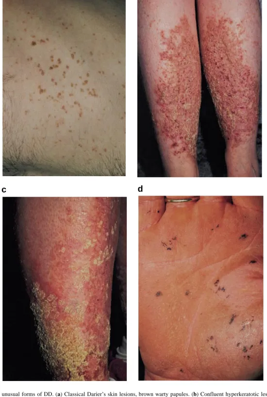
(6) 1626 Human Molecular Genetics, 1999, Vol. 8, No. 9. a. b. c. d. Figure 3. Classical and unusual forms of DD. (a) Classical Darier’s skin lesions, brown warty papules. (b) Confluent hyperkeratotic lesions of the lower legs (family UK-Py). (c) More erosive lesions of the lower legs (family UK-Pa). (d) The acral haemorrhagic variant: on palms and dorsal fingers, haemorrhage into acantholytic vesicles gives rise to black macules (family UK-O).. sense mutations, the frameshifting insertions/deletions and many of the splice site mutations. In probably all such cases, mRNA will be degraded rapidly by the nonsense-mediated RNA decay pathway, and no polypeptide synthesized (32). These mutations are therefore consistent with a model of. pathogenesis for DD in which haploinsufficiency results in abnormal responses, but where pathological effects are produced only under conditions of stress, e.g. UVB irradiation. It is uncertain whether acantholysis is a cause or consequence of the disordered keratinization, but it is well recognized that cal-.
(7) Human Molecular Genetics, 1999, Vol. 8, No. 9 1627. cium homeostasis is critical for proper desmosome assembly (33,34), and thapsigargin, an inhibitor of SERCA2, impairs cell adhesion in the Madin–Darby canine kidney cell model (35,36). The known functions of SERCA2 would also predict an effect on cardiac muscle, and indeed mice hemizygous for a null mutation in ATP2A2 exhibit impaired cardiac performance (37). The absence of a reported cutaneous phenotype in these mice may reflect the late onset of DD, or the need for additional stresses to precipitate skin disease. Conversely, there is no clinical evidence of cardiac insufficiency in human DD, so compensatory or alternative mechanisms may exist in man. In the present study, we found that 17 of the 40 mutations reported, or 42.5% of the total, were missense mutations, which is in close agreement with the 38% figure reported for missense mutations in an independent study of 19 families and six sporadic patients (38). In contrast to chain-terminating mutations, mutant proteins encoded by missense mutations potentially may further disrupt membrane localization or function of SERCA2. In vitro site-specific mutagenesis of SERCA2a disrupts sites of calcium binding (21), regulation by phospholamban (39) or phosphorylation (40). Although the effects of the exact mutations reported here are uncertain, substitutions at I348, K683, N767, G769 and C875 (but not C268) partially or completely abrogate SERCA2a activity (D.H. MacLennan, personal communication). In particular, the nonconservative substitution K683E, at a residue which is highly conserved across the superfamily of membrane ATPases, is predicted to disrupt formation of a phosphorylated intermediate with ATP or inorganic phosphate (41). Table 1 shows that missense mutations make a disproportionate contribution to DD pedigrees in whom unusual clinical features have been noted. Severe disease with cornifying lesions (as in UK-Py), and more erosive lesions in UK-Pa and UK-S, are recognized variant presentations. Although not all family members were severely affected, the associated missense mutations in these families (respectively G211D in the βstrand, L65S in the first transmembrane domain and M1V in the N-terminal domain; Fig. 2) may be a predisposing factor for developing such unusual features. A missense mutation, C875G, was also found in two Swiss brothers (CH-B20) with DD and retinitis pigmentosa (42), but this unique association is most likely to be due to the chance co-existence of a second mutation at another locus. The most consistent familial variant is the one with acral haemorrhagic lesions, and this is the one where we have demonstrated the clearest association between missense mutation and atypical phenotype. In the four families studied, all affected members had haemorrhagic lesions, and in all these families we were able to identify patient-specific missense mutations. Three families had an identical N767S mutation located in the fifth transmembrane domain, but mutations appear to have arisen independently, being associated with different marker haplotypes in geographically distant populations (from Scotland and two regions of Italy). The fourth, Swedish, family had a C268F substitution located in the neighbouring third transmembrane domain. These findings are particularly intriguing, since they imply that certain mutations may be specifically disruptive to SERCA2 function, perhaps in vascular endothelium cells as well as in keratinocytes, or that the mutant protein has a secondary effect in these sites.. The nature of the association of neuropsychiatric disorders with DD remains elusive. Mental handicap, sociopathic behaviour, schizophrenia and bipolar disorder have all been reported. A role for SERCA2 in neurological development or function remains highly plausible, since the gene is widely expressed in brain (43). In order to address this issue within our study population, we focused on a few pedigrees with the clearest evidence of concomitant neuropsychiatric disorder. Even so, we have found that these are associated with different mutation classes. A sporadic case with schizophrenia (UK-B2) and other individuals with learning difficulties or neuropsychiatric features in families UK-R, UK-Br and also in UK-P had mutations expected to cause nonsense-mediated mRNA decay, while others, as in UK-W, had missense mutations. This finding implies that any predisposition to neuropsychiatric disorder in DD is an inconsistent consequence of defective ATP2A2 expression which is not mutation specific, and depends on concomitant genetic and environmental factors. MATERIALS AND METHODS Immunocytochemistry Normal adult skin samples were fixed in formalin and paraffin embedded. Primary cultures of neonatal keratinocytes and fibroblasts were grown as monolayer cultures in KGM and DMEM, and cells fixed in acetone. Paraffin sections were dewaxed by heating at 60°C for 10 min and treated with Histoclear (National Diagnostics). Sections were rehydrated through decreasing concentrations of ethanol and rinsed in water and phosphate-buffered saline (PBS). Sections were permeabilized in PBS containing 0.2% Triton X-100 for 15 min as described previously (44). Non-specific binding sites were blocked with normal goat serum (1:60) for 20 min. After removal of excess serum, sections were incubated for 60 min with primary antibody diluted 1:500 or 1:1000 in PBS/0.1% bovine serum albumin. Polyclonal antibodies to SERCA2a and SERCA2b were a gift from Dr F. Wuytack and were reported previously (24,25). Following three washes in PBS, the secondary biotinylated goat anti-rabbit antibody (1:100) was applied for 30 min. After a further three washes in PBS, the biotin–avidin–peroxidase complex (Vectastain ABC kit; Vector Laboratories) was applied for 30 min. After three washes in PBS, sections were visualized using 0.05% DAB (3'3'-diaminobenzidine) and 0.01% hydrogen peroxide (44). The cultured keratinocyte sections were washed once in 0.1 M acetate buffer pH 6 in addition to the above washes, prior to detection using nickel–DAB solution (60 mM ammonium nickel sulfate, 0.05% DAB, 7.5 mM ammonium chloride, 0.2% β-D-glucose, 1 U/ml glucose oxidase) made up in 0.1 M acetate buffer pH 6, and nuclei counterstained with 1% methyl green for 1–2 min. All sections were dehydrated in acetone and histoclear before mounting. Omission of the primary antibody was used as a control, and isoform specificity of the antibodies was shown by control staining of adult heart muscle and by immunostaining of smooth pilar muscle in skin sections. Patients and families Patients with generalized forms of DD mostly were referred by dermatologists in the UK and The Netherlands (NL), but a.
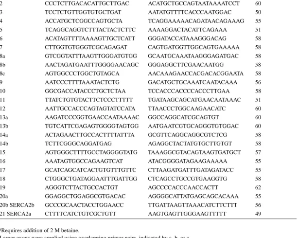
(8) 1628 Human Molecular Genetics, 1999, Vol. 8, No. 9. Table 2. Genomic primers for mutation analysis Exon. Forward primer sequence. 1a. GGAGTGCGAGGCGGAGGCGAGGAG GGTGGAGCCCGGTCAGCCATCTTC. Reverse primer sequence. 67. Temperature (°C). 2. CCCTCTTGACACATTGCTTGAC. ACATGCTGCCAGTAATAAAATCCT. 60. 3. TCCTCTGTTGGTGTGCTGAT. AATATGTTTTCACCCAATGGAC. 50. 4. ACCATGCTCGGCCAGTGCTA. TCAGGAAAAACAGATAACAGAAAG. 55. 5. TCAGGCAGGTCTTTACTACTCTTC. AAAAGGACTACATTCAGAAA. 51. 6. ACATAGTTTTAAAAGTTGCTCATT. GGGATACCATAAAGGGACAG. 58. 7. CTTGGTGTGGGTCGCAGAGAT. CAGTGATGGTTGGCAGTGAAAAA. 58. 8a. GTCGGTATTTAAGTTGGGATGTGG. GCAATGCAAATAAGGGAGATGAC. 58. 8b. AACTAGATGAATTTGGGGAACAGC. GGGAGGCTTCGAACAATGG. 58. 8c. AGTGGCCCTGGCTGTAGCA. AACAAAGAACCACGACACGGAATA. 58. 9. AATCCCTTTTAAATACTCTG. GACATGCTGCAAATCAATACAAA. 56. 10. GGCGACCATACCCTGCTCTAA. TCCACCCACCCCACCCTTGAA. 58. 11. TTATCTGTGTACTTCTCCCTTTTT. TGATAAGCAGCATGAACAATAAAC. 51. 12. AATTGCCACCCAGTAGTATCCATA. TTAACCCTGGCAAGAACATC. 60. 13a. AAGATCCCGGTGAACCAATAAAAC. GGCCAGGCATCGCAGTGT. 60. 13b. TGTCATTCGAGAGTGGGGTAGTGG. AATGAATCGTGCAGGGTGTGGAC. 60. 14a. ACTAGAACTTGCCACTTTTATTTA. GCGTTCAGGCAGGCGTCTCG. 58. 14b. TCTTCGGGCAGGATGAG. AGAGGCTACTATGTGCTTGTGT. 58. 15. AGTGGGCTTTTGCCTAGGGGTATG. TAAAGGCGTACAGTAAGTGATGCT. 57. 16. AAATAGTGGCCAGAAGTCAT. ATACGGGGATAGAAGAAAAA. 55. 17. GCATCAGCATCACTGTGTTTGTTC. CTTAAGATGATTTGATAGATACC. 55. 18. CTGGGCTGATAGGAATTTGATTGG. CTCAGCCTGCCGTGAAGGTG. 58. 19. AGGGTCTTACTGCCACTGT. AGCCCCACCCAACCACTT. 62. 20a. GGAGGCTGGAGGCGTGACAC. AGGGGCATTATGAGCAGCACAAA. 55. 20b SERCA2b. GCCCGCAACTACCTGGAACC. TTGATTAAGTTAAACATCTTCTTT. 56. 21 SERCA2a. CTTTTCATCTGTCGCTGTT. AAGTGAGTTGGGAAGTTTTT. 49. aRequires addition of 2 M betaine. Larger exons were amplied using overlapping primer pairs, indicated by a, b, or c.. minority were referred from Switzerland (CH), Sweden (S), Italy (I), Hungary (H) and Austria (O). The diagnosis was confirmed by a participating dermatologist (C.S.M., E.H., H.L., P.M.S., D.H., P.I., A.V., T.G., C.M., R.R. or G.N.) on the basis of clinical examination and was supported by histological examination of a skin biopsy in at least one member of each family. Diagnostic features included keratotic papules or plaques on the trunk, typical nail dystrophy and/or palmar pits (2). Clinical features in UK families B, F and G were reported by Munro (2); references to other published cases (42,45,46) are shown in Table 1. In the majority of families, including all of those from the UK, The Netherlands and Hungary, all available family members were ascertained to establish undiagnosed disease, consistency of phenotype and the presence of additional features. In other cases, information regarding variant clinical phenotype was sought from referring clinicians. Family UK-B11 comprises identical twins, only one of whom is affected with DD. The study was approved by the local Ethics Committee. Blood samples and skin biopsies were obtained after patients had given informed consent. Mutation analysis Genomic DNA was extracted from peripheral blood leucocytes using standard procedures. Primers were designed to amplify. all 21 coding exons and flanking intronic splice sites from genomic DNA (Table 2). PCRs were carried out using standard reaction mixes containing 1.5 mM magnesium chloride, except for the PCR to amplify exon 1 which was supplemented with 2 M betaine. After an initial denaturation step at 94°C for 4 min, we carried out 35 cycles of amplification consisting of 30–60 s at 94°C, 30–60 s at the optimally determined annealing temperature (51–67°C) and 30–60 s at 72°C. PCR products were screened for mutations by either SSCP or direct sequencing. SSCP gels consisted of 1× MDE solution (FMC), 5% glycerol and 0.6× TBE buffer, and were run at 300 V overnight. Following electrophoresis, gels were fixed in ethanol/ acetic acid and stained with silver nitrate, using standard methods. PCR fragments showing apparently patient-specific aberrant migration patterns or extra bands were validated by confirming their absence from 140 control chromosomes. The only nonpathogenic polymorphism detected in this study was a silent G to A substitution at the third base position of the codon GCG which specifies alanine at position 724. Patient-specific PCR products were sequenced in forward and reverse orientations. PCR (or RT–PCR) products which showed a single band on 2% agarose gel were purified directly using a Qiaquick PCR purification kit (Qiagen). PCR or RT–PCR products which showed more than one band were run in low melting point.
(9) Human Molecular Genetics, 1999, Vol. 8, No. 9 1629. agarose gel. The bands were separated and gel purified using a Qiaex gel purification kit (Qiagen). The purified products were sequenced using the ABI Prism AmpliTaq-reaction dye terminator cycle sequencing kit (PE Applied Biosystems) and an Applied Biosystems model 373A automated sequencer or a Thermosequenase [33P]ddNTP terminator cycle sequencing kit (Amersham) according to the manufacturer’s instructions. PCR products which showed ‘superimposed’ sequences were subcloned into pGEM-T vector (Promega) or the TA subcloning kit (Invitrogen), and mutiple subclones were sequenced. ACKNOWLEDGEMENTS We are grateful to the referring clinicians and the families who participated in the study, to Dr Giovanna Zambruno for help in obtaining specimens, to Dr Frank Wuytack for a gift of antibodies specific for the SERCA2a and SERCA2b isoforms of the ATP2A2 gene product, to Dr Chris Wright for providing sections of normal heart tissue and to Dr David Maclennan for helpful discussion and communication of unpublished data. We thank Dr Steve Bryce for his previous contribution to this project. This work was supported by a grant from the Wellcome Trust awarded to T.S., C.S.M. and J.L.R. V.L.R.-P. was supported by an EC TMR Fellowship. REFERENCES 1. Burge, S. and Wilkinson, D.J. (1992) Darier–White disease: a review of the clinical features in 163 patients. J. Am. Acad. Dermatol., 27, 40–50. 2. Munro, C.S. (1992) The phenotype of Darier’s disease: penetrance and expressivity in adults and children. Br. J. Dermatol., 127, 126–130. 3. Rongioletti, F., Cestari, R. and Rebora, A. (1990) Verrucous and malodorous vegetations on the legs. Darier’s disease, cornifying type. Arch. Dermatol., 128, 399–399. 4. Jones, W.N., Nix, T.E. and Clark, W.H. (1964) Haemorrhagic Darier’s disease. Arch. Dermatol., 89, 523–527. 5. Foresman, P.L., Goldsmith, L.A., Ginn, L. and Beck, A.L. (1993) Haemorrhagic Darier’s disease. Arch. Dermatol., 129, 511–512. 6. Boeck, C. (1916) Demonstration of a case of Darier’s disease with psychological aberration. In Proceedings of the Third Congress of the Nordic Dermatological Society. 7. Cockayne, E.A. (1933) Darier’s disease. In Inherited Abnormalities of the Skin and its Appendages. Oxford University Press, London, pp. 135–137. 8. Medansky, R.S. and Woloshin, A.A. (1961) Darier’s disease: an evaluation of its neuropsychiatric component. Arch. Dermatol., 84, 482–484. 9. Getzler, N.A. and Flint, A. (1966) Keratosis follicularis: a study of one family. Arch. Dermatol., 93, 545–549. 10. Craddock, N. et al. (1994) Familial cosegregation of major affective disorder and Darier’s disease (keratosis follicularis). Br. J. Psychiatry, 164, 355–358. 11. Svensen, I.B. and Albrechtsen, B. (1961) The prevalence of dyskeratosis follicularis (Darier’s disease) in Denmark. Acta Derm.-Venereol., 30, 256–269. 12. Burge, S. and Garrod, D.R. (1991) An immunohistological study of desmosomes in Darier’s disease and Hailey–Hailey disease. Br. J. Dermatol., 124, 242–251. 13. Bashir, R., Munro, C.S., Mason, S., Stephenson, A., Rees, J.L. and Strachan, T. (1993) Localisation of a gene for Darier’s disease. Hum. Mol. Genet., 2, 1937–1939. 14. Craddock, N., Dawson, E., Burge, S., Parfitt, L., Mant, B., Roberts, Q., Daniels, J., Gill, M., McGuffin, P., Powell, J. and Owen, M. (1993) The gene for Darier’s disease maps to chromosome 12q23–q24.1. Hum. Mol. Genet., 2, 1941–1943. 15. Carter, S.A., Bryce, S.D., Munro, C.S., Healy, E., Bashir, R., Weissenbach, J., LeBlanc-Straceski, J., Kucherlapati, R., Stephenson, A., Rees, J.L. and Strachan, T. (1994) Linkage analyses in British pedigrees suggest a single locus for Darier disease and narrow the location to the interval between D12S105 and D12S129. Genomics, 24, 378–382.. 16. Ikeda, S., Haake, W.A., Ewing, N., Polakowska, R., Sarret, Y., Trattner, A., David, M., Shohat, M., Schroeder, D.W., Epstein, E.H. and Goldsmith, L.A. (1994) Localization of the gene for Darier disease to a 5-cM interval on chromosome 12q. J. Invest. Dermatol., 103, 478–481. 17. Parfitt, E. et al. (1994) The gene for Darier’s disease maps between D12S78 and D12S79. Hum. Mol. Genet., 3, 35–38. 18. Wakem, P. et al. (1996) Localization of the Darier disease gene to a 2-cM portion of 12q23–24.1. J. Invest. Dermatol., 106, 365–367. 19. Monk, S. et al. (1998) Refined genetic mapping of the Darier locus to a <1-cM region of chromosome 12q24.1, and construction of a complete, high-resolution P1 artificial chromosome/bacterial artificial chromosome contig of the critical region. Am. J. Hum. Genet., 62, 890–903. 20. Sakuntabhai, A., Ruiz-Perez, V., Carter, S., Jacobsen, N., Burge, S., Monk, S., Smith, M., Munro, C.S., O’Donovan, M., Craddock, N., Kucherlapati, R., Rees, J.L., Owen, M., Lathrop, G.M., Monaco, A.P., Strachan, T. and Hovnanian, A. (1999) Mutations in ATP2A2, encoding a Ca (2+) pump, cause Darier disease. Nature Genet., 21, 271–277. 21. Maclennan, D.H., Rice, W.J. and Green, N.M. (1997) The mechanism of Ca2+ transport by sarco (endo)plasmic reticulum Ca2+-ATPases. J. Biol. Chem., 272, 28815–28818. 22. Lytton, J. and MacLennan, D.H. (1988) Molecular cloning of cDNAs from human kidney coding for two alternatively spliced products of the cardiac Ca2+-ATPase gene. J. Biol. Chem., 263, 15024–15031. 23. Berridge, M.J. (1997) Elementary and global aspects of calcium signalling. J. Physiol., 499, 291–306. 24. Wuytack, T., Eggermont, J.A., Raeymakers, L., Plessers, L. and Casteels, R. (1989) Antibodies against the non-muscle isoform of the endoplasmic reticulum Ca2+-transport ATPase. Biochem. J., 264, 765–769. 25. Eggermont, J.A., Wuytack, F., Verbist, J. and Casteels, R. (1990) Expression of endoplasmic reticulum Ca2+-pump isoforms and of phospholamban in pig smooth muscle tissues. Biochem. J., 271, 649–653. 26. Lytton, J., Westlin, M., Burk, S.E., Shull, G.E. and Maclennan, D.H. (1992) Functional comparisons between isoforms of the sarcoplasmic or endoplasmic reticulum family of calcium pumps. J. Biol. Chem., 267, 14483–14489. 27. Odermatt, A., Taschner, P.M., Khanna, V.K., Busch, H.M., Karpati, G., Jablecki, C.K., Breuning, M.H. and Maclennan, D.H. (1996) Mutations in the gene-encoding SERCA1, the fast-twitch skeletal muscle sarcoplasmic reticulum Ca2+ ATPase, are associated with Brody disease. Nature Genet., 14, 191–194. 28. Street, V.A., McKee-Johnson, J.W., Fonseca, R.C., Tempel, B.L. and Noben-Trauth, K. (1998) Mutations in a plasma membrane Ca2+-ATPase gene cause deafness in deafwaddler mice. Nature Genet., 19, 390–394. 29. Eggermont, J.A., Wuytack, F., De Jaegere, S., Nelles, L. and Casteels, R. (1989) Evidence for two isoforms of the endoplasmic-reticulum Ca2+ pump in pig smooth muscle. Biochem. J., 260, 757–761. 30. Campbell, A.M., Kessler, P.D. and Fambrough, D.M. (1992) The alternative carboxyl termini of avian cardiac and brain sarcoplasmic reticulum/ endoplasmic reticulum Ca+-ATPases are on opposite sides of the membrane. J. Biol. Chem., 267, 9321–9325. 31. Verboomen, H., Wuytack, F., Van den Bosch, L., Mertens, L. and Casteels, R. (1994) The functional importance of the extreme C-terminal tail in the gene 2 organellar Ca2+-transport ATPase (SERCA2a/b). Biochem. J., 303, 979–984. 32. Hentze, M.W. and Kulozik, A.E. (1999) A perfect message: RNA surveillance and nonsense-mediated decay. Cell, 96, 307–310. 33. Watt, F.M., Mattey, D.L. and Garrod, D.R. (1984) Calcium-induced reorganization of desmosomal components in cultured human keratinocytes. J. Cell Biol., 99, 2211–2215. 34. Duden, R. and Franke, W.W. (1988) Organization of desmosomal plaque proteins in cells growing at low calcium concentrations. J. Cell Biol., 107, 1049–1063. 35. Lytton, J., Westlin, M. and Hanley, M.R. (1991) Thapsigargin inhibits the sarcoplasmic or endoplasmic reticulum Ca2+-ATPase family of calcium pumps. J. Biol. Chem., 266, 17067–17071. 36. Stuart, R.O., Sun, A., Bush, K.T. and Nigam, S.K. (1996) Dependence of epithelial intercellular junction biogenesis on thapsigargin-sensitive intracellular calcium stores. J. Biol. Chem., 271, 13636–13641. 37. Periasamy, M., Reed, T.D., Liu, L.H., Ji, Y., Loukianov, E., Paul, R.J., Nieman, M.L., Riddle, T., Duffy, J.J., Doetschman, T., Lorenz, J.N. and Shull, G.E. (1999) Impaired cardiac performance in heterozygous mice with a null mutation in the sarco (endo)plasmic reticulum Ca2+-ATPase isoform 2 (SERCA2) gene. J. Biol. Chem., 274, 2556–2562..
(10) 1630 Human Molecular Genetics, 1999, Vol. 8, No. 9. 38. Sakuntabhai, A., Burge, S., Monk, S. and Hovnanian, A. (1999) Spectrum of novel ATP2A2 mutations in patients with Darier’s disease. Hum. Mol. Genet., 8, 1611–1619. 39. Toyofuku, T., Kurzydlowski, K., Tada, M. and Maclennan, D.H. (1994) Amino acids Lys-Asp-Asp-Lys-Pro-Val402 in the Ca2+-ATPase of cardiac sarcoplasmic reticulum are critical for functional association with phospholamban. J. Biol. Chem., 269, 22929–22932. 40. Toyofuku, T., Kurzydlowski, K., Narayanan, N. and Maclennan, D.H. (1994) Identification of Ser38 as the site in cardiac sarcoplasmic reticulum Ca2+-ATPase that is phosphorylated by Ca2+/calmodulin-dependent protein kinase. J. Biol. Chem., 269, 26492–26496. 41. Vilsen, B., Andersen, J.P. and Maclennan, D.H. (1992) Mutational analysis of the role of Lys684 in the Ca2+-ATPase of sarcoplasmic reticulum. Acta Physiol. Scand. Suppl., 146, 279–284.. 42. Itin, P., Büchner, S.A. and Bloor, G. (1988) Darier’s disease and retinitis pigmentosa: is there a pathogenetic relationship? Br. J. Dermatol., 119, 397–402. 43. Baba-Aissa, F., Raeymaekers, L., Wuytack, F., Dode, L. and Casteels, R. (1998) Distribution and isoform diversity of the organellar Ca 2+ pumps in the brain. Mol. Chem. Neuropathol., 33, 199–208. 44. Baba-Aissa, F., Raeymaekers, L., Wuytack, F., De Greef, C., Missiaen, L. and Casteels, R. (1996) Distribution of the organellar Ca2+ transport ATPase SERCA2 isoforms in the cat brain. Brain Res., 743, 141–153. 45. Nagy, G., Szabo, M. and Nyiro, I. (1990) Genetic and evolutionary analysis of cases of Darier’s disease. Orvosi Hetilap, 131, 469–473. 46. Regazzini, R., Zambruno, G., DeFilippi, C., Rosso, R. and Donadini, A. (1996) Isolated acral Darier’s disease with haemorrhagic lesions in a kindred. Br. J. Dermatol., 135, 495–496..
(11)
Figure
+2
Documents relatifs