From the *Department of Paediatrics, Fribourg Hospital HFR and Faculty of Science and Medicine, University of Fribourg, Switzerland; †Department of Paediatrics, The University of Melbourne, Parkville, Australia; ‡Infectious Diseases Research Group, Murdoch Children’s Research Institute, Parkville, Australia; and §Infectious Diseases Unit, The Royal Children’s Hospital Melbourne, Parkville, Australia.
Abstract: The novel severe acute respiratory syndrome coronavirus 2
(SARS-CoV-2) pandemic has spread rapidly across the globe. In contrast to initial reports, recent studies suggest that children are just as likely as adults to become infected with the virus but have fewer symptoms and less severe disease. In this review, we summarize the epidemiologic and clinical features of children infected with SARS-CoV-2 reported in pediat-ric case series to date. We also summarize the perinatal outcomes of neo-nates born to women infected with SARS-CoV-2 in pregnancy. We found 11 case series including a total of 333 infants and children. Overall, 83% of the children had a positive contact history, mostly with family mem-bers. The incubation period varied between 2 and 25 days with a mean of 7 days. The virus could be isolated from nasopharyngeal secretions for up to 22 days and from stool for more than 30 days. Co-infections were reported in up to 79% of children (mainly mycoplasma and influenza). Up to 35% of children were asymptomatic. The most common symptoms were cough (48%; range 19%–100%), fever (42%; 11%–100%) and phar-yngitis (30%; 11%–100%). Further symptoms were nasal congestion, rhi-norrhea, tachypnoea, wheezing, diarrhea, vomiting, headache and fatigue. Laboratory test parameters were only minimally altered. Radiologic find-ings were unspecific and included unilateral or bilateral infiltrates with, in some cases, ground-glass opacities or consolidation with a surrounding halo sign. Children rarely needed admission to intensive care units (3%), and to date, only a small number of deaths have been reported in chil-dren globally. Nine case series and 2 case reports described outcomes of maternal SARS-CoV-2 infection during pregnancy in 65 women and 67 neonates. Two mothers (3%) were admitted to intensive care unit. Fetal dis-tress was reported in 30% of pregnancies. Thirty-seven percent of women delivered preterm. Neonatal complications included respiratory distress or pneumonia (18%), disseminated intravascular coagulation (3%), asphyxia (2%) and 2 perinatal deaths. Four neonates (3 with pneumonia) have been reported to be SARS-CoV-2 positive despite strict infection control and prevention procedures during delivery and separation of mother and neo-nates, meaning vertical transmission could not be excluded.
Key Words: 2019 novel coronavirus, SARS-CoV-2, epidemiology,
symp-toms, clinical presentation, laboratory, imaging, infant, child, outcome, perinatal
T
he novel severe acute respiratory syndrome coronavirus 2
(SARS-CoV-2), which causes the disease termed coronavirus
disease 2019 (COVID-19), emerged in China in early December
2019.
1The outbreak was declared a public health emergency of
international concern by the World Health Organization on January
30, 2020.
2The virus has rapidly spread causing a global pandemic
with a major burden on the health care system and economy.
During the early stages of the outbreak, it was thought that
children were rarely affected by SARS-CoV-2 which could have
been as a result of their lower nosocomial exposure and less
fre-quent contact with animals.
3However, a number of reports suggest
that children are just as likely as adults to become infected with
SARS-CoV-2 but have fewer symptoms and less severe disease, as
well as a much lower case-fatality rate.
4,5Many of the initial
stud-ies in China were done in adults hospitals, so it is not surprising
that the numbers of children reported were small.
3,6Furthermore, as
many children with mild disease might not be tested, the true rate of
infection and viral carriage is likely underestimated.
In this review, we summarize the epidemiologic
character-istics and clinical features of children infected with SARS-CoV-2
reported in pediatric case series to date. We also summarize
perina-tal outcomes of infants born to women infected with SARS-CoV-2
during pregnancy. Understanding the clinical presentation of this
virus in this age group is important for early identification of
chil-dren with SARS-CoV-2 to provide optimal medical care and to help
control the pandemic.
PEDIATRIC CASE SERIES
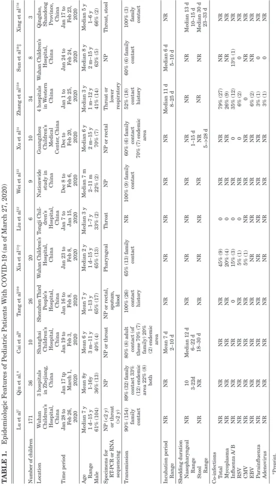
We found 11 case series, including a total of 333 children
(range 6–171 children) with confirmed SARS-CoV-2 infections
(Tables 1–4).
7–17All of the series are from China. One case series
included only infants
13and one only children who were admitted
to an intensive care unit.
16In 2 of the studies, there were patients
that overlapped
7,16and further duplicate reporting of patient could
not be excluded in 2 other studies.
7,11We did not include single
case reports,
18–23publications which did not give enough clinical
details
17,24–26or studies which were retracted.
27The age of the
chil-dren ranged from 1 day to 16 years, 55% (183) were male. The
majority of diagnoses were made by real-time polymerase chain
reaction on nasopharyngeal or other respiratory samples. Overall,
83% (275, range 52%–100%) of children had a positive contact
history, mostly with family members. Three studies reported
incu-bation periods which varied between 2 and 25 days (mean 7 days,
median 6 and 11 days, respectively).
9,15,16Several studies reported
that the nasopharyngeal or throat swabs can be positive before the
onset of symptoms.
7,11,14However, false-negative swabs have also
been described.
11There were 4 studies which did consecutive
sam-pling: real-time polymerase chain reaction on respiratory samples
remained positive between 1 and 22 days and in stool between 5
and over 30 days.
8,9,14,17Viral shedding from the gastrointestinal
tract might last longer and also be greater than that from the
res-piratory tract.
14Three studies investigated for co-infections (Table 1).
11,16One study only for influenza A and B, which was found in 1
of 8 children
16and the other 2 studies for a broader range of
pathogens, which were found in 45% and 79% of children.
11,15 P.Z. is supported by a Fellowship from the European Society of PaediatricInfec-tious Diseases.
The authors have no funding or conflicts of interest to disclose.
P.Z. drafted the initial manuscript. N.C. critically revised the manuscript and both authors approved the final manuscript as submitted.
Address for correspondence: Petra Zimmermann, MD, PhD, Faculty of Science and Medicine, University of Fribourg, Route des Arsenaux 41, 1700 Fri-bourg, Switzerland. E-mail: petra.zimmermann@unifr.ch.
COVID-19 in Children, Pregnancy and Neonates:
A Review of Epidemiologic and Clinical Features
Petra Zimmermann, MD, PhD*†‡ and Nigel Curtis, FRCPCH, PhD†‡§
http://doc.rero.ch
Published in "The Pediatric Infectious Disease Journal 39(6): 469–477, 2020"
which should be cited to refer to this work.
T
ABLE 1.
Epidemiologic F
eatures of P
ediatric P
atients
W
ith CO
VID-19 (as of Marc
h 27,
2020)
Lu et al 7 Qiu et al. 8 Cai et al 9 T ang et al 10* Xia et al 11† Liu et al 12 W ei et al 13 Xu et al 14 Zhang et al 15* Sun et al 16‡ Xing et al 17* Number of c hildren 1 7 1 3 6 1 0 2 6 2 0 6 9 1 0 3 4 8 3 Location W uhan Children’ s Hospital, China 3 hospitals in Zhejiang , China Shanghai Children’ s Hospital, China Shenzhen Third P eople’ s Hospital, China W uhan Children’ s Hospital, China T ongji Chil-dren’ s Hospital, China Nationwide study in China Guangzhou Children’ s Medical Center , China 4 hospitals in W estern China W uhan Children’ s Hospital, China Qingdao , Shandong Province , China Time period J a n 2 8 t o F e b 26, 2020 J an 17 tp Marc h 1 , 2020 J an 19 to F eb 3, 2020 J an 16 to F eb 8, 2020 J an 23 to F eb 8, 2020 J an 7 to Jan 15, 2020 Dec 8 to F eb 6, 2020 Dec to Feb 20, 2020 J an 1 to Feb 25, 2020 J an 24 to F eb 24, 2020 J an 17 to F e b 23, 2020 Age Median 7 y Mean 8y Mean 6 y Mean 7 y Median 2 y Median 3 y Median 7 m Median 6 y Median 3 y Median 8 y Median 5 y Range 1 d–15 y 1-16y 3 m–11 y 1–13 y 1 d–15 y 1–7 y 2–11 m 2 m–15 y 1 m–12 y 2 m–15 y 1–6 y Male 61% (104) 36% (13) 40% (4) 65% (17) 65% (13) 33% (2) 22% (2) 70% (7) 41% (14) 63% (5) 66% (2) Specimens for RT -PCR or RNA sequencing NP (<2 y) or throat (>2 y) NP NP or throat NP or rectal, sputum, blood Pharyngeal Throat NP NP or rectal Throat or lower respiratory NP Throat, stool Transmission 90% (154) family contact 89% (32) familycontact 33% (12) endemic area 22% (8)
both
80% (8) adult
contact (of
these 70% (7) family) 20% (2) endemic
area 100% (26) contact history 65% (13) family contact NR 100% (9) family contact 60% (6) family contact 70% (7) endemic area 52% (18) contact history 60% (6) family contact 100% (3) family contact Incubation period NR NR Mean 7 d N R N R N R N R N R Median 11 d Median 6 d N R Range 2–10 d 8–25 d 5–10 d
Shedding duration Nasopharyngeal
N R 1 0 Median 12 d 6–22 d 18–30 d NR NR NR NR NR NR NR NR NR Median 13 d Range 3-22d NR 1–15 d 10–15 d Median 30 d 23–33 d Stool NR NR NR NR NR NR Range 5–>28 d Co-infections T otal NR NR NR NR 45% (9) 0 N R N R 79% (27) NR NR Mycoplasma NR NR NR NR 20% (4) 0 N R N R 26% (9) NR NR Influenza A/ B N R N R N R 0 15% (3) 0 N R 0 35% (12) 13% (1) NR RSV NR NR NR NR 5% (1) 0 N R 0 6% (2) 0 N R CMV NR NR NR NR 5% (1) NR NR NR 0 N R N R EBV NR NR NR NR NR NR NR NR 6% (2) NR NR P arainfluenza NR NR NR NR NR NR NR NR 3% (1) 0 N R Adenovirus NR NR NR NR NR NR NR NR 3% (1) 0 N R *Preprint. †P
atients in this study are possibly also reported in the study by Lu et al.
7
‡Three patients overlap with the study by Lu et al.
7 CMV , cytomegalovirus; EBV , Epstein-Barr virus; NR, not reported; NP , nasopharyngeal; RSV
, respiratory syncytial virus;
RT
-PCR,
real-time polymerase c
hain reaction.
Mycoplasma (20%, 26%) and influenza A and B (15%, 35%)
were the most common co-infections, followed by
respira-tory syncytial virus (5%, 6%) and Epstein-Barr virus (6%).
Cytomegalovirus, parainfluenza and adenovirus were also
iso-lated.
11,15Depending on the study design, up to 35% of children were
asymptomatic (Table 2). The most common symptoms were cough
in 48% (160, 19%–100%), fever in 42% (140, 11%–100%, mean
duration 3–6 days, range 1–16 days) and pharyngitis in 30% (99,
11%–100%). Further symptoms were tachypnoea (0%–100%),
nasal congestions (0%–30%), rhinorrhea (0%–20%), wheezing
(33%), diarrhea (8%–23%), vomiting (8%–50%), headache (8%–
13%) and fatigue (8%–13%).
Typical laboratory findings were minor changes in white
blood cell counts (reports of both increased and decreased
lym-phocyte and, less commonly, neutrophil counts), as well as mildly
elevated inflammatory markers (erythrocyte sedimentation rate,
C-reactive protein or procalcitonin), liver enzymes, creatine kinase,
lactate dehydrogenase or D-dimers (Table 3).
Radiologic findings were unspecific and milder compared
with those in adults.
28They included unilateral or bilateral
infil-trates on chest radiograph or computer tomography and,
some-times, additional ground-glass opacities or consolidations with a
surrounding halo sign in the latter (Table 3).
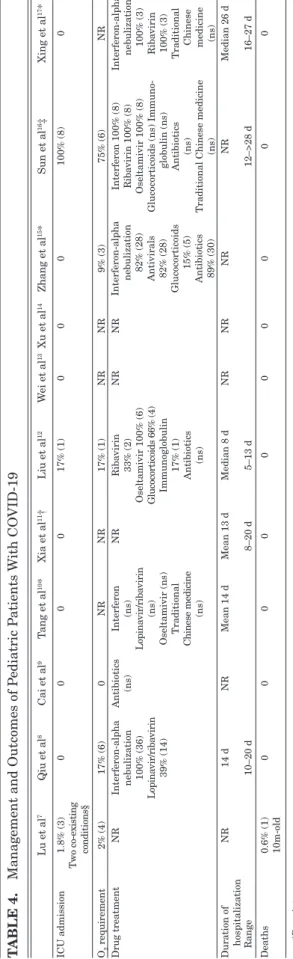
Twenty (6%) children were reported to require
oxy-gen (Table 4). Other treatments used were oseltamivir, ribavirin
(±lopinavir), interferon, glucocorticoids, immunoglobulin,
anti-biotics and traditional Chinese medicine.
8,10,12,15–17The hospital
stays ranged from 5 to more than 28 days with means of 13–14
days.
8,10–12,16Nine children (3%) needed admission to an intensive care
unit
7,12,16(there was an overlap of the reporting of 3 patients between
2 studies).
7,16Of these 9 children, only 2 were described to have a
preexisting condition (leukemia and hydronephrosis, respectively).
A 10-month-old girl admitted to an intensive care unit developed
intussusception, encephalopathy, septic shock and multiple organ
dysfunction, and died.
7A further death due to COVID-19 of a
14-year-old boy has been reported in an epidemiologic study from
China
29and further deaths have now been reported in Europe and
the USA.
SARS-COV-2 INFECTION DURING PREGNANCY,
VERTICAL TRANSMISSION AND PERINATAL
OUTCOMES
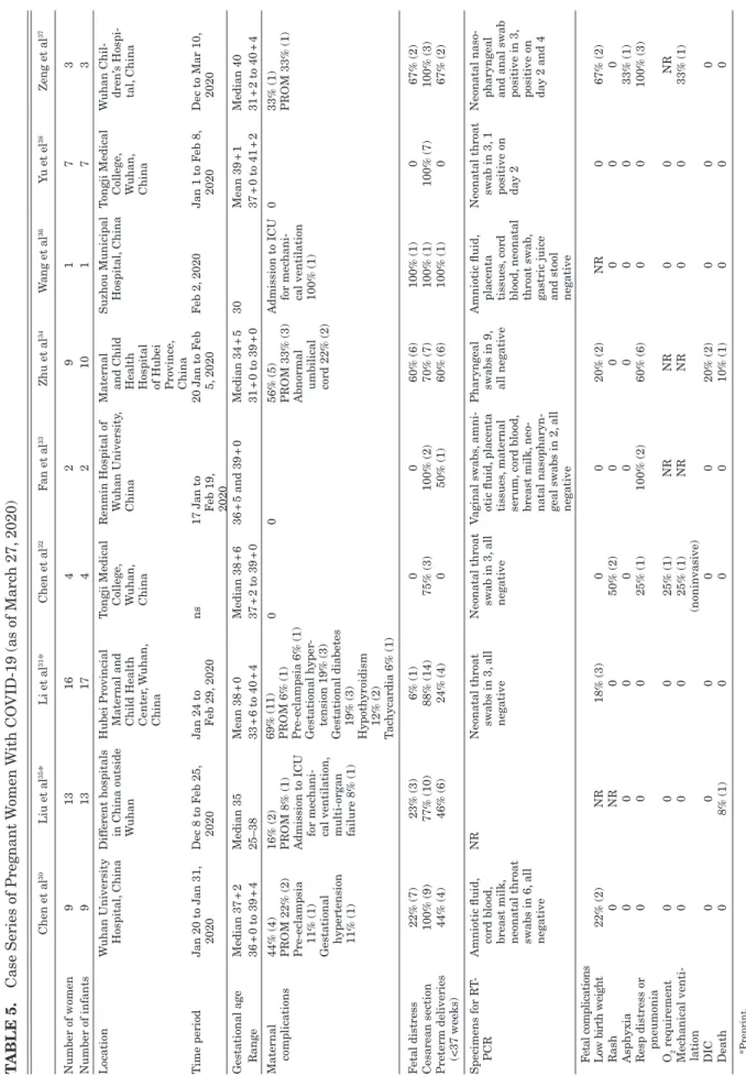
There are 9 small case series (all from China) and 2 case
reports including a total of 65 pregnant women (67 neonates) who
were infected with SARS-CoV-2 during pregnancy (Table 5).
30–39The number of women in each case series varied between 2 and
16 (median 7). Two women were infected at 25 and 27 weeks of
pregnancy, the remaining during the third trimester. Three women
were discharged, the remaining delivered between 30 and 40 weeks
of pregnancy, mostly by Cesarean section 88% (56). Fetal distress
was reported in 31% (20). A total of 38% (724) women delivered
preterm. Maternal complications included premature rupture of
membranes 12% (8), pre-eclampsia 3% (2), gestational
hyper-tension 6% (4), gestational diabetes 5% (3), hypothyroidism 3%
(2), tachycardia 2% (1) and abnormal umbilical cord 3% (2). Two
women (3%) were admitted to intensive care unit for
mechani-cal ventilation, one of whom developed multi-organ failure and
was still on extracorporeal membrane oxygenation at the time of
the publication.
35,36Neonatal complications included respiratory
distress or pneumonia 18% (12), low birth weight 13% (9), rash
3% (2), disseminated intravascular coagulation 3% (2), asphyxia
2% (1) and perinatal death 3% (2).
34,36SARS-CoV-2 could not be
isolated from amniotic fluid, placenta tissue, vaginal swabs, cord
blood or breast milk, or from neonatal nasopharyngeal and throat
swabs in 27 mother-infant pairs.
30–36,38,39However, 1 healthy neonate
and 3 neonates who developed pneumonia tested positive on throat,
nasopharyngeal and anal swabs on days 2 and 4 of life.
37This was
despite strict infection control and prevention procedures during
delivery and separation of mother and neonates. Additionally, three
neonates whose mother presented with COVID-19 infection 23
days before delivery were found to have immunoglobulin M and G
against SARS-CoV-2 at birth.
39,40Therefore, vertical transmission
could not be excluded.
DISCUSSION
This review confirms that, compared with adults, children
with SARS-CoV-2 infection have milder clinical symptoms and
fewer laboratory and radiologic abnormalities. The same findings
TABLE 2. Clinical Symptoms of Pediatric Patients With COVID-19
Lu et al7 Qiu et al8 Cai et al9 Tang et al10* Xia et al11† Liu et al12 Wei et al13 Xu et al14 Zhang et al15* Sun et al16‡ Xing et al17*
Asymptomatic 23% (39) (12/39 radiologic pneumonia) 28% (10) 0 35% (9) 10% (2) 0 11% (1) 10% (1) 0 0 NR Fever 32% (55) 11% (4) 70% (7) 42% (11) 60% (12) 100% (6) 44% (4) 60% (6) 76% (26) 75% (6) 100% (3) Definition >38°C >38.5°C ≥38°C ns >37.3°C >39°C ns >38°C ns ns >38.5°C Median duration 3 d 3 d 1 d 6 d Range 1–16 d 2–5 d 3–11 d Cough 49% (83) 19% (7) 60% (6) 46% (12) 65% (13) 100% (6) 22% (2) 50% (5) 59% (20) 75% (6) NR Pharyngitis 46% (79) 11% (4) 40% (4) 0 5% (1) 100% (6) 0 40% (4) 0 13% (1) NR Nasal congestion 5% (9) NR 30% (3) 0 0 0 0 20% (2) 0 0 NR Rhinorrhea 8% (13) NR 20% (2) 8% (2) 15% (3) 17% (1) 0 20% (2) 0 0 NR Tachypnoea 29% (49) 3% (1) 0 0 10% (2) 17% (1) 0 0 9% (3) 100% (8) NR Wheezing 0 0 0 0 33% (2) 0 0 0 0 NR Diarrhea 9% (15) 6% (2) 0 8% (2) 15% (3) 0 0 30% (3) 12% (4) 38% (3) NR Vomiting 0 6% (2) 0 8% (2) 10% (2) 67% (4) 0 0 12% (4) 50% (4) NR Headache NR 8% (3) NR NR NR NR NR NR NR 13% (1) NR Fatigue 8% (13) NR NR NR 5% (1) NR NR NR NR 13% (1) NR *Preprint.
†Patients of this study are possibly also reported in the study by Lu et al.7
‡Three patients overlaps with the study by Lu et al.7
NR, not reported; ns, not specified.
T
ABLE 3.
Laboratory and Radiologic F
indings of P
ediatric P
atients
W
ith CO
VID-19
Lu et al 7 Qiu et al 8 Cai et al 9 T ang et al 10 * Xia et al 11† Liu et al 12 W ei et al 13 Xu et al 14 Zhang et al 15* Sun et al 16‡ Xing et al 17 * Laboratory findings Leucocytosis 0 N R 20% (2) (>12 G/L) 15% (4) (ns) 10% (2) (>12 G/L) 0 N R 0 NR 13% (1) (>12 G/L) 0 Leukopenia 26% (45) (<5.5 G/L) 19% (7) (<4 G/L) 20% (2) (<5.5 G/L) 50% (13) (ns) 20% (5) (<5.5 G/L) 83% (5) (<5.5 G/L) NR 30% (3) (<5.5 G/L) NR 13% (1) (<5.5 G/L) 0 Neutrophilia 0 N R 10% (1) (>7 G/L) NR NR 0 N R 10% (1) (>7 G/L) NR 13% (1) (>7 G/L) 0 Neutropenia 0 N R 20% (2) (<1.5 G/L) NR NR 50% (3) (<1.5 G/L) NR 10% (1) (< 1.5 G/L) NR 26% (2) (<1.5 G/L) 33% (1) (<1.5 G/L) L ymphocytosis 0 N R 20% (2) (>4 G/L) NR§ 15% (3) (>65%) 0 N R 20% (2) (>4 G/L) 50% (17) (ns) 26% (2) (>4 G/L) 100% (3) (>4 G/L) L ymphopenia 4% (6) (<1.2 G/L) 31% (11) (<1.1 G/L) 0 NR§ 35% (7) (>45%) 100% (6) (<1.8 G/L) NR 40% (4) (<1.6 G/L) NR 13% (1) (>1.6 G/L) 0 Thrombocytosis NR NR 20% (2) (>350 G/L) Abnormal in 31% (8) NR 0 N R 0 NR 25% (2) (>350 G/L) 67% (2) (>350 G/L) Thrombopenia NR NR 10% (1) (<150 G/L) Abnormal in 31% (8) NR 0 N R 10% (1) (<150 G/L) NR 38% (3) (<150 G/L) 0 Elevated ESR NR 0 N R 27% (7) (>15 mm/h) NR 33% (2) (>20 mm/h) NR 30% (3) (>15 mm/h) NR 0 0 Elevated CRP 20% (33) (>10 mg/L) 3% (1) (>8 mg/L) 20% (2) (>10 mg/L) 19% (5) (>5 mg/L) 35% (7) (>3 mg/L) 83% (5) (>10 mg/L) NR 30% (3) (>5 mg/L) NR 63% (5) (>5 mg/L) 33% (1) (>10 mg/L) Elevated PCT 64% (105) (>0.05 ng/mL) 17% (6) (>0.5 ng/mL) 0 0 80% (16) (>0.05 ng/mL) NR NR 50% (5) (>0.1 ng/mL) NR 63% (5) (>0.05 ng/mL) 33% (1) (>0.1 ng/mL) Elevated ALA T 12% (21) (>45 U/L) 6% (2) (>40 U/L) 10% (1) (>45 U/L) 12% (3) (>45 U/L) 25% (5) (>40 U/L) 17% (1) (>40 U/L) NR 10% (1) (>45 U/L) NR 50% (4) (>45 U/L) NR Elevated ASA T 15% (25) (>50 U/L) 8% (3) (>40 U/L) 20% (2) (>45 U/L) 12% (3) (>45 U/L) NR 67% (4) (>40 U/L) NR 20% (2) (>45 U/L) NR 0 N R Elevated CK NR 3% (1) (>170 U/L) NR 0 N R 0 NR 0 N R 25% (2) (>170 U/L) 0 Elevated CK-MB NR 31% (11) (>18 U/L) 50% (5) (>25 U/L) NR 75% (15) (>25 U/L) NR NR NR NR NR NR Elevated LDH 0 N R 30% (3) (>300 U/L) 46% (12) (>250 U/L) NR 50% (3) (>300 U/L) NR 20% (2) (>300 U/L) 82% (28) (ns) 63% (5) (>300 U/L) 33% (1) (>250 U/L) Elevated D-dimers 14% (21) (>0.6 mg/L) 8% (3) (>0.5 mg/L) 0 N R N R 50% (3) (>0.6 mg/L) NR 10% (1) (>0.5 μ g/L¶) NR 25% (2) (>0.6 mg/L) 33% (1) (>0.6 mg/L) Prolonged PT 0 N R N R N R N R 0 NR 0 N R N R N R Chest CT findings Normal 0 N R N R 0 20% (4) 17% (1) NR 50% (5) 18% (6) 0 33% (1) Ground-glass opacities 33% (56) 53% (19) NR 31% (8) 60% (12) 33% (2) NR 50% (5) 3% (1) 75% (6) 33% (1) Unilateral infiltrates 19% (32) NR NR 42% (11) 30% (6) 0 N R N R 41% (14) 25% (2) 33% (1) Bilateral infiltrates 12% (21) NR NR 27% (7) 50% (19) 50% (3) NR NR 41% (14) 75% (6) 0 Interstitial abnormalities 2% (2) NR NR 0 0 0 N R N R 0 0 0Consolidation with surroundding halo sign
0 N R N R 0 50% (10) 0 N R N R 0 0 0 Nodules 0 N R N R 0 15% (3) 0 N R N R 0 0 0 Pleural effusion 0 N R N R 0 0 0 NR NR 0 13% (1) 0 ‘White-lung’ 0 N R N R 0 0 0 NR NR 0 13% (1) 0 *Preprint. †P
atients in this study are possibly also reported in the study by Lu et al.
7
‡Three patients overlap with the study by Lu et al.
7
§Specified norm values incorrect. ¶Likely the wrong unit w
as specified. ALA T , alanine aminotransferase; ASA T , aspartate aminotransferase; CRP , C-reactive protein; CK, creatinine kinase; CT , computer tomography; ESR,
erythrocyte sedimentation rate;
LDH, lactate dehydrogenase; NR, not reported; ns , not specified; PCT , procalcitonin; PT , prothrombin time .
http://doc.rero.ch
have previously been reported for SARS- and Middle East
respira-tory syndrome (MERS)-CoV.
41–46There are several hypotheses for why children infected
with SARS-CoV-2 have less severe symptoms (Table 6). One
potential explanation is differences in the immune system
between children and adults, especially elderly adults.
47Mice
models of infections with SARS-CoV show that both CD4 and
CD8 T cells, as well as antibodies, play an important role in virus
clearance.
48–50Children have a stronger innate immune response,
higher proportion of total lymphocytes and absolute numbers of
T and B cells, as well as natural killer cells, which might help to
fight the virus.
51However, children are often described to have
an ‘immature’ immune system and, for infections with other
res-piratory tract viruses, for example, resres-piratory syncytial virus
or influenza, infants and children are at higher risk for serious
disease and hospital admission.
52This suggests that protective
immunity against SARS-CoV-2 differs to that against other
com-mon respiratory viruses.
Furthermore, children have a less proinflammatory cytokine
response and are less prone to develop acute respiratory distress
syndrome.
51,53It is therefore possible that the cytokine storm which
plays an important role in the pathogenesis of severe COVID-19 in
adults, is attenuated in this age group.
54The second factor that may contribute to the reduced severity
of COVID-19 is the lower prevalence in children of the
co-morbidi-ties that have been associated with severe disease, such as diabetes,
chronic lung, heart and kidney problems or arterial hypertension.
55The third potential explanation for the milder symptoms
of SARS-CoV-2 infections in children is that common
circulat-ing coronaviruses are frequent in this age group, responsible for
approximately 8% of acute respiratory tract infections.
56–58Pre-existing immunity and cross-reacting antibodies to SARS-CoV-2
may play a protective role. Despite the fact that most individuals
develop antibodies to common circulation coronaviruses during
childhood,
59–62reinfections later in life occur,
56,63,64suggesting
wan-ing immunity against coronaviruses and increased susceptibility in
adults.
The fourth potential explanation is the higher mucosal
colo-nization by viruses and bacteria, which could limit colocolo-nization and
growth of SARS-CoV-2 through microbial interactions and
com-petition.
65,66A fifth hypothesis for the less severe symptoms in children is
that children are usually infected by an adult, which means that they
are infected by a second or third generation of the virus. For SARS-
and MERS-CoV, these following generations have been described
to have decreased pathogenicity.
67,68The sixth potential explanation related to
angiotensin-con-verting enzyme 2 (ACE2) receptors that are one of the main
recep-tors for the entry of SARS- and SARS-CoV-2 into human cells.
69,70It has been suggested that adults who are taking ACE inhibitors
or angiotensin receptor blockers for arterial hypertension might
have a higher number of ACE2 receptors, potential making them
more susceptible to SARS-CoV-2.
71,72However, this theory remains
controversial.
73It has been postulated that children have less ACE2
receptors with lower affinity compared with adults and therefore
might be less affected by SARS-CoV-2.
74ACE2 is important in
regulating the immune response, especially in the lungs. In
ani-mal studies, it has been shown to protect against SARS-CoV- and
influenza-associated lung injury.
75–77For Pseudomonal lung
infec-tions, it has been shown that a dynamic variation of pulmonary
ACE2 is required for protection against lung injury.
78The
interac-tion between ACE2 concentrainterac-tion and the number and affinity of
ACE2 receptor is likely complex and might also be influenced by
genetics.
79,80T
ABLE 4.
Management and Outcomes of P
ediatric P
atients
W
ith CO
VID-19
Lu et al 7 Qiu et al 8 Cai et al 9 T ang et al 10* Xia et al 11† Liu et al 12 W ei et al 13 Xu et al 14 Zhang et al 15* Sun et al 16‡ Xing et al 17* ICU admission 1.8% (3) Two co-existing conditions§ 0 0 0 0 17% (1) 0 0 0 100% (8) 0 O2 requirement 2% (4) 17% (6) 0 N R N R 17% (1) NR NR 9% (3) 75% (6) NR Drug treatment NR Interferon-alpha nebulization 100% (36) Lopina vir/riba virin 39% (14) Antibiotics (ns) Interferon (ns) Lopina vir/riba virin (ns) Oseltamivir (ns) Traditional Chinese medicine (ns) NR Riba virin 33% (2) Oseltamivir 100% (6) Glucocorticoids 66% (4) Immunoglobulin 17% (1) Antibiotics (ns) NR NR Interferon-alpha nebulization 82% (28) Antivirals 82% (28) Glucocorticoids 15% (5) Antibiotics 89% (30) Interferon 100% (8) Riba virin 100% (8) Oseltamivir 100% (8) Glucocorticoids (ns) Immuno-globulin (ns) Antibiotics (ns)Traditional Chinese medicine
(ns) Interferon-alpha nebulization 100% (3) Riba virin 100% (3) Traditional Chinese medicine (ns) Duration of hospitalization NR 14 d N R Mean 14 d Mean 13 d Median 8 d N R N R N R N R Median 26 d Range 10–20 d 8–20 d 5–13 d 12–>28 d 16–27 d Deaths 0.6% (1) 10m-old 00 0 0 0 0 0 0 0 0 *Preprint. †P
atients of this study are possibly also reported in the study by Lu et al.
7
‡Three patients overlaps with the study by Lu et al.
7 §Hydronephrosis 1, leukemia on maintenance c hemotherapy 1. NR, not reported; NP , nasopharyngeal; ns , not specified.
http://doc.rero.ch
T
ABLE 5.
Case Series of Pregnant
W
omen
W
ith CO
VID-19 (as of Marc
h 27,
2020)
Chen et al 30 Liu et al 35* Li et al 31* Chen et al 32 F an et al 33 Zhu et al 34 W ang et al 36 Y u et el 38 Zeng et al 37 Number of women 9 1 3 1 6 4 2 9 1 7 3 Number of infants 9 1 3 1 7 4 2 1 0 1 7 3 Location Wuhan University Hospital,
China
Different hospitals
in China outside Wuhan
Hubei Provincial
Maternal and Child Health Center
, W
uhan,
China
T
ongji Medical College
, W uhan, China Renmin Hospital of W uhan University , China Maternal
and Child Health Hospital of Hubei Province
, China Suzhou Municipal Hospital, China T
ongji Medical College
,
W
uhan,
China
W
uhan Chil- dren’
s Hospi-tal, China Time period J an 20 to J an 31, 2020 Dec 8 to F eb 25, 2020 J an 24 to F eb 29, 2020 ns 17 J an to F eb 19, 2020 20 J an to F eb 5, 2020 F eb 2, 2020 J an 1 to F eb 8, 2020 Dec to Mar 10, 2020 Gestational age Median 37 + 2 Median 35 Mean 38 + 0 Median 38 + 6 36 + 5 and 39 + 0 Median 34 + 5 3 0 Mean 39 + 1 Median 40 Range 36 + 0 to 39 + 4 25–38 33 + 6 to 40 + 4 37 + 2 to 39 + 0 31 + 0 to 39 + 0 37 + 0 to 41 + 2 31 + 2 to 40 + 4 Maternal complications 44% (4) PROM 22% (2) Pre-ec lampsia 11% (1) Gestational hypertension 11% (1) 16% (2) PROM 8% (1) Admission to ICU
for mec
hani-cal ventilation, multi-organ failure 8% (1) 69% (11) PROM 6% (1) Pre-ec lampsia 6% (1) Gestational hyper -tension 19% (3) Gestational diabetes 19% (3) Hypothyroidism 12% (2) T a chycardia 6% (1) 0 0 56% (5) PROM 33% (3) Abnormal umbilical cord 22% (2) Admission to ICU for mec hani-cal ventilation 100% (1) 0 33% (1) PROM 33% (1) F etal distress 22% (7) 23% (3) 6% (1) 0 0 60% (6) 100% (1) 0 67% (2) Cesarean section 100% (9) 77% (10) 88% (14) 75% (3) 100% (2) 70% (7) 100% (1) 100% (7) 100% (3) Preterm deliveries (<37 weeks) 44% (4) 46% (6) 24% (4) 0 50% (1) 60% (6) 100% (1) 0 67% (2) Specimens for RT -PCR Amniotic fluid,
cord blood, breast milk, neonatal throat sw
abs in 6, all negative NR Neonatal throat sw abs in 3, all negative Neonatal throat sw ab in 3, all negative V aginal sw abs , amni-otic fluid, placenta tissues , maternal serum, cord blood, breast milk,
neo-natal nasopharyn- geal sw
abs in 2, all negative Pharyngeal sw abs in 9, all negative Amniotic fluid, placenta tissues , cord blood, neonatal throat sw ab ,
gastric juice and stool negative
Neonatal throat sw ab in 3, 1 positive on da y 2 Neonatal
naso-pharyngeal and anal sw
ab positive in 3, positive on da y 2 and 4 F etal complications
Low birth weight
22% (2) NR 18% (3) 0 0 20% (2) NR 0 67% (2) Rash 0 N R 0 50% (2) 0 0 0 0 0 Asphyxia 0 0 0 0 0 0 0 0 33% (1)
Resp distress or pneumonia
0 0 0 25% (1) 100% (2) 60% (6) 0 0 100% (3) O2 requirement 0 0 0 25% (1) NR NR 0 0 NR Mec hanical venti-lation 0 0 0 25% (1) (noninvasive) NR NR 0 0 33% (1) DIC 0 0 0 0 0 20% (2) 0 0 0 Death 0 8% (1) 0 0 0 10% (1) 0 0 0 *Preprint. DIC , disseminated intra vascular coagulation; ICU
, intensive care unit;
ns
, not specified;
PROM,
premature rupture of membranes;
RT
-PCR,
real-time polymerase c
hain reaction.
The majority of children included in this review had a
reported adult or family contact infected with SARS-CoV-2. It is
still uncertain whether asymptomatic children transmit the virus
and therefore the role of children as a reservoir for SARS-CoV-2
and for transmission of the virus remains unclear. However, it has
been reported that even asymptomatic children can have high viral
loads of SARS-CoV-2
20and can excrete the virus in stool for a
pro-longed period.
9,14,17Unpublished data suggests that the clinical features of
COVID-19 in children varies in different countries. While in Asian
countries and Europe children have been reported to have milder
disease, recent data from the US reports that, by March 27, 2020,
at least 35 children needed mechanical ventilation and one infant
died. It has been suggested that this could be due to differences
in Bacillus-Calmette-Guérin vaccination policies, as this vaccine’s
off-target immunomodulatory effects might alter the immune
response to SARS-CoV-2.
81–83The influence of SARS-CoV-2 infection on pregnancy and
neonatal outcomes is also unclear. SARS- and MERS-CoV cause
more severe disease in pregnant women compared with
non-preg-nant women.
84,85To date, this has not been reported for
SARS-CoV-2.
25,86Nevertheless, 3% of pregnant women infected were
admitted to intensive care unit.
35,36There is no evidence that
SARS-CoV or MERS-SARS-CoV can be vertically transmitted to the fetus,
however, maternal infections have been associated with
intrauter-ine growth retardation, preterm delivery, stillbirths and perinatal
deaths.
85,87–91Similarly, low birth weight, preterm delivery and 2
perinatal deaths have been reported in association with
SARS-CoV-2.
30,31,33–37It is unclear if some of the reported maternal and
neonatal complications are due to the virus or were iatrogenic (eg,
decision for a Cesarean leading to preterm delivery and neonatal
respiratory problems). Nevertheless, 1 case-control study reported
that the number of pre-term deliveries were higher in
SARS-CoV-2-infected women compared with non-infected women.
31Further-more, fetal distress and preterm ruptures of membranes have been
reported in SARS-CoV-2 infected women.
30,31,34,37The one healthy neonate and 3 neonates who developed
pneumonia and tested positive for SARS-CoV-2 on day 2 of life
and the three neonates who had immunoglobulin M against
SARS-CoV-2 at birth, despite strict infection control and prevention
pro-cedures during delivery and separation of mother and infants,
sug-gests the possibility of vertical transmission of SARS-CoV-2.
37–40There is no evidence for the presence of SARS-CoV-2 in
genital fluids.
33However, the virus can be isolated from feces,
meaning it is possible that vaginal delivery poses a greater risk for
infection of the infant. Most of the women delivered by Cesarean
section as recommended in Chinese guidelines. It is still unclear
whether the virus can be transmitted through breast milk. However,
close contact during breast-feeding, might risk droplet or contact
transmission from the mother to the neonate.
REFERENCES
1. Zhu N, Zhang D, Wang W, et al. A novel coronavirus from patients with pneumonia in China, 2019. N Engl J Med. 2020;382:727–733.
2. WHO. Coronavirus disease 2019 (COVID-19) Situation Report - 11. https://www.who.int/docs/default-source/coronavir use/situation-reports/20200131-sitrep-11-ncov.pdf?sfvrsn=de7c0f7_4. Published 31 January 2020. Accessed March 23, 2020.
3. Wu Z, McGoogan JM. Characteristics of and important lessons from the coronavirus disease 2019 (COVID-19) outbreak in China: summary of a report of 72 314 cases from the Chinese center for disease control and pre-vention. JAMA. 2020.
4. Bi Q, Wu Y, Mei S, et al. Epidemiology and transmission of COVID-19 in Shenzhen China: analysis of 391 cases and 1,286 of their close contacts.
medRxiv. 2020. https://doi.org/10.1101/2020.03.03.20028423
5. Zimmermann P, Curtis N. Coronavirus infections in children including COVID-19: an overview of the epidemiology, clinical features, diagno-sis, treatment and prevention options in children. The Pediatric Infectious
Disease Journal. 2020.
6. Guan WJ, Ni ZY, Hu Y, et al. Clinical characteristics of coronavirus disease 2019 in China. N Engl J Med. 2020.
7. Lu X, Zhang L, Du H, et al. SARS-CoV-2 infection in children. N Engl J
Med. 2020.
8. Qiu H, Wu J, Hong L, et al. Clinical and epidemiological features of 36 children with coronavirus disease 2019 (COVID-19) in Zhejiang, China: an observational cohort study. The Lancet Infectious Diseases.
9. Cai J, Xu J, Lin D, et al. A case series of children with 2019 novel coro-navirus infection: clinical and epidemiological features. Clin Infect Dis. 2020; pii:ciaa198.
10. Tang A, Xu W, shen m, et al. A retrospective study of the clinical charac-teristics of COVID-19 infection in 26 children. medRxiv. 2020. https://doi. org/10.1101/2020.03.08.20029710
11. Xia W, Shao J, Guo Y, et al. Clinical and CT features in pediatric patients with COVID-19 infection: different points from adults. Pediatr Pulmonol. 2020. 12. Liu W, Zhang Q, Chen J, et al. Detection of covid-19 in children in early
january 2020 in Wuhan, China. N Engl J Med. 2020.
13. Wei M, Yuan J, Liu Y, et al. Novel coronavirus infection in hospitalized infants under 1 year of age in China. JAMA. 2020.
14. Xu Y, Li X, Zhu B, et al. Characteristics of pediatric SARS-CoV-2 infection and potential evidence for persistent fecal viral shedding. Nature Medicine. 2020. 15. Zhang C, Gu J, Chen Q, et al. Clinical characteristics of 34 children with
coronavirus disease-2019 in the West of China: a multiple-center case series. medRxiv. 2020. https://doi.org/10.1101/2020.03.12.20034686 16. Sun D, Li H, Lu XX, et al. Clinical features of severe pediatric patients with
coronavirus disease 2019 in Wuhan: a single center’s observational study.
World J Pediatr. 2020.
17. Xing Y, Ni W, Wu Q, et al. Prolonged presence of SARS-CoV-2 in feces of pediatric patients during the convalescent phase. medRxiv. 2020. https://doi. org/10.1101/2020.03.11.20033159
TABLE 6. Hypotheses Suggested to Date for Why
Children Infected With SARS-CoV-2 Have Less Severe
Symptoms
Hypothesis Details
1. Differences in the immune system
Children have stronger innate immune response, higher
proportion of total lymphocytes, absolute numbers of T and B and NK cells and lower proinflammatory cytokine responses
2. Lower prevalence of co-morbidities
Children have lower prevalence of diabetes, chronic lung, heart and kidney problems, arterial hypertension
3. Differences in pathogen exposure, e.g. higher prevalence of infections with common corona-viruses
Children are more likely to have preexisting immunity to common coronaviruses, includ-ing potential cross-reactinclud-ing antibodies to SARS-CoV-2
4. Microbial interactions and competition limit-ing colonization and growth of SARS-CoV-2
Children have higher mucosal colonization by viruses and bacteria
5. Infection with second or third generation of virus might have decreased pathogenic-ity
Children predominantly infected by transmis-sion from adults
6. Differences in ACE2 receptors
Children might have less ACE2 receptors with lower affinity
7. Protection through off-target effects of BCG vaccination
Possible correlation between BCG vaccination policies and severity of COVID-19 in children
ACE2, angiotensin-converting enzyme 2; NK, natural killer; SARS-CoV-2, severe acute respiratory syndrome coronavirus 2; BCG, Bacillus-Calmette-Guérin.
18. Cui Y, Tian M, Huang D, et al. A 55-Day-Old Female Infant infected with COVID 19: presenting with pneumonia, liver injury, and heart damage. J
Infect Dis. 2020 pii:jiaa113
19. Ji LN, Chao S, Wang YJ, et al. Clinical features of pediatric patients with COVID-19: a report of two family cluster cases. World J Pediatr. 2020. 20. Kam KQ, Yung CF, Cui L, et al. A well infant with coronavirus disease 2019
(COVID-19) with high viral load. Clin Infect Dis. 2020;pii:ciaa201. 21. Chan JF, Yuan S, Kok KH, et al. A familial cluster of pneumonia associated
with the 2019 novel coronavirus indicating person-to-person transmission: a study of a family cluster. Lancet. 2020.
22. Zhang YH, Lin DJ, Xiao MF, et al. [2019 novel coronavirus infection in a three-month-old baby]. Zhonghua Er Ke Za Zhi. 2020;58:182–184. 23. Cai JH, Wang XS, Ge YL, et al. [First case of 2019 novel coronavirus
infec-tion in children in Shanghai]. Zhonghua Er Ke Za Zhi. 2020;58:E002. 24. Lou XX, Shi CX, Zhou CC, et al. Three children who recovered from novel
coronavirus 2019 pneumonia. J Paediatr Child Health. 2020.
25. Liu H, Liu F, Li J, et al. Clinical and CT imaging features of the COVID-19 pneumonia: focus on pregnant women and children. J Infect. 2020;pii:S0163-4453(20)30118-3.
26. Xu XW, Wu XX, Jiang XG, et al. Clinical findings in a group of patients infected with the 2019 novel coronavirus (SARS-Cov-2) outside of Wuhan, China: retrospective case series. BMJ. 2020;368:m606.
27. Wang XF, Yuan J, Zheng YJ, et al. [Retracted: clinical and epidemiologi-cal characteristics of 34 children with 2019 novel coronavirus infection in Shenzhen]. Zhonghua Er Ke Za Zhi. 2020;58:E008.
28. Feng K, Yun YX, Wang XF, et al. [Analysis of CT features of 15 Children with 2019 novel coronavirus infection]. Zhonghua Er Ke Za Zhi. 2020;58:E007.
29. Dong Y, Mo X, Hu Y, et al. Epidemiological characteristics of 2143 pediatric patients with 2019 coronavirus disease in China. Pediatrics. 2020. 30. Chen H, Guo J, Wang C, et al. Clinical characteristics and intrauterine
verti-cal transmission potential of COVID-19 infection in nine pregnant women: a retrospective review of medical records. The Lancet.
31. Li N, Han L, Peng M, et al. Maternal and neonatal outcomes of pregnant women with COVID-19 pneumonia: a case-control study. medRxiv. 2020. https://doi.org/10.1101/2020.03.10.20033605
32. Chen Y, Peng H, Wang L, et al. Infants Born to Mothers With a New Coronavirus (COVID-19). Frontiers in pediatrics. 2020;8:104.
33. Fan C, Lei D, Fang C, et al. Perinatal transmission of COVID-19 associated SARS-CoV-2: should we worry? Clin Infect Dis. 2020.
34. Zhu H, Wang L, Fang C, et al. Clinical analysis of 10 neonates born to moth-ers with 2019-nCoV pneumonia. Transl Pediatr. 2020;9:51–60.
35. Liu Y, Chen H, Tang K, et al. Clinical manifestations and outcome of SARS-CoV-2 infection during pregnancy. J Infect. 2020.
36. Wang X, Zhou Z, Zhang J, et al. A case of 2019 Novel Coronavirus in a pregnant woman with preterm delivery. Clin Infect Dis. 2020.
37. Zeng L, Xia S, Yuan W, et al. Neonatal early-onset infection with SARS-CoV-2 in 33 neonates born to mothers with COVID-19 in Wuhan, China.
JAMA pediatrics. 2020.
38. Yu N, Li W, Kang Q, et al. Clinical features and obstetric and neonatal outcomes of pregnant patients with COVID-19 in Wuhan, China: a retro-spective, single-centre, descriptive study. The Lancet Infectious Diseases. 2020; pii:S1473-3099(20)30176-6
39. Dong L, Tian J, He S, et al. Possible vertical transmission of SARS-CoV-2 from an infected mother to her newborn. JAMA. 2020.
40. Zeng H, Xu C, Fan J, et al. Antibodies in infants born to mothers with COVID-19 pneumonia. JAMA. 2020.
41. Hon KL, Leung CW, Cheng WT, et al. Clinical presentations and outcome of severe acute respiratory syndrome in children. Lancet. 2003;361:1701– 1703.
42. Chiu WK, Cheung PC, Ng KL, et al. Severe acute respiratory syndrome in children: experience in a regional hospital in Hong Kong. Pediatr Crit Care
Med. 2003;4:279–283.
43. Bitnun A, Allen U, Heurter H, et al; Other Members of the Hospital for Sick Children SARS Investigation Team. Children hospitalized with severe acute respiratory syndrome-related illness in Toronto. Pediatrics. 2003;112:e261. 44. Leung CW, Kwan YW, Ko PW, et al. Severe acute respiratory syndrome
among children. Pediatrics. 2004;113:e535–e543.
45. Al-Tawfiq JA, Kattan RF, Memish ZA. Middle East respiratory syndrome coronavirus disease is rare in children: an update from Saudi Arabia. World
J Clin Pediatr. 2016;5:391–396.
46. Alfaraj SH, Al-Tawfiq JA, Altuwaijri TA, et al. Middle East respiratory syn-drome coronavirus in pediatrics: a report of seven cases from Saudi Arabia.
Front Med. 2019;13:126–130.
47. Simon AK, Hollander GA, McMichael A. Evolution of the immune system in humans from infancy to old age. Proc Biol Sci. 2015;282:20143085. 48. Zhao J, Zhao J, Perlman S. T cell responses are required for protection from
clinical disease and for virus clearance in severe acute respiratory syndrome coronavirus-infected mice. J Virol. 2010;84:9318–9325.
49. Chen J, Lau YF, Lamirande EW, et al. Cellular immune responses to severe acute respiratory syndrome coronavirus (SARS-CoV) infection in senescent BALB/c mice: CD4+ T cells are important in control of SARS-CoV infec-tion. J Virol. 2010;84:1289–1301.
50. Zeng LP, Ge XY, Peng C, et al. Cross-neutralization of SARS coronavirus-specific antibodies against bat SARS-like coronaviruses. Sci China Life Sci. 2017;60:1399–1402.
51. Valiathan R, Ashman M, Asthana D. Effects of ageing on the immune sys-tem: infants to elderly. Scand J Immunol. 2016;83:255–266.
52. Tregoning JS, Schwarze J. Respiratory viral infections in infants: causes, clinical symptoms, virology, and immunology. Clin Microbiol Rev. 2010;23:74–98.
53. Nye S, Whitley RJ, Kong M. Viral infection in the development and pro-gression of pediatric acute respiratory distress syndrome. Front Pediatr. 2016;4:128.
54. Mehta P, McAuley DF, Brown M, et al; HLH Across Speciality Collaboration, UK. COVID-19: consider cytokine storm syndromes and immunosuppres-sion. Lancet. 2020;395:1033–1034.
55. Zhou F, Yu T, Du R, et al. Clinical course and risk factors for mortality of adult inpatients with COVID-19 in Wuhan, China: a retrospective cohort study. Lancet. 2020.
56. Uddin SMI, Englund JA, Kuypers JY, et al. Burden and risk factors for coronavirus infections in infants in rural Nepal. Clin Infect Dis. 2018;67:1507–1514.
57. Taylor S, Lopez P, Weckx L, et al. Respiratory viruses and influenza-like illness: Epidemiology and outcomes in children aged 6 months to 10 years in a multi-country population sample. J Infect. 2017;74:29–41.
58. Gaunt ER, Hardie A, Claas EC, et al. Epidemiology and clinical presen-tations of the four human coronaviruses 229E, HKU1, NL63, and OC43 detected over 3 years using a novel multiplex real-time PCR method. J Clin
Microbiol. 2010;48:2940–2947.
59. Dijkman R, Jebbink MF, El Idrissi NB, et al. Human coronavirus NL63 and 229E seroconversion in children. J Clin Microbiol. 2008;46:2368–2373. 60. Hasony HJ, Macnaughton MR. Prevalence of human coronavirus antibody
in the population of southern Iraq. J Med Virol. 1982;9:209–216. 61. Kaye HS, Marsh HB, Dowdle WR. Seroepidemiologic survey of
corona-virus (strain OC 43) related infections in a children’s population. Am J
Epidemiol. 1971;94:43–49.
62. Leung TF, Li CY, Lam WY, et al. Epidemiology and clinical presenta-tions of human coronavirus NL63 infecpresenta-tions in hong kong children. J Clin
Microbiol. 2009;47:3486–3492.
63. Isaacs D, Flowers D, Clarke JR, et al. Epidemiology of coronavirus respira-tory infections. Arch Dis Child. 1983;58:500–503.
64. Monto AS, Lim SK. The Tecumseh study of respiratory illness. VI. Frequency of and relationship between outbreaks of coronavirus infection. J
Infect Dis. 1974;129:271–276.
65. Gonzalez AJ, Ijezie EC, Balemba OB, et al. Attenuation of influenza a virus disease severity by viral coinfection in a mouse model. J Virol. 2018;92.
66. Nickbakhsh S, Mair C, Matthews L, et al. Virus-virus interactions impact the population dynamics of influenza and the common cold. Proceedings of
the National Academy of Sciences of the United States of America. 2019.
67. Chowell G, Abdirizak F, Lee S, et al. Transmission characteristics of MERS and SARS in the healthcare setting: a comparative study. BMC Med. 2015;13:210.
68. Breban R, Riou J, Fontanet A. Interhuman transmissibility of Middle East respiratory syndrome coronavirus: estimation of pandemic risk. Lancet. 2013;382:694–699.
69. Zhou P, Yang XL, Wang XG, et al. A pneumonia outbreak associated with a new coronavirus of probable bat origin. Nature. 2020;579:270–273.
70. Letko M, Marzi A, Munster V. Functional assessment of cell entry and receptor usage for SARS-CoV-2 and other lineage B betacoronaviruses. Nat
Microbiol. 2020;5:562–569.
71. Perico L, Benigni A, Remuzzi G. Should COVID-19 concern nephrologists? Why and to What Extent? The emerging impasse of angiotensin blockade.
Nephron. 2020:1–9.
72. Fang L, Karakiulakis G, Roth M. Are patients with hypertension and dia-betes mellitus at increased risk for COVID-19 infection? The Lancet
Respiratory medicine. 2020.
73. Tignanelli CJ, Ingraham NE, Sparks MA, et al. Antihypertensive drugs and risk of COVID-19? The Lancet Respiratory Medicine. 2020.
74. Fang F, Luo XP. [Facing the pandemic of 2019 novel coronavirus infections: the pediatric perspectives]. Zhonghua Er Ke Za Zhi. 2020;58:81–85. 75. Imai Y, Kuba K, Rao S, et al. Angiotensin-converting enzyme 2 protects
from severe acute lung failure. Nature. 2005;436:112–116.
76. Zou Z, Yan Y, Shu Y, et al. Angiotensin-converting enzyme 2 protects from lethal avian influenza A H5N1 infections. Nat Commun. 2014;5:3594. 77. Wang J, Zhao S, Liu M, et al. ACE2 expression by colonic epithelial cells is
associated with viral infection, immunity and energy metabolism. medRxiv. 2020. https://doi.org/10.1101/2020.02.05.20020545
78. Sodhi CP, Nguyen J, Yamaguchi Y, et al. A dynamic variation of pul-monary ACE2 is required to modulate neutrophilic inflammation in response to pseudomonas aeruginosa lung infection in mice. J Immunol. 2019;203:3000–3012.
79. Luo Y, Liu C, Guan T, et al. Association of ACE2 genetic polymor-phisms with hypertension-related target organ damages in south Xinjiang.
Hypertens Res. 2019;42:681–689.
80. Liu D, Chen Y, Zhang P, et al. Association between circulating levels of ACE2-Ang-(1-7)-MAS axis and ACE2 gene polymorphisms in hyperten-sive patients. Medicine (Baltimore). 2016;95:e3876.
81. Miller A, Reandelar MJ, Fasciglione K, et al. Correlation between uni-versal BCG vaccination policy and reduced morbidity and mortality for COVID-19: an epidemiological study. medRxiv. 2020. https://doi. org/10.1101/2020.03.24.20042937
82. Moorlag SJCFM, Arts RJW, van Crevel R, et al. Non-specific effects of BCG vaccine on viral infections. Clin Microbiol Infect. 2019;25:1473–1478. 83. Messina NL, Zimmermann P, Curtis N. The impact of vaccines on
heterolo-gous adaptive immunity. Clin Microbiol Infect. 2019;25:1484–1493. 84. Lam CM, Wong SF, Leung TN, et al. A case-controlled study comparing
clinical course and outcomes of pregnant and non-pregnant women with severe acute respiratory syndrome. BJOG. 2004;111:771–774.
85. Assiri A, Abedi GR, Al Masri M, et al. Middle east respiratory syndrome coronavirus infection during pregnancy: a report of 5 Cases From Saudi Arabia. Clin Infect Dis. 2016;63:951–953.
86. Liu D, Li L, Wu X, et al. Pregnancy and perinatal outcomes of women with coronavirus disease (COVID-19) pneumonia: a preliminary analysis. AJR
Am J Roentgenol. 2020:1–6.
87. Wong SF, Chow KM, Leung TN, et al. Pregnancy and perinatal outcomes of women with severe acute respiratory syndrome. Am J Obstet Gynecol. 2004;191:292–297.
88. Shek CC, Ng PC, Fung GP, et al. Infants born to mothers with severe acute respiratory syndrome. Pediatrics. 2003;112:e254.
89. Payne DC, Iblan I, Alqasrawi S, et al; Jordan MERS-CoV Investigation Team. Stillbirth during infection with Middle East respiratory syndrome coronavirus. J Infect Dis. 2014;209:1870–1872.
90. Alserehi H, Wali G, Alshukairi A, et al. Impact of Middle East Respiratory Syndrome coronavirus (MERS-CoV) on pregnancy and perinatal outcome.
BMC Infect Dis. 2016;16:105.
91. Malik A, El Masry KM, Ravi M, et al. Middle east respiratory syndrome coronavirus during pregnancy, Abu Dhabi, United Arab Emirates, 2013.
Emerg Infect Dis. 2016;22:515–517.