ARTICLE
Management and outcome of primary CNS
lymphoma in the modern era
An LOC network study
Caroline Houillier, MD,* Carole Soussain, MD, PhD,* Herv´e Ghesqui`eres, MD, PhD, Pierre Soubeyran, MD, PhD, Olivier Chinot, MD, PhD, Luc Taillandier, MD, PhD, Thierry Lamy, MD, PhD, Sylvain Choquet, MD, PhD, Guido Ahle, MD, Gandhi Damaj, MD, PhD, Philippe Agap´e, MD, C´ecile Moluçon-Chabrot, MD, Alexandra Amiel, MD, Vincent Delwail, MD, Michel Fabbro, MD, Fabrice Jardin, MD, PhD, Adrien Chauchet, MD, Marie-Pierre Moles-Moreau, MD,
Franck Morschhauser, MD, PhD, Olivier Casasnovas, MD, R´emy Gressin, MD, Luc-Matthieu Fornecker, MD, PhD,
Julie Abraham, MD, Jean-Pierre Marolleau, MD, PhD, Adrian Tempescul, MD, PhD, Chantal Campello, MD, Philippe Colin, MD, J´erˆome Tamburini, MD, PhD, Kamel Laribi, MD, Caroline Serrier, MD, Corinne Haioun, MD, PhD, Safia Chebrek, MD, Anna Schmitt, MD, Marie Blonski, MD, Roch Houot, MD, Eileen Boyle, MD, Jacques-Olivier Bay, MD, PhD, Lucie Oberic, Emeline Tabouret, MD, PhD, Agathe Waultier, MD, Nadine Martin-Duverneuil, MD, Val´erie Touitou, MD, PhD,
Nathalie Cassoux, MD, PhD, Aur´elie Kas, MD, PhD, Karima Mokhtari, MD, Frederic Charlotte, MD, Agusti Alentorn, MD, PhD, Lo¨ıc Feuvret, MD, Magali Le Garff-Tavernier, PharmD, PhD, Myrto Costopoulos, PharmD, Bertrand Mathon, MD,
Matthieu Peyre, MD, PhD, Daniel Delgadillo, PhD, Hassen Douzane, MSc, Diane Genet, MD, PhD, Bachir Aidaoui, MD, Khˆe Hoang-Xuan, MD, PhD,* and Emmanuel Gyan, MD, PhD*
Neurology
®
2020;94:e1027-e1039. doi:10.1212/WNL.0000000000008900
Correspondence Dr. Houillier
caroline.houillier@aphp.fr
Abstract
Objective
Real-life studies on patients with primary CNS lymphoma (PCNSL) are scarce. Our objective was to analyze, in
a nationwide population-based study, the current medical practice in the management of PCNSL.
Methods
The French oculo-cerebral lymphoma network (LOC) database prospectively records all newly diagnosed
PCNSL cases from 32 French centers. Data of patients diagnosed between 2011 and 2016 were retrospectively
analyzed.
Results
We identified 1,002 immunocompetent patients (43% aged >70 years, median Karnofsky Performance Status
[KPS] 60). First-line treatment was high-dose methotrexate-based chemotherapy in 92% of cases, with an
increasing use of rituximab over time (66%). Patients <60 years of age received consolidation treatment in 77%
of cases, consisting of whole-brain radiotherapy (WBRT) (54%) or high-dose chemotherapy with autologous
stem cell transplantation (HCT-ASCT) (23%). Among patients >60 years of age, WBRT and HCT-ASCT
consolidation were administered in only 9% and 2%, respectively. The complete response rate to initial
chemotherapy was 50%. Median progression-free survival was 10.5 months. For relapse, second-line
chemo-therapy, HCT-ASCT, WBRT, and palliative care were offered to 55%, 17%, 10%, and 18% of patients,
respectively. The median, 2-year, and 5-year overall survival was 25.3 months, 51%, and 38%, respectively (<60
years: not reached [NR], 70%, and 61%; >60 years: 15.4 months, 44%, and 28%). Age, KPS, sex, and response
to induction CT were independent prognostic factors in multivariate analysis.
Conclusions
Our study confirms the increasing proportion of elderly within the PCNSL population and shows comparable
outcome in this population-based study with those reported by clinical trials, reflecting a notable application of
recent PCNSL advances in treatment.
*These authors contributed equally to this work.
From Service de Neurologie 2-Mazarin, Sorbonne Universit´e, IHU, ICM (C. Houillier, A.A., D.D., H.D., D.G., B.A., K.H.-X.), Service d’H´ematologie (S.Choquet),Service deNeuro-Radiologie (N.M.-D.),Serviced’Ophtalmologie (V.T.),Servicede M´edecine Nucl´eaire (A.K.), Service de Neuro-Pathologie (K.M.), Service d’Anatomie et Cytologies Pathologiques (F.C.), Service de Radioth´erapie (L.F.), Service d’H´emato-Biologie (M.L.G.-T., M.C.), and Service de Neurochirurgie (B.M., M.P.), APHP, Groupe Hospitalier Piti´e-Salpˆetri`ere, Paris; Service d’H´ematologie (C. Soussain), Institut Curie, Site Saint-Cloud; Service d’H´ematologie (H.G.), CHU Lyon Sud; Service d’H´ematologie (P.S., A.S.), Institut Bergoni´e, Bordeaux; Service de Neuro-Oncologie (O. Chinot), Aix-Marseille Universit´e, CNRS, INP, AP-HM, CHU de la Timone, Marseille; Service de Neurologie (L.T., M.B.), CHU de Nancy; Service d’H´ematologie (R.H.), Inserm U1236 Universit´e de Rennes 1 (T.L.), CHU de Rennes; Service de Neurologie (G.A.), Hˆopitaux Civils, Colmar; Service d’H´ematologie (G.D.), CHU de Caen; Service d’Oncologie M´edical (P.A.), Institut de Canc´erologie de l’Ouest, Saint Herblain; Service d’H´ematologie (C.M.-C.), CHU de Clermont-Ferrand; Service de Neurologie (A.A.), CHU de Toulouse; Service d’Oncologie H´ematologique et de Th´erapie Cellulaire (V.D.), CHU de Poitiers, INSERM, CIC 1402, Centre d’Investigation Clinique, Universit´e de Poitiers; Service d’Oncologie M´edicale (M.F.), Institut du Cancer de Montpellier Val d’Aurelle; Service d’H´ematologie (F.J.), Centre Henri Becquerel, Rouen; Service d’H´ematologie (A.C.), CHU de Besançon; Service d’H´ematologie (M.P.M.-M.), CHU d’Angers; Service d’H´ematologie (F.M., E.B.), CHRU de Lille; Service d’H´ematologie (O. Casasnovas), CHU de Dijon; Service d’Onco-H´ematologie (R.G.), CHU de Grenoble; Service d’H´ematologie (L.M.F.), CHU de Strasbourg; Service d’H´ematologie (J.A.), CHU de Limoges; Service d’H´ematologie (J.-P.M.), CHU d’Amiens;Serviced’H´ematologie (A.T.),CHU deBrest;Service deNeurologie(C.C.) andService d’H´ematologie (A.W.),CHUde Nˆımes;CliniqueCourlancy(P.C.),Reims;Service d’H´ematologie (J.T.),HˆopitalCochin,APHP,Paris;Serviced’H´ematologie Clinique (K.L.), Centre Hospitalier, Le Mans; Service d’H´ematologie (C. Serrier), Centre Hospitalier de Perpignan; Service d’H´ematologie (C. Haioun), Hˆopital Henri Mondor, Cr´eteil, APHP; Service d’H´ematologie Clinique (S. Chebrek), Centre Hospitalier d’Avignon; Service d’H´ematologie (J.O.B.), CHU de Clermont-Ferrand; Service d’H´ematologie (L.O.), Institut Universitaire du Cancer de Toulouse; Service de Neuro-Oncologie (E.T.), Aix-Marseille Univ, CNRS, INP, AP-HM, CHU de la Timone; Service d’Ophtalmologie (N.C.), Institut Curie, Universit´e Paris V Descartes et PSL (Paris Science et Lettre), Paris; and Service d’H´ematologie et Th´erapie Cellulaire (E.G.), Centre d’Investigations Cliniques INSERM U1517, Centre Hospitalier Universitaire, Universit´e de Tours, France.
Go to Neurology.org/N for full disclosures. Funding information and disclosures deemed relevant by the authors, if any, are provided at the end of the article.
Primary CNS lymphoma (PCNSL) is a rare lymphoid neoplasm
whose management is challenging, with a poor overall outcome
despite major advances. Indeed, the treatment of PCNSL has
substantially evolved during the last 3 decades, resulting from the
findings of retrospective series, single-arm phase II trials, and
a few randomized trials, contributing to the establishment of
guidelines.
1–4Because of the infiltrative and diffuse nature of
PCNSL, surgery has traditionally been considered to have no
role in the treatment. Whole-brain radiotherapy (WBRT) alone
was the standard treatment for a long time,
5but it was replaced in
the 1990s by high-dose methotrexate-based chemotherapy
(HD-MTX-CT) associated with consolidation WBRT,
6allowing
a 2- to 3-fold improvement in median survival. However, this
combined treatment exposes the patients to a high risk of delayed
neurotoxicity with potential devastating consequences on quality
of life, especially in the elderly.
7This circumstance has led several
authors to avoid WBRT in
first-line treatment in this vulnerable
population in order to preserve neurocognitive function and
quality of life.
8In younger patients, the use of WBRT for
con-solidation remains more controversial, as the phase III trial
that addressed this question failed to reach a consensus.
9,10Moreover, high-dose chemotherapy with autologous stem cell
transplantation (HCT-ASCT), which was
first shown as an
ef-ficient therapeutic approach to recurrence,
11has been proposed
as a valuable alternative to WBRT for consolidation in
first-line
treatment in single-arm studies
12,13and recently in randomized
trials.
14,15Several studies have aimed to
find the best partners
to combine with high-dose methotrexate, which remains the key
drug for PCNSL. High-dose cytarabine is the one for which the
most convincing evidence has been provided to date,
4while the
use of rituximab, despite encouraging preliminary results,
16–18remains controversial, especially in elderly patients.
19Our objective was to analyze, in a nationwide
population-based study, the current medical practice in the management
of PCNSL in order to assess how the advances of recent years
summarized above have been taken into account and to assess
their effect on outcome in a real-life setting. The management
of relapses will only be discussed briefly, as it was already the
subject of a previous study.
20Methods
Database
The French oculo-cerebral lymphoma network (LOC) is
a French network created in 2011 dedicated to primary ocular
and cerebral lymphomas and supported by the Institut
Na-tional du Cancer in the setting of its rare cancers program. LOC
includes 32 certified expert centers throughout the country and
has set up a prospective database recording all newly diagnosed
PCNSL cases. Based on the nationwide population-based study
on all newly diagnosed and histologically confirmed primary
cerebral tumors,
21we can estimate that more than 80% of
newly diagnosed PCNSL are managed in these 32 centers. The
database is hosted on a secured website using Webtrial
soft-ware. It includes demographic information, data on
comor-bidities, clinical characteristics, and radiologic presentation,
diagnostic workup, treatment, response to treatment, side
effects, and relapse. The database is prospectively updated and
is able to provide real-time information on the outcome of the
cohort. To guarantee the quality of the database, all the
in-formation implemented was double-checked by a research
technician and a neurooncologist after a review of the patient’s
medical chart.
Standard protocol approvals, registrations,
and patient consents
The database was approved by the Institutional Ethical
Committee of the coordinating center and by the French
Commission Nationale de l’Informatique et des Libert´es. All
patients gave written informed consent.
Eligibility
Patients enrolled in the present study had to fulfill the following
criteria: (1) PCNSL diagnosis from January 1, 2011, and
thereafter; (2) pathologic or cytologic (CSF or vitreous biopsy)
confirmed diagnosis; (3) negative full-body CT scan or
FDG-PET scan; (4) age greater than 18 years; (5)
immu-nocompetence and negative HIV status. Patients with primary
vitreoretinal lymphoma were excluded from the present
study. Patients included in prospective trials were not
ex-cluded. The patients were selected in September 2016 and the
data were analyzed in September 2018.
Endpoints and statistical considerations
The tumor response was assessed according to International
PCNSL Collaborative Group criteria.
22The overall objective
response rate (ORR) rate was defined as the sum of the
complete response (CR), unconfirmed complete response
(CRu), and partial response (PR) rates. Progression-free
survival (PFS) was defined as the time between the diagnosis
and the progression of the disease or the death of the patient,
and overall survival (OS) was defined as the time between the
Glossary
CI
= confidence interval; CR = complete response; CRu = unconfirmed complete response; HCT-ASCT = high-dose
chemotherapy with autologous stem cell transplantation; HD-MTX-CT = high-dose methotrexate-based chemotherapy;
IELSG
= International Extranodal Lymphoma Study Group; KPS = Karnofsky Performance Status; LDH = lactate
dehydrogenase; LOC = French oculo-cerebral lymphoma network; MSKCC = Memorial Sloan Kettering Cancer Center;
ORR
= objective response rate; OS = overall survival; PCNSL = primary CNS lymphoma; PFS = progression-free survival;
PR
= partial response; WBRT = whole-brain radiotherapy.
diagnosis and the death of the patient. The
χ
2test was used to
test the association between variables. Probability estimates
for PFS and OS were calculated using the Kaplan-Meier
method. The log-rank test was used to test for equality of the
PFS and OS distributions. A multivariate analysis, including
the variables with significant prognostic value in the univariate
analysis, was performed with the multivariate Cox
pro-portional hazards regression model. Two-sided p values <
0.05 were considered significant. Analyses were performed
using SPSS 17.0 software (SPSS Inc., Chicago, IL).
Data availability
Data requests can be directed to author Caroline Houillier
(caroline.houillier@aphp.fr).
Results
Patient characteristics at diagnosis
In total, 1,002 patients with newly diagnosed PCNSL fulfilling
the inclusion criteria were identified in the LOC database.
Their main characteristics at diagnosis are reported in table 1.
The median age was 68 years (range 18–91 years) and the
median Karnofsky Performance Status (KPS) was 60 (range
10–100). The main presenting symptoms were cognitive
impairment and gait disorders, observed in 61% and 58% of
patients, respectively. On cerebral MRI, PCNSL displayed
contrast enhancement in 97% of cases with a unique lesion
and diffuse/multiple lesions in 54% and 46% of cases,
re-spectively. Of the patients who had an ophthalmologic
ex-amination (61%) or a lumbar puncture (69%) in their
pretherapeutic workup, lymphomatous ocular or CSF
in-volvement could be diagnosed in 15% and 21%, respectively.
The median delay between the
first symptoms and diagnosis
was 35 days (range 0–6.7 years). The diagnostic confirmation
was obtained from a brain biopsy in 84% of cases and from
vitrectomy or CSF analysis in only 3% of cases each, with
a diagnosis of diffuse large B-cell lymphoma in 94% of cases.
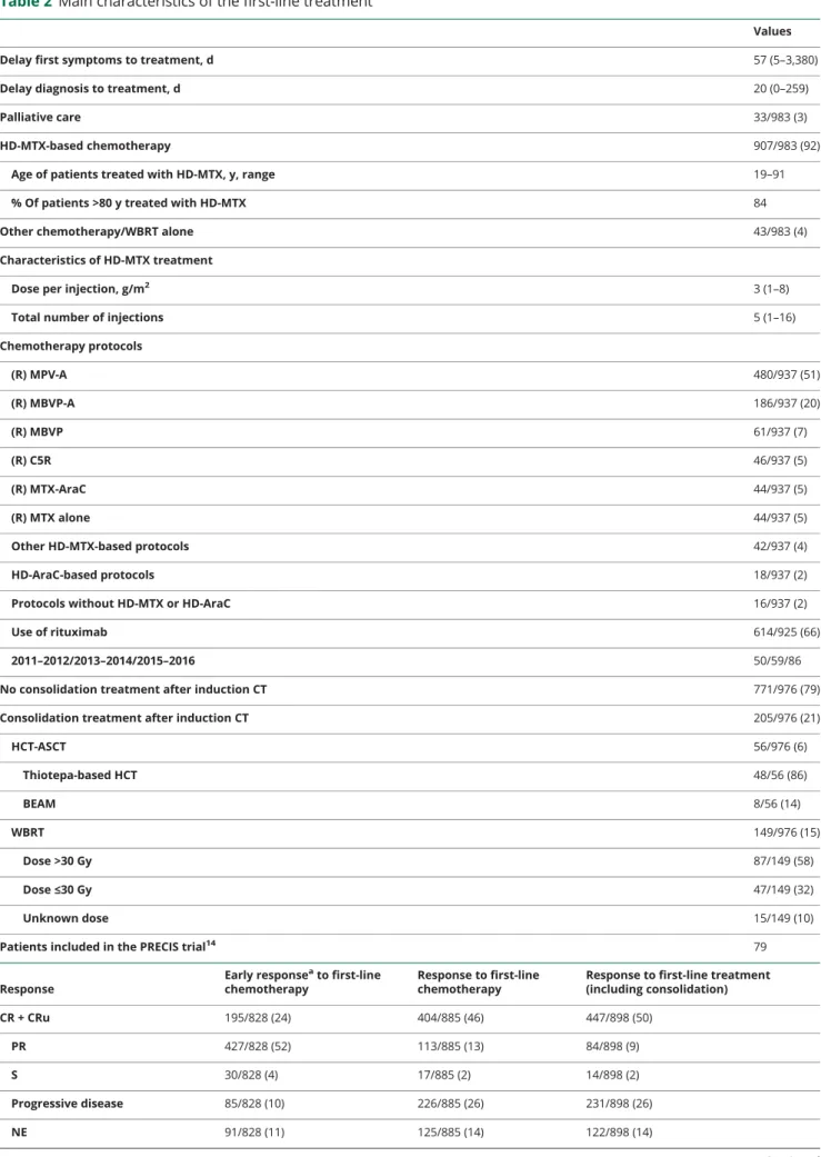
First-line treatment
The main characteristics of the
first-line treatment are reported
in table 2. Ninety-two percent of the patients (aged 19–91
years) received HD-MTX-CT. The methotrexate dose ranged
from 1 to 8 g/m
2with 78% of patients receiving at least 3 g/
m
2/injection. Interestingly, HD-MTX-CT was also
adminis-tered in the vast majority of the oldest patients aged over 80
years (84%); however, it was frequently administered at a
re-duced dose (39% with a dose
≥3 g/m
2/injection). Only 3% of
the patients received palliative care as their sole treatment.
Rituximab was used in 66% of the patients, with an increase in
use over time (from 50% in 2011–2012 to 86% in 2015–2016).
Twenty-one percent of the patients (median age 57 years)
received consolidation treatment after induction
chemother-apy, consisting of either WBRT (15%) or HCT-ASCT (6%).
In patients who received WBRT, the dose ranged from 18 to
56 Gy. However, the use of lower doses (≤30 Gy) increased
over time (from 13% in 2011–2012 to 83% in 2015–2016).
Outcome
After completion of
first-line treatment, the ORR rate was
59% (CR/Cru: 50%; PR: 9%), and 26% of the patients had
progressive disease. For the patients who received
consoli-dation treatment, the
final ORR was 92% (CR/CRu: 83%;
PR: 10%; HCT-ASCT: ORR: 96%; CR/CRu: 87%; PR:9%;
WBRT: ORR: 89%; CR/CRu: 79%; PR: 10%).
The median follow-up was 44.4 months (95% confidence
interval [CI] 41.2–47.6). The median PFS was 10.5 months
(95% CI 8.9–12.1), with a 2-year PFS rate of 36%. The
me-dian OS was 25.3 months (95% CI 18.3–32.3), with 1-, 2-, and
5-year OS rates of 62%, 51%, and 38%, respectively. In
patients treated with
first-line HCT-ASCT, the 5-year OS rate
was 76% (figure e-1, doi.org/10.5061/dryad.kc27cm8).
Of note, as illustrated by the survival curves (figure 1), a
sub-stantial subgroup of patients (n = 239), corresponding to 25%
of the whole cohort, experienced an early death in the
first 6
months after diagnosis. This group of patients was significantly
older than the other patients (p < 0.001) and had a worse KPS
at diagnosis (p < 0.001). In 44% of cases, the cause of death was
multifactorial (impaired neurologic status due to lymphoma
combined with several complications such as infections and
treatment-related side-effects). In other cases, death was related
to lymphoma before treatment (13%), progression of disease
despite treatment (31%), treatment-related toxicity (5%),
and unknown causes (7%) (table e-1, doi.org/10.5061/dryad.
kc27cm8).
The main differences between the older and younger
patients with PCNSL are summarized in table 3. Among the
patients responding to induction chemotherapy (CR or PR),
77% of the patients <60 years old received consolidation
treatment (23% HCT-ASCT and 54% WBRT), whereas
only 11% of the patients >60 years old received
consolida-tion treatment. The ORR to the
first-line treatment was
significantly higher in patients younger than 60 years (73%
vs 54%, p < 0.001). The outcome was better in younger
patients, with a median PFS of 8 months in patients >60
years old and 28.4 months in patients <60 years old (p <
0.001), and a median OS of 15.4 months in patients >60
years old and not reached in patients <60 years old (p <
0.001) (figure 1).
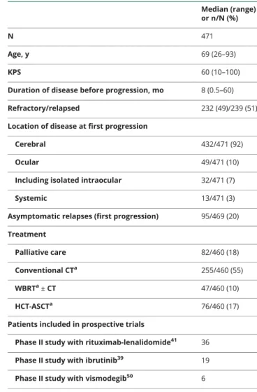
Relapse/progression
At the time of analysis, 471 patients in the cohort had
pro-gressed (232 refractory, 239 relapsed). Their main
character-istics are indicated in table 4. The median age at progression
was 68 years and the median KPS was 60. Treatments for
progression consisted of palliative care (18%), conventional
chemotherapy without RT or HCT-ASCT (55%), treatment
with WBRT at any time after progression but without
HCT-ASCT (10%), and treatment with HCT-HCT-ASCT at any time
after progression (17%). The patients who received
HCT-ASCT were younger and in better health than those who did
not, with a median age of 60 years (range 26–73) and a median
KPS of 80, and 85% of the patients achieved CR, CRu, or PR
before receiving HCT-ASCT.
The median OS from relapse was 6.8 months (95 CI 5.3–8.3),
with 1-, 3-, and 5-year OS rates of 38%, 25%, and 18%,
re-spectively. In patients treated with HCT-ASCT, the median
OS from relapse was not yet reached, with 1-year, 3-year, and
5-year OS rates of 74%, 57%, and 52%, respectively (figure 2).
Prognostic factors
We chose to study the prognostic effect of the main
charac-teristics of the patients as well as the factors previously
reported in other studies. The main prognostic factors are
indicated in table 5. Age <60 years, female sex, KPS at
di-agnosis
≥70, and response to initial chemotherapy were
as-sociated with increased OS in univariate and multivariate
analyses. CSF protein, blood lactate dehydrogenase (LDH),
ocular involvement, and tumor resection were not associated
with prognosis. The use of rituximab was associated with
prolonged OS in the univariate analysis but not in the
mul-tivariate analysis, most likely because patients treated with
rituximab were significantly younger and had a higher KPS.
When using the Memorial Kettering Cancer Center (MSKCC)
prognostic model,
23we could separate the population into 3
prognostic groups: patients aged
≤50 years, patients aged >50
years with KPS
≥70, and patients aged >50 years with KPS <50
(p < 0.001) (figure e-2, doi.org/10.5061/dryad.kc27cm8). The
International Extranodal Lymphoma Study Group (IELSG)
score could not be replicated, as the database does not give
information about the involvement of deep regions.
24Among patients <60 years old with CR to initial chemotherapy,
the patients who received consolidation treatment (WBRT or
HCT-ASCT) with
first-line treatment (n = 104) had a
signifi-cantly prolonged PFS (p < 0.001) and OS (p = 0.004)
com-pared to the patients who received no consolidation treatment
(n = 35), despite both populations being balanced in terms of
age and KPS (figure e-3, doi.org/10.5061/dryad.kc27cm8).
Table 1
Main patient characteristics at diagnosis
Median (range) or n/N (%) No. 1,002 Age, y 68 (18–91) 18–60 285/1,002 (28) 60–70 290/1,002 (29) 70–80 307/1,002 (31) 80–90 116/1,002 (12) >90 y 4/1,002 (0.4) Sex ratio, M/F 1.1 KPS 60 (0–100) Symptoms at diagnosis Cognitive impairment 586/966 (61) Walking disorders 551/951 (58) Motor/sensory deficit 369/955 (39) Headache/intracranial hypertension 255/968 (26) Epilepsy 112/967 (12) MRI
Unique vs multiple/diffuse lesions 519/961 (54) vs 442/ 961 (46)
Contrast enhancement 889/912 (97) Infratentorial involvement 242/858 (28) Ophthalmologic workup
Ophthalmologic workup done 573/935 (61) Ocular involvement 85/573 (15) No ocular involvement 488/573 (85) CSF workup CSF workup done 594/861 (69) CSF protein >0.5 g/L 383/524 (73); 0.73 (0.17–7.89) Low CSF glucose 5
CSF cell count: >5 cells 245/491 (50); 5 (0–1,800) Lymphomatous meningitis (positive
cytology or positive flow cytometry)
123/594 (21)
Elevated serum LDH 109/747 (23) Delay first symptoms to diagnosis 35 d (0–6.7 y) Diagnostic method
Cerebral biopsy 818/976 (84) Tumor resection 98/976 (10)
Vitrectomy 27/976 (3)
CSF 33/976 (3)
Table 1
Main patient characteristics at diagnosis
(continued) Median (range) or n/N (%)Histopathologic diagnosis
Diffuse large B-cell lymphoma 938/1,002 (94) Other high-grade B-cell lymphoma 14/1,002 (1.4) Low-grade B-cell lymphoma 14/1,002 (1.4) Unclassifiable B-cell lymphoma 31/1,002 (3) T-cell lymphoma 4/1,002 (0.4) Hodgkin lymphoma 1/1,002 (0.1) Abbreviations: KPS = Karnofsky Performance Status; LDH = lactate dehydrogenase.
Table 2
Main characteristics of the first-line treatment
Values
Delay first symptoms to treatment, d 57 (5–3,380)
Delay diagnosis to treatment, d 20 (0–259)
Palliative care 33/983 (3)
HD-MTX-based chemotherapy 907/983 (92)
Age of patients treated with HD-MTX, y, range 19–91
% Of patients >80 y treated with HD-MTX 84
Other chemotherapy/WBRT alone 43/983 (4)
Characteristics of HD-MTX treatment
Dose per injection, g/m2 3 (1–8)
Total number of injections 5 (1–16)
Chemotherapy protocols (R) MPV-A 480/937 (51) (R) MBVP-A 186/937 (20) (R) MBVP 61/937 (7) (R) C5R 46/937 (5) (R) MTX-AraC 44/937 (5) (R) MTX alone 44/937 (5)
Other HD-MTX-based protocols 42/937 (4)
HD-AraC-based protocols 18/937 (2)
Protocols without HD-MTX or HD-AraC 16/937 (2)
Use of rituximab 614/925 (66)
2011–2012/2013–2014/2015–2016 50/59/86
No consolidation treatment after induction CT 771/976 (79)
Consolidation treatment after induction CT 205/976 (21)
HCT-ASCT 56/976 (6) Thiotepa-based HCT 48/56 (86) BEAM 8/56 (14) WBRT 149/976 (15) Dose >30 Gy 87/149 (58) Dose≤30 Gy 47/149 (32) Unknown dose 15/149 (10)
Patients included in the PRECIS trial14 79
Response
Early responseato first-line
chemotherapy
Response to first-line chemotherapy
Response to first-line treatment (including consolidation) CR + CRu 195/828 (24) 404/885 (46) 447/898 (50) PR 427/828 (52) 113/885 (13) 84/898 (9) S 30/828 (4) 17/885 (2) 14/898 (2) Progressive disease 85/828 (10) 226/885 (26) 231/898 (26) NE 91/828 (11) 125/885 (14) 122/898 (14) Continued
Discussion
To our knowledge, the present study analyzed the most
com-prehensive cohort of patients with newly diagnosed PCNSL
treated in the modern era. In addition to clinical trials, which
enroll selected patients and contribute to the establishment of
standards of care, population-based studies are of interest
be-cause of their ability to better reflect real-world patient
man-agement and outcome and to inform on how advances are
implemented in daily practice.
25An important
finding of the present nationwide study
in-cluding patients diagnosed in a short period of time between
2011 and 2016 was the demographics of the population. We
found a higher proportion of older patients compared to
previous single-center or collaborative group series until the
1990s or early 2000s.
23,26–28Hence, patients aged over 60
years represented 72% of the study population, including 43%
of patients aged over 70 years. This age distribution has also
been reported in another recent cohort
29and is in line with
epidemiologic studies reporting a continuously increasing rate
in the elderly over last decades.
30,31This trend remains
unelucidated and is likely to continue in the near future with
population aging and the increasing need for brain biopsy in
the elderly. This demographic change is important as age
represents not only the strongest independent prognostic
factor of the disease
23,24,26but also a major risk factor for
severe treatment-related neurotoxicity.
7This issue should
stimulate specific studies devoted to the elderly to optimize
the therapeutic management of this growing vulnerable
population. Of note, the elderly patients were particularly
susceptible to early deaths, which occurred in up to 25% of
cases in the
first 6 months after diagnosis. In most cases, those
deaths were not related to resistance to chemotherapy but
Table 2
Main characteristics of the first-line treatment
(continued) ResponseEarly responseato first-line
chemotherapy
Response to first-line chemotherapy
Response to first-line treatment (including consolidation) Median KPS before treatment 60
Median KPS after treatment 70 Median KPS after treatment in
responder patients
80
Abbreviations: A or AraC = cytarabine; BEAM = carmustine, etoposide, cytarabine, melphalan; CR = complete response; CRu = unconfirmed complete response; CT = chemotherapy; HD-MTX = high-dose methotrexate; KPS = Karnofsky Performance Status; MBVP = methotrexate, carmustine, VP16, pred-nisone, cytarabine; C5R49; MPV = methotrexate, procarbazine, cytarabine; NE = not evaluable; PR = partial response; R = rituximab; S = stable ; WBRT =
whole-brain radiotherapy.
Values are median (range) or n/N (%).
aEvaluation 2 months after the onset of the treatment.
Figure 1
Outcome according to age
rather to comorbidity complications and treatment-related
toxicity favored by precarious health and poor neurologic
condition. This observation
first underlines the need to
shorten the pretreatment diagnostic workup as much as
possible once PCNSL is suspected in order to start treatment
with minimal delay (median of 57 days from the
first
symp-toms in our cohort) and second, to better adapt treatment
according to baseline oncogeriatric evaluation to reduce
tox-icities. As leptomeningeal and ocular involvement are not rare,
systematic lumbar puncture and ophthalmologic assessment
are recommended even in asymptomatic patients, as positive
cytology in the CSF or in the vitreous body may allow
avoidance of brain biopsy for diagnostic confirmation.
1Numerous factors have been reported to have prognostic
effect in PCNSL. We investigated the main prognostic
factors reported in the literature (table 5). Age <60 years,
KPS
≥70, tumor resection, use of rituximab, CR/CRu/PR
rates to initial chemotherapy, and use of consolidation
treatment in the
first line were associated with better
out-come in the univariate analysis, but only age, KPS at
di-agnosis, sex, and the response to
first-line treatment
remained statistically significant in the multivariate analysis.
Concerning prognostic scoring, the cohort could be divided
into prognostic subgroups according to the MSKCC
model,
23confirming the major prognostic effect of age and
KPS already reported in other studies.
9,26,32Neither
in-creased CSF protein level nor blood LDH, reported in the
IELSG score,
24were correlated with outcome. We also failed
to confirm the prognostic effect of early vs delayed CR after
methotrexate-based induction chemotherapy,
33of tumor
resection vs biopsy,
34or of intraocular involvement.
35In terms of
first-line treatment approaches, the main finding
was that the large majority of the patients (92%) received
HD-MTX-CT, regardless of age (84% in patients older than
Table 3
Main differences between patients <60 years and
patients >60 years
Patients <60 years old (n = 285) Patients >60 years old (n = 717) Men/women 1.6 1 Median KPS at diagnosis 70 60 Consolidation treatment afterinduction CT in responder patients
138/179 (77) 36/335 (11)
HCT-ASCT 42/179 (23) 6/335 (2)
WBRT 96/179 (54) 30/335 (9)
Response to first-line treatment
CR + CRu 170/263 (65) 277/635 (44) PR 21/263 (8) 63/635 (10) S 4/263 (2) 10/635 (2) Progressive disease 59/263 (22) 172/635 (27) NE 9/263 (3) 113/635 (18) PFS PFS, mo 28.4 (12.4–44.4) 8 (6.9–9.1) 2-y PFS 51 28 OS OS, mo NR (NR-NR) 15.4 (11.9–18.9) 1-y OS 78 54 2-y OS 70 44 5-y OS 61 28
Abbreviations: CR = complete response; CRu = unconfirmed complete re-sponse; CT = chemotherapy; HCT-ASCT = high-dose chemotherapy with autologous stem cell transplantation; KPS = Karnofsky Performance Status; NE = not evaluable; OS = overall survival; PFS = progression-free survival; PR = partial response; S = stable; WBRT = whole-brain radiotherapy. Values are median (95% confidence interval) or n/N (%).
Table 4
Main characteristics at progression
Median (range) or n/N (%)
N 471
Age, y 69 (26–93)
KPS 60 (10–100)
Duration of disease before progression, mo 8 (0.5–60) Refractory/relapsed 232 (49)/239 (51) Location of disease at first progression
Cerebral 432/471 (92)
Ocular 49/471 (10)
Including isolated intraocular 32/471 (7)
Systemic 13/471 (3)
Asymptomatic relapses (first progression) 95/469 (20) Treatment
Palliative care 82/460 (18) Conventional CTa 255/460 (55)
WBRTa± CT 47/460 (10)
HCT-ASCTa 76/460 (17)
Patients included in prospective trials
Phase II study with rituximab-lenalidomide41 36
Phase II study with ibrutinib39 19
Phase II study with vismodegib50 6
Abbreviations: CT = chemotherapy; HCT-ASCT = high-dose chemotherapy with autologous stem cell transplantation; KPS = Karnofsky Performance Status; WBRT = whole-brain radiotherapy.
aWhatever line of treatment after relapse.
80 years), with methotrexate doses ranging from 1 to 8 g/m
2(≥3 g/m
2in 78% of cases). WBRT has almost disappeared in
first-line treatment in patients aged over 60 years, probably
due to the fear of delayed neurotoxicity. This result amplifies
the decrease in the use of WBRT in elderly patients reported
by the MSKCC between 1986 and 2008.
36It is also
in-teresting to note the trend to use lower WBRT according to
Morris et al.,
37who reported encouraging results both in
terms of efficacy and tolerance with 23.4 Gy WBRT in
complete responders after HD-MTX-CT. The results of the
ongoing randomized Radiation Therapy Oncology Group 1114
trial using this modality of WBRT are expected in the coming
months. Consolidation was more highly debated in younger
patients, with 23% receiving HCT-ASCT, 54% receiving
WBRT, and 23% receiving no consolidation in our study,
reflecting the controversy in the field.
9,10,38In terms of
out-come, the omission of consolidation treatment (HCT-ASCT
or WBRT) in younger patients with CR after initial
chemo-therapy was correlated to worse outcome both in PFS and OS
in our series. The practices in the coming years will probably
be strongly influenced by the results of the 2 recent
ran-domized phase II studies
14,15supporting HCT-ASCT as
a valuable alternative to WBRT, both in terms of efficacy and
neurotoxicity.
Concerning rituximab, an important increase in its use in
first-line treatment was noted between 2011 and 2012 (50%) and
2015 and 2016 (86%), in parallel to the cumulative publications
of several studies supporting its use in PCNSL.
16–18However,
in a recent phase III study, Bromberg et al.
19showed that the
use of rituximab was not associated with better outcome,
es-pecially in older patients, in line with the results of the present
study; therefore, the trend in the use of rituximab might be
inverted in the coming years. The potential interest in
ritux-imab in younger patients could not be studied in the present
work, as 90% of patients <60 years old received rituximab.
In this study, despite a substantial overall response rate to
first-line treatment (59%), the prognosis of the disease
remained poor globally, with a median PFS of 10.2 months,
a median OS of 25.3 months, and a median OS after relapse
of 6.1 months. Regardless of age, there is a high rate of
refractory disease, and there is a need for developing a new
generation of induction treatment regimens. Ibrutinib,
im-munomodulatory drugs, or immune checkpoint inhibitors
are promising new agents to be possibly combined with
chemotherapy, given their efficacy as single agent
treat-ments in PCNSL.
39–43However, most of the responding
patients benefitted clinically from the treatment, with
a median KPS of 60 at the beginning of treatment that
increased to 80 at the end of the treatment. Furthermore,
there is a real hope of long-term survival and cure, which
concerned 38% of the patients at 5 years, mainly in younger
patients (61% OS at 5 years). In terms of prognosis, our
results are in line with the majority of the prospective
studies published recently, in elderly patients
8,17,44,45as well
as in younger patients.
9,12,13,37,46Although involving a minority of patients (6% in the
first line,
17% at relapse) in our study, an increasing use of HCT-ASCT
in
first-line treatment or at relapse was observed with
prom-ising results (76% 5-year OS in the
first line, 57% 3-year OS
after relapse), in line with previously published studies.
11–13,47This strategy is now offered to patients older than 60 years
and older than 65 years as well, who nevertheless remain
highly selected (high KPS and adequate response to salvage
chemotherapy before HCT-ASCT), as recently reported in
a European retrospective study.
48In the coming years, it will
be useful to develop evaluation scales in order to better target
patients potentially eligible for HCT-ASCT.
Population-based studies are scarce. Two larger cohorts
28,29have recently been published but with limited data on clinical
presentation and treatments. Fallah et al.
29reported a cohort
of more than 9,000 HIV-negative patients with PCNSL
di-agnosed in the United States between 2004 and 2013. A
proportion of 27.2% of patients did not receive any
chemo-therapy in the
first line. The prognosis was worse than in our
cohort (median OS of 1.3 years) but tended to improve over
time, with a 3-year OS of 40.9% in the patients diagnosed
during 2010–2012, close to the 3-year OS of our study, and an
increase in the use of multiagent chemotherapy over time.
Van der Meulen et al.
28reported a Dutch population-based
cohort of 1,673 patients with PCNSL diagnosed between
1989 and 2015. An increasing incidence of PCNSL in patients
aged over 60 was noted. OS improved over time but only in
the patients <70 years old, probably because approximatively
40% of elderly patients did not receive any antineoplastic
therapy, even in the more recent period (2009–2015). There
Figure 2
Overall survival (OS) from relapse according to
treatment at relapse
CT = chemotherapy; HCT-ASCT = high-dose chemotherapy with autologous stem cell transplantation; WBRT = whole-brain radiotherapy.
Table 5
Prognostic factors in terms of overall survival (OS) (univariate and multivariate analysis)
N
Univariate analysis Multivariate analysis
Median OS p Value HR p Value
Age <60 y 285 NR <0.001 2.3 <0.001 Age >60 y 717 15.4 Female sex 466 31.5 0.1 1.2 0.03 Male sex 535 22.4 KPS at diagnosis <70 444 11.7 <0.001 0.6 <0.001 KPS at diagnosis≥70 454 55.6 Unifocal lesion 519 27.4 0.8 Plurifocal/diffuse lesions 442 23.9 No ocular involvement 488 35 0.4 Ocular involvement 85 45.7 No lymphomatous meningitis 471 42.2 0.3 Lymphomatous meningitis 123 25.3 Normal CSF protein 176 43.6 0.2 Elevated CSF protein 348 30.5 Normal blood LDH 365 40 0.9 Elevated blood LDH 109 37.6 No tumor resection 98 23.8 0.05 0.9 0.5 Tumor resection 881 37.2
Delay first symptoms–treatment, d 0.8
≤57 407 25.3
>57 405 30.9
No rituximab in first line 311 12.6 <0.001 1 1
Rituximab in first line 614 53.1
Response to initial CT <0.001 0.1 <0.001
NE/S/P 402 5.2
CR/CRu/PR 517 NR
Early responseato initial CT 0.04
PR 426 57.7
CR + CRu 194 NR
Among patients in CR to initial CT 0.08
Early CRato initial CT 164 NR
Early PRato initial CT 188 NR
Consolidation in first line 206 14.6 <0.001 0.9 0.7
No consolidation in first line 771 NR
Among patients <60 y 0.6
Patients included in the PRECIS trial14 79 NR
Other patients 75.3
Abbreviations: CR = complete response; CRu = unconfirmed complete response; CT = chemotherapy; HR = hazard ratio; KPS = Karnofsky Performance Status; LDH = lactate dehydrogenase; NE = not evaluable; P = progressive; PR = partial response; S = stable.
aEvaluation 2 months after the onset of the treatment.
was 56% OS at 5 years in patients <60 years old from 2009 to
2015, in line with our results.
This work had several limitations, mainly due to the inherent
biases of a retrospective study. There were some missing data
and loss of follow-up, but not exceeding 10% for most items.
Although important, notably in elderly patients, data on
tox-icity, especially neurotoxtox-icity, and quality of life are lacking in
this study. A longer follow-up will be necessary to describe the
population of long-term survivors and late relapses.
Our study confirms the increasing proportion of elderly
patients within the PCNSL population, who are associated
with poor outcomes, are frequently disabled, and have a high
risk of early death, raising the need for a specific pretreatment
evaluation and therapeutic management. In contrast, there is
a higher rate of long-term survival and hope for a cure in
younger patients, who can benefit from vigorous
consolida-tion therapies such as HCT-ASCT. Our results provide
evi-dence that management advances in PCNSL from recent
years have been applied in the real life and stress the need to
further implement guidelines and develop multidisciplinary
networks in daily practice for this rare and complex disease.
Acknowledgment
The authors thank the patients and their families for their
participation, all members of the LOC network, and the
Institut National du Cancer.
Study funding
This study was supported by the Institut National du Cancer.
Disclosure
The authors report no disclosures relevant to the manuscript.
Go to Neurology.org/N for full disclosures.
Publication history
Received by Neurology April 12, 2019. Accepted in
final form
September 5, 2019.
Appendix
Authors
Name Location Role Contribution Caroline
Houillier, MD
CHU Piti´ e-Salpˆetri`ere, Paris, France
Author Designed and conceptualized study, analyzed the data, drafted the manuscript for intellectual content, major role in the acquisition of data, interpreted the data Carole Soussain, MD, PhD Institut Curie, Saint Cloud, France
Author Designed and conceptualized study, analyzed the data, drafted the manuscript for intellectual content, major role in the acquisition of data, interpreted the data
Appendix
(continued)Name Location Role Contribution Herv´e
Ghesqui`eres, MD, PhD
CHU Lyon Sud, France
Author Major role in the acquisition of data, revised the manuscript for intellectual content Pierre Soubeyran, MD, PhD Institut Bergoni´e, Bordeaux, France
Author Major role in the acquisition of data, revised the manuscript for intellectual content Olivier Chinot, MD, PhD CHU de la Timone, Marseille, France
Author Major role in the acquisition of data, revised the manuscript for intellectual content Luc Taillandier, MD, PhD CHU de Nancy, France
Author Major role in the acquisition of data, revised the manuscript for intellectual content Thierry Lamy, MD, PhD CHU de Rennes, France
Author Major role in the acquisition of data, revised the manuscript for intellectual content Sylvain Choquet, MD, PhD CHU Piti´ e-Salpˆetri`ere, Paris, France
Author Major role in the acquisition of data, revised the manuscript for intellectual content Guido Ahle, MD Hˆopitaux Civils,
Colmar, France
Author Major role in the acquisition of data, revised the manuscript for intellectual content Gandhi Damaj, MD, PhD CHU de Caen, France
Author Major role in the acquisition of data, revised the manuscript for intellectual content Philippe Agap´e, MD Institut de Canc´erologie de l’Ouest, Saint Herblain, France
Author Major role in the acquisition of data, revised the manuscript for intellectual content C´ecile Moluçon-Chabrot, MD CHU de Clermont-Ferrand, France
Author Major role in the acquisition of data, revised the manuscript for intellectual content Alexandra Amiel, MD CHU de Toulouse, France
Author Major role in the acquisition of data, revised the manuscript for intellectual content Vincent Delwail, MD CHU de Poitiers, France
Author Major role in the acquisition of data, revised the manuscript for intellectual content
Appendix
(continued)Name Location Role Contribution Michel Fabbro, MD Institut du Cancer de Montpellier Val d’Aurelle, France
Author Major role in the acquisition of data, revised the manuscript for intellectual content Fabrice Jardin, MD, PhD Centre Henri Becquerel, Rouen, France
Author Major role in the acquisition of data, revised the manuscript for intellectual content Adrien Chauchet, MD CHU de Besançon, France
Author Major role in the acquisition of data, revised the manuscript for intellectual content Marie-Pierre Moles-Moreau, MD CHU d’Angers, France
Author Major role in the acquisition of data, revised the manuscript for intellectual content Franck Morschhauser, MD, PhD CHRU de Lille, France
Author Major role in the acquisition of data, revised the manuscript for intellectual content Olivier Casasnovas, MD CHU de Dijon, France
Author Major role in the acquisition of data, revised the manuscript for intellectual content R´emy Gressin, MD CHU de Grenoble, France
Author Major role in the acquisition of data, revised the manuscript for intellectual content Luc-Matthieu Fornecker, MD, PhD CHU de Strasbourg, France
Author Major role in the acquisition of data, revised the manuscript for intellectual content Julie Abraham, MD CHU de Limoges, France
Author Major role in the acquisition of data, revised the manuscript for intellectual content Jean-Pierre Marolleau, MD, PhD CHU d’Amiens, France
Author Major role in the acquisition of data, revised the manuscript for intellectual content Adrian Tempescul, MD, PhD CHU de Brest, France
Author Major role in the acquisition of data, revised the manuscript for intellectual content Chantal Campello, MD CHU de Nˆımes, France
Author Major role in the acquisition of data, revised the manuscript for intellectual content
Appendix
(continued)Name Location Role Contribution Philippe Colin,
MD
Clinique Courlancy, Reims, France
Author Major role in the acquisition of data, revised the manuscript for intellectual content J´erˆome Tamburini, MD, PhD Hˆopital Cochin, Paris, France
Author Major role in the acquisition of data, revised the manuscript for intellectual content Kamel Laribi, MD Centre Hospitalier, Le Mans, France
Author Major role in the acquisition of data, revised the manuscript for intellectual content Caroline Serrier, MD Centre Hospitalier de Perpignan, France
Author Major role in the acquisition of data, revised the manuscript for intellectual content Corinne Haioun, MD, PhD Hˆopital Henri Mondor, Cr´eteil, France
Author Major role in the acquisition of data, revised the manuscript for intellectual content Safia Chebrek, MD Centre Hospitalier d’Avignon, France
Author Major role in the acquisition of data, revised the manuscript for intellectual content Anna Schmitt, MD Institut Bergoni´e, Bordeaux, France
Author Major role in the acquisition of data, revised the manuscript for intellectual content Marie Blonski, MD CHU de Nancy, France
Author Major role in the acquisition of data, revised the manuscript for intellectual content Roch Houot, MD, PhD CHU de Rennes, France
Author Major role in the acquisition of data, revised the manuscript for intellectual content Eileen Boyle, MD CHRU de Lille, France
Author Major role in the acquisition of data, revised the manuscript for intellectual content Jacques-Olivier Bay, MD, PhD CHU de Clermont-Ferrand, France
Author Major role in the acquisition of data, revised the manuscript for intellectual content Lucie Oberic, MD Institut Universitaire du Cancer de Toulouse, France
Author Major role in the acquisition of data, revised the manuscript for intellectual content
Continued
References
1. Hoang-Xuan K, Bessell E, Bromberg J, et al. Diagnosis and treatment of primary CNS lymphoma in immunocompetent patients: guidelines from the European Association for Neuro-Oncology. Lancet Oncol 2015;16:e322–e332.
2. Grommes C, DeAngelis LM. Primary CNS lymphoma. J Clin Oncol 2017;35: 2410–2418.
3. Rubenstein JL, Gupta NK, Mannis GN, Lamarre AK, Treseler P. How I treat CNS lymphomas. Blood 2013;122:2318–2330.
4. Ferreri AJ, Reni M, Foppoli M, et al. High-dose cytarabine plus high-dose metho-trexate versus high-dose methometho-trexate alone in patients with primary CNS lymphoma: a randomised phase 2 trial. Lancet 2009;374:1512–1520.
5. Nelson DF, Martz KL, Bonner H, et al. Non-Hodgkin’s lymphoma of the brain: can high dose, large volume radiation therapy improve survival? Report on a prospective trial by the Radiation Therapy Oncology Group (RTOG): RTOG 8315. Int J Radiat Oncol Biol Phys 1992;23:9–17.
6. DeAngelis LM, Yahalom J, Thaler HT, Kher U. Combined modality therapy for primary CNS lymphoma. J Clin Oncol 1992;10:635–643.
7. Omuro AM, Ben-Porat LS, Panageas KS, et al. Delayed neurotoxicity in primary central nervous system lymphoma. Arch Neurol 2005;62:1595–1600.
Appendix
(continued)Name Location Role Contribution Emeline Tabouret, MD, PhD CHU de la Timone, Marseille, France
Author Major role in the acquisition of data, revised the manuscript for intellectual content Agathe Waultier, MD CHU de Nˆımes, France
Author Major role in the acquisition of data, revised the manuscript for intellectual content Nadine Martin-Duverneuil, MD CHU Piti´ e-Salpˆetri`ere, Paris, France
Author Major role in the acquisition of data, revised the manuscript for intellectual content Val´erie Touitou, MD, PhD CHU Piti´ e-Salpˆetri`ere, Paris, France
Author Major role in the acquisition of data, revised the manuscript for intellectual content Nathalie Cassoux, MD, PhD Institut Curie, Paris, France
Author Major role in the acquisition of data, revised the manuscript for intellectual content Aur´elie Kas,
MD, PhD
Groupe Hospitalier Piti´ e-Salpˆetri`ere, Paris, France
Author Major role in the acquisition of data, revised the manuscript for intellectual content Karima Mokhtari, MD Groupe Hospitalier Piti´ e-Salpˆetri`ere, Paris, France
Major role in the acquisition of data, revised the manuscript for intellectual content Fr´ed´eric Charlotte, MD Groupe Hospitalier Piti´ e-Salpˆetri`ere, Paris, France
Author Major role in the acquisition of data, revised the manuscript for intellectual content Agusti Alentorn, MD, PhD Groupe Hospitalier Piti´ e-Salpˆetri`ere, Paris, France
Author Major role in the acquisition of data, revised the manuscript for intellectual content Lo¨ıc Feuvret, MD Groupe Hospitalier Piti´ e-Salpˆetri`ere, Paris, France
Author Major role in the acquisition of data, revised the manuscript for intellectual content Magali Le Garff-Tavernier, PharmD, PhD Groupe Hospitalier Piti´ e-Salpˆetri`ere, Paris, France
Author Major role in the acquisition of data, revised the manuscript for intellectual content Myrto Costopolous, PharmD Groupe Hospitalier Piti´ e-Salpˆetri`ere, Paris, France
Author Major role in the acquisition of data, revised the manuscript for intellectual content
Appendix
(continued)Name Location Role Contribution Bertrand
Mathon, MD
Groupe Hospitalier Piti´ e-Salpˆetri`ere, Paris, France
Author Major role in the acquisition of data, revised the manuscript for intellectual content Matthieu Peyre, MD, PhD Groupe Hospitalier Piti´ e-Salpˆetri`ere, Paris, France
Author Major role in the acquisition of data, revised the manuscript for intellectual content Daniel Delgadillo, PhD Groupe Hospitalier Piti´ e-Salpˆetri`ere, Paris, France
Author Major role in the acquisition of data, revised the manuscript for intellectual content Hassen Douzane, Msc Groupe Hospitalier Piti´ e-Salpˆetri`ere, Paris, France
Author Major role in the acquisition of data, revised the manuscript for intellectual content Diane Genet, Msc, PhD Groupe Hospitalier Piti´ e-Salpˆetri`ere, Paris, France
Author Major role in the acquisition of data, revised the manuscript for intellectual content Bachir Aidoui, MD Groupe Hospitalier Piti´ e-Salpˆetri`ere, Paris, France
Author Major role in the acquisition of data, revised the manuscript for intellectual content Khˆe Hoang-Xuan, MD, PhD Groupe Hospitalier Piti´ e-Salpˆetri`ere, Paris, France
Author Designed and conceptualized study, analyzed the data, drafted the manuscript for intellectual content, major role in the acquisition of data, interpreted the data Emmanuel
Gyan, MD, PhD
CHU Tours, France
Author Designed and conceptualized study, analyzed the data, drafted the manuscript for intellectual content, major role in the acquisition of data, interpreted the data
8. Omuro A, Chinot O, Taillandier L, et al. Methotrexate and temozolomide versus methotrexate, procarbazine, vincristine, and cytarabine for primary CNS lymphoma in an elderly population: an intergroup ANOCEF-GOELAMS randomised phase 2 trial. Lancet Haematol 2015;2:e251–259.
9. Thiel E, Korfel A, Martus P, et al. High-dose methotrexate with or without whole brain radiotherapy for primary CNS lymphoma (G-PCNSL-SG-1): a phase 3, randomised, non-inferiority trial. Lancet Oncol 2010;11:1036–1047.
10. Ferreri AJ, DeAngelis L, Illerhaus G, et al. Whole-brain radiotherapy in primary CNS lymphoma. Lancet Oncol 2011;12:118–119.
11. Soussain C, Hoang-Xuan K, Taillandier L, et al. Intensive chemotherapy followed by hematopoietic stem-cell rescue for refractory and recurrent primary CNS and in-traocular lymphoma: Soci´et´e Française de Greffe de Mo¨elle Osseuse-Th´erapie Cel-lulaire. J Clin Oncol 2008;26:2512–2518.
12. Kasenda B, Schorb E, Fritsch K, Finke J, Illerhaus G. Prognosis after high-dose che-motherapy followed by autologous stem-cell transplantation asfirst-line treatment in primary CNS lymphoma: a long-term follow-up study. Ann Oncol 2015;26:608–611. 13. Omuro A, Correa DD, DeAngelis LM, et al. R-MPV followed by high-dose
chemo-therapy with TBC and autologous stem-cell transplant for newly diagnosed primary CNS lymphoma. Blood 2015;125:1403–1410.
14. Houillier C, Taillandier L, Dureau S, et al. Radiotherapy or autologous stem-cell transplantation for primary CNS lymphoma in patients 60 years of age and younger: results of the intergroup ANOCEF-GOELAMS randomized phase II PRECIS study. J Clin Oncol 2019;37:823–833.
15. Ferreri AJM, Cwynarski K, Pulczynski E, et al. Whole-brain radiotherapy or autolo-gous stem-cell transplantation as consolidation strategies after high-dose methotrexate-based chemoimmunotherapy in patients with primary CNS lym-phoma: results of the second randomisation of the International Extranodal Lym-phoma Study Group-32 phase 2 trial. Lancet Haematol 2017;4:e510–e523. 16. Birnbaum T, Stadler EA, von Baumgarten L, Straube A. Rituximab significantly
improves complete response rate in patients with primary CNS lymphoma. J Neurooncol 2012;109:285–291.
17. Fritsch K, Kasenda B, Hader C, et al. Immunochemotherapy with rituximab, meth-otrexate, procarbazine, and lomustine for primary CNS lymphoma (PCNSL) in the elderly. Ann Oncol 2011;22:2080–2085.
18. Ferreri AJ, Cwynarski K, Pulczynski E, et al. Chemoimmunotherapy with methotrexate, cytarabine, thiotepa, and rituximab (MATRix regimen) in patients with primary CNS lymphoma: results of thefirst randomisation of the International Extranodal Lymphoma Study Group-32 (IELSG32) phase 2 trial. Lancet Haematol 2016;3:e217–e227. 19. Bromberg JEC, Issa S, Bakunina K, et al. Rituximab in patients with primary CNS
lymphoma (HOVON 105/ALLG NHL 24): a randomised, open-label, phase 3 in-tergroup study. Lancet Oncol 2019;20:216–228.
20. Langner-Lemercier S, Houillier C, Soussain C, et al. Primary CNS lymphoma atfirst relapse/progression: characteristics, management, and outcome of 256 patients from the French LOC network. Neuro Oncol 2016;18:1297–1303.
21. Darlix A, Zouaoui S, Rigau V, et al. Epidemiology for primary brain tumors: a na-tionwide population-based study. J Neurooncol 2017;131:525–546.
22. Abrey LE, Batchelor TT, Ferreri AJ, et al. Report of an international workshop to standardize baseline evaluation and response criteria for primary CNS lymphoma. J Clin Oncol 2005;23:5034–5043.
23. Abrey LE, Ben-Porat L, Panageas KS, et al. Primary central nervous system lym-phoma: the Memorial-Kettering Cancer Center prognostic model. J Clin Oncol 2006; 24:5711–5715.
24. Ferreri AJM, Blay JY, Reni M, et al. Prognostic scoring system for primary CNS lymphomas: the international extranodal lymphoma study group experience. J Clin Oncol 2003;21:266–272.
25. Zeremski V, Koehler M, Fischer T, Schalk E. Characteristics and outcome of patients with primary CNS lymphoma in a“real-life” setting compared to a clinical trial. Ann Hematol 2016;95:793–799.
26. Bessell EM, Graus F, Lopez-Guillermo A, et al. Primary non-Hodgkin’s lymphoma of the CNS treated with CHOD/BVAM or BVAM chemotherapy before radiotherapy: long-term survival and prognostic factors. Int J Radiat Oncol Biol Phys 2004;59:501–508. 27. Ferreri AJ, Reni M, Pasini F, et al. A multicenter study of treatment of primary CNS
lymphoma. Neurology 2002;58:1513–1520.
28. Van der Meulen M, Dinmohamed AG, Visser O, Doorduijn JK, Bromberg JEC. Improved survival in primary central nervous system lymphoma up to age 70 only: a population-based study on incidence, primary treatment and survival in The Netherlands, 1989-2015. Leukemia 2017;31:1822–1825.
29. Fallah J, Qunaj L, Olszewski A. Therapy and outcomes of primary central nervous system lymphoma in the United States: analysis of the National Cancer Database. Blood Adv 2016;1:112–121.
30. Villano JL, Koshy M, Shaikh H, Dolecek TA, McCarthy BJ. Age, gender, and racial differences in incidence and survival in primary CNS lymphoma. Br J Cancer 2011; 105:1414–1418.
31. Shiels MS, Pfeiffer RM, Besson C, et al. Trends in primary central nervous system lymphoma incidence and survival in the U.S. Br J Haematol 2016;174:417–424. 32. Schultz C, Scott C, Sherman W, et al. Preirradiation chemotherapy with
cyclophos-phamide, doxorubicin, vincristine, and dexamethasone for primary CNS lymphomas: initial report of Radiation Therapy Oncology Group protocol 88-06. J Clin Oncol 1996;14:556–564.
33. Pels H, Juergens A, Schirgens I, et al. Early complete response during chemotherapy predicts favorable outcome in patients with primary CNS lymphoma. Neuro Oncol 2010;12:720–724.
34. Weller M, Martus P, Roth P, Thiel E, Korfel A; German PCNSL Study Group. Surgery for primary CNS lymphoma? Challenging a paradigm. Neuro Oncol 2012;14: 1481–1484.
35. Kreher S, Strehlow F, Martus P, et al. Prognostic impact of intraocular involvement in primary CNS lymphoma: experience from the G-PCNSL-SG1 trial. Ann Hematol 2015;94:409–414.
36. Ney DE, Reiner AS, Panageas KS, Brown HS, DeAngelis LM, Abrey LE. Character-istics and outcomes of elderly patients with primary central nervous system lym-phoma: the Memorial-Kettering Cancer Center experience. Cancer 2010;116: 4605–4612.
37. Morris PG, Correa DD, Yahalom J, et al. Rituximab, methotrexate, procarbazine, and vincristine followed by consolidation reduced-dose whole-brain radiotherapy and cytarabine in newly diagnosed primary CNS lymphoma:final results and long-term outcome. J Clin Oncol 2013;31:3971–3979.
38. Birnbaum T, Bochmann K, Straube A. Early relapses in patients with primary CNS lymphoma treated with methotrexate-based chemotherapy without consolidating whole brain irradiation. J Neurooncol 2013;112:233–239.
39. Soussain C, Choquet S, Blonski M, et al The LYSA/LOC network iLOC phase II study with ibrutinib for relapse/refractory primary CNS or vitreoretinal lymphoma. Eur J Cancer 2019;117:121–130.
40. Grommes C, Pastore A, Palaskas N, et al. Ibrutinib unmasks critical role of Bruton tyrosine kinase in primary CNS lymphoma. Cancer Discov 2017;7:1018–1029. 41. Ghesquieres H, Chevrier M, Laadhari M, et al. Lenalidomide in combination with
intravenous rituximab (REVRI) in relapsed/refractory primary CNS lymphoma or primary intraocular lymphoma: a multicenter prospective“proof of concept” phase II study of the French oculo-cerebral lymphoma (LOC) network and the Lymphoma Study Association (LYSA). Ann Oncol 2019;30:621–628.
42. Tun HW, Johnston PB, DeAngelis LM, et al. Phase 1 study of pomalidomide and dexamethasone for relapsed/refractory primary CNS or vitreoretinal lymphoma. Blood 2018;132:2240–2248.
43. Nayak L, Iwamoto FM, LaCasce A, et al. PD-1 blockade with nivolumab in relapsed/ refractory primary central nervous system and testicular lymphoma. Blood 2017;129: 3071–3073.
44. Fritsch K, Kasenda B, Schorb E, et al. High-dose methotrexate-based immuno-chemotherapy for elderly primary CNS lymphoma patients (PRIMAIN study). Leukemia 2017;31:846–852.
45. Hoang-Xuan K, Taillandier L, Chinot O, et al. Chemotherapy alone as initial treat-ment for primary CNS lymphoma in patients older than 60 years: a multicenter phase II study (26952) of the European Organization for Research and Treatment of Cancer Brain Tumor Group. J Clin Oncol 2003;21:2726–2731.
46. Rubenstein JL, Hsi ED, Johnson JL, et al. Intensive chemotherapy and immuno-therapy in patients with newly diagnosed primary CNS lymphoma: CALGB 50202 (Alliance 50202). J Clin Oncol 2013;31:3061–3068.
47. Soussain C, Choquet S, Fourme E, et al. Intensive chemotherapy with thiotepa, busulfan and cyclophosphamide and hematopoietic stem cell rescue in relapsed or refractory primary central nervous system lymphoma and intraocular lymphoma: a retrospective study of 79 cases. Haematologica 2012;97:1751–1756.
48. Schorb E, Fox CP, Fritsch K, et al. High-dose thiotepa-based chemotherapy with autologous stem cell support in elderly patients with primary central nervous system lymphoma: a European retrospective study. Bone Marrow Transpl 2017;52: 1113–1119.
49. Ghesqui`eres H, Ferlay C, Sebban C, et al. Long-term follow-up of an age-adapted C5R protocol followed by radiotherapy in 99 newly diagnosed primary CNS lymphomas: a prospective multicentric phase II study of the Groupe d’Etude des Lymphomes de l’Adulte (GELA). Ann Oncol 2010;21:842–850.
50. Houot R, Soussain C, Tilly H, et al. Inhibition of Hedgehog signaling for the treatment of lymphoma and CLL: a phase II study from the LYSA. Ann Oncol 2016;27: 1349–1350.