Approaches to Determining the
Three-Dimensional Structure and Dynamics of
Bacterial Chromosomes
by
Matthew A. Wright
B.A. Chemistry, B.M. Music Performance University of Southern Maine, 1999
Submitted to the Department of Chemistry
in Partial Fulfillment of the Requirements for the Degree of
DOCTOR OF PHILOSOPHY
in Physical Chemistry at the
Massachusetts Institute of Technology
September 2005
© Massachusetts Institute of Technology, 2005
All rights reserved
A/ A
I
A Signature of Author Department of Chemistry September 1,2005 Certified by Accepted by. A-l
N
MASSACHUSETTS INSTIT OF TECHNOLOGY OCT ! 1 2005 LIBRARIES ) George M. ChurchProfessor of Genetics, Harvard Medical School Thesis Supervisor Robert W. Field
Chairman, Departmental Committee on Graduate Students
OiL,
Nt/ke I _ _ _ _ IV
/J7
(This doctoral thesis has been examined by a Committee of the Department of Chemistry as follows:
Professor Keith A. Nelson
Professor George M. Church
X/ nCommittee Chairman
(
Prssor of - Genetics, Harvard Medical School I Professor of Genetics, Harvard Medical School
A
Thesis SupervisorProfessor Leonid Mirny
Associate Professor of Physics, Massa tts Institute of Technology Committee Member
-1 _ _
--- .
Approaches to Determining the
Three-Dimensional Structure and Dynamics of
Bacterial Chromosomes
by
Matthew A. Wright
Submitted to the Department of Chemistry
in Partial Fulfillment of the
Requirements for the Degree of Doctor of Philosophy in Chemistry
Abstract
The information in genomes is only partially contained in the linear sequence of their nucleotides. Their folding into dynamic three-dimensional structures creates spatial relationships between loci that likely play important functional roles. Yet so far only the broad outlines of this spatial organization have been discerned.
In chapter 2 of this thesis I describe a general constraint-based framework for defining the configuration space of chromosomes. Analogous to protein structure determination through NMR, such a framework allows the quantitative reduction of the conformation
space down to the level of a single structure or an ensemble of structures. It is compatible
with both experimentally determined and theoretical constraints, particularly those motivated by evolutionary optimality.
In chapter 3., I describe the first method to search for signals of large-scale three-dimensional structure in genome sequences. The results suggest that there is strong selection for three-dimensional relationships within the chromosome, particularly those related to transcription. The signals generated recapitulate both known structural data from microscopy and functional data on genome-wide transcription levels. Moreover, a detailed analysis of these signals in E. coli suggests previously unknown structural features including chromosome-long periodic looping and an axis of high transcriptional activity. There are immediate applications to other bacteria and potentially to eukaryotes.
Thesis Supervisor: George M. Church
Title: Professor of Genetics, Harvard Medical School
Table of Contents
Chapter 1 Introduction 10 Chapter 2Constraint-based determination of
27
chromosome structure
Chapter 3"Just in place" functional organization
59
of bacterial chromosome structure
Chapter 4
Conclusions and future directions
112
Appendix 1
On the complete determination of
122
biological systems
Appendix 2
An open-source oligomicroarray
138
standard for human and mouse
I dedicate this thesis to my parents, Bonnie and Tom Wright, for their love and encouragement, to my grandfather, Robert E. Arnot, and uncle, Robert B. Arnot, who ignited in me a passion for medical science, and to my undergraduate chemistry advisor, Henry J. Tracy, who first showed me the beauty of basic scientific research.
Acknowledgements
It was my grandfather, Robert Arnot, who introduced me to science, through my
first scientific tool, a calculator, given to me at age 2. Although it was the buttons that
fascinated me, I like to think that a bit of arithmetic wore off during the months that followed while the calculator never left my side. My grandfather was an enormous influence on me, through his example as a fine physician and remarkable human being, and through his passion for science as a means for human good. He brought the mindset of a researcher to the care of patients long after he left his own formal work in
psychopharmacology.
It is my father from whom I inherited a love for mathematics. He is among the
few people I know who takes on a glow at the mere mention of the words "linear algebra" and can spend an evening discussing abstract multidimensional spaces. For better or for worse I seem to have inherited these traits. Moreover, he has a seemingly insatiable curiosity, evidenced by a constant and ever-changing stack of scientific texts
heaped upon his desk. His curiosity serves as a reminder to always inquire.
I thank my mother who has been a solid support for me and for my entire family through many trials; additionally she lovingly guided me from music into science. I thank my piano teacher Laura Kargul who both enriched my life through 7 years of teaching and helped to open me to possibilities outside of music. I thank my family as a whole, my grandmother, Mary Arnot, my siblings,Tom Doug and Emily, and my uncle Robert, an important support. I thank also many friends, in particular Brad Coull, David Goodman, Andrew Butterworth, Tracey Orick, and Janice Sullivan, Cathy O'Neil and Jen Johnson,
close friends and inspiring mathematicians, and Serhan Altunata, an excellent physicist. I
thank the entire Sievers family, quite a scientific clan, in particular Kathryn for her quick
mind, LeRoy, for his wonderful practicality as a physicist/engineer, and Charlie, for
frequent conversations on physics and Fourier analysis.
Stephen MacDonald first introduced me to the beauty of higher mathematics, in a calculus course that I originally took as a core math requirement in college. His brilliant
teaching, somehow allowed his students to always see one step ahead in the web of
mathematical logic and convinced me to take one, then two, then three more courses with
him while still a piano performance major. Ultimately this led me to embrace science as a career.
John Ricci's, patient and clear teaching of general chemistry and then physical chemistry introduced me to the mathematics of chemical structure. And his long talks
with me illuminated the possibilities of a life in science.
It was Henry Tracy, though, who first introduced me to scientific research. He
inspired in me a true passion for science by his enormously enthusiastic classroom teaching and through his guidance in my first independent research project, a year-long organo-metallic synthesis, which, for him, given my lack of synthetic ability was a true labor of love. Outside of chemistry, he exposed me to a broad range of science, from quantum mechanics to astronomy, which he introduced me to through his own telescope
from the fields outside his home in Maine. He became a mentor and a good friend.
In my brief stay at Caltech, I had the privilege to work with a truly great theorist, Rudolf Marcus, and to listen as he worked through the complex problem of ion-transport across interfaces. During this time, I gained an appreciation for the multiple angles a
theorist must approach a problem from and the multiple fields a theorist must therefore draw upon. I was also fortunate to work with Niles Pierce and ErikWinfree, both highly
interdisciplinary scientists who encouraged me to be broad in choosing problems. It is George Church and all of the scientists of the Church lab who have been the enablers of my doctoral work and who have been the teachers that have nurtured my growth as a scientist over the past 5 years. I thank everyone I've known in the lab but must mention a few in particular. Tzachi Pilpel, was my first mentor here and offered me important encouragement and guidance. He is a wonderful scientist, with a keen mind, amazing clarity of thought, and an amazing ability to communicate. He continues to serve as a model and inspiration. Kun Zhang, a fabulous experimentalist, has been both a
patient teacher and friend. Farren Isaacs, has been an excellent editor and a sharp mind. Xliao Xia Lin has been a great support. Peter Kharchenko, an excellent and critical
colleague. Kyriacos Leptos, an avid conversationalist on language and music. Dana Pe'er has been both an inspiring example, and a source of wonderful encouragement. Mark
Umbarger, a strong support, and a rigorous critic. Doug Selinger a fellow philosopher and important friend. Yonatan Grad, an inspiration as a scientist, as a humanist, and also as
friend. Jay Shendure, has been an important example, a startlingly quick mind which both critically grasps and will not relinquish the important details of a problem but which also views these details within the compass of the whole. His is a mind simultaneously critical and visionary, and practical. Nikos Reppas has managed to somehow be both a roommate and a bay-mate for three years. He has been my educator in the lab, fielding an average of 1 question every 5 minutes while I attempted experimental work, a constant editor and a fabulous example of both an amazing work ethic and extraordinary scientific rigor.
Daniel Segr& deserves pages of his own. He has been my closest mentor. Indeed much of the work of this thesis is as much his as it is my own. His mind is a philosopher scientist's and serves as a model of creativity. He fosters a work environment which simultaneously encourages free expression of ideas and hones these ideas critically to maturity. From him I have learned the process of theoretical science and been guided through this process during our chromosome structure work. His like-mindedness has made him both a scientific colleague and an intellectual colleague for talks on differential geometry, language, the vast span of physics, and music. He has become among my closest friends.
And finally there is George Church whose leadership for the lab consists in both his example, and in his creation of a place where it is possible to pursue almost any question of interest from sequencing to quantum computing. He fosters an environment of immense scientific possibility. More than this, he is a true model of the enlightened scientist, aware of the problems of the world and of science's intersection with them, always choosing problems within this broadest of contexts based, not on a narrowing of skills and interests, but on a broadening of them, on a rigorous examination of the current state of the world, identification of the problems of importance, and an incredibly
expansive, inclusive, and creative set of approaches to solving them. I can think of no better example of what a scientist should strive to be. And I thank him for this time in his lab, his mentorship, and his inspiration.
Chapter
1
Introduction
Our current ways of thinking about the cell have been enormously influenced by the success of molecular biology and genetics. We think of genomes as sequences of letters with subsequences that can be deleted, inserted, inverted, or mutated. We represent the flow of information from DNA through mRNA through protein by strings where T's move to U's, and triplets are transformed to an alphabet of K's and Y's and W's. We represent interactions among genes, proteins, and metabolites by lines whose colors and ends delineate repression or activation.
Even as biology is broadening from the study of individual cellular components to system-wide measurements and models, our thinking is still heavily influenced by these abstractions. DNA binding sites for transcription factors are represented by linear weight matrices [ 1 ] genetic circuits are represented by diagrams similar to electrical circuits, and proteome-wide interaction maps are represented by exploding tangles of lines [2]. Our mathematical models too are colored by these abstractions. Reaction dynamics are still often modeled in the same way they were in the 1960's when we had not yet abandoned the conception of the cell as a homogeneously stirred mixture and thought of reactions as simple bimolecular A + B - AB. [3]
In many cases these abstractions are extraordinarily useful, allowing us to discern logic that might have escaped our notice in more complex representations. And often these abstractions contain our full knowledge of the system since so much of biology is, of necessity, learned from purifying molecular components and analyzing them outside of their cellular context or from making genetic manipulations with simple phenotypic
readouts. Yet still these abstractions are divorced from the notion of the cell as a physical
object. We know that the cellular space is exquisitely organized. Macromolecular
crowding can be so extreme that the cellular space is closer to liquid crystal than to aqueous solution [4]. DNA natively is not a simple string, but a double helix where subsequent nucleotides are rotated relative to each other.. Proteins are highly convoluted objects with marvelously intricate folds. Without attention to the true nature of this cellular space, we are robbed of important intuition - our intuition about arrows and lettered strings is very different from our intuition about physical objects moving and rotating and interacting. This kind of visual intuition has played a pivotal role in the history of science. Newton, it is said, first conceived of orbital motion by imagining the
trajectory of a sphere thrown such that it falls at the same rate its horizontal motion takes
it around the body it orbits. Maxwell conceived of electrodynamics by imagining tubes of fluid as the lines of a field. Kekule solved the structure of benzene in a dream about a snake biting its tail. And most famously, Einstein conceived of relativity in a series of thought experiments involving elevators and clocks and traveling on light beams.
Fortunately, the appreciation of cellular spatial organization is growing rapidly. Scientists talk now of interconnected networks of protein "machines" containing tens of
proteins [3]. For example, in addition to the ribosome, we now refer to a replisome for
DNA replication, large holoenzymes for transcription, and even complexes of metabolic
enzymes as machines for metabolic pathways. Some have referred to the entire cell as a
massive macromolecular organelle [5]. Advances in structural biology are yielding atomic resolution structures of complexes as large as the ribosome while, simultaneously, imaging from electron microscopy is reaching levels of resolution that allows known
atomic domain structures to be fit into density maps of large cellular regions [6]. Such
techniques are creating images of the cell components in their true space at particular
moments in time, both revealing new complexes and discriminating between the many interaction partners found in techniques such as tandem affinity purification mass spectrometry and yeast two-hybrid analysis .
A change in our understanding of the spatial organization of prokaryotic cells is also underway. In vivo fluorescence imaging is revealing that these organisms which were until little more than a decade ago thought to be unstructured, have intricate patterns of protein localization that are carefully choreographed in time [7]. These dynamics have begun to reveal the precise mechanisms underlying processes such as the cell division cycle and the creation of cellular asymmetries [8]. Interestingly, such insights also suggest that the differences between prokaryotes and eukaryotes are much smaller than once thought:; the finding that bacteria have cytoskeletal proteins and rapid chromosome segregation systems not unlike the spindle apparatus of eukaroyotic mitosis seems to indicate that many of the mechanisms governing fundamental cell processes are conserved across prokaryotes and eukaryotes [9].
Chromosome Folding
The genomic DNA that we envision as a linear sequence of letters is embedded in this complex cellular space, supercoiled or wound around nucleosomes and folded into higher order domains, compacted thousands of times its contour length. It intrinsically rotates an entire helical turn every -10.6 base pairs such that contacts within and between binding proteins are affected by changes in the local helical twist. The entire information content of the genome is thus not fully realized without its embedding in this space
including the local alterations of structure along the helix and the global coiling and
looping that organizes genes and promoters and other loci in three-dimensions. It is this spatial organization that is the topic of this thesis.
The spatial organization of genomes has only recently come under scrutiny. In eukaryotes, the nucleus is beginning to be understood three-dimensionally in terms of distinct functional territories, and chromosomes in terms of domains with non-random spatial distributions [10]. An intricate relationship between chromosome structure and transcription has been revealed, extending from the level of nucleosome positioning, through the level of larger scale loops, to the positioning of entire domains [11]. In
human cells, it has been shown that certain alleles on separate chromosomes are "kissing"
- clustered together in space such that they regulate each other's transcription [12] - and
that the repositioning of chromosomal loci can lead to transcriptional activation or
silencing [ 11 ]. Within the nucleus there are recognizable structures which play known functional roles, the nucleolus for rRNA synthesis, structures for storage of splicing components, "factories" for transcription, and other structures of unknown function [11, 13]. Additionally, there are known proteins, lamins, that anchor various loci to the nuclear envelope and constrain their movement. All of this is yielding a view of the nucleus as an elaborately scaffolded or marvelously self-assembling entity, and of
chromatin as a three-dimensional mesh whose geography is highly regulated and through which binding proteins diffuse and exchange, generating a highly interconnected
network. The "codes" specifying nucleosome positioning and the compaction of DNA into condensed heterochromatin are under intense investigation, as are the ways in which larger chromosome loops are formed and regulated [14]. There are even indications of a broad relationship between chromosome spatial organization and disease; chromosome
structure plays a role in triplet repeat disorders, malfunction of lamin disorders, and also
in cancer [11 ].
While this global spatial organization is tremendously exciting, it appears that for
now it will be difficult to unravel in eukaryotes. We do not yet know the rules governing either the folding or the positioning of genomic loci. Indeed, while the positioning of domains in territories is non-random, it also appears to be not completely deterministic
[ 11 ]. Although this view may be complicated by cell lines used, and the anecdotal nature
of the measurements, for now, until either the rules are better understood or better global means of measuring are found, descriptions of nuclear organization will likely be
probabilistic.
Bacterial Chromosome Organization
In bacteria, where the cellular substructures are simpler, transcription and translation are coupled, and no envelope separates chromosome from cytoplasm, we
expect the spatial organization of the genome to be particularly strongly related to function and the rules of folding perhaps simpler. Bacteria should therefore serve as a logical testing ground to derive the rules of chromosome folding. Notably, because of the recent commonalities discovered between mechanisms involved in prokaryotic and eukaryotic subcellular architecture, we may expect that at least certain rules governing bacterial chromosome folding may also be shared with eukaryotes. For these reasons, the folding of bacterial chromosome is the focus of this thesis.
The existence of significant spatial organization of the bacterial chromosome fold has been long under-appreciated. The same initial experiments that contributed to views of the bacterial cell as an unstructured space shaped the initial views of the chromosome
[15]. In transmission electron microscopy images acquired over thirty years ago, the
chromosome appeared as an amorphous mass, identifiable primarily by the exclusion of ribosomes. We now know that the harsh specimen preparation for these images
-replacing solvents and soaking in heavy metals - were prone to generating artifacts. Electron microscopic images of chromosomes from lysed cells also showed seemingly disordered structures, notable for large numbers of loops, extending from the lysed membrane. This led to the view that the chromosome was effectively a disordered rosette
of loops, extending from a central core. Consistent with this view, much in vivo
experimental work has involved classification of "topological domains" of supercoiling which were thought to be equivalent to these loops [ 16]. The properties of these domains are important for both the nature of local chromosome compaction and for its relationship to transcription and replication. Therefore I discuss them in some detail below.
Topological Domains
In vivo, bacterial DNA is, topologically, a negatively supercoiled circle. The twist
of the DNA along its helical axis (Tw) is supplemented with a "writhe" (Wr) which
causes larger loops of the entire double helix to form [ 17]. This writhe is constrained within the structure by the joined ends of the circle and thus without a break in one of the DNA strands,, the total amount of Tw and Wr is constant (called Lk, the linking number.) Negatively supercoiled DNA is energetically less stable than relaxed double helical DNA
and thus, if a single stranded break allowing the DNA to freely rotate about the other strand is introduced, the supercoils will spontaneously be released from the structure. This relaxation should propagate until the entire structure is fully relaxed [ 17].
In nicking experiments in vivo, where a single stranded break is introduced at a particular position along the bacterial chromosome, however, the relaxation of
supercoiling does not propagate through the entire structure but rather is constrained to certain regions [18]. These regions are referred to as topological domains because their local topology is insulated from the rest of the structure.
Variations in the local linking number forms a first level of conformational regulation above the level of the double helix. Without any topological change, the total linking number of the chromosome remains constant [17]. However, the local linking number can be changed by transcription, replication, or binding of DNA binding proteins
such as nucleoid-structuring proteins, RNA or DNA polymerase, or transcription factors which may constrain local structure or unwind the double helix, thereby transforming twist into writhe. The edges of a topological domain are formed by such constraints and can alter the local linking number by constraining certain amounts of the global linking number within their boundaries to promote various effects. The potential energy of supercoiling can be used, for instance, to locally unwind the positively twisted DNA of
the double helix necessary for both transcription and replication [ 17].
A recent set of discoveries suggest that there is an elaborate interplay between this topological structure of the chromosome and transcription. In particular, the expression
level of hundreds of genes are affected by changes in supercoiling [ 19]. In some genes, the promoter regions are underwound (local Lk too high) so that RNA polymerase and
initiating factors are misaligned. In other regions the promoters are overwound (local Lk
too low) leading to a similar misalignment. Alteration of local negative supercoiling writhe by the binding of nucleoid-associated proteins like HU, HNS, or FIS can twist the promoter into the proper configuration, aligning the binding faces of the helix and initiating transcription [20] Additional local supercoiling can also contribute to
transcription initiation in promoters where the elongation step is energetically disfavored because of local high energy GC sequence content (called discriminator sequences.) [20] Intriguingly, since supercoils can propagate until they reach a topological barrier,
formation of a new supercoiling restraint (by a binding event say) can send local
supercoiling to a neighboring site, allowing the DNA to act as a sort of telegraph [ 17].
Amazingly, it: seems that the global level of supercoiling (the global linking number which is modulated by a set of topoisomreases capable of removing negative supercoils and a DNA gyrase capable of adding them) is also a global control on transcriptional state. The amount of negative supercoiling appears to be used to precisely titrate the amount of ribosomes produced to meet the requirements of given nutrient conditions [20]. Topological domains have one further important consequence in solving a problem of replication: the isolation of positive supercoils generated by the unwinding of
replicating DNA in front of DNA polymerase from the rest of the structure [ 16]
Since the dynamic nature of topological domains is consistent with the disordered loop structures seen in lysed cells, until very recently the entire chromosome structure was believed to consist of a disordered, dynamic collection of supercoiled domains [21 ]. However, some intriguing observations suggested otherwise. Chromosomal inversions between certain regions of the chromosome were found to be disallowed [22] Synthetic
constructions of these inversions that yielded viable cells suggested that the disallowed inversions were structurally disallowed. Thus these experiments indicated that there was additional structure to the chromosome or that certain regions of topological domains were not totally fluid.
Improvements in the resolution of fluorescence microscopy, however, yielded the most radical reevaluation of bacterial chromosome structure, in a startling set of
observations. First, fluorescently labeled origins and termini were observed to occupy reproducible positions along the cellular axis in E. coli, C. crescentus and B. subtilis [23]. Moreover, these regions exhibited reproducible spatiotemporal dynamics during
replication and cell division. Subsequently, in E. coli, Niki et al. measured the positions of a set of chromosomal loci between the origin and terminus using fluorescence in situ hybridization (FISH) and found that their positions along the longitudinal axis of the cell corresponded linearly with their distance from the origin along the genome sequence
[24]. Finally, in C. crescentus, Viollier et al. measured a set of 141 different
chromosomal loci using both FISH in formaldehyde-fixed cells and GFP-lac fusions in
vivo and found that this linear relationship held for every locus tested [25]. Furthermore,
they followed several loci simultaneously with the origin of replication throughout the cell cycle and observed specific replication dynamics and a rapid movement after replication back to cellular locations occupied before replication initiation. These experiments indicated that the bacterial chromosome is in fact highly organized into a structure that is symmetric about the origin of replication and which compacts the DNA such that it preserves genetic distance from the origin. Additionally, they showed that this structure has tightly controlled dynamics.
Evolutionary Optimality of Chromosome Structure
While fluorescence microscopy indicates in broad outlines a very large-scale structural order to the chromosome, and topological domains illustrate ways in which
supercoiling properties affect the local promoter level and suggest a highly dynamic local level structure to the genome, the large-scale folding bridging these two structural
regimes is unknown. In other words, how are topological domains of genomic DNA packaged into the symmetric linear arrangement of chromosomal arms observed by microscopy? This is the structural level at which long-range spatial interactions between genes would occur and which would determine the nature of intermediate compaction which may be of profound importance for replication. It is this scale that I focus on in this
thesis.
Because of coupled transcription-translation, this folding has the potential to organize groups of functionally related genes such that their protein products are translated and assembled into complexes in the region where they are transcribed, forming a scaffold for the assembly of the many large protein machines which have begun to enter our conception of the cell.. It may likewise organize highly active genes
around transcription factories similar to those in the eukaryotic nucleus [26]. During replication this folding must allow segregation without extreme entanglement.. Indeed, this is perhaps the reason for the linear correlation of genomic distance from the origin and longitudinal position in the cell viewed bymicroscopy; such compaction would help prevent regions yet to be replicated from becoming entangled
with newly replicated regions. The paired fork model of replication by which the set of DNA polymerases replicating the chromosome bidirectionally from the origin of replication remain fixed together in space and pull the DNA through, would explain the symmetry observed between chromosome halves about the origin of replication as well [27]; these points must be close together when moving through the polymerase machine. Additionally, since this model extrudes daughter DNA naturally to opposite sides of the
cell, it offers an elegant solution to the segregation problem even with some small level
of entanglement. By generating a biased movement of DNA in opposite directions, it allows the resolution of entangled DNA strands by topoisomerases (itself unbiased) to ultimately separate the daughter chromosomes [ 16]
Although the intermediate-scale folding has been inaccessible to direct
microscopic measurement, several intriguing observations have been made of long-range positional correlations in transcription both in absolute expression level and in the
expression correlation of gene pairs in E. coli. These correlations extend far beyond the
-10kb level of topological domains to 100kb and even to 600 kb [28-30]. Moreover they
seem to change as a function of environmental state, thus indicating potentially that there are long-range spatial contacts between various chromosome regions that are modulated by transcriptional state. Positioning of certain transcription factors and their binding sites
also have been reported to be periodic.[31] However, it is not yet clear to what extent these correlations and periodicities are confined to particular regions of the chromosome - the 600kb correlation extends the entire chromosome length but some studies have found the smaller periodicities confined to particular regions [28] - and it is not clear how they relate to a global folding of the chromosome.
In this thesis I approach the problem of chromosome folding from the perspective
of function. The fundamental ideas are three-fold. First, the three-dimensional spatial
organization of the chromosome is likely to have been optimized by the process of evolution. Evolutionary selection on genomes should be working not only at the level of
gene content and controlling elements, but also at the level of structure. Thus insight into
the structure can be gained by an understanding of the "optimization function" that evolution is optimizing in the same way that understanding the "objective function" for
metabolic fluxes or for gene expression levels has yielded accurate predictions of these
quantities [32, 33]. Indeed, by understanding even a subset of the optimization criteria or a few constraints we can restrict the space of possible conformations immensely. And a constraint-based framework allows for the testing both of the implications of a set of constraints and for their compatibility. Chapter 2 describes work on constraints and optimization in detail, detailing the theoretical and experimental observations underlying
constraints and optimization criteria for chromosome structure. It outlines a general
constraint-based method for describing feasible conformations given hard distance bounds which is based on the method of distance geometry and also portions of the likely global optimization functions operating on the chromosome. Furthermore, it descibes a set of montecarlo methods, one parametric, the other non-parametric, for finding optimal configurations within the constrained configuration space.
The second fundamental idea of this thesis is that, as a result of evolutionary
optimization, we expect to see signals of selection for three dimensional-spatial
relationships recorded in the hundreds of bacterial genomes that have now been sequenced. Chapter 3 describes this work. Within it, I describe the first evidence of
evolutionary selection for spatial relationships in bacterial genomes. The signals we find are based on a simple but novel method to identify points that are likely evolutionarily
selected for spatial vicinity and a method to extract structural information from them. I
describe an analysis of these signals in E. coli where they show unambiguous periodicities that span the entire length of the chromosome and a strong position preference for a single phase of the period. The signals strongly suggest a periodically
looped possibly helical organization of the E. coli chromosome with a single
chromosome--long longitudinal axis along which most of the pairing occurs. The pairing is also strongly correlated with transcriptional level indicating likely functional relevance. The end of this chapter describes a first attempt to elucidate structural features of the E.
coli chromosome fold based on fitting of the pair data derived from comparative
genomics to explicit models. Even fitting at this simple level of complexity reveals interesting functional consequences of structure like the preference of highly expressed
genes for a single helical face.
The appendices on complete determination of biological systems and selection of oligonucleotide probes relate to constraint-based determination and potential
experimental measurements that could generate a large set of constraints for chromosome folding. Extensions of the oligonucleotide probe work in particular are discussed in the last chapter on conclusions and future directions.
It is the final contention of this thesis that solving the structure of a chromosome fold, whether bacterial or human, should be approached quantitatively using the same mathematical framework used for solving the structure of a protein - instead of
minimizing the error between observed and calculated electron densities, we minimize
more generally the error between a broad set of constraints gathered from diverse experimental methodologies and a model of the fold at some given level of resolution, whether the 10kb, kb, b, 00bp or atomic scale With enough constraints, it will be possible
to solve both the structure and its dynamics as a function of cell state. This 4D
chromosome trajectory will undoubtedly contain a wealth of information about cell function and the spatial organization of the nucleus, nucleoid, and the cell.
References
1. Zhu, Z., J. Shendure, and G.M. Church, Discoveringfunctional transcription-factor combinations in the human cell cycle. Genome Res, 2005. 15(6): p. 848-55. 2. Scholtens, D., M. Vidal, and R. Gentleman, Local modeling of global interactome
networks. Bioinformatics, 2005. 21(17): p. 3548-57.
3. Alberts, B., The cell as a collection ofprotein machines: preparing the next
generation of molecular biologists. Cell, 1998. 92(3): p. 291-4.
4. Ovadi, J. and P.A. Srere, Macromolecular compartmentation and channeling. Int Rev Cytol, 2000. 192: p. 255-80.
5. Sali, A., et al., From words to literature in structuralproteomics. Nature, 2003.
422(6928): p. 216-25.
6. Aloy, P., et al., Structure-based assembly ofprotein complexes in yeast. Science, 2004. 303(5666): p. 2026-9.
7. Jensen, R.B. and L. Shapiro, Cell-cycle-regulated expression and subcellular
localization of the Caulobacter crescentus SMC chromosome structural protein. J Bacteriol, 2003. 185(10): p. 3068-75.
8. Gitai, Z., The new bacterial cell biology: moving parts and subcellular
architecture. Cell, 2005. 120(5): p. 577-86.
9. Gitai, Z., M. Thanbichler, and L. Shapiro, The choreographed dynamics of bacterial chromosomes. Trends Microbiol, 2005. 13(5): p. 221-8.
10. Taddei, A., et al., The function of nuclear architecture: a genetic approach. Annu
Rev Genet, 2004. 38: p. 305-45.
11. Misteli, T., Concepts in nuclear architecture. Bioessays, 2005. 27(5): p. 477-87.
12. Spilianakis, C.G., et al., Interchromosomal associations between alternatively
expressed loci. Nature, 2005. 435(7042): p. 637-45.
13. Phair., R.D., et al., Global nature of dynamic protein-chromatin interactions in
vivo: three-dimensional genome scanning and dynamic interaction networks of
chromatin proteins. Mol Cell Biol, 2004. 24(14): p. 6393-402.
14. Dekker, J., et al., Capturing chromosome conformation. Science, 2002. 295(5558): p. 1306-11.
15. Travers, A. and G. Muskhelishvili, Bacterial chromatin. Curr Opin Genet Dev, 2005.
16. Hardy, C.D., et al., Disentangling DNA during replication: a tale of two strands.
Philos Trans R Soc Lond B Biol Sci, 2004. 359(1441): p. 39-47.
17. Hatfield, G.W. and C.J. Benham, DNA topology-mediated control of global gene
expression in Escherichia coli. Annu Rev Genet, 2002. 36: p. 175-203.
18. Postow, L., et al., Topological domain structure of the Escherichia coli
chromosome. Genes Dev, 2004. 18(14): p. 1766-79.
19. Peter, B.J., et al., Genomic transcriptional response to loss of chromosomal
supercoiling in Escherichia coli. Genome Biol, 2004. 5(11): p. R87.
20. Travers, A. and G. Muskhelishvili, DNA supercoiling - a global transcriptional
regulatorfor enterobacterial growth? Nat Rev Microbiol, 2005. 3(2): p. 157-69.
21. Thanbichler, M., P.H. Viollier, and L. Shapiro, The structure andfunction of the
bacterial chromosome. Curr Opin Genet Dev, 2005. 15(2): p. 153-62.
22. Segall, A., M.J. Mahan, and J.R. Roth, Rearrangement of the bacterial
chromosome:forbidden inversions. Science, 1988. 241(4871): p. 1314-8.
23. Webb, C.D., et al., Bipolar localization of the replication origin regions of
chromosomes in vegetative and sporulating cells of B. subtilis. Cell, 1997. 88(5): p. 667-74.
24. Niki, H., Y. Yamaichi, and S. Hiraga, Dynamic organization of chromosomal
DNA in Escherichia coli. Genes Dev, 2000. 14(2): p. 212-23.
25. Viollier, P.H., et al., Rapid and sequential movement of individual chromosomal
loci to specific subcellular locations during bacterial DNA replication. Proc Natl Acad Sci U S A, 2004. 101(25): p. 9257-62.
26. Cook. P.R., Predicting three-dimensional genome structure from transcriptional
activity. Nat Genet, 2002. 32(3): p. 347-52.
27. Dingrnan, C.W., Bidirectional chromosome replication: some topological
considerations. J Theor Biol, 1974. 43(1): p. 187-95.
28. Jeong, K.S., J. Ahn, and A.B. Khodursky, Spatialpatterns of transcriptional activity in the chromosome of Escherichia coli. Genome Biol, 2004. 5(11): p. R86.
29. Allen, T.E., et al., Genome-scale analysis of the uses of the Escherichia coli
genome: model-driven analysis of heterogeneous data sets. J Bacteriol, 2003.
185(21): p. 6392-9.
30. Carpentier, A.S., et al., Decoding the nucleoid organisation of Bacillus subtilis
and Escherichia coli through gene expression data. BMC Genomics, 2005. 6(1): p. 84.
31. Kepes, F., Periodic transcriptional organization of the E.coli genome. J Mol Biol,
2004. 340(5): p. 957-64.
32. Edwards, J.S. and B.O. Palsson, Metabolic flux balance analysis and the in silico
analysis of Escherichia coli K-12 gene deletions. BMC Bioinformatics, 2000. 1(1): p. 1.
33. Dekel, E. and U. Alon, Optimality and evolutionary tuning of the expression level
of a protein. Nature, 2005. 436(7050): p. 588-92.
Chapter 2
Constraint-based determination of chromosome
structure
with Daniel Segre
Abstract
We outline a method to integrate data on the three-dimensional structure of whole chromosomes and genomes. The method is based on evolutionary optimality. We hypothesize that the three dimensional structure of the chromosome is optimal for many of the functions that it performs. These functions determine geometrical and dynamical constraints that can be expressed mathematically. Our goal is to gather enough constraints to create a 4D model of the chromosome. Changes in the structure of the chromosome over time (the 4th dimension) will reflect the varying functional constraints on the genome during different phases of the cell cycle.
How to fold a chromosome
Massive effort has been devoted to studying cellular structure at the nanometer and micrometer scales. Very little success, however, has been achieved in studying
structure at intermediate scales. These mesoscopic scales are important in many
biological processes. For instance, aggregation of molecular components in specific cellular regions is involved in processes as diverse as mitosis and bacterial chemotaxis, spatial gradients are crucial for the development of many multicellular organisms, and the geometry of cells is exquisitely tailored to their function.
The structure of the chromosome is of particular importance. It forms the spatial framework for transcription and determines the genes that are accessible to the
transcription machinery (6). Moreover, its replication and spatial segregation into daughter cells are necessary for successful reproduction. The extended DNA of most organisms is hundreds of times longer than the diameter of the nuclear or cellular
membrane and must be folded extensively to fit inside the cell. This fold can be described
as a configuration in space with regions of varying flexibility. Some regions may be quite rigid while others may diffuse freely throughout most of the cell. Still other regions may sample the space unevenly, spending most of their time in particular areas. Our goal is to understand and characterize these folds.
A viable fold must allow the DNA to fulfill its functions for the cell: transcription
of the necessary genes and replication. We hypothesize that evolutionary pressure has selected those configurations that are optimal for these processes. The functions
determine geometrical and dynamical constraints that can be expressed mathematically. Varying constraints on the genome during different phases of the cell cycle and in different environmental conditions change the structure of the chromosome over time (the 4th dimension.) We have developed methods to generate structures based on these
constraints.
4D information in the genome?
There are two major factors we expect to determine the folds (structures) of the
chromosome: constraints from the physical chemical properties of DNA and optimization from the process of evolution. The energy of a particular structure is determined by chemical properties such as bond lengths, bond angles, and electrostatic interactions. Thus, physics and chemistry define the space of feasible structures of the chromosome. Evolution chooses particular feasible structures from this space, by altering local sequence properties, generating sites for geometry altering binding proteins, or, in part, by changing the linear position of genes along the chromosome which, through their
interactions with the transcription machinery and various proteins may mechanically and dynamically shape the chromosome fold.
Since evolution may influences structure by altering the linear sequence of the genome, we expect genome sequences and annotation to contain information about the
structure of the chromosome. The situation is analogous to protein folding where the primary amino acid sequence gives information about the three dimensional structure of
the protein (7). In chromosome folding, gene function and other experimental data may give information about the three dimensional structure of the chromosome.
Many research efforts have been aimed at predicting the structure of proteins from sequence data. These theories use the physical chemical properties of amino acid
sequences to find the most energetically stable structure. (7). With chromosomes, the
physical properties of the nucleic acid sequence leave a large amount of flexibility in
what structures are formed. Indeed, at mesoscopic scales, the atomic forces are observed
mainly in the elastic properties of DNA: its persistence length (the distance within which points on the strand feel the presence of each other and which determines its bendability), and the energy of supercoiling (8). The information from functional genome annotation, which reflects evolutionary selection, however, is potentially far more constraining in that certain folds will be more or less efficient for the cell. Our methods use such functional information to predict aspects of chromosome structure.
Theoretical and Experimental Distance Constraints
The methods we have developed are constraint based. We establish a set of criteria (constraints) and then search for structures that satisfy these criteria. Constraint-based models have been immensely successful in modeling metabolic fluxes and are generally useful in several ways: (i) the degrees of freedom of the constrained system (or the dimensionality of the "feasible" space) provide information about the degree of understanding of the system, (ii) tests of the consistency of multiple constraints offer the possibility of critically revising data or our interpretations of data, (iii) computer
simulations or optimization algorithms can be applied within constrained feasible spaces to search for solutions that correspond to specific configurations; optimal configurations are particularly interesting as they may capture important properties of evolutionary adaptation.
The constraints on chromosomes can be motivated by experiment or by theory. Experimental data sources include confocal microscopy, cross-linking, and other visualization techniques. Confocal microscopy can be used to follow particular
chromosomal loci in time (9) and measure the distance between these loci. Cross-linking experiments can provide genome scale information about the distances between many chromosomal loci simultaneously. Recently, a cross-linking technique determined the large scale morphology of the entire yeast chromosome III (10). Other visualization techniques, particularly electron tomography promise to give detailed views of the entire chromosome.
The two main categories of theoretical constraints in our current model are transcriptional constraints, and replication constraints. Transcriptional constraints focus
on the configurations of the chromosome that are energetically efficient for the cell in
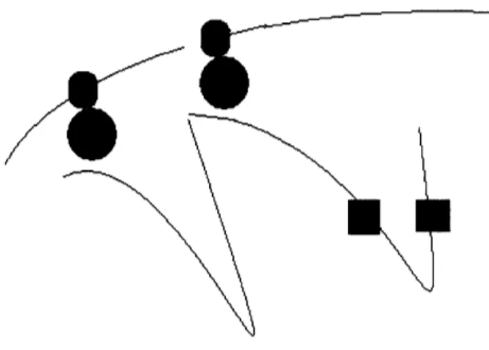
terms of the spatial locations of transcribed genes. For complexes to form, for example, the components must diffuse into the same region of space and encounter each other in the proper configuration (1 1). This probability is increased when the loci of translation (for proteins) or transcription (for RNAs) are closer together. In bacteria, this can be achieved by having the physical locations on the genome where the components are transcribed close together in space (figure 2.1, black squares.) Another possible
Figure 2.1 Two types of distance constraints motivated by transcription in bacteria:
membrane genes (blue) close to the membrane and genes that code for components of a protein complex (black)
Figure 2.2 Highly transcribed genes (yellow - one labeled i) will be located close to
transcriptional constraint in bacteria is that membrane protein coding genes be close to the membrane where they can be transcribed, translated, and then directly inserted into the membrane in a process called transertion which may itself play a large role in
structuring the chromosome (figure 2.1 red circles.) (12). A third transcriptional
constraint is that genes with high levels of transcription be located close to regions of high transcription activity ("transcription factories") thought to be located at specific positions in the cell (figure 2.2.) For fixed transcription factories, genes that are highly
transcribed will, of necessity, spend much of their time attached to the factories. It is believed that the transcriptional factories are one of the most important organizing factors of chromosome structure (6). There are many other possible transcriptionally motivated
constraints such as those that locate transcription factors close to their binding sites and
those that optimize the relative spatial positions of enzymes involved in metabolism (13). It is useful to note two possible ways in which transcriptional constraints could act upon chromosome structure. One is through the evolutionary positioning of genes linearly along the genome and the selection of specific sequences that may control the geometry of small regions of the chromosome (for instance AT tracts cause bends or binding sites for specific chromosome conformation altering proteins can cause regions
to take on specific geometries.) Another is the product of the "mechanical forces"
between proteins or RNAs that are still attached to their genomic loci. In bacteria, when proteins that are attached through ribosomes and mRNAs to their sites of transcription interact, they may cause these sites to spend more time close together thus enforcing particular structures. We can imagine this happening with ribosomal RNAs in the
eukaryotic nucleolus, for instance, where all of the ribosomal RNAs are colocalized and transcribed.
Modes of Bacterial Chromosome Replication
During reproduction, the entire genome must be copied and segregated into the
daughter cells. In bacteria, this process is often depicted with simple diagrams using circles to represent the replicating chromosome. However, these diagrams ignore the compactness of the genome inside the cell. The situation is instead one where a circle, folded hundreds of times, is duplicated and segregated. The fact that the genome must be so compact during this process places many constraints on the structure and these
constraints can be expressed mathematically given a replication model.
Topological and spatial factors affecting genome replication have been discussed many times in the literature, and theoretical and experimental evidence for a bidirectional
paired fork process has accumulated. (8, 14, 15). We have used the paired fork process as
a basis for formulating replication constraints for our chromosome model. The paired fork is a set of four linked DNA polymerases, which is probably fixed in space.
Replication proceeds in both directions (with two polymerases in one direction and the
other two in the opposite directions.) Since the polymerases are linked and fixed, it is the
chromosome, not the polymerases that move. The chromosome is pulled through the polymerase complex and daughter DNA emerges on either side. The effect is that while
the DNA is replicated, it is separated into different sides of the cell (figure 2.3.)
There are several ways in which replication constraints can be incorporated into
models. If the diffusion of the chromosome through space is faster than the rate of replication, we can expect folding of the newly generated chromosome as it grows from the replication forks. However, the constraints on the fold at any given moment will only be those that are derived from the portion of the new chromosome that has been already replicated or its interaction with the other daughter chromosome or the remaining
unreplicated chromosome. As the replicated chromosome grows, new constraints will be active, but these new constraints will be acting on a structure that is already partially folded. Thus the space of possible folds that meet the constraints at later times is
dependent on the space of possible folds at earlier times. This results in final folds (after replication) that reflect the history of the growing replicated chromosome (figure 2.4.) The effects are analogous to known dynamic effects in protein folding where the final conformation of the protein is affected by folding of the partially translated protein intermediates before translation is completed.
Replication yields constraints related to the ability to segregate and disentangle structures as they go through the replication process. Daughter structures that are more entangled, are more difficult to separate, requiring greater topoisomerase activity. It is possible to quantify entanglement by examining overlap between replicating structures using various methods, for example support vector machines which can determine the optimal surface separating two structures Furthermore, since loci symmetrically opposite the origin of replication are pulled through the polymerase complex simultaneously, it is possible to impose constraints that reflect this symmetry, for instance by enforcing a
Figure 2.3 A diagram of possible chromosome replication mechanism. Left: the classical view; Right: the paired fork model; Center: an intermediate state, shown to emphasize the transformation from one model to the other.
Figure 2.4 At left, representations of the matrices of distances between points on a
growing chromosome and at right, the structures.
(--I
PIk-~
L
.
.
symmetric flattened circle shape (flattened about the origin and terminus) for the entire chromosome.
Distance Geometry at the Genome Level
The static constraints on chromosome structure can be represented as upper and lower bounds on the distances between chromosomal loci. Proximity of a membrane
protein-coding gene to the membrane (modeled as a sphere), for instance, can be
enforced by setting a lower bound on the distance between the center of the cell and the membrane protein-coding gene. Structures can then be found which satisfy these constraints.
The constraints do not completely determine the structure of the chromosome, rather, they limit the space of possible conformations. This conformation space effectively defines both the number of distinct structure classes consistent with the
constraints and the flexibility of the chromosomal loci in each structure class. It is
therefore important to have a means, not only of generating structures consistent with the constraints, but also of performing an unbiased sampling of the conformation space to determine the ensemble of structure classes. Fortunately, nuclear magnetic resonance spectroscopists have faced similar problems determining the structure of proteins from NOE (Nuclear Overhauser Effect) data (16, 17). We have adapted one of their methods based on the mathematics of distance geometry for our models.
Method of Distance Geometry
Mathematically, the upper and lower bounds on distances between loci define the conformation space. However, this space is defined in terms of distances and not in terms of the coordinates necessary to visualize a structure and examine many of its properties. Distance geometry takes the distance bounds and finds coordinates that are consistent with them in three-dimensional space. For an under-constrained structure, the problem of choosing a distance matrix that satisfies the bounds is NP hard (non -polynomial time) but heuristic algorithms exist that are quite efficient at finding solutions.
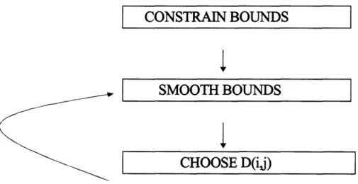
Our algorithm is an adaptation of the EMBED algorithm of Timothy Havel (16,
17). It consists of four steps. 1. Bound Smoothing, 2. Metrization, 3. Projection, and 4. Optimization. The bound smoothing step addresses the issue that although the initial
distance bounds constrain only a small subset of pairs of loci, Euclidean distances are not independent of each other and thus the bounds contain much more information than the
specific distances that they constrain. Bound smoothing makes some of these
dependences into explicit bounds. The best known example of a distance dependence which is a property of the geometry of a space is the triangle inequality which restricts
the feasible distance between a triplet of points i, j, and k. (dij < dik + djk)
There are additional higher order distance relations between the distances of 4, 5, 6, ..., n points from each other in three-dimensional space. These higher order relations are mathematically complex and computationally time consuming and therefore
infeasible to apply iteratively to the problem of distance geometry as existing algorithms require. Our algorithm performs bound smoothing with only the triangular inequality, a
good approximation that is standard practice in the field. This bound smoothing serves to significantly increase the number of explicit constraints on the structure.
The second step in the algorithm is to choose a set of distances that are consistent
with the bounds. This step, called metrization, results in the selection of a random
distance matrix that is representative of a single structure. One at a time, distances are
chosen randomly between the bounds and after each choice, the algorithm performs an
additional step of bound smoothing to incorporate the changes in the constraints that depend on the new distance (figure 2.6)
Metrization results in a distance matrix that represents a single structure. The next step, projection, is used to generate a set of coordinates that are consistent with the
distance matrix. Since our bound smoothing algorithm only smoothes based on the triangular inequality, the distance matrix generated after metrization is guaranteed to be consistent with distances between the n points in n -1 dimensional space not necessarily
in three dimensions. The projection step finds coordinates in three dimensions with distances that are closest to the original metrization distances (figure 2.7 and box 2.1).
This projection into three-dimensional space often results in minor violation of
the initial constraints. The final step in the algorithm is optimization of the coordinates so
that they minimally violate the initial distance bounds. We perform the optimization
using a simple error function and the method of steepest descent.
The entire distance geometry process results in an unbiased sampling of the conformation space defined by the initial distance constraints. Additionally, the bound smoothing step produces several useful results. First, it allows the consistency of the constraints to be determined in n- dimensions. This is useful in determining if any
Figure 2.5 The pro cess of distance geometry generates a matrix of distances (pictured on
the right) that are consistent with a set of distance bounds (pictured on the left) Blue in the left matrix indicates a distance that is unconstrained (except for along the diagonal where it indicates zero distance from a point to itself.) The other colors represent upper bounds on the distances, red indicating the largest.
CONSTRAIN BOUNDS
SMOOTH BOUNDS
CHOOSE D(ij)
Figure 2.6 Diagram of the process of metrization using distance geometry
x
y z
Coordinates
Figure 2.7 The projection process takes a matrix of distances and gene rates a set of coordinates in three-dimensional space.
Givenna r ix of all pirwise distans betehe n poits, D, one can gensate a
matrix of .mn roduds M by i od -ing anext ra pnt, the origin, which is usuallyr takenm be the ceru d ofthe dtac dat This matrix can be faoredusing SVD cr
rincipl wzoaiedrt ansis to ie the corinavts oftthe oginal structue (up to an
artrary$trMW andxttion) This canbe eyqrssed;k
He, U is an nx n ntrix ofones, andI is tbe n insional idetity matrix
FactringM into
U S V =US, Sm2 V' =L Lt.- X X
where Xis ann x 3 matrix of the coodinates. The last eulity follows from the fact that L* L is a bw triangi matrix times its Uanspo, we L will have all zros except
for ie top 3 rows (if we ae Llking about atee dior ucna re.) Since there is only one fact on ofa matrix into lor andupper triangular matrices andwe can cho:se X cea such that X is an upper triangular matrix. Then X andL must be ide,izcat
subsets of the constraints are impossible to simultaneously satisfy. Second, bound smoothing generates a set of lower bounds and upper bounds that take into account all triangular inequalities. These lower and upper bound matrices are themselves
independent distance matrices which represent the most expanded and most contracted structures in n-1 dimensions that are consistent with the constraints.
Results from Mycoplasma
We used the distance geometry method to model the chromosome structure of the bacterium Mycoplasma pneumoniae (18). It is nearly a minimal cell with a genome that is
816 kbp long and only 688 genes. It has limited metabolism, no known regulation, and very few DNA binding proteins (figure 2.8). Since it is so simple, we expect chromosome
structure to be under strong evolutionary selection. We constrained the 110 membrane
protein coding genes to be close to a spherical membrane with the diameter of an average
Mycoplasma cell. We also constrained the 52 annotated ribosomal protein-coding genes
to be close to each other. Furthermore, we constrained the origin and terminus of
replication to be at opposite poles of the cell, an observation that has been made in many microscopy experiments. Finally, to reflect the geometry of the paired fork model, we modeled the genome as a flattened circle (with origin and terminus of replication as poles.)
This resulted in a model with 165 loci (13,530 distances) and 1547 constraints.
With this input, the implementation of our algorithm in MATLAB takes about twenty
minutes to generate a structure. It is therefore possible to generate hundreds of structures
in a few days time. Below we present some statistical data from analysis of 160 structures
generated using the Mycoplasma constraints. Figure 2.9 shows a particular structure.
Note that the membrane protein coding genes (in blue) are all close to the membrane and
the ribosomal protein coding genes (in red) cluster together. No connecting DNA segments are shown because the chromosomal distance between subsequently modeled
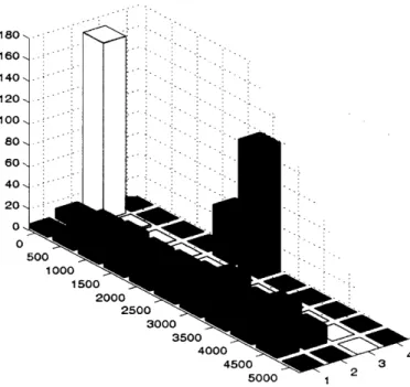
loci is sufficient to stretch across the cell. Figure 2.10 shows the distribution of distances
between two membrane proteins, a membrane protein and a ribosomal protein, two ribosomal proteins, and between a ribosomal protein and the center of the cell. These distributions show that the constraints have been satisfied and show evidence of clusters of distinct structure classes. For instance, the membrane - membrane protein distribution appears to be bimodal. Further statistical analysis is necessary before deciding if the
peaks are significant.
Optimization Algorithms
Distance geometry is extraordinarily useful as a rigorous way of defining the
conformational space accessible to a structure given a set of known constraints and also
in allowing the implications of these constraints to be derived. However, within the framework of distance geometry, the constraints implemented are hard (they must be
satisfied) and, in the case of chromosomes, there are many aspects of a fold that may
reflect optimal, desirable properties but not necessarily required properties. For example the spatial colocalization of genomic loci encoding components of large protein
complexes may allow for maximally efficient assembly of complexes but such complexes
816 Kbp
90% Coding688 Genes
110 Membrane Proteins
52 Ribosomal Proteins
No Active Transport
No Regulation
Limited Metabolism
Few DNA Binding Proteins
.5 ptm diameter (1470 bp)
Ui
! - WBlue Membrane
Red Ribosomal
Figure 2.8 Fundamental data about M. pneumon iae (left), and a diagram (right) with the
positions of membrane and ribosomal genes along the chromosome
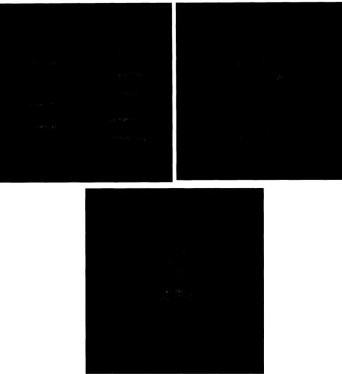
l t I i / ma !a # .--- --- ' gov`
Figure 2.9 A visualization of a particular structure. Green sphere: membrane. Blue dots:
180-.
160
-140-.
120-100 .
80 60 40 20 0 0 Membrane-Membrane _ Membrane-Ribosome | Ribosome-Ribosome _ Ribosome-Center 500 1000 1500 2000 2500 3000 3500 4000 4500 2Figure 2.10 Results of the statistical analysis of 160 runs of Distance Geometry and structure refinement programs for the chromosome of M. pneumon iae.
Each histogram displays the distribution of distances (in Angstrom) between two chosen points.
. ,
4
might still be assembled less efficiently without spatial clustering of the coding loci. These sorts of properties may be optimized subsequent to a hard constraint method like distance geometry, by constructing a cost function reflecting the degree to which a particular structure satisfies the desired properties and using an optimization process to
find structures which minimize the cost. For example we can write the degree to which a
conformation spatially colocalizes the ribosome components and places membrane
protein coding genes close the membrane in the cost function below.
C=
w-
Ed(Mi)
+w
2'd,R)
i ij
Equation 2
(where d(Mj,t) is the distance between transmembrane gene i and the membrane,
and d(Rj, Rj) is the distance between ribosomal genes i and j.)
Therefore, we can use distance geometry to produce an ensemble of structures satisfying an initial set of hard constraints and then optimization based on a cost function with high weights maintaining the hard constraints of the distance metrization to find optimal structures within this reduced ensemble. We developed two algorithms to find configurations that represent optima.
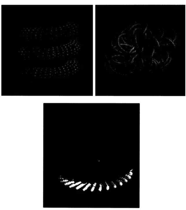
Helichrome
Our first algorithm uses a set of six parameters to describe the chromosome
structure. Here, the constraints are not derived from an initial distance geometry
metrization but rather from the set of parameters that describe the space of feasible structures. The parameters define a supercoiled helix which can assume a diversity of structures from simple helices to complex structures such as the structure pictured in figure 2.11 top right panel. Supercoiled helices are consistent with known chromatin structure in higher organisms and have several other desirable characteristics: they automatically enforce the polarity of the origin and terminus of replication, and add a degree of order to the structure which should solve some problems of entanglement associated with replication. Box 2 (top) lists the six parameters. Large and small refer to the primary helix and supercoil, and the large helix frequency is an extra oscillation that
changes the overall radius of the large helix as a function of the arc length.
Mathematically these structures are described by the equations for a local helix in the frenet frame defined by the large helix. The equations are listed in vector form in the bottom of box 2.
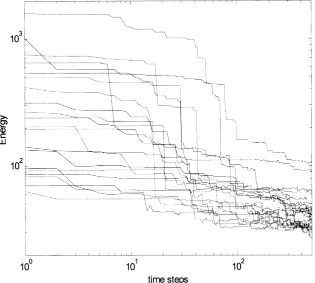
Using helichrome we perform a random walk in the parameter space, beginning with randomly generated initial structures and then randomly change parameters to generate test structures for each iteration. The cost of the test structure is compared with the previous structure. If lower, the new structure is accepted. If higher, it is accepted
with probability
e
C where AC is the difference in cost between the new structure andthe old and
P3
is the 1/kT, a Botzmann factor that varies inversely with the temperature. The temperature is lowered according to an "annealing schedule" beginning with high temperatures at early iterations so that structures of higher energy are frequently acceptedand decreasing so that in later iterations the structures "anneal" to a final state.