HAL Id: tel-01444454
https://tel.archives-ouvertes.fr/tel-01444454
Submitted on 24 Jan 2017
HAL is a multi-disciplinary open access
archive for the deposit and dissemination of sci-entific research documents, whether they are pub-lished or not. The documents may come from teaching and research institutions in France or abroad, or from public or private research centers.
L’archive ouverte pluridisciplinaire HAL, est destinée au dépôt et à la diffusion de documents scientifiques de niveau recherche, publiés ou non, émanant des établissements d’enseignement et de recherche français ou étrangers, des laboratoires publics ou privés.
tumor targeting using optical imaging
Ioanna Theodorou
To cite this version:
Ioanna Theodorou. Evaluation of nanoparticles and aptamers for in vivo tumor targeting using optical imaging. Cancer. Université Paris-Saclay, 2016. English. �NNT : 2016SACLS247�. �tel-01444454�
NNT : 2016SACLS247
T
HESE DE DOCTORAT
DE
L’U
NIVERSITE
P
ARIS
-S
ACLAY
PREPAREE A
“CEA
F
ONTENAY AUX
-
ROSES
”
E
COLED
OCTORALE N°
577
Structure et dynamique des systèmes vivants
Sciences de la Vie et de la Santé
Par
Mlle Ioanna Theodorou
Evaluation of nanoparticles and aptamers for in vivo tumor
targeting using optical imaging
Thèse présentée et soutenue à Fontenay aux-Roses, le 13 Septembre 2016 : Composition du Jury :
Pr. Fabrice Confalonieri, Professeur de Univ. Paris-Sud, (I2BC) (Orsay, France) Président Dr. Jean-Luc Coll, Directeur de recherche, INSERM-UJF U823 (Grenoble, France) Rapporteur Dr. Giannis Zacharakis, Chercheur, FORTH– IESL (Heraklion, Crete, Grèce) Rapporteur Pr. Elias Fattal, Professeur de Univ. Paris-Sud, CNRS UMR 8612 (Châtenay-Malabry, France) Examinateur Dr. Thomas Pons, Chercheur, LPEM/ESPCI ParisTech & CNRS UMR 8213 (Paris, France) Examinateur Dr Frédéric Ducongé, Chercheur, CEA/I2BM/MIRCen/LMN Directeur de thèse
Knowledge is a tree. Is growing up just like me.
Acknowledgments
The works of this doctoral thesis were carried out partly at the Service Hospitalier Frédéric Joliot (SHFJ) and were completed at the Molecular Imaging Research Center (MIRCen). I want to thank Dr. Philippe Hantraye for having me in the premises of MIRCen.
Firstly, I want to address a warmly thank you to the members of my thesis committee: To Proffesor Fabrice Confalonieri for accepting to chair this jury commitee.
To Mr. Jean-Luc Coll, Mr. Giannis Zacharakis, Mr. Elias Fattal and Mr. Thomas Pons for the honor of being referee reviewers and examinators of this work.
I want to express my sincere gratitude to Frédéric Ducongé, my supervisor, for his supervision and quidance, for being open minded and willing to consider new ideas, for always being available for long or short discussions, not only about the scientific context of the thesis, but also for other subjects outside the lab. I would like to equaly thank him for his time and patience with me during these three and a half years.
Special thank you to Eric Doris and his group members, notably to Edmond Gravel, for their work on the nanoparticles studied here, for our excellent, fruitful collaboration and for all the help they provided during my thesis. A sincere thank you to Parambath Anilkumar, who co –authored the article on the zwitterionic micelles.
I also want to thank Bernard Rousseau for being my tutor and Regis Tournebize for his participation to my annual thesis committee.
To all the collaborators of the European META project, especially to Günter Mayer, Laia Civit, Juergen Groll, Ilona Zilkowski, Michael Kubbutat, Andreas Lingnau, Holger Weber and Jerzy Silberrin. I really enjoyed our scientific meetings during trips to Krakow, Wurtzbourg and Fribourg.
I want to thank everyone on our team for their kindness and their friendship during these three and a half years in the lab. Karine, Benoît J., Benoît T., former lab members, thank you for your valuable help and long time friendship outside the lab.
Anikite, thank for your friendship, your advices during my thesis, your support and … for being my concert – going partner !
Nam, Benoît L., and Chloe, for the great pleasure of working together, your warmth, your humor and for enriching the everyday athmosphere in our office. I could not have asked for better friends and lab mates. I want to thank all the other students, post-docs and researchers for sharing the first and second floor of MIRCen, it was a great pleasure to have shared so many good times in the lab, the animal facilities, around coffee, champagne and beers! Thank you Susannah, Nad, Marco, Michel and Yael for setting up a MIRCen climbing and cooking « club » and the good times spend during. Thank you for all these unforgettable evenings, the laughter, the long discussions and many other things! Special thank you to my friends outside the lab for their continuous support and love: Semina, Aggelina, Igor, Ellen, Clement and Lefteris.
Απόλλωνα, Λεάνδρε και Αλέξανδρε ευχαριστώ για την συνεχόμενη στήριξη, κατανόηση, ενθάρρυνση και αγάπη. Xωρίς εσάς δε θα μπορούσα ποτέ να ξεπεράσω της δύσκολες στιγμές που αντιμετώπισα κατά τη διάρκεια του διδακτορικού. Τάνια, ευχαριστώ για την υποστήριξη, τις ατελείωτες συζητήσεις και τις βόλτες μας στο Παρίσι. Τέλος, θέλω να αφιερωσω αυτή τη διατριβή στους γονείς μου Λένια και Θεόδωρο, τα αδέλφια μου Ανδρέα, Φρόσω, Μαρία και στα ανίψια μου Θεόδωρο και Λούκα. Η ενθάρρυνση και η αγάπη σας αποτέλεσε καταλυτικό παράγοντα για την ολοκλήρωση της διατριβής μου.
Table of contents
1. Foreword ... 8
1.1 Cancer ... 8
1.2 Hallmarks of cancer ... 10
2. Tumor targeting pathways ... 12
2.1 Motivation for the development of nanomedicine for cancer therapy ... 12
2.2 Passive targeting ... 15
2.2.1 Factors that affect the EPR effect and the passive tumor targeting of nanoparticles ... 18
2.2.2 Polyethylene glycol (PEG) coating ... 20
2.2.3 Zwitterionic coating ... 21
2.2.4 Different endocytosis pathways ... 22
2.3 Active tumor targeting ... 23
2.3.1 Mechanisms of active tumor targeting ... 24 3. Targeting objects studied during the thesis ... 27 3.1 Polymeric nanomicelles ... 27 3.2 Nanogels ... 31 3.3 Aptamers ... 33 3.3.1 General Selection principle ... 35 3.3.2 Aptamers selected against cell surface biomarkers ... 37 3.3.3 Aptamers conjugated to nanoparticles for cancer targeting and therapy ... 40 3.3.4 Aptamers for molecular imaging applications ... 43 3.3.5 Chemical modification of aptamers for in vivo use ... 45 4. Optical imaging in vivo: an asset for the development of new therapeutic molecules ... 47
4.1 In vivo molecular imaging: The various techniques ... 47
4.2 Optical imaging: Principles ... 52
4.2.1 Planar Imaging and tomography. ... 56
5. Objectives ... 62
6. Results ... 66
6. 1 Passive tumor targeting by nanoparticles ... 66
6.1.1 Zwitterionic Micelles ... 66
6.1.2 Nanogels ... 72
6.2 Active tumor targeting by aptamers selected from in vivo SELEX ... 100
6.2.1 Oligonucleotide evaluation by whole body Planar Imaging ... 101 7. General discussion and Perspectives ... 128 8. Materials and methods ... 136 9. Rèsumè ... 145 10. Annex ... 159 10. Bibliography ... 168
ABREVIATIONS
ART : Algebraic Reconstruction TechniqueA. U. : Arbitrary unit
CDD: Charge-Coupled Device CMC: Critical Micelle Concentration CT : Computed Tomography Cy 7 : Cyanine 7 dye
Da : Dalton
DHX9 : Human RNA helicase A DNA : Deoxyribonucleic acid Dtxl : Docetaxel
ECNG : Ester-Containing nanogel
ECPGSH : Ester Containing thiofunctional polymer EDTA : Ethylenediaminetetraacetic acid
EFNG : Ester-Free nanogel
EFPGSH : Ester-Free thiofunctional polymer EPR : Enhanced Permeability and Retention effect ER : Endoplasmic Reticulum
eV : Electronvolt
fDOT : fluorescence Diffuse Optical Tomography FDA : Food and Drug Administration
FMT : Fluorescence molecumar tomography FRI : Fluorescence Reflectance Imaging GFP : Green Fluorescent Protein
HPMA : N-(2-hydroxypropyl)methacrylamide copolymer ID : Injected Dose
i.v : Intravenous
IFP : Interstitial Fluid Pressure KD : Dissociation constant LED : Light-Emitting Diode MI : Molecular Imaging
MPS : Mononuclear Phagocyte System MRI : Magnetic resonance imaging NIR : Near InfraRed
NTA : Nitrilotriacetic acid
PI3K : Phosphoinositide 3-kinase inhibitor PCB : Poly(carboxybetaine)
PCR : Polymerase Chain Reaction PDA : Polydiacetylene
PDLLA : Poly(d,l-lactide) block copolymer PEG : Polyethylene glycol
PET : Positron Emission Tomography PG : Poly(glycidol)
PLGA-b-PEG : Poly(D,L-lactic-co-glycolic acid)-block-poly(ethylene glycol) PLLA : Poly-D,L- lactic acid
PSMA : Prostate-Specific Membrane Antigen PTX : Paclitaxel
PVA : Poly(vinyl alcohol) RES :Reticuloendothelial System RNA : Ribonucleic acid
RNAi : RNA interference ROI : Region of Interest RT-PCR : Reverse transcription
SELEX : Systematic Evolution of Ligands by EXponential enrichment siRNA : Small interfering RNA
sP(EO-stat-PO) : Poly(ethylene oxide-stat-propylene oxide TAM : Tumor-Associated Macrophages
US : Untrasound UV : Ultraviolet
GENERAL INTRODUCTION
1. Foreword
1.1 Cancer
Cancers represent one of the leading causes of morbidity and mortality worldwide, with 14 million new cases and claiming the life of 8.2 million people in 2012 (World Cancer Report 2014 from World Health Organization). Furthermore, the number of new cases over the next two decades is expected to rise by around 70% according to the World Health Organization (http://www.who.int/mediacentre/factsheets/fs297/en/).
Cancer, is a generic term for a group of diseases that can start almost anywhere in the human body. Malignant tumours and neoplasms are other terms used to describe cancer. It’s defining feature is the creation and rapid division of abnormal cells that grow or/and travel beyond their usual boundaries and often spread into adjacent tissues or organs with a processes that is referred to as metastasizing. These distant settlements of tumor cells are the cause of 90% of deaths (Sporn 1996).
More than 100 types of cancer exist (Hanahan and Weinberg 2000) and they are usually named from the organs or tissues where they develop. For example, cervical cancer starts in cells of the cervix, and hepatic cancer originates from cells in the liver. Cancers can also be described by the type of cell that formed them, such as an epithelial or a squamous cell
(http://www.cancer.gov/). Below are some categories of cancers that derive from specific cell
types:
Carcinomas
Carcinomas make up the most common type of cancer and they are developed by epithelial cells. Depending on the different epithelial cell types, carcinomas are divided to different categories. Some of those are :
1. Adenocarcinomas. Cancer that are developed in epithelial cells of the glandular tissues. Breast, colon, and prostate cancers are some examples of adenocarcinomas.
2. Basal cell carcinoma. Cancer that begins in the lower or basal layer of the epidermis, the outer layer of skin.
3. Squamous cell carcinoma. Cancer that develops in squamous cells, which are epithelial cells that are found either just beneath the outer layer of the skin, or in the lining of many other organs, such as the stomach, urinary bladder, cervix, intestines, lungs, and kidneys. 4. Sarcomas. Sarcomas are malignant tumours that appear in bone and soft tissues, including muscle, fat, blood vessels, lymph vessels, and fibrous tissue (tendons and ligaments). Osteosarcoma is the most common cancer of the bone. Other common types of soft tissue sarcomas are: Kaposi’s sarcoma and liposarcoma.
Leukemia
Also known as hematopoietic cancers, leukemias and lymphomas begin in the blood-forming tissue of the bone marrow or the lymph, and lymphatic system respectively (World Cancer Report 2014 from World Health Organization) and do not form solid tumors. Instead, there is a build up of abnormal white blood cells (leukemia cells and leukemic blast cells) in the blood and bone marrow, displacing normal blood cells, resolving low level of the latter.
Brain and Spinal Cord Tumors
Similar to other cancer types, these tumors are named based on the cell type of the central nervous system in which they formed. Gliomas and glioblastomas belong in this category of brain tumors.
Other Types of Tumors:
Germ Cell Tumors
This type of cancer develops from the cells that give rise to sperm cells and oocytes. They can occur almost anywhere in the body and can be either benign or malignant.
Neuroendocrine Tumors
Neuroendocrine tumors arise from cells that produce and release hormones into the blood stream in response to a signal from the nervous system. These tumors, produce higher than
needed concentrations of hormones, causing different symptoms. Neuroendocrine tumors may be benign or malignant.
1.2 Hallmarks of cancer
Douglas Hanahan and Robert A. Weinberg in their important review article « the hallmarks of cancer » suggested that tumorigenesis in humans is a multiscale process that starts from genetic alterations or DNA lesions that drive the progressive transformation of normal human cells into malignant derivatives. More precicely, they argue that the presence of six cell physiology alterations lead to tumor growth. Those are self-sufficiency in growth signals, insensitivity to growth-inhibitory (antigrowth) signals, evasion of programmed cell death (apoptosis), limitless replicative potential, sustained angiogenesis, and tissue invasion and metastasis. Each one of these physiologic changes are acquired during tumor development (Figure 1) and disrupt the anticancer defense mechanism of normal cells and tissues.
Figure 1 Acquired capabilities of cancer. Most types of human tumors have acquired and share the same set of
functional capabilities during their development (Hanahan and Weinberg 2000)
Besides genetic factors, cancer might also arise from external agents for instance infections from viruses, bacteria or parasites, ultraviolet and ionizing radiation, many chemical carcinogens such as asbestos dust, components of tobacco smoke, arsenic, etc. Age is another fundamental factor for the development of cancer because of the accumulation of risks for certain types of cancers combined with the increasing weakening of cellular repair
mechanisms as the person is growing older
Nowdays, there is increasing knowledge of the causes of cancer and thus the disease can be sometimes reduced and controlled by the development and implementation of strategies for prevention, early detection and patient managment. As a result, many types of cancer can now be treated and the mortality can be decreased, especiallyif they are detected and treated in early stages. Two main strategies of early detection include the early diagnosis of cancer from rising awareness for signs and symptoms (for detecting skin, oral, breast and cervical cancer for example) and screening methods for identifing abnormalities that could lead to malignacies.
Effective cancer treatment requires a correct diagnosis mostly because each cancer type needs a specific treatment plan that might include one or more modalities such as surgery, and/or radiotherapy, and/or chemotherapy. Additional treatment regimens are immunotherapy and hormone therapy for certain tumor types.
The primary goal of tumor treatment is to increase the possibility for tumor regression, prolonging and improving the quality of the patient’s life. In the same context, the development of new therapies, tumor targeting agents, biomarkers and systems for controlled drug delivey at the site of the disease is under intense investigation worldwide.
Currently, one of the most appealing and fast developing areas of research for drug delivery is the design and characterisation of nanomedicines. Nanomedicine is a hybrid field. Briefly, it is the medical application of nanotechnology. Nanomedicine extents from the medical applications of miniaturized biosensors to the design of nanomaterials for improvement of diagnosis and therapy. Part of the applications of nanomedicine concerns the development of multimodal nanosystems able to deliver simultaneously drugs and contrast agents to tumors (theranostic application).
In the first part of this introductory chapter we present the different pathway of tumor targeting. More precisely, we focus on two classes of targeting agents that were under investigation during this study: 1- different nanoparticles intented to improve the delivery of drug and/or contrast agent to tumor site, 2- aptamers that represent a new class of targeting agents able to bind specific markers expressed at the surface of cancer cells. Then, we will present small animal photonic imaging techniques that were used during the thesis for studying the biodistribution and tumor targeting of the two different targeting agents mentioned before.
2. Tumor targeting pathways
2.1 Motivation for the development of nanomedicine for cancer
therapy
When a drug is administered into the organism, it faces a series of obstacles that limit its efficacy. The drug will have to traverse different biological barriers such as cell membrane, vascular walls and intestinal mucosa. In addition, drugs can suffer from instability, poor availability and short half-life in vivo, resulting in quick elimination by the body. In addition cancer cells can develop resistance to drugs. But most importanly, they have limited tumor selectivity and can also accumulate in healthy tissues introducing many harmfull side effects such as hair loss, nausea, vomiting etc. For this reason, improving the specific delivery of chemotherapeutics to tumors is of great importance.
Over the past few years, nanomedicine has been greately involving in the development of nanoparticles, a term concerned with materials and systems whose structures have a length
scale between 0,1 to 100 nanometer (1 nm = 10−9
m). Such nanoparticles have been showing great promise to improve the drug delivery to tumors and, as a result, to increase the efficacy of treatments and simultaneously to decrease side effects (Wicki, Witzigmann et al. 2015). Nanoparticles developed for medicine, have particular properties such as nanoscale size, high surface-to-volume ratio, and favorable physico-chemical characteristics. They are often named nanocarriers or nanovectors (terms used to describe nanoparticles able to transport and eventually deliver one or more therapeutic agents or/and contrast agents for molecular imaging). These nanocarriers can be built from a wide range of components (Figure 2). They are usualy designed to modify the pharmacokinetic and pharmacodynamic profiles of drugs and therefore enhance their therapeutic index. Therefore, once loaded into nanocarriers, therapeutic agents can have higher in vivo stability and blood circulation time. Furthermore, nanocarriers can promote a controlled release of drugs.
Figure 2. Illustration of various nanotherapeutic platforms. There are different nanomedicine products such as
drug-conjugates, lipid-based nanocarriers, polymeric nanocarriers, inorganic nanoparticles and viral nanoparticles that are currently used in clinic (Wicki, Witzigmann et al. 2015).
There are several convincing arguments in favour of the development of nano-sized therapeutics (for review (Cho, Wang et al. 2008) and (Wicki, Witzigmann et al. 2015))
1- Many anti-cancer drugs have poor water solubility and chemical stability that can limit their bioavailability and may hinder their development. Nanoparticles may help to overcome these problems. For instance, the uptake and delivery of poorly soluble drugs may be increased by encapsulating the compound in a hydrophilic nanocarrier. Wortmannin (PI3K inhibitor and radiosensitizer), is an example for a drug whose development was hampered because of poor solubility and its toxicity. By encapsulating it into a lipid-based nanocarrier system, the solubility of wortmannin was increased from 4 mg/L to 20 g/L and its toxicity was three- to five-times lower compared with that of wortmannin alone (Karve, Werner et al. 2012).
2- Nanocarriers can protect anti-cancer compounds from biodegradation or early excretion (from liver or kidneys) changing their pharmacokinetic profiles. For example, drugs that are cleaved enzymatically (e.g., siRNA by RNAses in the plasma, proteins by pepsin or trypsin in the stomach) can escape enzymatic degradation by either encapsulation into nanocarriers or coupling to synthetic polymers.
3- Nanotechnology can help to improve the biodistribution and targeting of anti-cancer compounds. The in vivo distribution and tumor targeting of anti-cancer drugs is govern by their physico-chemical properties (charge, size, water solubility). Most of the time, anti-cancer drugs do not have specific accumulation in tumors and can be accumulated in several healthy tissues leading to harmful side effects. Nanocarriers can be used to improve the delivery of drugs to tumors while decreasing their undesirable diffusion to healthy tissue. 4- Nanotechnology has developed stimuli-responsive systems that release their payload upon physical, chemical, or biological stimuli. Such stimuli can promote release of drugs by interfering with the phase, structure, or conformation of the nanocarriers. For example, Du et
al. reported the increased cellular drug uptake and release of doxorubicin from a pH- sensitive
nanoparticle that can be degraded in the acidic environment of endosomes (Du, Du et al. 2011).
Today, many nanocarriers have made their way in clinical trials and some have already been approved to be used in clinic (Table 1). Notable examples are Doxil®, a PEGylated liposome encapsulating Doxorubicine (Muggia, Hainsworth et al. 1997; Soloman and Gabizon 2008) and Abraxane®, which is Paclitaxel bound to the protein Albumin (Fader and Rose 2009). Such nanocarriers are able to change the biodistribution and targeting of drugs by permitting them to accumulate preferably at the tumor site or proximity. This phenomenon is known as the enhanced permeability and retention effect (EPR) (Wicki, Witzigmann et al. 2015) and will be described in detail in the following section.
Table 1.
Approved drug conjugates and nanocarriers for cancer therapy. Table adapted from (Stylianopoulos and Jain 2015)
Nanomedicines Material Drug Indication
Abraxane Nanoparticle
albumin-bound
Paclitaxel Breast cancer, Pancreatic
cancer, non-small-cell lung cancer
DaunoXome Liposome Daunorubicin HIV-related Kaposi's sarcoma
Myocet Liposome Doxorubicin Metastatic breast cancer
Doxil/Caelyx PEGylated
liposome
Doxorubicin Kaposi's sarcoma, Ovarian
cancer, Breast cancer
Genexol-PM PEG-PLA
polymeric micelle
Paclitaxel Breast cancer, Lung
cancer, Ovarian cancer
Lipusu Liposome Paclitaxel Breast and non-small-cell lung
cancer
MM-398 Liposome Irinotecan Pancreatic ductal
adenocarcinoma
PICN paclitaxel injection
concentrate for nanodispersion
Paclitaxel Metastatic breast cancer
2.2 Passive targeting
Once the nanoparticles are administered (e.g., intravenous injection), they are distributed to different organs through blood circulation. At the same time they undergo elimination by the liver and/or excretion by the kidneys. They are also removed by cells of the reticuloendothelial system (RES) (macrophages, Kupffer cells) (Moghimi and Davis 1994). The delivery of nanoparticles from the injection site to target sites within a tumor, involves multiple kinetic steps starting with their transport through blood to tumors, extravasation from tumor vasculature, interstitial transport, binding to cell membrane, internalization and finally intracellular trafficking (Li, Wang et al. 2012). Additionally, the delivery of nanoparticles to a solid tumor is not only driven by physiological factors including tumor blood vasculature, lymphatic drainage and tumor interstitial fluid pressure (IFP), but also by the physicochemical properties of the nanoparticles. These properties include surface characteristics (charge and hydrophilicity) and particle size among others.
Pathophysiological features of cancers and their environment have been exploited for passive targeting. In particular, Matsumura and Maeda (Matsumura and Maeda 1986) in 1986
investigated, described and validated these features in detail several times (Matsumura, Oda et al. 1987; Maeda 2001; Maeda, Sawa et al. 2001; Maeda, Bharate et al. 2009; Maeda and Matsumura 2011). These studies demonstrated that most solid tumors have blood vessels with defective architecture and they usually produce increased amounts of various vascular permeability factors (Fang, Nakamura et al. 2011). As a result, intratumoral blood vessel endothelium is fenestrated with wide gaps between 100 nm and 780 nm of size (Hobbs, Monsky et al. 1998; Wicki, Witzigmann et al. 2015) and even up to 2 µm depending on the type of the tumor (Hobbs, Monsky et al. 1998). In addition, tumors, have defective lymphatic function and thus they exhibit poor lymphatic drainage. The combination of these two phenomena was termed as the Enhanced Permeability and Retention effect (EPR). This enhanced vascular permeability also ensures a sufficient supply of nutrients and oxygen to tumor tissues for rapid growth. By the same time, it has been observed that the EPR effect
does not occur in normal tissues (Maeda, Bharate et al. 2009).
This unique anatomical–pathophysiological nature of the tumor’s blood vessels, can be exploited to facilitate the transport of macromolecules and nanocarriers into tumor tissues for eventual release of therapeutic drugs locally in the extracellular area. Indeed, macromolecules larger than 40 kDa and several nanoparticles in the 70-200 nm size range could leak out from tumor vessels and passively accumulate in tumor tissues (Figure 3) (Lammers, Hennink et al. 2008). The discovery of the EPR effect was considered as one of the greatest breakthrough for antitumor therapy (Torchilin 2011). Accordingly, it is a landmark principle in tumor-targeting chemotherapy and it is now increasingly used as a standard for anticancer drug development.
Figure 3. Passive tumor targeting. Particles passively extravasate through the leaky vasculature and accumulate
in tumors through the EPR effect. Image adapted from (Farokhzad and Langer 2009)
The vast majority of nanomedicines developed for drug targeting to tumors rely on the EPR effect and it is generally referred to as ‘passive drug targeting’. These include long-circulating liposomes, polymers and micelles. Examples of passively targeted nanomedicines approved for clinical use are Myocet (non-PEGylated liposomal doxorubicin), Doxil (Caelyx in Europe; PEGylated liposomal doxorubicin), Daunoxome (non-PEGy- lated liposomal daunorubicin), Abraxane (albumin-based paclitaxel) and Genexol-PM (paclitaxel-containing polymeric micelles; pre-approved in Korea) (see Table 1). Additionaly, there are several other passively tumor-targeted nanomedicines currently in clinical trials as well as in early- and late-stage
preclinical development (Jain and Stylianopoulos 2010). When compared with conventional
anticancer drugs, most of which are small molecular drugs, these macromolecular drugs and nanocarriers experience greater in vivo pharmacokinetics and prolonged plasma half-life, but more important, greater tumor selectivity. Thus, they can improve antitumor effects with no or less adverse reactions (Fang, Nakamura et al. 2011).
However, the EPR effect is much more complex than initially defined. It encloses dozens of complex biological processes such as angiogenesis, vascular permeability, hemodynamic regulation, heterogeneities in tumor genetic profile, heterogeneities in the tumor microenvironment and lymphangiogenesis (Bertrand, Wu et al. 2014). In fact, it is a highly heterogeneous phenomenon. It varies to a large degree from tumor model to tumor model, as well as from patient to patient (Jain and Stylianopoulos 2010; Bae and Park 2011).
Furthermore, even within a single tumor, there are huge differences especially in vascular permeability. In many cases, there are parts of tumors in which particles as large as 200 nm are able to extravasate, while in many other parts, molecules of the size of albumin (3–4 nm), are unable to enter the interstitium. This is a result of either an intact endothelial lining, or because vascular leakiness is compromised by the presence of a dense perivascular lining, constituted of pericytes, smooth muscle cells, fibroblasts and/or tumor associated macrophages (Lammers, Kiessling et al. 2012). Similarly, as mentioned above, the distribution and accumulation of nanoparticles in tumors are affected by their physicochemical properties (Bertrand, Wu et al. 2014).
2.2.1 Factors that affect the EPR effect and the passive tumor targeting of
nanoparticles
The physical and chemical properties of nanoparticles affect their extravasation by influencing diffusivity, permeability through the vascular wall, their interactions with the cancer cells and the extracellular matrix. They eventually determine their pharmacokinetic behavior and tumor accumulation capacity. Furthermore, the physicochemical characteristics of materials used for therapeutic or diagnostic applications have an impact on how the organism eliminates them from blood circulation (Alexis, Pridgen et al. 2008; Bertrand and Leroux 2012). Additionally, the total blood exposure to the nanoparticles is a key factor influencing their distribution and their tumor targeting via the EPR effect (Maeda 2001).
Maintaining elevated blood concentrations of nanoparticles ensures diffusion toward the tumor (Dreher, Liu et al. 2006). Therefore, longer circulation times in the blood result in higher amounts extravasated into the tumor interstitium.
Moreover, the biodistribution and accumulation of nanoparticles in tumors is also affected by their size, chemical coating, charge and shape. More precisely, many in vitro studies demonstrated that nanoparticle size and surface chemistry considerably impact the interaction with plasma proteins (Lundqvist, Stigler et al. 2008) cellular uptake (Chithrani, Ghazani et al. 2006), toxicity (Ding, Stilwell et al. 2005) and molecular response (Jiang, Kim et al. 2008).
Size
The penetration of nanoparticles within the tumor tissue is highly dependent on the global size of the nanoparticle. Larger nanoparticles appear to stay near the vasculature while smaller nanoparticles can rapidly diffuse in the tumor matrix (Perrault, Walkey et al. 2009).
Charge
Like other physicochemical parameters, the global charge of nanoparticles can alter both systemic circulation times and intratumoral interactions. Hence, the presence of surface charge can change the opsonisation profile of the material, its recognition by cells in the organs of the Mononuclear Phagocyte System (MPS) and its overall plasma circulation profile
(Alexis, Pridgen et al. 2008). Negative surface charges can either increase, decrease or have
no impact on the blood clearance of nanoparticles (Levchenko, Rammohan et al. 2002), but
positive charges are generally recognized as having a negative effect on the plasma exposure to the nanomaterial (Xiao, Li et al. 2011) For example, Juliano et al. (Juliano and Stamp 1975), reported that neutral and positively charged liposomes were eliminated less rapidly than their negatively charged counterparts, which could be explained by the tendency of negatively charged liposomes to associate in the presence of proteins and calcium ions in blood plasma. On the contrary, Yamamoto et al. (Yamamoto, Nagasaki et al. 2001), demonstrated that there was no important difference in the blood clearance kinetics for neutral and negatively charged PEG-PDLLA micelles. Nevertheless, negatively charged micelles reduced significantly the non-specific uptake by the liver and spleen, when compared with neutral micelles, which was attributed to the electrostatic repulsion between the negatively charged micelles and the cellular surface. While those results from the above studies seem to be inconsistent, an explaination for it may lay to the difference of nanoparticle types, the variation in stability of nanoparticles resulted from surface charge, the nature of charged groups, and other factors such as inhomogeneous particle sizes.
Shape
The shape of nanoparticles can influence their interactions with the MPS (Bertrand and Leroux 2012). For example, single-wall carbon nanotubes with high aspect ratio (length divided by width, i.e., from 100:1 to 500:1) were shown to be eliminated efficiently by the kidneys despite their dimensions (i.e., 100–500 nm) 10–20 times above the usual glomerular filtration threshold, suggesting that elongated shapes could provide benefits to the filtration
process through porous structures (Ruggiero, Villa et al. 2010). Recently, the tumor
distribution kinetics of nanorods with a length of 44 nm (aspect ratio: 10) were compared to those of 35 nm nanospheres showing the same hydrodynamic radius (Chauhan, Popovic et al.
2011). Regardless of similar blood circulation profiles, the nanorods were shown to
together, these findings suggest that elongated shapes might present beneficial EPR properties (Bertrand, Wu et al. 2014).
Coating
Nanoparticulate systems are highly susceptible to association with plasma proteins, lipids and other biomolecules, leading to formation of a dynamic biomolecular « corona » making it difficult to predict and/or control their behavior in vivo (Walczyk, Bombelli et al. 2010). To overcome this issue, the surface of the particles is usually functionalized with molecules that can provide « stealth » properties to the nanoparticle and avoid the corona formation. There are different possibilities for fuctionalization with polymers like PEG, PVA, and Dextran. Below we will present two types of surface functionalization that were studied during this thesis project. Those were PEG and zwitterionic coating.
2.2.2 Polyethylene glycol (PEG) coating
Polyethylene glycol (PEG) has been extensively used to modify the pharmacokinetic properties and in turn modifying pharmacodynamics of drugs and/or nanoparticles (Harris, Martin et al. 2001). PEGylated nanoparticles experience long circulation times, and this appears to be dependent upon the molecular weight of the PEG chain used. When PEG of different molecular weights were intravenously injected into mice, it was found that the circulation half-life of PEG 6000 (molecular weight of 6000 g/ mol) was only less than 30 min, while it was extended to a day with PEG 190,000 (Yamaoka, Tabata et al. 1994).
PEGylation reduces the renal clearance of compounds increasing their size to be higher than 8-10 nm. Moreover, hydrophilic polymers, such as PEG, can provide steric stabilization by hindrance and award « stealth » properties to the nanoparticle’s surface and prevent protein absorption onto it. The absorption of serum proteins (opsonins) on the surface of nanoparticles or foreign organisms is known as opsonization. Obsonization could lead to quick aggregation that makes the nanoparticles detectable to phagocytic cells resulting their rapid clearance of the nanoparticles from the bloodstream (Owens and Peppas 2006). Indeed, the opsonized particles can be cleared by receptor-mediated mechanism due to the high concentration of phagocytic cells in the liver (Kupffer cells) spleen, lungs, and bone marrow (Moghimi, Hunter et al. 2001).
serum protein absorption, still the fraction of nanoparticles entering tumors is quite limited. Generally, only around 5% of the administered particles circulates after 12 h with about 80% of the initial dose being cleared in few hours (Hong, Zhu et al. 2009). Nonetheless, several studies demonstrated a >100% increase in tumor accumulation of nanoparticles following PEGylation (Litzinger, Buiting et al. 1994; Hong, Zhu et al. 2009). The majority of i.v. administered nanoparticles will still end up in the liver and spleen, with a lesser extent being in the lungs and kidneys. In addition, >90% of the PEGylated nanoparticles are removed from the bloodstream within several hours and only around 2% of the total administered dose is accumulated in the tumor, resulting in low targeting specificity (as defined by the amount of nanoparticles in tumor than that in liver) (Bae and Park 2011; Liu, Yu et al. 2013).
2.2.3 Zwitterionic coating
Most efforts to achieve biocompatibility of nanoparticles have focused on functionalizing their surface with polyethylene glycol chains (PEGylation). However, in order to avoid phagocytosis, PEG chains need to have high molecular weight (Owens and Peppas 2006) which increases the overall hydrodynamic diameter of the nanoparticles.
Recently, the use of zwitterionic coatings as an alternative strategy for escaping non-specific adsorption of biomolecules onto nanoparticles, has been proposed (for review, (Pombo Garcia, Zarschler et al. 2014). Zwitterions are polyelectrolytes that contain both positively and negatively charged groups, but they are overall neutraly charged. They hold anti-fouling properties through strong ionic structuring of water, creating a highly hydrophilic surface (Kane, Deschatelets et al. 2003). Additionally, the global surface charge of a zwitterionic coating is internally balanced (Zeta potential close to zero).
Cell membranes served as inspiration for zwitterionic entities, and several of them have been investigated as biocompatible coating materials for different types of nanoparticles such as Quantum Dots and gold nanoparticles. These coatings promote the production of nanoparticles that are stable over a wide range of pHs and salt concentrations, with little increase of their hydrodynamic diameter in vitro and/or in vivo. Furthermore, they protect the nanoparticle’s surface from non-specific biomolecule absorption (Figure 4), provide minimal non-specific cellular adhesion and, minimal uptake by macrophages. Such chemical coatings can be assembled from low-molecular weight materials and can potentially provide a more adaptable means with desirable in vivo behavior compared to PEGylation. Most importantly,
they offer the opportunity to develop nanoparticle that are small enough to be eliminated into the urine via the kidneys, a highly desirable advantage for in vivo imaging (for review (Pombo Garcia, Zarschler et al. 2014)).
Figure 4. Graphic representation of resistance to non-specific protein absorption of a zwitterionic-coated
nanoparticle. When nanoparticles are exposed to any biological environment, biological (macro)molecules are absorbed non-specifically onto their surface. Coating with biocompatible zwitterionic moieties minimizes or prevents non-specific biomolecule absorption and cellular adhesion, and reduces elimination by the MPS (Pombo Garcia, Zarschler et al. 2014).
To conclude, this class of nanoparticle coating represents a particularly significant development, since this strategy provides the possibility of producing diagnostic/therapeutic agents with promising targeting properties. For example Li et al., developed zwitterionic poly(carboxybetaine) (PCB) modified liposomes for systemic delivery of siRNA therapeutics. The zwitterionic liposomes demonstrated extended blood circulation time -compared to PEGylated counterparts- and enhanced the tumor accumulation of siRNA following injection (Li, Liu et al. 2015).
2.2.4 Different endocytosis pathways
Increasing the passive tumor targeting of a drug is not enough to acquire effective therapeutic outcome. The efficacy of nanoparticles depends also on their ability to reach various sites of action including tumor vasculature (e.g., anti-angiogenics), tumor interstitium (e.g., drugs targeting extracellular proteins), cell membrane (e.g., antibodies), and intracellular compartments such as the cytosol (Kobayashi, Nakase et al. 2009) (e.g., RNAi) nucleus (e.g., DNA-active drugs) (Wagstaff and Jans 2009), and other intracellular compartments including
mitochondria, Golgi apparatus and endoplasmic reticulum (ER) (Tarrago-Trani and Storrie 2007; Yamada and Harashima 2008)
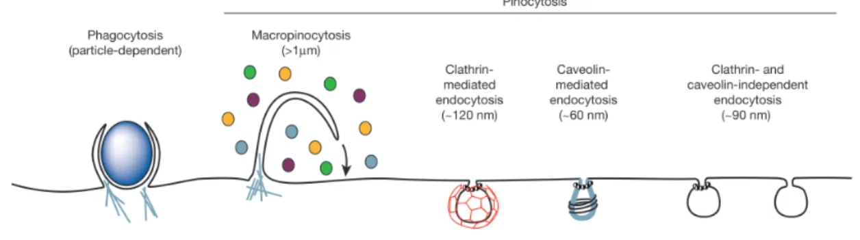
Investigations of the cellular uptake mechanisms of nanoparticles have shown that particles with sizes between 10 nm and 5 μm could typically be internalized into cells via endocytosis. Endocytosis is an active transport process, where the cell transports molecules (e.g., proteins) or particles, by engulfing and encapsulating them within a lipid bilayer in an energy-dependent manner. The resulting endosomes often maturate to late endosomes and lysosomes. However, this process is also highly dependent on the cell type, the internalization mechanism, and the properties of the interacting materials. Usually, endocytosis is classified into two categories: phagocytosis (cell eating), by which cells encapsulate only solid material, and pinocytosis (cell drinking or fluid endocytosis), where cells uptake a significant amount of liquid from the extracellular environment along with the internalized material. Phagocytosis is mostly limited to specialized cells such as macrophages and dendritic cells that interact directly with large material (>250 nm). On the other hand, pinocytosis occurs in almost all cell types. Pinocytosis can be subclassified into a number of separated mechanisms, including macropinocytosis, clathrin-mediated endocytosis, caveolae-mediated endocytosis, and clathrin/caveolae- independent pathways (Figure 5). Similar to phagocytosis, macropinocytosis is connected with the uptake of large material and can form endocytic vesicles up to 5 μm in diameter. On the other hand, clathrin and caveolae pathways are usually limited to smaller (<150 nm) particles (Doherty and McMahon 2009).
Figure 5. Representation of the endocytic pathways that differ regarding the size of the endocytic vesicle, the
nature of the cargo (ligands, receptors, lipids) and the mechanism of vesicle formation (Conner and Schmid 2003).
2.3 Active tumor targeting
Although the EPR phenomenon can increase the distribution of the drug-carrier to the tumor, it does not necessarily increase the ability of the drug to reach its pharmacological target.
Therefore, the optimization of a nanoparticle’s efficacy can also involve optimal drug release rates either through controlled diffusional release (Karnik, Gu et al. 2008). Without specific affinity of the nanomaterial for the cancer cells, the chemotherapeutic agents will have to find their pharmacological targets by their own or risk diffusing back into the vasculature (Dreher, Liu et al. 2006).
To overcome the above hurdles, the development of next generation nanomedicines with advanced functionalities is encouraged. Towards this goal, second-generation nanomedicines are based on drug delivery technologies with active targeting vectors and they hold the promise of improved targeting and increased efficacy (Farokhzad and Langer 2006).
2.3.1 Mechanisms of active tumor targeting
In the case of active targeting, a high-affinity ligand is attached to the surface of a nanocarrier. Then the ligand actively binds to a receptor on target/specific cells after extravasation (Peer, Karp et al. 2007). Specific binding on target cells may be achieved by attaching targeting agents on the surface of the nanocarrier. Such targeting ligands (molecules that bind to specific receptors on the cell surface) are attached to the surface by a variety of conjugation chemistries (Torchilin 2005). Nanocarriers will recognize and bind to target cells through ligand–receptor interactions, and bound carriers are internalized before the drug is released inside the cell (Figure 6). In general, when using a targeting agent to deliver nanocarriers to cancer cells, it is important that the ligand binds with high selectivity to receptors that are uniquely expressed on the cell surface.
A wide range of ligands have been used for such purposes, including small molecules such as folic acid, carbohydrates, or macromolecules such as peptides, proteins, antibodies, antibody fragments and aptamers. In general, the ligand must be chosen in such a way so it allows binding to the target cells while minimizing binding to healthy cells. Higher binding affinity of the ligand increases targeting efficacy. Nevertheless, in the case of solid tumors, there is evidence that high binding affinity can decrease penetration of nanocarriers due to a « binding-site barrier », where the nanocarrier binds to its target so strongly that the uptake from the tissue is hindered.
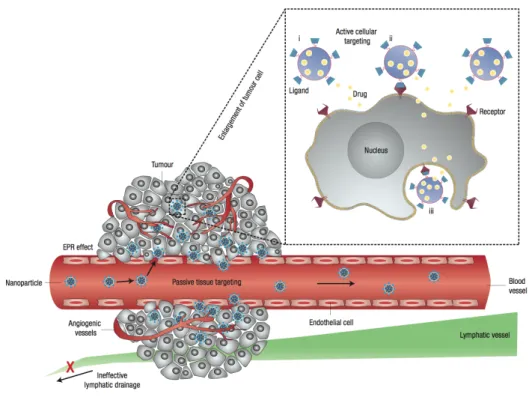
Figure 6. Schematic representation of different mechanisms by which nanocarriers can deliver therapeutic
agents to tumors. Passive tissue targeting is achieved by extravasation of nanoparticles through increased permeability of the tumour vasculature and ineffective lymphatic drainage (EPR effect). Active cellular targeting (inset) can be achieved by functionalizing the surface of nanoparticles with ligands that promote cell-specific recognition and binding. Thus the nanoparticles can (i) release their contents in close proximity to the target cells; (ii) bind to the membrane of the cell and act as an extracellular sustained drug release; or (iii) internalize into the cell (Peer, Karp et al. 2007).
However, the EPR-mediated passive extravasation remains the dominant phenomenon. Most of the time, nanocarriers do not accumulate into tumor tissues to a higher degree upon integration of specific ligands that bind to cancer cells. This happens because when nanomedicines leave the leaky tumor blood vessels and permeate into the interstitium, they need first find the tumor cells before being able to bind to them. This proceduce can be either more or less easy, depending on the tumor type and how they allow for extravasation. Therefore, it is very important to mention that it is wrong to speculate that active tumor targeting will increase target site accumulation (Lammers, Kiessling et al. 2012). Nevertheless, actively (cancer cell-) targeted nanomedicines can be taken up by cancer cells much more efficiently. For example, Davis and colleagues, demonstrated that overall, transferrin-targeted PEGylated gold nanoparticles did not accumulate in tumors to a higher extent than their untargeted counterparts. But, they could provide greater intracellular delivery of therapeutic agents to the cancer cells within solid tumors (Choi, Alabi et al. 2010).
The functionalization of ligands on the surface of nanoparticles can change the properties of both the targeting molecules and the nanomaterial (Kamaly, Xiao et al. 2012). The size,
geometry, surface properties (charge and hydrophobicity), and composition of nanoparticles can also alter their biodistribution and tumor targeting capacity (Figure 7). In some cases, nanoparticles have displayed benefits that go beyond simple delivery of drug. For example, strands of nucleic acids immobilized on the surface of nanomaterials are more resistant to nuclease degradation (Wu, Phillips et al. 2008; Seferos, Prigodich et al. 2009). Therefore it is crucial to first determine how the physicochemical properties of the nanoparticles affect the interactions with their targets in order to understand the properties of actively-targeted nanoparticles (Bertrand, Wu et al. 2014).
Figure 7. The physical and chemical properties of the ligands and nanoparticles affect the blood circulation
profiles, their biodistribution and their capacity to be internalized by cancer cells (Bertrand, Wu et al. 2014).
The density of the targeting molecules on the surface of nanoparticles affects their affinity for their target. In thermodynamic terms, the binding of a ligand to its substrate facilitates the subsequent binding of its neighbors and thus increased valency allows cooperative effects
(Bertrand, Wu et al. 2014). In biological terms, the multiple interactions of the nanoparticles
with the cell membrane causes the clustering and local concentration of receptors. This triggers wrapping of the membrane and leads to internalization by endocytosis (Mukherjee, Ghosh et al. 1997). Combined together, the detachment of the nanoparticles from the cell surface is hindered resulting in increased avidity. Therefore, the use of multiple relatively low affinity ligands is allowed to efficiently bind targets with high avidity. In vitro, improved
cellular uptake is a result of increased ligand density (Gu, Zhang et al. 2008; Stefanick, Ashley et al. 2013). For example, dendrimer nanocarriers conjugated to 3–15 folate molecules showed a 2,500–170,000-fold enhancement in dissociation constants (KD) over free folate when attaching to folate-binding proteins immobilized on a surface. This was due to the avidity of the multiple folic acid groups (Hong, Leroueil et al. 2007). However, ligand density should be carefully controlled because increase in affinity is not always linear. In some cases, the cooperative effect of the ligand can saturate and increase in ligand density can lead to harmful effects on cell binding (Elias, Poloukhtine et al. 2013). Improper orientation of the ligand, steric hindrance of neighboring molecules or competitive behaviors for the binding of the receptor are factors that cause those negative effects. For example, similar negatively cooperative systems have been observed with folic acid-targeted micelles, where the ligands
are arranged in patchy clusters (Poon, Chen et al. 2010).In this study, the architecture of the
targeting ligands on the micelles influenced the extent of receptor-mediated tumor uptake in cancer cells in vitro and in vivo. In other cases, high densities of hydrophobic ligands increased macrophage uptake of the nanoparticles, without impoving the receptor-mediated
internalization of polymeric nanoparticles (Valencia, Hanewich-Hollatz et al. 2011).
3. Targeting objects studied during the
thesis
There is great advancement of next-generation nanomedicine carriers with advanced functionalities. Among the various types of nanomedicine compounds that have been developed, are viral vectors, drug conjugates, lipid-based nanocarriers, polymer-based nanocarriers, and inorganic nanoparticles. However, not all of these different types of nanomedicine products are going to be described in this manuscript. Instead, special emphasis will be given to different types of nanoparticles (polymerized polymeric micelles and nanogels) and one type of active targeting agent (aptamers).
3.1 Polymeric nanomicelles
Micelles are colloidal dispersions of amphiphilic polymers (or amphiphilic molecules consisting of two distinct regions with opposite affinities regarding a solvent) made from a hydrophilic and hydrophobic domains. They spontaneously self-assemble to colloidal particles with a hydrophobic core and hydrophilic shell upon exposure to aqueous
environment. Concentration and temperature are crucial factors for the self-assembly of the micelles. In fact, at low concentrations, the amphiphilic polymers are individualized species and as the concentration increases, the polymers self organize to micellar structures upon reaching the critical micellar concentration. The formation of micelles also depends on the temperature, that is also known as the critical micellar temperature (Ogier, Arnauld et al. 2009). Their core–shell structure, and their highly hydrophobic inner domain, allows polymeric micelles to carry insoluble hydrophobic pharmaceuticals into aqueous solutions. When used as aqueous carriers, micelles can efficiently solubilize hydrophobic pharmaceuticals in their core (Torchilin 2004; Torchilin 2007).
Micellar structures hold some advantages over other delivery systems. Those include smaller size (allowing deeper diffusion into target tissues), surface and structure modularity, straightforward assembly, and high-loading capacity. Yet, micelles often suffer from poor stability, because their assembly depends mainly by the critical micelle concentration (CMC). Below the CMC, amphiphilic molecules exist as unimers, but as their concentration increases, they assembly into colloidal micelles. This process is reversible and the disassembling the micelles likely occurs in dilute conditions (for example, in the blood stream), leading to altered integrity of the carrier (Gravel, Ogier et al.). To solve this issue, the group of Dr. Eric Doris (CEA/IBITECS/Service de Chimie Bioorganique et de Marquage) developed stable micellar carriers, produced from the assembly of diacetylenic amphiphile monomers and subsequently photopolymerized to reinforce the micelle supramolecular architecture and stability (Ogier, Arnauld et al. 2010).
Encouraged by the above results, our group studied the potential of small polymerized micelles (with diameter of aproximately 10 nm), for imaging and drug delivery (Mackiewicz, Gravel et al.). The micelles were composed of diacetylenic (polydiacetylene, PDA) amphiphiles that upon UV irradiation at 254 nm, undergo polymerization forming polymerized/crosslinked micellar systems (Figure 8).
Figure 8. Schematic illustration of the synthetic steps followed for the production of photopolymerized PDA
micelles. CMC represents the critical micelle concentration. Image adapted from (Mackiewicz, Gravel et al.).
Following the above synthetic steps, the pharmacokinetics and in vivo biodistribution of polymerized micelles with three different chemical coatings was investigated in mice bearing subcutaneous tumors from human breast cancer MDA MB 231 GFP cells. The three micelles were synthesised by polydiacetylenic amphiphiles and decorated by polar heads made by either hydrophilic nitrilotriacetic (NTA) moieties, or PEG chains of two different molecular weights and lengths (Figure 9). Those were PEG350 (8 ethylene glycol units) or PEG2000 (45 ethylene glycol units). All micelles were labeled fluorescently with the Near InfraRed (NIR) dye FP730 and passive tumor targeting by the EPR effect was evaluated by optical imaging. Their results demostrated that the chemical coating of the micelles heavily influences their pharmacokinetics, biodistribution and tumor targeting properties (Figure 10).
Figure 9. Schematic illustration of structures of the different amphiphiles. Image adapted from (Mackiewicz,
Gravel et al.).
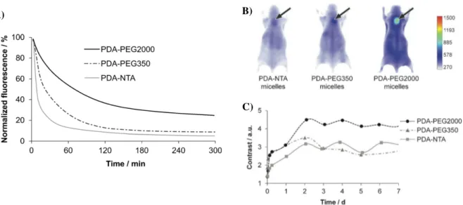
Figure 10. Pharmacokinetics and in vivo biodistribution of polymerized micelles with three different chemical
coatings. A) Evolution of the three FP730-labeled micelle concentration in blood as a function of time. B) Whole body NIR fluorescence images 48 h post i. v injection of the three fluorescent micelles in mice bearing MDA-MB-231 xenografted tumors (dorsal side comparison with arrows indicating tumors) and C) fluorescence contrast evolution over a period of 7 days. Image adapted from (Gravel, Ogier et al.).
The results showed EPR-mediated tumor accumulation of all micelles but strong differences were observed between the different micelle types. In particular, PEG2000-coated micelles experienced longer blood residence time, better tumor targeting and better imaging contrast than the other two micelles (Mackiewicz, Gravel et al.). The potential use of PDA-PEG2000 micelles as a drug delivery system was then assessed by loading the hydrophobic anticancer drug paclitaxel (PTX) into the micelles. Interestingly, the tumor growth was significantly decreased in the two groups of mice treated with Taxol® and PTX-loaded micelles which had more than 3-fold higher EC50. These results demonstrated that PDA-micelles are promising
systems for theranostic applications (Figure 11).
A) B)
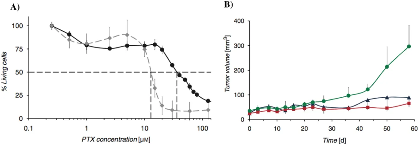
Figure 11. Therapeutic effect of Paclitaxel (PTX)-loaded PDA-PEG2000 micelles. A) In vitro cytotoxicity of
PTX-loaded PDA-PEG2000 micelles (black circles) compared to Taxol (in grey rectangular) on MDA MB 231 cells demostrated that the drug remains active in the micellar cargo. B) Evolution of tumor volume over 8 weeks for mice injected with NaCl 0,9% (green circles), Taxol® (blue triangles) and PTX-loaded PDA-PEG2000 micelles (red squares). Image adapted from (Mackiewicz, Gravel et al.).
The above study shows the important influence of the physicochemical properties on the pharmacokinetics, biodistribution and tumor targeting of different nanoparticles. Moreover, it is demonstrated that photopolymerized diacetylene micelles hold some advantageous features such as size, ease of synthesis and functionality and overcome important drawbacks of classical nonpolymeric micelles such as low stability, short circulation half-life and unstable drug retention.
3.2 Nanogels
Nanogels are nano-sized networks composed of hydrophilic or amphiphilic polymeric chains, which can be non-ionic or ionic (Kabanov and Vinogradov 2009). Initially they have been developed as drug carriers for controlled delivery. Additionally they can be designed to absorb biologically active molecules through formation of salt bonds, hydrogen bonds, or hydrophobic interactions. More precicely, polyelectrolyte nanogels can easily incorporate biomacromolecules such as oligonucleotides, siRNA, DNA and proteins, which bind with the nanogel ionic chains and phase separate within the finite nanogel volume. Because of that, the loading capacity of nanogels is superior to other drug carriers. Nanogels hold many advantages such as the possibility to make multiple chemical functionalities for introduction of imaging labels and targeting molecules. Several studies suggested a number of promising applications of nanogels, such as delivery of DNA into cells (Lemieux, Vinogradov et al. 2000), or siRNA (Lee, Mok et al. 2007), encapsulation of bioactive drugs such as doxorubicin (Missirlis, Kawamura et al. 2006), etc.
There are several approaches for preparing nanogels (for review (Kabanov and Vinogradov 2009). One of these approaches utilizes inverse microemulsions (water in oil, w/o) as a media for polymerization of monomers with bifunctional monomers added as cross-linkers to ensure formation of stable nanoscale networks (Figure 12).
Figure 12. Synthesis of nanogels by co-polymerization of monomers (1) and bifunctional cross-linkers (2) in
w/o microemulsions stabilized by surfactants (3). Image adapted from (Kabanov and Vinogradov 2009)
The utility of inverse microemulsion was demonstrated by the Matyjaszewsk group, where a disulfide-functionalized cross-linker was used to synthesize biodegradable nanogels (Oh, Tang et al. 2006). The same approach (inverse microemulsion) has been used by Groll et al., where they reported the formation of redox-sensitive and biodegradable nanogels with hydrodynamic diameter of 350 ± 50nm, using hydrophilic thiol functionalized prepolymers based on star shaped poly(ethylene oxide-stat-propylene oxide) (sP(EO-stat-PO)) and linear poly(glycidol) (PG). Nanogels were prepared by oxidative cross-linking of thiol groups to disulfide bonds in inverse miniemulsion using hydrogen peroxide (Groll, Singh et al. 2009). However, with this method there is poor control over the nanogel’s size and in addition the use of hydrogen peroxide as oxidation catalyst is unfavourable for embedding of peptides and proteins, as under certain conditions, disulphides are not the sole reaction products (Luo, Smith et al. 2005). Therefore, the same group also used alloxan (2, 4, 5, 6-tetraoxypyrimidine) as an alternative to the direct use of hydrogen peroxide for the formation of disulphide linked poly(glycidol) (PG) nanogels. The introduction of redox sensitive disulphide cross-links into the nanogels matrix, resulted in particles that were sensitive to degradation within reductive environments (Figure 13). Further studies demonstrated that these thiofunctional poly(glycidol) had no cytotoxic effect on L929 fibroblasts after incubation with
concentrations up to 10mg mL-1
(Singh, Zilkowski et al. 2013). However, they have not yet been tested for in vivo biodistribution experiments.
Figure 13. Synthesis of nanogels using alloxan for the formation of disulphide cross-linked nanogels and
resulting degradation within reductive environments such as the cytosol. Image adapted from (Singh, Zilkowski et al. 2013).
3.3 Aptamers
Recently, aptamers have emerged as a new class of tumor targeting agents. Aptamers are nucleic acid based structures obtained by directed molecular evolution process from a library
of 1014-1015 oligonucleotides containing a region of random base composition. This procedure
is called SELEX (Systematic Evolution of Ligands by EXponential enrichment) (Ellington and Szostak 1990; Tuerk and Gold 1990) and it consists of repetitive cycles of selection and amplification. Aptamers are made of DNA, RNA or non-natural oligonucleotides. The term «aptamer» was first introduced about 25 years ago (Ellington and Szostak 1990), and is derived from the Latin word aptus (meaning «to fit»). Aptamers can fold into three-dimensional structures (G-quartet, bulge loop, pseudoknot, hairpin, etc.) via intramolecular interactions such as ionic, hydrogen bond and van der Waals forces. These specific secondary and tertiary structures, enable aptamers to bind a target (Figure 14) with high affinity (with dissociation constants (Kd) in picomolar to low nanomolar range) and with a very high specificity since they are able to discriminate between closely related molecules (Gijs, Aerts et al. 2015). For example, an RNA aptamer directed against theophylline has binding affinity 10,000-fold greater than for caffeine, which differs from theophylline only by a methyl group (Jenison, Gill et al. 1994).
Figure 14. Schematic illustration of the functionality of aptamers (Stoltenburg, Reinemann et al. 2007).
Alloxan Reductive environments (e.g. cytosol)
Aptamer use is growing for several reasons (for reviews, (Pestourie, Tavitian et al. 2005; Chauveau, Pestourie et al. 2006; Keefe, Pai et al. 2010)) listed below :
1. In contrast to antibodies aptamers can be selected for almost any target, like small molecules, poorly immunogenic antigens, toxic or non-immunogenic targets. Moreover, their small size and structural flexibility allows them to bind hidden epitopes which otherwise cannot be reached by antibodies (Gijs, Aerts et al. 2015).
2. They can be produced by solid-phase chemical synthesis and are easy to amplify in
vitro. They can be produced in large amounts, at a relative low cost and with high
reproducibility. In addition, chemical synthesis allows the incorporation of various base modifications to improve their pharmacokinetics (Gijs, Aerts et al. 2015).
3. Aptamers can be coupled with a large number of compounds and may be used as tools for the targeted delivery of active drug substances (for review (Xiang, Shigdar et al. 2015). They can also be easily labeled with dyes or functional groups for imaging applications.
4. They are considered to be non-toxic and poorly immunogenic since they resemble endogenous molecules.
5. They are stable under a wide range of pH and temperature and can be selected and functionalized in organic solvents.
6. Long term storage does not affect their stability.
7. There is the possibility to increase their affinity or specificity to their targets since aptamers can be linked together (avidity). Combining identical or non-identical aptamers may increase the affinity or specificity, respectively. However, attention is needed during the linking procedure because it can result in loss of function by steric hindrance or by disruption of the folding of the aptamers (Gijs, Aerts et al. 2015).
8. Finally they can be selected against extracellular targets that are easier to access in
Nowadays, aptamers have been used in several applications from basic to applied research. For example, to study interactions between RNA and proteins, to regulate gene expression, to develop biosensors, to purify specific molecules, to inhibit the function of a protein and to develop drugs (for review, (Rimmele 2003)).
In the following section, we briefly present the history and the general principle of aptamer selection (SELEX). Then, we describe more precisely the different methods used for selecting aptamers directed against cell surface markers and their use for research, diagnosis and therapy. Finally, we present the different possible strategies to improve aptamer stability and resistance to nucleases and improve their pharmacokinetic properties.
3.3.1 General Selection principle
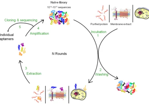
The traditional aptamer selection technology, Systematic Evolution of Ligands by Exponential Enrichment (SELEX), is an in vitro screening technique based on iterative rounds of binding, separation and amplification (Figure 15). It was developed in the early 1990s simultaneously by Tuerk and Gold (Tuerk and Gold 1990), Ellington and Szostak (Ellington and Szostak 1990) and Robertson and Joyce (Robertson and Joyce 1990).
The basic steps of a SELEX process are presented in Figure 15. Iterative cycles of in vitro selection and enzymatic amplification by PCR mimic a darwinian type process leading the selection towards relatively few, but optimized structural motifs as result for ligand binding (Goringer, Homann et al. 2003). Starting point of a SELEX process, is a chemically synthesized random DNA oligonucleotide library, consisting of about 1014
to 1015
different sequence motifs. Each oligonucleotide is composed of a random sequence that is obtained under conditions that allow the introduction, with the same probability, of adenine, thymine,
guanine or cytosine at each position. Therefore, for a sequence of n nucleotides, there are 4n
possibilities. The extremities of this random sequence are flanked by two constant sequences essential for the various enzymatic steps of the selection process. The DNA pool can be transcripted in RNA. Then the library is incubated directly with the target. The binding complexes are subsequently separated from unbound and weakly bound oligonucleotides. This is the most crucial step of aptamer selection process and it greatly affects the binding properties of the aptamers to be selected. Target bound oligonucleotides are then eluted before being amplified by PCR (for DNA SELEX) or reverse transcription (RT)-PCR (for RNA SELEX). The resulting double-stranded DNA has to be transformed into a new