HAL Id: hal-02358923
https://hal.archives-ouvertes.fr/hal-02358923
Submitted on 18 Nov 2020HAL is a multi-disciplinary open access
archive for the deposit and dissemination of sci-entific research documents, whether they are pub-lished or not. The documents may come from teaching and research institutions in France or abroad, or from public or private research centers.
L’archive ouverte pluridisciplinaire HAL, est destinée au dépôt et à la diffusion de documents scientifiques de niveau recherche, publiés ou non, émanant des établissements d’enseignement et de recherche français ou étrangers, des laboratoires publics ou privés.
Metal-based redox-responsive MRI contrast agents
Sara Pinto, Vanessa Tomé, Mário J.F. Calvete, M. Margarida C.A. Castro,
Éva Tóth, Carlos F.G.C. Geraldes
To cite this version:
Sara Pinto, Vanessa Tomé, Mário J.F. Calvete, M. Margarida C.A. Castro, Éva Tóth, et al.. Metal-based redox-responsive MRI contrast agents. Coordination Chemistry Reviews, Elsevier, 2019, 390, pp.1-31. �10.1016/j.ccr.2019.03.014�. �hal-02358923�
1
Metal-based redox-responsive MRI contrast agents
Sara M. Pintoa,b, Vanessa Tomé, a,b Mário J. F. Calvetea,b, M. Margarida C.A. Castro,a,d Éva Tóthc* and Carlos F. G. C. Geraldesa,d*
aCentro de Química de Coimbra (CQC), bDepartment of Chemistry and dDepartment of Life Sciences, Faculty of Science and Technology, University of Coimbra, Rua Larga, 3004-535, Coimbra, Portugal.
cCentre de Biophysique Moléculaire, CNRS, UPR 4301, Université d’Orléans, Orléans, France
*Email: [email protected] and [email protected]
Dedicated to Professor Armando J.L. Pombeiro for his outstanding achievements in Coordination Chemistry and Catalysis
Highlights
Use of metal-based complexes and nanoparticles as relaxation and PARACEST redox responsive contrast agents for MRI is reviewed
Ligand-based and metal-based T1 and PARACEST redox responsiveness was reported, in some cases in small animals in vivo.
Mn-based nanoparticles provide strong redox-active relaxation responses in in vivo animal studies.
Ratiometric methods provide in some cases quantitative redox evaluation of cells in vitro.
Keywords: MRI contrast agents; Metal chelates; Inorganic nanoparticles; Redox responsive probes; Hypoxia probes; Relaxation agents; PARACEST agents
List of abbreviations
ASTM: Diacetyl-bis(N4-methylthiosemicarbazone) BOLD: Blood Oxygenation Level Dependent BPG: 2,3-D- bisphosphoglycerate
2
CA= contrast agent
CBT: 2-cyanobenzothiazole
CEST: Chemical Exchange Saturation Transfer CYCLAM: 1,4,8,11-tetraazacyclotetradecane
CYCLEN: 1,4,7,10-tetraazacyclododecane
CyHBET N-(2-hydroxybenzyl-N,N′,N′-trans-1,2-cyclohexylenediaminetriacetic acid
CySH/CySSCy: Cysteine/Cystine CT: Computed Tomography
DO3A: 1,4,7,10-tetraazacyclododecane-1,4,7-triacetic Acid DOTA: 1,4,7,10-tetraazacyclododecane-1,4,7,10-tetraacetic Acid
DOTAM: 1,4,7,10-tetrakis(carbamoylmethyl)-1,4,7,10-tetraazacyclododecane
DOTAM-Gly4: N,N',N'',N'''-[1,4,7,10-Tetraazacyclododecane-1,4,7,10-tetrayltetrakis(1-oxo-2,1-ethanediyl)]tetrakis[glycine]
DOTMA = 1R, 4R, 7R, 10R)-α, α’, α”, α’”-tetramethyl-1,4,7,10-tetraazacyclododecane-1,4,7,10-tetraacetic Acid
DOTP: 1,4,7,10-tetraazacyclododecane-N,N′,N″,N‴-tetrakis- (methylenephosphonate) DO3A: 1,4,7,10-tetraazacyclododecane-1,4,7-triacetic Acid
DO3ABA: DO3A-monobutylamide
DO3NI: DO3A-nitroimidazole
DTPA:1,1,4,7,7-pentakis-(carboxymethyl)-1,4,7-triazaheptane DTT: Dithiotreitol
EPR: Electron Paramagnetic Resonance
EPT: Exofacial Protein Thiol
GSH/GSSG: Glutathione/Glutathione Disulfide HbA: Adult Hemoglobin
HBED: Dihydroxybenzylethylenediamine diacetic acid
HBET: Hydroxybenzylethylenediamine triacetic acid
HbF: Fetal Hemoglobin
hepPDMS: Heparin–poly(dimethylsiloxane)
HP-DO3A: 1,4,7-tris(carboxymethyl)- 10-(2-hydroxypropyl)-1,4,7,10-tetraazacyclododecane
HSA: Human Serum Albumin HSAB: Hard–soft acid–base
3
HIF: Hypoxia Inducible Transcription Factor 5HT: 5-hidroxitryptamin
ICP-Ms: Inductively Coupled Plasma Mass Spectrometry
MT: Magnetization Transfer MR: Magnetic Resonance
MRI: Magnetic Resonance Imaging
NADH/NAD+: Nicotinamide Adenine Dinucleotide
NADPH/NADP+: Nicotinamide Adenine Dinucleotide Phosphate NHE: Normal Hydrogen Electrode
NMR: Nuclear Magnetic Resonance NOX1: NADPH Oxidase 1
NSF: Nephrogenic Systemic Fibrosis
ODDA=1,4,10,13-tetraoxa-7,16-diazacyclooctadecane-7,16-diacetate
ODDM= 1,4,10,13-tetraoxa-7,16-diaza-cyclooctadecane-7,16-dimalonate
PAI: Photoacoustic Imaging
PAMAM: Polyaminoamide
PARACEST: Paramagnetic Chemical Exchange Saturation Transfer PEG: Polyethylene Glycol
PET: Positron Emission Tomography PLGA: Poly lactic-co-glycolic Acid
pO2: Partial Oxygen Pressure Poly-β-CD: Poly-β-cyclodextrin RBC: Red Blood Cells
RE: Redox Environment
ROI: Region of Interest
ROS: Reactive Oxygen Species RNS: Reactive Nitrogen Species SOD: Superoxide Dismutase
SPECT: Single-Photon Emission Computed Tomography
SPION: Superparamagnetic Iron Oxide Nanoparticle
TACN: 1,4,7-triazacyclononane TCEP: Tris(2-carboxyethyl)phosphine
4
TPPS4: 5,10,15,20-tetrakis-(p-sulfonato phenyl) porphinate Trx(SH)2/TrxSS: Thioredoxin/Thioredoxin Disulfide
Table of contents
1.Introduction 05
2. Intra- and extracellular redox environment 06
3.Background on relaxation-based and PARACEST-based MRI CAs 09
3.1. Relaxation-based MRI contrast agents 09
3.2. PARACEST-based MRI probes 11
3.3. General considerations on redox-responsive, metal-based MRI agents 13
4. Redox responsive MRI contrast agents 14
4.1. T1 contrast agents 14
4.1.1. T1 probes based on redox active ligands 14
4.1.2. T1 probes based on metal redox states 28
4.2. T2/T2*contrast agents… 46
5. Redox responsive PARACEST MRI contrast agents…… 47
5.1. LanthanideIII PARACEST agents…… 47
5.1.1. Ln-PARACEST probes based on redox active ligands………… 48 5.1.2 Ln-PARACEST probes based on redox active metal centers… 48
5.2. Transition metal ion PARACEST agents……… 55
6. Concentration independent redox-responsive MRI probes… 61
7. Conclusions and perspectives… 63
8. Acknowledgements……… 64
9. References…… 65
Abstract
Given their potential in a better characterization and diagnosis of major pathologies like cancer or chronic inflammation, redox-activated Magnetic Resonance Imaging (MRI) probes have recently attracted much interest from chemists. Such redox responsive probes are capable of reporting on specific biomarkers that are related to tissue redox potential disruption or hypoxia. Lately, this research area has experienced remarkable development, including redox-responsive metal complexes and nanoparticles. Here we critically review the progress with a specific focus on metal-based probes and some nanoparticle examples. We demonstrate, via representative cases, the different molecular mechanisms that can generate a redox-modulated MRI response. They can be based on the redox activity of either the ligand or the metal center, provided the different oxidation states of the metal ion are endowed with different magnetic properties. A particular emphasis is given to recent advances and to the imaging probes that have attained in vivo validation. In overall, we aim to provide the reader with a comprehensive view of how intracellular or extracellular redox buffer systems can be assessed by using MRI contrast
5
agents based on lanthanide or transition metal ions using T1-weighted, T2-weighted, PARACEST 1H or 19F MRI.
1. Introduction
Thanks to its excellent resolution, non-invasiveness and the lack of ionizing radiation, Magnetic Resonance Imaging (MRI) has become an essential tool in current clinical radiology. For three decades, GdIII complexes used to enhance image contrast has largely contributed to the diagnostic potential of MRI. Once injected, commercial contrast agents distribute non-specifically in the extracellular space and their role in delineating different pathologies is solely related to a better visualization of an altered morphology or physiology.1,2,3
Bringing MRI from anatomical to molecular imaging opens opportunities for characterizing tissues not only by their morphology but also by their molecular features.4 One can identify a multitude of molecules or tissue parameters as biologically relevant biomarkers that carry important information about the physiological or pathological status of a tissue. The unique capability of MRI agents to modulate the generated MRI signal in response to a specific biomarker or stimulus represents a great advantage, in contrast to other clinical modalities such as X-ray Computed Tomography (CT) or nuclear imaging. Consequently, chemists have dedicated important efforts to design smart or responsive molecular imaging probes that could enable non-invasive, repeatable and real-time in vivo mapping of such biomarkers.
Among those, the detection of the tissue redox state has received remarkable interest.5 This is first of all due to the great biological importance of this biomarker in many physiological and pathological processes, but also because of the very diverse chemical approaches that chemists can exploit for the design of redox-responsive MRI probes. First, redox-dependent MRI response can be generated by inducing redox reactions centered either on the metal center or on the ligand. While GdIII itself is not redox active, many other metal ions can be potentially used and have been explored in this context, such as Mn, Eu, Fe, Cu, Co, etc. Metal-based redox switches can be further extended to nanoparticle systems, leading to signal amplification. Also, in addition to the classical T1/T2 weighted imaging, other contrast mechanisms can be adapted to redox mapping, including PARACEST (Paramagnetic Chemical Exchange Saturation Transfer) or 19F MRI. For all these reasons, the field of redox MRI probes is very rich from the
6
coordination chemistry point of view. The versatility of the approaches also holds promise of potentially successful in vivo translation of the probes in the future.
In addition to MRI agents, redox sensitive probes have been also extensively studied for optical imaging, positron emission tomography (PET) or single-photon emission computed tomography (SPECT).2,5 For optical detection, the probe can undergo redox reactions depending on the environment and this will trigger changes in the fluorescence properties. In contrast, the principle of redox-active PET and SPECT probes is fundamentally different. Indeed, once injected, a radioactive isotope will decay in the same way independently of its chemical environment. Here the principle of detection is based on specific trapping of the probes in hypoxic cells leading to accumulation of the radioactive isotope in hypoxic tumors. One should also mention photoacoustic imaging (PAI) where the oxygenation level can be quantified based on the different absorption coefficients of oxy- and deoxyhemoglobin. Each of these imaging modalities presents advantages and disadvantages. While optical imaging is excellent to produce high resolution images at the cellular level, it is less adapted to macroscopic imaging. PAI is so far limited to small animals. PET and SPECT provide low resolution images and represent an ionization burden, but have great sensitivity and hypoxia PET probes are already in clinical use. In the future, MRI probes could nicely complement the radiologists’ and biologists’ toolbox for redox mapping.
The scope of this review is to introduce the different molecular mechanisms that can provide redox-dependent MRI response by focusing exclusively on metal-based probes. Each mechanism will be illustrated by representative literature examples, without the pretention of being exhaustive. If available, we present examples that have been validated in vivo. Previously, several reviews have been dedicated to smart MRI agents in general,6,7,8,9 or to redox-probes in particular.10,11 In this quickly growing field, our intention is to survey the recent developments and to provide the reader with a comprehensive view of how intracellular or extracellular redox buffer systems can be assessed by using MRI contrast agents based on lanthanide or transition metal ions with a read-out in T1-weighted, T2-weighted, PARACEST 1H or 19F MRI.
2. Intra- and extracellular redox environment
The biological aspects of redox-potential homeostasis have been described in several reviews.12,13,14 Here, we will summarize the most important aspects in the context of molecular imaging.
7
In any living organism, both extracellular and intracellular redox environments are tightly regulated15,16,17 and thus are important health biomarkers. In fact, their dysregulation can be associated with a wide range of pathophysiological conditions, including chronic inflammation,18,19 neoplastic growth12 and ischemia,20 since several triggered biochemical cascade events can damage cellular or tissue components, enabling disease progression.13,14,21 In healthy tissue, intracellular environments tend to be reducing whereas extracellular environments tend to be more oxidizing and subject to much larger fluctuations.17 For example, in necrotic tissue, dead cells leach components of the cytosol into the extracellular space to create a relatively reducing environment.22 Hence, the development of imaging techniques capable of monitoring changes in redox activity is of utmost interest in biomedical or clinical research as well as in medical practice.23,24,25
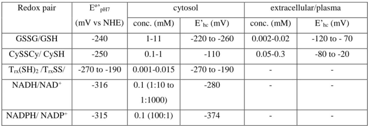
The redox environment in a biological fluid, organelle, cell or tissue is maintained by a number of redox couples present at relatively high concentrations, which are linked to each other by biochemical redox homeostasis processes (Table 1). Any single redox couple contributes to the redox state, with a half-cell redox potential (E’hc) described by a Nernst equation (1):
𝐸′ℎ𝑐 = 𝐸º′𝑝𝐻7− 59.1
𝑛 log 𝑄 (1) where 𝐸º′
𝑝𝐻7 is the standard biochemical redox potential for the couple against the normal hydrogen electrode (pH 7, 298.15 K), n is the number of electrons involved in the chemical process and Q is the respective mass action ratio, eg. [NADH]/[NAD+] and [NADPH]/[NADP+] for nicotinamide adenine dinucleotide and its phosphorylated derivative, respectively, [Trx(SH)2]/[TrxSS] for thioredoxin, [GSH]2/[GSSG] for glutathione (GSH=(Glu-Cys-Gly)) and [CySH]2/[CySSCy] for cysteine (CySH). There are also contributions from other couples, such as the exofacial free thiols (EPTs) of proteins with surface cysteinyl residues. The redox environment (RE) is defined by the summation of the products of the reduction potentials weighted with the concentration of the respective reduced species for all the redox couples:22
𝑅𝐸 = 𝛴𝑖 𝐸º′𝑖 [𝑋𝑖 𝑟𝑒𝑑 ] (2)
Among these, glutathione (GSH) is the major thiol-disulfide redox buffer of the intracellular space with an average concentration of 1–11 mM, much higher than the other redox active compounds.17 Therefore, the redox state of the glutathione disulfide-glutathione couple (GSSG/2GSH) can serve as an indicator of the intracellular redox
8
environment. Changes of the half-cell reduction potential (Ehc) of the GSSG/2GSH couple appear to correlate with the biological status of the cell: proliferation Ehc ~ -240 mV; differentiation Ehc ~ - 200 mV; apoptosis Ehc ~ -170 mV, with even less negative potentials for necrotic cells.22 The extracellular space is typically more oxidized than the cytosol and its redox environment is dominated by the cystine-cysteine couple (CySSCy/2CySH).
Table 1. Some properties and compartmentation of biological redox buffer pairs.22
Redox pair Eº’pH7 (mV vs NHE)
cytosol extracellular/plasma conc. (mM) E’hc (mV) conc. (mM) E’hc (mV) GSSG/GSH -240 1-11 -220 to -260 0.002-0.02 -120 to - 70 CySSCy/ CySH -250 0.1-1 -110 0.05-0.3 -80 to -20 Trx(SH)2 /TrxSS/ -270 to -190 0.001-0.015 -270 to -190 - - NADH/NAD+ -316 0.1 (1:10 to 1:1000) -280 - - NADPH/ NADP+ -315 0.1 (100:1) -374 - -
Besides the biological redox buffers, the local tissue concentration and reactivity of molecular oxygen (O2), expressed by its partial pressure (pO2), and its reactivity products (reactive oxygen species -ROS – such as O2- .and H2O2, or reactive nitrogen species – RNS- such as NO or peroxynitrite ONO2-.) are important targets for the design of MRI redox probes. 26 Oxygen has a relatively large concentration dissolved in arterial blood (pO2 ~ 100 mmHg) or bound to hemoglobin and less in venous blood (pO2 ~ 40 mmHg). pO2 values vary substantially in different tissues depending on their vascular architecture and tissue demand, with typical average pO2 ~ 40 mmHg for normal muscle. When local oxygen pressures are low (pO2 < 10-15 mmHg), the condition known as hypoxia substantially affects cell metabolism. Hypoxia is important in tumors because rapid proliferating tumor cells require high levels of oxygen, which drives the cancer cells to promote angiogenesis. The abnormal signaling pathways in the tumor lead to formation of underdeveloped blood vessels which are chaotic in nature, leading to regions of hypoxia in non-necrotic parts and especially in necrotic cores that are severely lacking oxygen (pO2 < 2-3 mmHg). Hypoxia has a significant influence on the progression of cancer and is considered as a marker of poor prognosis. This is because hypoxia has several biochemical consequences, including high expression levels of the transcription
9
factor HIF-1 (hypoxia inducible factor 1) and increased reductive stress, promoting tumor proliferation and resistance to therapy.
Several important enzymes contribute to the redox status of the extracellular matrix, which are of interest for MRI redox probes.12,13,14 ROS and NOS, important as signaling molecules, can be produced in hyperoxia or hypoxia, in the second case due to accumulation of reduced species such as NADH. The activity of the membrane enzyme NADPH oxidase 1(NOX1) produces the superoxide radical O2-.. In the extracellular space, superoxide dismutase (SOD) catalyzes the conversion of O2-. to H2O2 and O2, while glutathione peroxidases catalyze the reduction of peroxide and lipid hydroperoxides. Most ROS and RNS are formed inside the cell, but may be exported into the extracellular space, where they may oxidize the exofacial protein thiols (EPTs), which are highly sensitive reporters of the extracellular environment.
3. Background on relaxation-based and PARACEST-based MRI CAs
3.1. Relaxation-based MRI CAs
Magnetic resonance images are based on the density and the relaxation properties of water proton nuclei in tissues. Paramagnetic metal complexes used as contrast agents will reduce the longitudinal T1 and transverse T2 relaxation times in the various tissues leading to an increased contrast on the images. The efficiency of a contrast agent is commonly expressed by its proton relaxivity, r1, which is defined as the paramagnetic relaxation enhancement of the longitudinal water proton relaxation rate in the presence of 1 mM concentration of the agent (eq. 3):
1 𝑇1,𝑜𝑏𝑠=
1
𝑇1,𝑑𝑖𝑎+ 𝑟1 𝑐 (3)
where T1,obs and T1,dia are the longitudinal relaxation times observed in the presence and in the absence of the contrast agent, respectively, c is the concentration of the paramagnetic species and r1 is the longitudinal water proton relaxivity.27,28,29
Nuclear relaxation phenomena in the proximity of a paramagnetic metal are governed by dipole-dipole interactions between the electron spin of the metal and the proton nuclear spin, and are directly linked to the microscopic parameters of the metal chelate as described by the Solomon-Bloembergen-Morgan theory. The overall relaxivity is the sum of two major contributions, classified as outer sphere and inner sphere
10
relaxivity, originating from interactions of the electron spin with protons on water molecules diffusing in the surroundings of the complex, or with protons on the inner sphere water molecule, respectively. In some cases, second sphere water molecules can represent an additional contribution to relaxivity (eq. 4). Although for commercial agents, inner- and outer-sphere mechanisms have comparable contributions to relaxivity, chemists are more interested in the inner sphere term which can be modulated by ligand design, whereas the outer sphere contribution is dependent only on diffusion effects and is not amenable to improvement. All three relaxivity terms (inner sphere, second sphere and outer sphere) in eq. 4 increase with increasing electron spin, S, of the metal.
𝑟1 = 𝑟1𝐼𝑆+ 𝑟1,2𝑛𝑑𝑆+ 𝑟1𝑂𝑆 (4)
𝑟1𝐼𝑆 = 𝑓(𝑆, 𝑞, 𝑘𝑒𝑥, 𝜏𝑅, 𝑇1,2𝑒) (5)
The inner sphere relaxivity term is linearly proportional to the number of coordinated water molecules, q, and depends on the rate of water exchange between the metal-bound state and the bulk, kex, as well as on the rotational dynamics of the chelate (rotational correlation time, R) (eq. 5). All these three factors are dependent on the ligand structure and therefore they are the most accessible to modification via rational ligand design. In addition, electron spin relaxation times, T1,2e, also influence relaxivity, but their dependence on chelate structure is much less understood for Gd3+ or Mn2+ and their effect vanishes at clinically important magnetic fields.
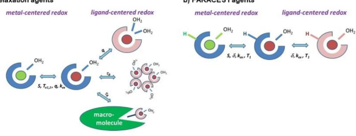
The chemical design of redox responsive probes can be based on redox reactions occurring on the metal centre or on the ligand.11
The first approach takes benefit of the redox capabilities of the metal ion. Several metal ions (Eu, Mn, Fe, Co, …) have at least one redox state which is a strong relaxation agent. A change in the redox state of the metal ion might impact the relaxivity via essentially all parameters, namely the electron spin, the electronic relaxation, the water exchange rate and potentially the hydration number.
When the redox reaction occurs on the ligand, this can lead to the modulation of (i) the hydration number, (ii) the rotational dynamics, or (iii) the water exchange rate; the first two strategies being by far easier to realize and therefore more representative.
11
Concerning changes in the hydration number, variations between 0 and maximum q=2 can be reasonably exploited for a Gd3+-, and q=0 and 1 for a Mn2+-complex; higher hydration numbers would lead to substantial decrease in complex stability and to a risk of ternary complex formation with endogenously available coordinating functions (carboxylates, hydrogenocarbonate, etc). As the effect of the hydration number is independent of the magnetic field, responsive probes based on q changes can operate equally well at any field and will produce the same relative relaxivity changes. In contrast, the effect of the rotational dynamics on relaxivity is strongly field-dependent, and it might even depend on the water exchange rate (even if for typical Gd3+ or Mn2+ complexes water exchange has no limiting effect on relaxivity, thus changes in the rotational correlation time will lead to relaxivity variation). The Solomon-Bloembergen-Morgan theory predicts that the influence of rotational dynamics on relaxivity is maximum in the 20-60 MHz proton Larmor frequency range (the slower the rotation, the higher the relaxivity). Then this effect vanishes with increasing frequencies and above ~200 MHz,
the r1 – R relationship is even inversed (the slower the rotation, the lower the relaxivity). As a consequence, responsive probes based on variation of the rotational dynamics typically work well at 20-60 MHz (several-fold relaxivity increase), but can produce limited relaxivity changes at higher frequencies. These considerations should not be neglected when designing responsive relaxation agents. Modifications in the rotational correlation times can be achieved via dimerization, aggregation, binding to biomacromolecules (proteins, typically serum albumin) by either non-covalent or covalent linkage.
3.2. PARACEST-based MRI probes
Chemical Exchange Saturation Transfer (CEST) agents possess protons in slow to intermediate exchange with bulk water protons, typically -NH of amides, amines or –OH of water or alcohols (kex ≤ , where is the chemical shift difference between the two proton pools). In a CEST experiment, the slowly exchanging protons of the agent are saturated by selective radiofrequency pulses, which results in the disappearance of their proton NMR signal, and, given the chemical exchange of these and bulk water protons, the water proton signal intensity will be also decreased. This water proton signal intensity decrease can be then visualized on an MR image. Complexes of paramagnetic metal ions that induce an important paramagnetic shift of the surrounding protons but do not have a
12
strong relaxation effect have specific advantages for CEST. The large chemical shifts allow for exploiting higher proton exchange rates yet remaining in the slow exchange region, and the selective saturation of the protons on the probe is also easier if its signal is far from the water proton signal. In the lanthanide series, all ions except for the highly relaxing GdIII and EuII and the diamagnetic LaIII and LuIII, while among transition metals, CoII, NiII, FeII can typically be exploited to create PARACEST probes. The parameters that determine the CEST effect involve the proton exchange rate, kex, the power of the pre-saturation pulse applied, B2, and the proton relaxation times in the two exchanging pools. Provided that the saturation of the exchangeable protons on the CEST probe is complete, the net magnetization of the bulk water protons (Mz/M0) at steady state can be estimated as: 𝑀𝑧 𝑀0 = (1 + 𝑐𝑛𝑇1 111 𝜏𝑀) −1 (6)
where M (= kex-1) is the proton residence lifetime, c is the concentration of probe, n is the number of chemically equivalent exchanging protons per molecule that are saturated and T1 is the spin-lattice relaxation time of bulk water protons. The optimal exchange rate providing a maximum CEST effect is related to B2 according to kex = 2B2.27,28
PARACEST (and CEST) imaging has advantages over classical relaxation-based MRI. The contrast is turned on by the application of the selective saturation of the exchanging protons, and thanks to the frequency-encoding of CEST, several protons in the same agents or several probes can be simultaneously visualized on a single image, if their saturation frequencies are different. CEST is well adapted to the design of responsive agents, for which the modulation of any of the parameters in eq. 6 can be used, even if the concentration of the exchangeable protons and their exchange rate are the most accessible. CEST agents are also easily amenable to ratiometric approaches, given the possibility to incorporate two exchangeable proton sites into the same molecule.30 Finally, we should note that for PARACEST, higher magnetic fields are advantageous as they lead to better signal separation between the exchanging sites.
As for relaxation probes, the redox process that initiates the detectable PARACEST changes can also occur either on the metal centre or on the ligand. In the first case, the magnetic properties of the metal ion change, leading to a modification of
13
the resonance frequency and of the relaxation rate of the ligand protons. Redox reactions on the ligand might also lead to chemical shift changes.
The various mechanisms exploitable to generate a redox-dependent MRI signal for relaxation and PARACEST agents are schematically summarized in Figure 1.
Figure 1. The main strategies to modulate a) r1 relaxivity and b) PARACEST signal of redox probes through ligand-centred and metal-centred redox activity.
3.3. General considerations on redox-responsive, metal-based MRI agents
Any metal-based imaging probe designed for in vivo applications has to be biocompatible, non-toxic and water soluble. For typical polyamino-carboxylate complexes used as imaging agents, the non-toxicity is directly linked to the thermodynamic stability and kinetic inertness of the chelate which both need to be high enough to prevent release of free metal ions in the body. These issues have become particularly important following the recent concerns about Gd-toxicity in relation to Nephrogenic Systemic Fibrosis (NSF)31,32 and Gd-deposition in the brain.33 In general terms, one can consider that macrocyclic complexes are more stable and inert than the open-chain analogues and that increased ligand rigidity results in higher kinetic inertness. As for any responsive probe, a strong MRI signal change, if possible a signal increase, is expected upon activation (“turn on” probe). For redox responsive probes, the redox half-cell potential has to be adapted to the potential of biological redox buffers and the redox reaction should be reversible and its kinetics has to be fast enough as compared to the time scale of the imaging experiment and of the probe elimination.
14
4. Redox responsive MRI contrast agents
Here, we will describe MRI contrast agents based on redox active ligands and or on changes of metal redox states that can potentially respond to an alteration in the redox environment in the organism.
4.1 T1 contrast agents
4.1.1 T1 probes based on redox active ligands
One of the most frequently used strategies to obtain redox active MRI probes is the modulation of GdIII chelates that, under a reductive/oxidative environment, undergo a change in their structure that affects their r1 relaxivity, creating a “pseudo switch” on/off or off/on signal (better described as “ON/on” – relaxivity decrease, or “on/ON” relaxivity increase, as the loss of MRI signal is never complete).5 Here we will discuss characteristic examples from the literature, highlighting the structure modulation and the corresponding changes in the molecular parameters determining the relaxivity changes. The most frequently used methods involve changes a) in the number (q) and/or exchange kinetics (kex) of the water molecule(s) coordinated to the GdIII center, or b) changing the rotational correlation time (R) of the complex by reversible or irreversible attachment to macromolecular structures.
a) Changes of hydration state
The first mechanism for changing q based on redox active ligands was reported by Louie and collaborators34 who used the known capacity of the photochromic dyes spiropyrans and spirooxazines35 to isomerize under UV irradiation to the merocyanine forms. They linked GdIIIDO3A (DO3A= 1,4,7,10-tetraazacyclododecane-1,4,7-triacetic acid)) to the dye moiety to obtain potential multimodal magnetic-resonance/optical-imaging contrast agents (Figure 2).33,36,37,38
15
Figure 2. The conversion of the merocyanine derivative 1 into the spyrooxazine form 2
by NADH increases q, and hence r1.33
In the dark or under UV light, the probes are present in the colored merocyanine
1 form which can be converted into the spiropyran derivative 2 by irradiation with visible
light, leading to decreased fluorescence intensity and increased r1 relaxivity.36,37 The spyrooxazine derivatives have higher r1 values than the corresponding merocyanine forms (e.g. r1 = 8.60 versus 5.56 mM-1 s-1, respectively, for the spironaphthoxazine probe) due to the cleavage of the coordination bond of the phenoxide oxygen with the GdIII ion, allowing the coordination number q to increase from one to two, as determined by 17O NMR.33,38 The spirooxazine and dinitrospyran-bound complexes also isomerize from the form 1 to the form 2, upon reduction with NADH, although the mechanism is unclear, as it appears that no electron transfer occurs. While the reduction of the spirooxazine derivative is irreversible, the spirooxazine isomer could be partially converted back into the merocyanine derivative by H2O2 (Figure 2). Interestingly, the probes were not responsive to other biological reducing agents, like L-cysteine and L-ascorbic acid, showing their potentiality as NADH responsive probes.33,38
Nagano and collaborators39 proposed a family of redox active ligand probes obtained by linking a nitrobenzenesulfonamide moiety to a GdIIIDO3A complex (see e.g.
3 in Figure 3). Under hypoxia conditions (pH = 5.8-7.6), the nitro group was reduced to
an amino group with a concomitant 1.8-fold increase of the r1 value. This is a consequence of the shift in acidity of the sulfonamide nitrogen atom upon reduction, allowing its protonation and the dissociation of the GdIII-N bond, thus leaving two sites for water binding (q changes from 0 to 2). Reduction of 3 was observed in the presence of rat liver microsomes, under hypoxic conditions, to give an increased contrast on T1-weighted MR images.
16
Figure 3. Redox-activated MRI probes 339 and 4.40
Another strategy towards detection of reducing thiols in the extracellular microenvironment used redox probe 4,40 based on GdIII-DO3A bearing a flexible linker ending in a 2-pyridyl-dithio moiety(Figure 3) that can promptly react with free thiol groups to form mixed disulfides in a reversible reaction. In the case of biological reductants also containing a carboxyl group like glutathione (GSH) or cysteine, the resulting product had about half of the relaxivity of the starting GdIII complex (r1 drop from 8.1 mM−1 s−1 to 4.1 mM−1 s−1 at 20 MHz, 25 °C and pH 7.4). This has been explained by the coordination of the carboxylate group, hence a decrease of the GdIII hydration number from q=2 to q=1, on the basis of 1H NMRD and 17O relaxation studies. Larger relaxivity differences could be achieved by forming an inclusion complex between the
GdIII complex and cyclodextrin (CD) or poly--CD.40
b) Changes of rotational dynamics
Reversible or irreversible binding of low molecular weight GdIII complexes to macromolecules has been often used to increase R and to obtain higher r1 values. This has been exploited by Raghunand and collaborators,41,42,43,44,45 whosynthesized a series of GdIII-DO3A 5, 6 and 7 and GdIII-DOTA-monoamide complexes 8 and 9 (DOTA = 1,4,7,10-tetraazacyclododecane-1,4,7,10-tetraacetic acid) bearing alkyl linkers with terminal thiol groups (Figure 4). These probes interacted with the Cys-34 residue of human serum albumin (HSA) via formation of an S-S bond, enhancing r1 by slowing down the rotational dynamics of the GdIII complex.40,41,44 In reducing environments, the GdIII chelates were released and r1 decreased. The authors have demonstrated high in vivo
17
MRI contrast enhancement in mice bearing Mia-PaCa-2 or NCI-N87 tumor xenografts when using the GdIII complex with a thiol moiety on a hexyl arm (8). This was correlated with tumor thiol content, after the animals were treated with a gluthatione synthesis inhibitor or the thiol-oxidizing anticancer drug Imexon, confirming the potentiality of this probe as an MRI reporter of the tumor redox status.41,43,44
Figure 4. Redox-activated MRI probes based on thiol oxidation of the ligand followed
by protein conjugation.41,42,43,44,45
Exofacial protein thiols (EPTs) are a pool of thiol groups attached to membrane proteins on the external surface of various types of cells. They are present as free -SH or in the oxidized -S-S- form depending on the extracellular redox state. As the extracellular space is more oxidizing, most of them are present in the form of disulfide bridges. Aime et al. 46, 47, 48 studiedthe relaxometric properties of derivatives of GdIII-DO3A 10, 11 and
12 or GdIII-DTPA 13 and 14, bearing a flexible linker of varying length ending in a 2-pyridyl-dithio moiety (Figure 5).
18
Figure 5. Redox-activated MRI probes based on S-S rearrangement of the ligand.46,47,48
At 20 MHz and 25 ºC, the bishydrated complex 11 had r1= 9.0 mM-1s-1, while the other, monohydrated complexes had lower r1 values (6.3-6.9 mM-1s-1). 46,48 It was shown that it is possible to bind these complexes to EPTs for potential application as markers for tissue hypoxia. Upon formation of a disulfide bridge, the GdIII-DO3A derivatives 11 and
12 were internalized efficiently and accumulated in endosomes of different types of cells
(as many as 1.2-1010 Gd atoms per single cell, a number similar to the number of EPTs at the cell surface) as demonstrated by in vitro MR imaging.46,47,48 It was also possible to label B16 melanoma tumor cells in vivo with this type of chelates (chelate 11) (Figure 6) and an increased uptake was correlated with increasing levels of EPTs, which was observed in the case of more reductive extracellular microenvironment.47
19
Figure 6. T1w-SE axial images at 1 T (TR/TE/NEX 250/8/10) of mice subcutaneously grafted with a B16 tumor. The ROIs used to measure the signal enhancement in tumor are shown. Time course of the signal enhancement from precontrast to 48 h post intratumor injection (at 0.12 μmol) of (A) 11; (B) GdIII-DO3A.Reproduced with permission from Ref. 47, Copyright (2011) by American Chemical Society.
On the other hand, GdIII-DTPA derivatives 13 and 14, despite their capacity to react with the thiol groups of EPTs, showed a very low or non-detectable uptake. This was attributed to their negative charge, while the neutral GdIII-DO3A-based ligands had efficient uptake.48
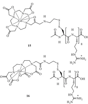
Meade and collaborators49 developed GdIII-DOTA 15 and GdIII-DTPA 16 derivative chelates linked to an octamer of arginine residues through a disulfide bond (Figure 7). The addition of an arginine polypeptide improved the transport of the probe across cell membranes. Upon cell internalization, the disulfide bond was efficiently cleaved by GSH, the biological reducing agent which attains higher concentration inside the cells. The separation from the cell-penetrating peptide transduction domain increased the cell-associated lifetime of the GdIII probes, leading to enhanced MRI signal in labeled NIH/3T3 cells.
Figure 7. Redox-activated MRI probes based on S-S rearrangement of the ligands 15 and 16.49
20
Figure 8. Redox-activated MRI probes based on reductive isomerization of the ligand 17.50
Probes containing nitro groups have been commonly used for imaging hypoxia. For instance, 2-nitroimidazoles are known to accumulate in hypoxic tissues due to reduction of the nitro to an amine group in reducing environments.51,52 Sherry and collaborators50 developed a GdIII-DO3A type 17 chelate bearing a nitroimidazole moiety for MRI imaging of hypoxia (Figure 8). They observed that chelate 17, although of lower stability than GdIII-DO3A-monobutylamide, showed a similar r1 value corresponding to
q = 1. Importantly, it was selectively trapped inside hypoxic 9L rat glioma cells, as evidenced by an increase of contrast enhancement in the in vitro MRI images as compared to normoxic cells. In an in vivo study using rats bearing prostate adenocarcinoma AT1 tumor, the same authors53 clearly evidenced that 17 accumulated in the central, poorly perfused and hypoxic regions of the tumor while it showed complete clearance from the surrounding muscle tissue (Figure 9). These results demonstrated the utility of this nitroimidazole derivative complex to image tumor hypoxia in vivo.
Figure 9. Grayscale MR T2-weighted (a,d) and color T1-weighted (b, c, e, f) images of Copenhagen rat thighs bearing syngeneic Dunning prostate R3327-AT1 tumors following injection of 0.1 mmol GdDO3NI per kg body weight (a–c) or 0.1 mmol GdDO3ABA (17) per kg body weight (d–f) before injection (b, e) and 145 min after injection (c, f). The
21
tumor and thigh muscle are labeled T and M, respectively.Reproduced with permission from Ref. 51, Copyright (2013) by Springer Nature.
The potential of a GdIII complex containing phosphonate groups (18) (Figure 10) was evaluated as a hypoxia responsive agent by Aime and collaborators54,55. This complex has different affinities for the two human adult (HbA) and fetal hemoglobin (HbF) conformations (R and T), acting as an allosteric effector by favoring an heterotropic interaction with the 2,3-D- bisphosphoglycerate (BPG) binding site, allowing a change in the thermodynamic equilibrium stabilizing the T conformation. The r1 relaxivities obtained for the probe linked to R-Hb (oxy-Hb) and to T-Hb (deoxy-Hb) were quite different from the unbound chelate, suggesting that this type of probe could potentially be used as a detector of hypoxia environments. Later, the same authors56 proposed to map tumour hypoxia via the combined use of the pO2-responsive GdIII -DOTP- and the non-responsive GdIII-HPDO3A-labeled red blood cells (RBC). In fact, with this strategy, it was possible both to detect the vascular oxygen status and to assess the local labeled RBC concentration in a transplanted breast tumor mouse model. This circumvents an important barrier in the development of responsive agents, which is the difficulty to separate the effect arising from local concentration changes from that associated with the relaxivity response of the probe to hypoxia.
Figure 10. Redox-activated MRI probes based on Red blood Cells binding: Gd-DOTP
(18).54,55,56
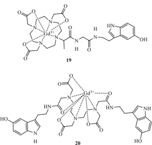
Several GdIII complexes containing ligands with peripheral redox-active hydroxyl groups have been studied as H2O2 sensors of myeloperoxidase activity (Figure 11). GdIII -DOTA-5HT (19), a conjugate of serotonin (or 5-hidroxitryptamin = 5HT), containing a peripheral catechol pendant group57 undergoes a myeloperoxidase-mediated reduction of the catechol in the presence of peroxide, yielding oligomers with a 70% increase in r1 at
22
1.5 T. A similar study was undertaken with GdIII- DTPA-bis-5HT (20), leading also to a 70% relaxivity increase.58
Figure 11. GdIII-based MRI probes to detect myeloperoxidase activity. a) GdIII -DOTA-5HT (19);57 b) GdIII-DTPA-bis 5HT (20).58
Although GdIII is the most widely used paramagnetic metal to develop MRI probes, other metal ions with similarly favorable magnetic properties, like MnII have also been investigated59, 60, 61. Most literature examples of redox responsive contrast agents involving manganese take advantage of the different oxidation states of this metal, as can been seen in the next section. Albeit, Goldsmith and collaborators62, 63, 64 reported a series of non-cyclic MnII complexes that contain ligands with an increasing number of peripheral redox-active hydroxyl groups as H2O2 sensors (Figure 12). While the monohydrated and heptacoordinate MnII complex 21 is stable in the presence of oxygen, addition of H2O2 leads to an intermolecular oxidative cross-coupling reaction between two carbon atoms of the 2-hydroxy-5-methylbenzyl rings, yielding a binuclear complex.62 The slightly lower relaxivity of this binuclear as compared to the monomer complex (r1= 3.59 mM-1 s-1 vs r1= 4.39 mM-1 s-1) results from the hydration water molecule shared by the two MnII that counterbalance the effect of the increased R.
A drawback of this probe was the non-reversibility of the oxidation. Later the same authors synthetized ligand 22, containing a hydroquinone moiety that, in an oxidizing environment, is transformed into a p-benzoquinone with lower coordinating capacity. This allows to more easily displace the quinone part of the ligand by hydration water molecules increasing q; however, it also leads to decreased complex stability.63 The
23
r1 increase observed upon addition of H2O2 excess was modest (r1 = 0.8 mM−1 s−1), due to incomplete oxidation of the ligand. However, the reaction was reversed in the presence of a reductant. Upon addition of a second hydroquinone moiety (23), a MnII complex was obtained with a two-fold increase of r1 = 1.71 mM−1 s−1.64
Figure 12. Redox-activated MnII based MRI probes based on hydroxyl group oxidation of the ligand: 21, 62 2263 and 23. 64
There are relatively few examples of nanoparticle-based T1 MRI redox probes. To date, examples are primarily based on changes in the rotational correlation time (τR), resulting either from dissociation or association of a small GdIII chelate from/with a larger molecule.
Figure 13. Nanoscale redox-activated MRI probe 24, based on S-S rearrangement.
Reproduced with permission from Ref. 65, Copyright (2011) by Royal Society of Chemistry.
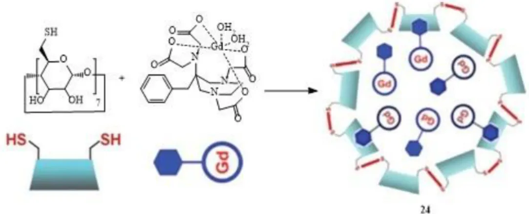
Several case studies are based on a thiol/disulfide redox couple. When dissociation occurs through the S-S bonds, r1 decreases (“turn-off“ probes). The first example was a redox sensor made by encapsulating a functionalized GdIII-AAZTA probe (24)
(AAZTA=6-amino-6-methylperhydro-1,4-diazepinetetraacetic acid) in crosslinked thiolated -CD nanocapsules via hydrophobic pendant aromatic residues (Figure 13).65
24
This incorporation resulted in a high r1 value of the GdIII chelate (15.2 mM−1 s−1 at 70 MHz) due to restricted mobility and good water permeability through the β-CD shell. The addition of tris(2-carboxyethyl)phosphine (TCEP) reducing agent led to S-S bond cleavage and degradation of the nanocapsules, releasing the β-CD monomers and the GdIII-complexes. As a result, τR was shortened and relaxivity decreased from 20.7 to 9.0 mM−1 s−1 (0.47 T, 25ºC).50 Similar work was published using PAMAM dendrimers conjugated to β-CD incorporating GdIII chelates, where the redox sensitive S-S bonds have been introduced in the dendrimer arms or in the hydrophobic arm of the GdIII ligand responsible for its encapsulation in the CD structure.66,67
A similar disassembly-based redox example was a fluorescence/19F-MRS/1 H-MRI triple-functional probe consisting of Gd nanoparticles based on a precursor (25) GdIII-DOTA monoamide linked with a quenched amino oxyluciferin fluorophore and a 19F-bearing moiety attached to the amino oxyluciferin through a disulfide linker (Figure
14) which self-assembled through hydrophobic interactions.68 Biothiols, like GSH, that are abundant in reducing biological environments were able to cleave the disulfide bond in probe 25 to induce disassembly of the nanoparticles and lead to fluorescence activation (~70-fold), 19F-MRS signal amplification (~30-fold) and significant r1 relaxivity reduction from 23.6 mM−1 s−1 to 7.6 mM−1 s−1 (~68% at 0.5 T, 37 ºC). Molecular imaging of reducing environment in HeLa cells and in vivo mice bearing subcutaneous Hela tumors was achieved using this probe.
25
Figure 14. Nanoscale redox-activated MRI probes based on S-S rearrangement.68,69,70
71,72
In another example, porous silica microparticles were surface-decorated with a GdIII-HP-DO3A-like chelate (26) through a disulfide bond providing MRI responsivity to redox potential (HP-DO3A: 1,4,7-tris(carboxymethyl)- 10-(2-hydroxypropyl)-1,4,7,10-tetraazacyclododecane) and rhodamine B allowing for optical imaging (Figure
14). Addition of TCEP to a suspension of GdIII-SiMSS and reductive cleavage of the disulfide bond led to a 30% decrease of r1 (20 MHz) form the initial value of 5.7 mM−1 s−1 due to the loss of the macromolecule effect upon release of the GdIII-SH chelate. Such microparticles were interspersed with therapeutic cells (human Mesenchymal Stem Cells - hMSCs) within a biocompatible hydrogel. The onset of reducing conditions within the hydrogel was paralleled by an increased clearance of the monomer GdIII complex that can be detected by MRI.72
Liang et. al.69 developed a “turn-on“ probe from a precursor (27), Acetyl-Arg-Val-Arg-Arg-Cys(StBu)-Lys(Gd-DOTA)-CBT, for self-assembling gadolinium nanoparticles (GdIII-NPs) under the action of furin in living tumor cells. (Figure 14). It contains a RVRR peptide sequence for cleavage and cell membrane translocation by
26
furin, a trans-Golgi protease overexpressed in tumors, a GdIII–DOTA monoamide for MRI, disulfided Cys for supplying the 1,aminothiol group for condensation, and 2-cyanobenzothiazole (CBT).After entering MDA-MB-468 cells, the disulfide bond of the Cys motif of the probe is reduced by the intracellular GSH and subsequently its RVRR motif is cleaved by furin on the site of this enzyme (i.e., Golgi body), resulting in an active intermediate.. Two of these condense quickly, activated by disulfide reduction of the cyano group of CBT to free thiol, followed by intramolecular cyclization to yield an amphiphilic dimer which has a hydrophobic macrocyclic core for self-assembling the GdIII-NPs via π–π stacking. At 25.6 MHz (ca. 0.6 T), the proton relaxation effect of the probe increased by 160% after self-assembly, due to the elongated τR, while at 64 MHz (1.5 T), the relaxation rate increased by 110%. T1-weighted MRI of MDA-MB-468 tumor xenografts showed that the probe enhanced contrast within the tumors.
A potential limitation of the previous system is the competition from the reaction between the 2-cyanobenzothiazole (CBT) moiety in the monomer and the endogenous free cysteine that stops polymerization and self-assembly. In order to avoid the
competition from free cysteine and generate a larger r1 value, the self-assembly strategy was optimized by switching the concentration-dependent intermolecular polymerization to concentration-independent intramolecular macrocyclization and self-assembly, using an acyclic precursor B containing two Gd-DOTA amide chelates and a biothiol sensitive disulfide bond (28, Figure 14).70 This precursor is reduced to form a reactive intermediate, which subsequently undergoes intramolecular cyclization to generate a more rigid and hydrophobic macrocyclic product. The following self-assembly into 28 generated a ∼60% increase in r1 from 25 to 34.2 mM−1 s−1 per molecule at 0.5 T and brighter MRI contrast.
These examples demonstrate that effective r1 responses can be observed using a redox stimulus, even if reduction in r1 and in subsequent MRI signal intensity resulting from dissolution of aggregates are less useful than relaxivity (and signal) increase due to longer τR.
27
Figure 15. Nanoscale redox-activated MRI probe 29 based on S-S rearrangement.
Reproduced with permission from Ref. 71, Copyright (2011) by John Wiley and Sons.
Mesoporous silica nanospheres (MSNs) surface-functionalized with cleavable GdIII chelates have also been studied as redox “turn-off” MRI agents. GdIII-DTPA monoamide complexes were conjugated to MSNs through a cleavable S-S linker.71 The particles were further decorated with polyethylene glycol (PEG) and an anisamide (AA) ligand 29, Figure 15, to improve their biocompatibility and target specificity. The AA-PEG–GdIII-DTPA–MSN particles had an r
1 of 25.7 mM−1 s−1 (at 3 T). Reductive S-S bond cleavage by cysteine and subsequent release of the Gd chelate, with a half-life t1/2 ≈ 35 h, caused a large decrease in r1. The probe efficiency and its targeting ability were demonstrated in vitro using human HT-29 colon adenocarcinoma and pancreatic cancer cells, as well as in vivo in nude mice xenografts using a 3T scanner. The GdIII chelate was quickly cleaved by the blood pool thiols and eliminated through the renal excretion pathway.71
28
Figure 16. Other nanoscale redox-activated MRI probes. Probe 31 representation was
reproduced with permission from Ref. 73,74, Copyright (2016) by Royal Society of Chemistry.
Other redox triggering mechanisms have been also explored.73,74 An activation mechanism was based on ultrasmall (< 5 nm) gadolinium oxide NPs encapsulated in capsules made up of a bioresponsive poly lactic-co-glycolic acid (PLGA) polymer bearing boronic ester groups (30, Figure 16) that degrades by quinone methide rearrangement upon exposure to H2O2, triggering the release of the NPs in response to this chemical marker of disease. Inside the hydrophobic polymeric matrix, the Gd-NPs are shielded from the aqueous environment, silencing their ability to enhance water proton relaxation (r1 = 0.7 mM−1 s−1. Upon disassembly of the polymeric particles, the released NPs have a r1 of 6.7 s−1·mM−1, yielding a strong positive contrast enhancement of one order of magnitude. Thus, this system could be a useful H2O2 MRI biosensor.73
Palivan and collaborators reported an interesting redox responsive nanoplatform of 45 nm size.74 (31, Figure 16) It results from self-assembling of an amphiphilic heparin–poly(dimethylsiloxane) (hepPDMS) block copolymer with complexation sites for GdIII. A disulfide-linked amphiphilic peptide interacts with hepPDMS-Gd via its polyhistidine sequence and co-assembles to peptide-hepPDMS-Gd NPs 31 (p-hepPDMS-Gd-NPs), with a high r1 = 44.2 mM−1 s−1 (at 3 T). In a reductive environment, such as exposure to GSH or dithiotreitol (DTT), the peptides are released, increasing the accessibility of GdIII for water molecules, with a 20% increase of r1 to 54.4 mM−1 s−1. In
vitro cellular assays demonstrated low cellular uptake and the absence of cell toxicity of the assembled NPs. The relaxivity increase observed in a reductive environment can be used to obtain increased MRI contrast for cancerous tissue based on the elevated GSH levels in the extracellular space of tumors.74
In conclusion, the strategy of introducing a redox sensitive moiety into the ligand of GdIII or MnII- based chelates or nanosystems provides relaxivity changes which are solely modulated through alteration of the ligand structure or molecular rotation. However, neither the reduced nor the oxidized forms are completely silent in terms of T1 relaxation, thereby limiting the change in MR signal intensity obtainable upon the redox process.
29
For an effective clinical application, it is preferable that redox probes exhibit a completely silent signal either in the oxidized or in the reduced form (“OFF/ON” or “ON/OFF”), thus maximizing the r1 relaxivity change (r1) and consequently the change of contrast in T1-weighted images. This is not the case in the ligand-based redox Gd-probes described above (they are “on/ON” or “ON/on”). Alternatively, complexes of redox-active metal ions that present different magnetic properties in their reduced and oxidized forms can be used to design MRI agents that directly sense redox changes of their environments. For some metal ion redox systems, the relaxivity of either the reduced or oxidized form is zero or negligible, and, in these cases, on/off sensors can be designed with maximum sensitivity.5,10
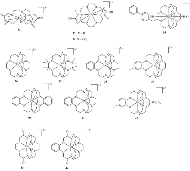
The EuIII/EuII redox couple could be a good candidate for such systems since EuII, with seven unpaired f electrons in an 8S7/2 ground state, is isoelectronic with GdIII. Thus, the GdIII and EuII complexes can be expected to have similar proton relaxivities. EuIII is a very poor relaxing agent due to its small magnetic moment, resulting from contributions from the thermally accessible 7F1 and 7F2 states above the diamagnetic 7F0 ground state, and also to its very fast electronic relaxation (S ~ 0.01 – 0.05 ps).75 Therefore, the paramagnetic relaxation effect of EuIII is negligible relative to EuII.
In some early studies towards developing EuII-based MRI agents,76,77,78 EuII complexes have been found to have faster (more favorable) water exchange kinetics than the corresponding GdIII analogues, due to the larger ionic radius (1.25 Å for EuII vs. 0.938 Å for GdIII) and lower charge (lower charge density) of EuII. On the other hand, EuII has a shorter electronic relaxation time than GdIII in most of the complexes investigated, leading to r1 relaxivities often somewhat lower than that those of typical GdIII chelates.
However, the use of EuII complexes as in vivo redox probes requires high thermodynamic stability and redox stability. The thermodynamic stability constants of the EuII polyaminopolycarboxylate (eg. [EuII(DTPA)(H2O)]3−) (32) and macrocyclic azacrown ether carboxylate derivatives ([EuII(ODDA)(H2O)] (33) and [EuII(ODDM)]2− (34) ODDA2−=1,4,10,13-tetraoxa-7,16-diazacyclooctadecane-7,16-diace tate, ODDM4− = 1,4,10,13-tetraoxa-7,16-diaza-cyclooctadecane-7,16-dimalonate, see Figure 17) initially studied are very close to those of the SrII analogues and far below those of the corresponding GdIII chelates.78 Additionally, most EuII polyaminopolycarboxylate complexes have redox potentials significantly lower than that of the EuII(H2O)82+ aqua ion (EIII/II = -0.39 V vs. NHE), reflecting the instability of the EuII oxidation state. A small improvement has been found for the azacrown derivatives, with EIII/II values of -0.58 V
30
and -0.68 V for the [EuII(ODDA)(H
2O)] (33) and [EuII(ODDM)]2− (34) complexes, respectively, but their negative values severely limit the potential for their use in vivo.77,78
Figure 17. Chemical structures of EuII complexes discussed.
EuII cryptates (Figure 17) proved to be more stable toward oxidation and offered further advantages. For example, [EuII(2.2.2)(H
2O)2]2+ (35) (2.2.2 = 4,7,13,16,21,24-hexaoxa-1,10-diazabicyclo[8.8.8]hexacosane), with a better size match of its cryptand 2.2.2 cavity (1.4 Å) to the EuII ion (1.25 Å) relative to the EuIII ion (1.07 Å), showed a thermodynamic stability seven orders of magnitude higher than for the corresponding EuIII cryptate and a better redox stability (EIII/II = -0.39 V vs. NHE) than the EuII(H2O)82+ aqua ion. It was also shown to have a fast water-exchange rate (108 s–1), a 10-fold slower electron-spin relaxation than 32 that does not limit the relaxivity and two inner-sphere water molecules in a ten-coordinate complex affording a r1= 8 mm-1 s-1 at 0.47 T and 298 K.79,80
31
While this EuII cryptate has some promising properties, its propensity to be oxidized in water is still too high, preventing in vivo MRI applications. Further improvements on the redox stability of the EuII cryptates have been achieved by the group of Allen. They modified the structure of the cryptand 35 using coordination chemistry principles to stabilize the electron-rich EuII ion,81,82 to obtain cryptands 36–41 (Figure
17).83 The first goal was to increase the steric bulk surrounding cryptand 36 to minimize interactions between EuII and its environment, by adding methyl groups to the ethylene carbon atoms between the oxygen atoms, resulting in ligand 37. This substitution pattern was chosen to block the metal–environment interactions occurring between the unmodified ethylene groups. The second goal was to reduce the Lewis basicity of cryptand 36 to favor the electron rich EuII over EuIII. Phenyl rings were introduced to decrease the electron donating ability of the adjacent oxygen atoms of ligands 38–40 by a resonance withdrawing effect, which was modulated by varying the electron density of the phenyl ring through the addition of a fluorine atom (39) or by increasing the number of rings (40). The third goal was to decrease cavity size of the cryptand to better match the size of the EuII ion, due to the effect of each phenyl ring in cryptands 38–40. The final goal was to modify the hard–soft acid–base (HSAB) properties of cryptand 36, which was done by introducing relatively soft sulfur-atom donors in cryptand 41 in place of oxygen-atom donors to explore the HSAB preferences for the softer EuII ion relative to the harder EuIII ion. The redox potential of these EuII cryptates could be tuned over a range of 300 mV. Cryptates 39 and 41 had the highest redox potential increases, with EIII/II values that were 0.622 V (EIII/II = +0.329 V vs NHE) and 0.666 mV (EIII/II = +0.283 V vs NHE), respectively, more positive than the aqueous EuII(H
2O)82+ ion.
The relaxivity of several EuII-containing cryptates (35, 36, 38, Figure 17) was studied at different field strengths (1.4, 3, 7, 9.4, and 11.7 T), which showed higher r1 values at higher fields (7 and 9.4 T) than at lower fields (1.4 and 3 T), unlike common GdIII-containing contrast agents, eg. [Gd(DOTA)(H2O)]-.84,85 Their higher relaxivities relative to [Gd(DOTA)(H2O]- at ultra-high fields are due to the ability to accommodate two water molecules in the inner sphere, the increase in water-exchange rates and changes in rotational correlation rates compared to [Gd(DOTA)(H2O]-. The differences in relaxivities among the different cryptates arise mainly from the changes in the rotational correlation rates that are proportional to molecular weight differences.
The biphenyl derivative EuII-cryptate (35) is capable of interacting with human serum albumin (HSA). The influence of HSA binding on the r1 relaxivity was investigated
32
at different field strengths (1.4, 3, 7, 9.4, and 11.7 T). Relaxometric measurements showed that HSA binding led to a r1 enhancement of 35 of 170 % at the lower field of 1.4 T, as expected from the increased rotational correlation time (R) upon protein binding, while had no effect at high fields (≥ 3T).86 It was also shown that the number of metal-bound water molecules (q) decreases from two in the free complex to one in its bound form, while the water exchange of the remaining bound water molecule was not hindered by the protein side chains.
Besides high relaxivity, other requirements for effective MRI CAs, including kinetic stability, need to be considered for in vivo use. EuII-containing cryptates 36 and
38 were found to be stable in the pH 3-10 region and also to transmetallation in the
presence of endogenous ions such as Ca2+, Mg2+ and Zn2+.84 That was not the case for cryptates 43 and 44, where weak metal binding can be attributed to the presence of the amide groups in the cryptand structure, which, by lowering the basicity of the neighboring nitrogen atoms, affect their metal coordination capacity.
The increased redox stability in water and air-sensitivity of these EuII cryptates led to their proposal as pO2 sensitive MRI redox probes. This was recently tested in vivo for the first time by performing T1-weighted MRI before and after intra-tumoral injection of the EuII cryptate (39) into a 4T1 mammary carcinoma xenograft mouse model (Figure
18, a–d).87 Positive contrast enhancement was observed during the whole experiment, but the contrast location changed with time. While heterogeneous contrast was observed in the whole tumor immediately post injection, after 120 min it was only observed in a localized core of the tumor, indicating a lack of oxygen and the persistence of the reduced (EuII) probe in that part for at least 120 min.
33
Figure 18. T1-weighted in vivo sagittal plane images of a 4T1 tumor in a xenograph mouse carcinoma model injected with 39. Images are recorded a) pre-injection and b) 3 min, c) 20 min, and d) 120 min post-intratumoral injection. e) The difference between the 120 min and pre-injection images (image (d) minus image (a)) colored using the ImageJ green lookup table.f) Hematoxylin- and eosin-stained slice of the tumor imaged in (a–e) and g) the sum of images (e) and (f). All images are on the same scale. Selected imaging parameters: echo time =1.5 ms, repetition time =11 ms, flip angle = 40º, field of view =30 mm x 90 mm, in-plane resolution = 0.352 mm x 0.352 mm. Reproduced with permission from Ref. 87, Copyright (2015) by John Wiley and Sons.
The presence of necrotic tissue in the tumor was confirmed by histological staining, excised after the MRI experiment, with hematoxylin and eosin dyes (Figure 18,
f).87 Hematoxylin stains nuclei (blue) and eosin stains elements of the cytoplasm (pink) to differentiate nuclei abundant nuclei-deficient areas. Areas associated with necrosis (in the mid-to-upper half of the tumor) stained pink to a greater extent than non-necrotic areas, because of the lack of cells and their corresponding nuclei in necrotic regions. Consistent with the staining results, most of the positive contrast enhancement was observed in the mid-to-upper half of the tumor, suggesting that EuII-39 provided positive contrast enhancement in the necrotic core of the tumor (Figure 18, g).87 This probe retention was confirmed by analysis of the tumor Eu content as a function of time by inductively coupled plasma mass spectrometry (ICP-MS). The parallel decrease of MRI tumor contrast (85%) and of Eu content (80%) at 3h post-injection showed the slow clearance of the EuII probe. The clearance process could not be observed by MRI because
34
of the fast probe oxidation in tissues or fluids of relatively higher oxygen content to the EuIII form, which is MRI contrast silent.
The dependence of the in vivo contrast enhancement on the type of probe injection was studied by performing intravenous, intraperitoneal and subcutaneous injections of the EuII cryptate 36 in mice.88 The observed contrast was very sensitive to the route of administration. An inverse correlation was observed between tissue oxygen content and expected rates of diffusion with the persistence of EuII-based contrast enhancement. Biodistribution studies showed Eu clearance through the liver and kidneys for intravenous and intraperitoneal injections, but no contrast enhancement was observed in organs associated with clearance. This study helped understand the behavior of EuII-based complexes in vivo and might be helpful in their preclinical application.
Given the in vivo correlation between tissue pO2 and the persistence of EuII-based positive contrast enhancement,87,88 oxidation reactions of a set of EuII complexes with O2, bromate (BrO3-) and glutathione disulfide (GSSG) have been studied in vitro.89 These helped rationalize the observed EuII oxidation responses and understand the possible products of probe oxidation in vivo. The reaction between EuII(aq) and O2 readily led to superoxide formation, which was detected using electron paramagnetic resonance (EPR) spectroscopy and spin traps. The oxidation kinetics of three EuII-containing complexes, including EuII
(aq) and the cryptate 39, with BrO3- and GSSG was also studied. The reactions with BrO3-, with negative Gibbs free energies of electron transfer were all thermodynamically allowed. However, the oxidation rate of cryptate 39 was relatively low (one order of magnitude lower than with EuII(aq)), indicating that the ligand responsible for the oxidative stability of the cryptate also inhibits the access of water to the metal center by decreasing the available coordination sites from 9-10 in EuII(aq) to 1 in 39.
In contrast, oxidation was not observed for any EuII-containing complex with GSSG. The lack of reactivity between 39 and GSSG can be explained by a positive Gibbs free energy, indicating that spontaneous reactions should not be observed. However, in the case of EuII(aq), spontaneous reaction is expected from the negative free energy of the reaction.87,88 This lack of reactivity points to a kinetic activation barrier, possibly related to the inability of the disulfide bond to access the EuII metal center, preventing easy disulfide reduction. Altogether, the GSSG data and in vivo imaging experiments87,88 indicate that disulfide bonds or other bulky oxidizing agents are likely to have a minor contribution to EuII redox probe oxidation in in vivo MRI experiments of short duration.