HAL Id: inserm-00175279
https://www.hal.inserm.fr/inserm-00175279
Submitted on 19 Mar 2012
HAL is a multi-disciplinary open access
archive for the deposit and dissemination of sci-entific research documents, whether they are pub-lished or not. The documents may come from teaching and research institutions in France or abroad, or from public or private research centers.
L’archive ouverte pluridisciplinaire HAL, est destinée au dépôt et à la diffusion de documents scientifiques de niveau recherche, publiés ou non, émanant des établissements d’enseignement et de recherche français ou étrangers, des laboratoires publics ou privés.
Autophagy and CD4+ T lymphocyte destruction by
HIV-1.
Lucile Espert, Mélanie Denizot, Marina Grimaldi, Véronique
Robert-Hebmann, Bernard Gay, Mihayl Varbanov, Patrice Codogno, Martine
Biard-Piechaczyk
To cite this version:
Lucile Espert, Mélanie Denizot, Marina Grimaldi, Véronique Robert-Hebmann, Bernard Gay, et al.. Autophagy and CD4+ T lymphocyte destruction by HIV-1.. Autophagy, Taylor & Francis, 2007, 3 (1), pp.32-4. �inserm-00175279�
©2006 LANDES BIOSCIENCE
. DO NO
T DISTRIBUTE
.
[Autophagy 3:1, 32-34, January/February 2007]; ©2007 Landes Bioscience
32 Autophagy 2007; Vol. 3 Issue 1
Lucile Espert
1,*
Mélanie Denizot
1Marina Grimaldi
1Véronique Robert-Hebmann
1Bernard Gay
1Mihayl Varbanov
1Patrice Codogno
2Martine Biard-Piechaczyk
11Laboratoire “Infections Rétrovirales et Signalisation Cellulaire;” CNRS/UM1 UMR
5121; Institut de Biologie; Montpellier, France
2INSERM U756; Faculté de Pharmacie; Université Paris XI; Châtenay-Malabry,
France
*Correspondence to: Lucile Espert; Laboratoire “Infections Rétrovirales et Signalisation Cellulaire,” CNRS UMR 5121; Institut de Biologie; 4, Bd Henri IV; Montpellier 34000 France; Tel.: 33.4.67.60.86.60; Fax: 33.4.67.60.44.20; Email: lucile.espert@univ-montp1.fr
Received 08/10/06; Accepted 08/14/06
Previously published online a an Autophagy E-publication:
http://www.landesbioscience.com/journals/autophagy/abstract.php?id=3275
KEY WORDS
autophagy, apoptosis, HIV-1, HIV-1 envelope glycoproteins, cell signaling, Beclin 1
ACKNOWLEDGEMENTS
This work was supported by institutional funds from the Centre National de la Recherche Scientifique (CNRS) and the University of Montpellier 1 (UM1), and grants from SIDACTION and Agence Nationale de Recherches sur le SIDA (ANRS). Lucile Espert was a recipient of a fellowship from SIDACTION.
Addendum to:
Autophagy is Involved in T Cell Death after Binding of HIV-1 Envelope Proteins to CXCR4
L. Espert, M. Denizot, M. Grimaldi, V. Robert-Hebmann, B. Gay, M. Varbanov, P. Codogno and M. Biard-Piechaczyk
J Clin Invest 2006; 116:2161-72.
Addenda
Autophagy and CD4
+
T Lymphocyte Destruction by HIV-1
ABSTRACT
The first step of HIV-1 infection is mediated by the binding of envelope glycoproteins (Env) to CD4 and two major coreceptors, CCR5 or CXCR4. The HIV-1 strains that use CCR5 are involved in primo-infection whereas those HIV-1 strains that use CXCR4 play a
major role in the demise of CD4+T lymphocytes and a rapid progression toward AIDS.
Notably, binding of X4 Env expressed on cells to CXCR4 triggers apoptosis of uninfected
CD4+ T cells. We now have just demonstrated that, independently of HIV-1 replication,
transfected or HIV-1-infected cells that express X4 Env induce autophagy and accumulation
of Beclin 1 in uninfected CD4+ T lymphocytes via CXCR4. Moreover, autophagy is a
prerequisite to Env-induced apoptosis in uninfected bystander T cells, and CD4+T cells
still undergo an Env-mediated cell death with autophagic features when apoptosis is inhibited. To the best of our knowledge, these findings represent the first example of autophagy triggered through binding of virus envelope proteins to a cellular receptor, without viral replication, leading to apoptosis. Here, we proposed hypotheses about the
significance of Env-induced Beclin 1 accumulation in CD4+ T cell death and about the
role of autophagy in HIV-1 infected cells depending on the coreceptor involved.
INTRODUCTION
HIV-1 infection usually leads to progressive decline in functionality and number of CD4+T lymphocytes, resulting in AIDS development.1As early as 1991, T cell apoptosis has been proposed as a possible mechanism responsible for T cell depletion in patients infected with HIV-1,2,3and an extensive body of literature since then has supported this
hypothesis. In addition, there is a correlation between the extent of apoptosis and disease progression,4,5and highly active antiretroviral therapy (HAART) is associated with a lower level of CD4+ T cell apoptosis in HIV-1-infected patients.6-8
In HIV-1-infected patients, both infected and uninfected cells undergo accelerated apoptosis. However, HIV-1-induced apoptosis in bystander uninfected immune cells is likely the event leading to the depletion of T lymphocytes since the degree of cell loss largely exceeds the number of infected cells. Furthermore, the vast majority of T cells undergoing apoptosis in peripheral blood and lymph nodes of HIV patients are uninfected.9,10
Several HIV-1 proteins, such as HIV envelope glycoproteins (Env), Tat, Vpr, Nef, Vpu and the protease can induce T cell apoptosis. Cumulative data demonstrate a major role of Env in the death of uninfected lymphocytes.11,12The mature HIV-1 Env is composed of gp120, the exterior envelope glycoprotein, and gp41, the transmembrane one. In most cases, to enter a target cell, HIV-1 must bind two cell surface receptors on its surface. gp120 first interacts with CD4, which triggers conformational changes leading to increased exposure of the gp120 V3 loop that is then able to bind to several coreceptors, essentially CCR5 and CXCR4, determining the tropism of the virus (R5 and X4, respec-tively) for particular cell types.13These events trigger the formation of a coiled-coil structure
in the gp41 ectodomain that places the hydrophobic amino-terminal region of gp41 in close proximity to the cellular membrane, thereby inducing cell fusion.14Env receptors (CD4, CCR5 and CXCR4) are able to transduce signals within cells after interaction with their physiological ligands. Thus, HIV-1 Env can be considered as a pathological ligand and constitutes the primary interface between viruses and T cells. It has been shown that binding of HIV-1 Env to either CXCR4 or CCR5 induces CD4+ T cell death.15,16
Whereas the R5 HIV-1 strains are involved in the first steps of infection, the X4 HIV-1 strains are associated with a stronger depletion of CD4+T lymphocytes and a rapid
www.landesbioscience.com Autophagy 33
AUTOPHAGY IS TRIGGERED BY HIV-1 ENV
BINDING TO CXCR4 AND IS INVOLVED IN
UNINFECTED CD4
+T CELL DEATH
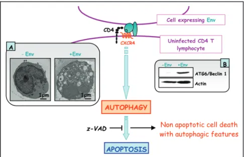
We have recently demonstrated that autophagy is involved in T-cell death after binding of HIV-1 Env to CXCR4.18 Indeed, independently of HIV
replication, transfected or HIV-infected cells that express Env induce autophagy in uninfected CD4+
T lymphocytes through CXCR4, and accumulation of Beclin 1 is rapidly observed in these target cells. Furthermore, Env-mediated autophagy is required to trigger CD4+T cell apoptosis because blockade
of autophagy at different steps, by either drugs (3-methyladenine or bafilomycin A1) or short interfering RNAs specific for the beclin 1/atg6 and atg7 genes, totally inhibits the apoptotic process. In
addition, CD4+T cells still undergo an Env-mediated cell death with autophagic features when apoptosis is inhibited by zVAD. These results suggest that HIV-1-infected cells can induce autophagy in bystander CD4+T lymphocytes through contact of
Env with CXCR4, leading to apoptotic cell death, a mechanism most likely contributing to immunode-ficiency (see Fig. 1). Naive CD4+T cells, which
cannot be productively infected by HIV, may thus
take different routes to die after contact with a cell infected by an X4 HIV-1 strain and sequential, but not exclusive, cell death pathways are triggered by Env binding to CXCR4.
What is the signification of Env-induced Beclin 1 accumu-lation in CD4+T cell death? Beclin 1 is accumulated at the
early steps of the Env-induced signaling cascade, and this phenomenon precedes autophagic vacuolization. Accumulation of Beclin 1 has also been reported in response to drugs that trigger cell death with autophagic features.19-21
Beclin 1, which is involved in the very early steps of autophagosome formation, was first identified as a Bcl-2 interacting protein. Recently, Pattingre et al. have shown that Bcl-2 negatively regulates Beclin 1-dependent autophagy and Beclin 1-dependent autophagic cell death.22This study suggests that the modulation of Beclin 1/Bcl-2 interaction could act as a switch to determine the cell fate between survival and death. Thus the accumulation of Beclin 1 may lead to autophagic cell death. Deciphering the molecular mechanism of Beclin 1 accumulation is a crucial point to understand the role of autophagy in HIV-1 pathogenicity.
What is the role of autophagy in HIV-1 infected cells?
Interactions exist between the autophagic pathway and the intracellular multiplication of microorganisms. Even if autophagy is implicated in bacterial clearance in many cases, some bacteria have developed strategies to circumvent this mechanism of innate immunity and are able to divert this cellular process to their own benefit to replicate inside the autophagosomes.23-25Recent studies show that viruses are
also able to modulate autophagy. Indeed, it has been shown that RNA viruses such as poliovirus require autophagic membranes to assemble their replication complexes in the host cell cytoplasm.26In contrast, autophagy can also be an antiviral mechanism, as demon-strated after Herpes Simplex Virus-1 (HSV-1) infection.27
Nothing is currently known, however, about autophagy induced in HIV-infected CD4+T cells. Nevertheless, at least two hypotheses can be suggested (see Fig. 2).
• HIV-1 infection triggers an autophagic program in infected T cells. In this case autophagy may lead to apoptosis. However this seems unlikely because it is known that viruses are able to maintain the viability of infected cells in order to replicate efficiently and produce new infectious particles. The autophagic program may thus be triggered in order to maintain cell integrity, and to permit HIV-1 replication and propagation.
• Alternatively, it is possible that autophagy is not induced in infected cells. In this case, a viral determinant may be involved in the repression of autophagy, to block the death of infected T cells and, consequently, to permit HIV-1 replication and propagation.
Can R5 HIV-1 strains induce autophagy? The R5 HIV-1 strains
are found in primary infection while the X4 HIV-1 strains emerge in the late and most aggressive stages of the pathology, leading to AIDS. We have demonstrated that X4 Env expressed on
Autophagy and CD4+T Lymphocyte Destruction by HIV-1
Figure 1. Autophagy is needed for Env-induced apoptosis of uninfected CD4+ T lymphocytes after binding to CXCR4. Nonapoptotic cell death with autophagic features is observed when apoptosis is inhibited. A. Transmission electron microscopy showing autophagosome accu-mulation in CD4+ T lymphocyte cytoplasm after Env-binding to CXCR4. B. Immuno-blotting experiments showing Beclin 1 accumulation in uninfected CD4+ T lymphocytes after Env-bind-ing to CXCR4.
Figure 2. Possible scenarios for autophagy in HIV-1 infected cells. If autophagy is triggered in infected cells, this may lead to apoptosis, or may benefit viral replica-tion. If autophagy is repressed, the virus may replicate more efficiently and avoid apoptosis. In this case, viral proteins may be involved.
Autophagy and CD4+T Lymphocyte Destruction by HIV-1
34 Autophagy 2007; Vol. 3 Issue 1
HIV-1-infected cells can induce autophagy and cell death in bystander uninfected CD4+T cells.18Even if CD4+T cell infection
by the R5 HIV-1 strain can also induce CD4+T cell death, the
putative role of autophagy in this case has not been investigated. It would be important to analyze if the two main coreceptors, CCR5 or CXCR4, present differences in triggering and/or regulating autophagy and if CCR5-dependent autophagy and CXCR4-dependent autophagy have different outcomes on cell survival and cell death. This process may determine the course of the HIV-1 associated pathology.
The emerging development of novel therapeutic strategies based on modulation of autophagy in cancer and neurodegenerative diseases attests to the increasing importance of the autophagy field. A number of essential issues, however, remain to be addressed, especially the connections between autophagy, cell death and viral infections. It appears fundamental to better understand the autophagic process and its regulation in the case of HIV-1 infection in order to develop new and more adapted therapeutics that target not only the viral replication but also the cellular response to infection.
References
1. Fauci AS. The human immunodeficiency virus: Infectivity and mechanisms of pathogene-sis. Science 1988; 239:617-22.
2. Ameisen JC, Capron A. Cell dysfunction and depletion in AIDS: The programmed cell death hypothesis. Immunol Today 1991; 12:102-5.
3. Terai C, Kornbluth RS, Pauza CD, Richman DD, Carson DA. Apoptosis as a mechanism of cell death in cultured T lymphoblasts acutely infected with HIV-1. J Clin Invest 1991; 87:1710-5.
4. Cotton MF, Ikle DN, Rapaport EL, Marschner S, Tseng PO, Kurrle R, Finkel TH. Apoptosis of CD4+ and CD8+ T cells isolated immediately ex vivo correlates with disease severity in human immunodeficiency virus type 1 infection. Pediatr Res 1997; 42:656-64. 5. Gougeon ML, Lecoeur H, Dulioust A, Enouf MG, Crouvoiser M, Goujard C, Debord T, Montagnier L. Programmed cell death in peripheral lymphocytes from HIV-infected per-sons: Increased susceptibility to apoptosis of CD4 and CD8 T cells correlates with lym-phocyte activation and with disease progression. J Immunol 1996; 156:3509-20. 6. Lederman MM, Connick E, Landay A, Kuritzkes DR, Spritzler J, St Clair M, Kotzin BL,
Fox L, Chiozzi MH, Leonard JM, Rousseau F, Wade M, Roe JD, Martinez A, Kessler H. Immunologic responses associated with 12 weeks of combination antiretroviral therapy consisting of zidovudine, lamivudine, and ritonavir: Results of AIDS Clinical Trials Group Protocol 315. J Infect Dis 1998; 178:70-9.
7. Badley AD, Parato K, Cameron DW, Kravcik S, Phenix BN, Ashby D, Kumar A, Lynch DH, Tschopp J, Angel JB. Dynamic correlation of apoptosis and immune activation dur-ing treatment of HIV infection. Cell Death Differ 1999; 6:420-32.
8. Bohler T, Walcher J, Holzl-Wenig G, Geiss M, Buchholz B, Linde R, Debatin KM. Early effects of antiretroviral combination therapy on activation, apoptosis and regeneration of T cells in HIV-1-infected children and adolescents. Aids 1999; 13:779-89.
9. Debatin KM, Fahrig-Faissner A, Enenkel-Stoodt S, Kreuz W, Benner A, Krammer PH. High expression of APO-1 (CD95) on T lymphocytes from human immunodeficiency virus-1-infected children. Blood 1994; 83:3101-3.
10. Finkel TH, Tudor-Williams G, Banda NK, Cotton MF, Curiel T, Monks C, Baba TW, Ruprecht RM, Kupfer A. Apoptosis occurs predominantly in bystander cells and not in productively infected cells of HIV- and SIV-infected lymph nodes. Nat Med 1995; 1:129-34.
11. Heinkelein M, Sopper S, Jassoy C. Contact of human immunodeficiency virus type 1-infected and uninfected CD4+ T lymphocytes is highly cytolytic for both cells. J Virol 1995; 69:6925-31.
12. Laurent-Crawford AG, Krust B, Riviere Y, Desgranges C, Muller S, Kieny MP, Dauguet C, Hovanessian AG. Membrane expression of HIV envelope glycoproteins triggers apoptosis in CD4 cells. AIDS Res Hum Retroviruses 1993; 9:761-73.
13. Berger EA. HIV entry and tropism: The chemokine receptor connection. Aids 1997; 11(Suppl A):S3-16.
14. Weissenhorn W, Dessen A, Harrison SC, Skehel JJ, Wiley DC. Atomic structure of the ectodomain from HIV-1 gp41. Nature 1997; 387:426-30.
15. Ahr B, Robert-Hebmann V, Devaux C, Biard-Piechaczyk M. Apoptosis of uninfected cells induced by HIV envelope glycoproteins. Retrovirology 2004; 1:12.
16. Roggero R, Robert-Hebmann V, Harrington S, Roland J, Vergne L, Jaleco S, Devaux C, Biard-Piechaczyk M. Binding of human immunodeficiency virus type 1 gp120 to CXCR4 induces mitochondrial transmembrane depolarization and cytochrome c-mediated apopto-sis independently of Fas signaling. J Virol 2001; 75:7637-50.
17. Lusso P. HIV and the chemokine system: 10 years later. Embo J 2006; 25:447-56. 18.Espert L, Denizot M, Grimaldi M, Robert-Hebmann V, Gay B, Varbanov M, Codogno P,
Biard-Piechaczyk M. Autophagy is involved in T cell death after binding of HIV-1 enve-lope proteins to CXCR4. J Clin Invest 2006; In press.
19. Shimizu S, Kanaseki T, Mizushima N, Mizuta T, Arakawa-Kobayashi S, Thompson CB, Tsujimoto Y. Role of Bcl-2 family proteins in a nonapoptotic programmed cell death
dependent on autophagy genes. Nat Cell Biol 2004; 6:1221-8.
20. Furuya D, Tsuji N, Yagihashi A, Watanabe N. Beclin 1 augmented cisdiamminedichloro-platinum induced apoptosis via enhancing caspase-9 activity. Exp Cell Res 2005; 307:26-40.
21. Scarlatti F, Bauvy C, Ventruti A, Sala G, Cluzeaud F, Vandewalle A, Ghidoni R, Codogno P. Ceramide-mediated macroautophagy involves inhibition of protein kinase B and upreg-ulation of beclin 1. J Biol Chem 2004; 279:18384-91.
22. Pattingre S, Tassa A, Qu X, Garuti R, Liang XH, Mizushima N, Packer M, Schneider MD, Levine B. Bcl-2 antiapoptotic proteins inhibit Beclin 1-dependent autophagy. Cell 2005; 122:927-39.
23. Gutierrez MG, Master SS, Singh SB, Taylor GA, Colombo MI, Deretic V. Autophagy is a defense mechanism inhibiting BCG and Mycobacterium tuberculosis survival in infected macrophages. Cell 2004; 119:753-66.
24. Nakagawa I, Amano A, Mizushima N, Yamamoto A, Yamaguchi H, Kamimoto T, Nara A, Funao J, Nakata M, Tsuda K, Hamada S, Yoshimori T. Autophagy defends cells against invading group A Streptococcus. Science 2004; 306:1037-40.
25. Ogawa M, Yoshimori T, Suzuki T, Sagara H, Mizushima N, Sasakawa C. Escape of intra-cellular Shigella from autophagy. Science 2005; 307:727-31.
26. Jackson WT, Giddings Jr TH, Taylor MP, Mulinyawe S, Rabinovitch M, Kopito RR, Kirkegaard K. Subversion of cellular autophagosomal machinery by RNA viruses. PLoS Biol 2005; 3:e156.
27. Tallóczy Z, Jiang W, Virgin HW IV, Leib DA, Scheuner D, Kaufman RJ, Eskelinen EL, Levine B. Regulation of starvation- and virus-induced autophagy by the eIF2α kinase sig-naling pathway. Proc Natl Acad Sci USA 2002; 99:190-5.