HAL Id: inserm-01848512
https://www.hal.inserm.fr/inserm-01848512
Submitted on 24 Jul 2018HAL is a multi-disciplinary open access
archive for the deposit and dissemination of sci-entific research documents, whether they are pub-lished or not. The documents may come from teaching and research institutions in France or abroad, or from public or private research centers.
L’archive ouverte pluridisciplinaire HAL, est destinée au dépôt et à la diffusion de documents scientifiques de niveau recherche, publiés ou non, émanant des établissements d’enseignement et de recherche français ou étrangers, des laboratoires publics ou privés.
phenotype: the case of Claudin11deficiency in testis.
Séverine Mazaud-Guittot, Emmanuelle Meugnier, Sandra Pesenti, X. Wu, H
Vidal, A Gow, Brigitte Le Magueresse-Battistoni
To cite this version:
Séverine Mazaud-Guittot, Emmanuelle Meugnier, Sandra Pesenti, X. Wu, H Vidal, et al.. Loss of tight junction integrity abolishes the epithelial phenotype: the case of Claudin11deficiency in testis. : Claudin11 and loss of the Sertoli cell epithelial phenotype. Biology of Reproduction, Society for the Study of Reproduction, 2010, 82 (1), pp.202-13. �10.1095/biolreprod.109.078907�. �inserm-01848512�
Loss of tight junction integrity abolishes the epithelial phenotype: the case
1of Claudin11 deficiency in testis.
23 4
Running title: Claudin11 and loss of the Sertoli cell epithelial phenotype
5 6
Mazaud-Guittot S.1,2,3,4,5,9,10, Meugnier E.1,2,3,4,5, Pesenti S.1,2,3,4,5, Wu X.6, Vidal H1,2,3,4,5, Gow 7 A.6,7,8, Le Magueresse-Battistoni B.1,2,3,4,5 8 9 Address : 10 1
Inserm, U870, Oullins, France; 2INRA, UMR1235, Oullins, France; 3INSA-Lyon, RMND, 11
Villeurbanne, France; 4Université Lyon 1, Lyon, France; 5Hospices Civils de Lyon, Lyon, 12
France. 13
14
6
Center for Molecular Medicine and Genetics, 7Karman and Ann Adams Department of 15
Pediatrics, 8Department of Neurology, Wayne State University, 3217 Scott Hall, 540 E 16
Canfield, Detroit, MI, U.S.A. 17
18
Present address : 9 Inserm, U625, Rennes, France ; 10 Univ Rennes I, Campus de Beaulieu, 19
IFR-140, GERHM, Rennes, F-35042, France. 20
21
Corresponding authors : Brigitte Le Magueresse-Battistoni at 22
Brigitte.lemagueresse@inserm.fr and Séverine Mazaud-Guittot at severinemazaud@yahoo.fr
23 24 25
ABSTRACT
26
Tissue integrity relies on barriers formed between epithelial cells. In the testis, the 27
barrier is formed at the initiation of puberty by a tight junction complex between adjacent 28
Sertoli cells, thereby defining an adluminal compartment where meiosis and spermiogenesis 29
occur. Claudin11 is an obligatory protein for tight junction formation and barrier integrity in 30
the testis. It is expressed by Sertoli cells, and spermatogenesis does not proceed beyond 31
meiosis in its absence, resulting in male sterility. Sertoli cell maturation – arrest of 32
proliferation and expression of proteins to support germ cell development – is known to 33
parallel tight junction assembly; however, the pathophysiology underlying the loss of tight 34
junctions in the mature testis remains largely undefined. Herein, we use 35
immunohistochemistry and microarrays in wild-type and Claudin11-/- testes from mice to 36
demonstrate that adult Claudin11-/- Sertoli cells re-enter the cell cycle while maintaining 37
expression of several differentiation markers. Dividing Sertoli cells lose polarity, detach from 38
the basement membrane and are eliminated through the lumen together with apoptotic germ 39
cells that they have phagocytosed. Thus, Claudin11-/- Sertoli cells exhibit a unique phenotype 40
whereby loss of tight junction integrity results in loss of the epithelial phenotype but not the 41
progression to a tumorigenic phenotype. 42
43
INTRODUCTION
44 45
The integrity of epithelial cell layers is maintained by intercellular junctional 46
complexes composed of adhesive (adherens junction, desmosomes, hemidesmosomes) and 47
occluding (tight) junctions, and gap junctions promote intercellular communication. The 48
transmembrane proteins constituting these junctions are linked to components of the actin and 49
intermediate filament cytoskeletons, and a growing number of cytoplasmic scaffolding 50
molecules associated with these junctions are involved in regulating such diverse processes as 51
transcription, cell proliferation, cell polarity and the assembly of regulated diffusion barriers. 52
Typically, tight junctions (TJs) define the boundary between the apical and basolateral 53
domains of epithelial cell membranes and function as the primary barrier to diffusion of 54
macromolecules, ions, and small non-charged solutes through the paracellular pathway (22, 55
47). 56
57
Most blood-tissue barriers, such as the blood-brain and blood-retinal barriers, are 58
generated by endothelial cell TJs in specialized microvessels. By contrast, the blood-testis 59
barrier (BTB) is generated and maintained by Sertoli cells in the seminiferous epithelium and 60
is physically remote from microvessels of the interstitium. The BTB is also unique because it 61
is cyclically restructured during spermatogenesis when preleptotene spermatocytes migrate 62
into the adluminal compartment and enter meiosis. Thus, the BTB divides the seminiferous 63
epithelium into two compartments: the basal compartment, which forms the niche for 64
spermatogonia proliferation and renewal, and the adluminal compartment, where meiosis and 65
spermiogenesis occur. 66
67
Both the structural integrity of the seminiferous epithelium and the BTB are 68
maintained by the highly specialized actin filament network of the Sertoli cell cytoskeleton, 69
known as the ectoplasmic specialization (ES). Ectoplasmic specializations are complex 70
cytoskeletal structures occurring in submembranous regions adjacent to TJs (BTB) and the 71
apical adhesion sites of spermatogenic cells (14, 15, 38, 43). They consist of bundles of actin 72
filaments sandwiched between the plasma membrane and cisternae of the endoplasmic 73
reticulum (7, 14, 15, 38, 43). Because they are closely associated with junctional sites, ESs 74
are thought to play a major role in maintaining and regulating intercellular junction assembly 75
(14, 34, 42, 44, 58-60, 62). 76
77
The molecular composition of the BTB has been the subject of numerous studies 78
reviewed in (32) and Sertoli cell TJs are composed, at least, of the transmembrane proteins 79
claudin 11, claudin 3, occludin, junction associated molecule (JAM-A) and the coxsackie 80
virus and adenovirus receptor (CAR) (32, 36). The phenotypes of mice deficient in various 81
components of these TJs varies from normal (no apparent phenotype) as revealed in JAM-A -/-82
mice (12), to slowly degenerative as for Occludin-/- mice (45), to sterility in Claudin11-/- mice 83
(21). Claudin11-/- mice exhibit neurologic, auditory and reproductive deficits, including 84
slowed central nervous system (CNS) nerve conduction, conspicuous hind limb weakness, 85
profound sensorineural deafness and male sterility (13, 20, 21). 86
87
In the testis of Claudin11-/- mice, spermatogenesis does not proceed beyond the 88
spermatocyte stage and cell clusters are observed in the seminiferous lumen. To understand 89
the relationship between claudin11 loss and seminiferous tube disorganization, we have 90
determined the aetiology of this phenotype, in particular during the formation of the BTB. 91
Our comprehensive survey reveals that in the absence of claudin11, Sertoli cells do not form a 92
mature BTB which induces a spermatogenesis defect in neighbouring germ cells. 93
Furthermore, Sertoli cells lose polarity, detach from the basement membrane, undergo an 94
epithelial-to-fibroblastic cell shape transformation and re-enter the cell cycle while 95
maintaining expression of differentiation markers. These changes are associated with TJ 96
regulation as well as actin-related and cell cycle gene expression. Thus, Claudin11-/- Sertoli 97
cells exhibit a unique phenotype in that the loss of TJ integrity induces the loss of an 98
epithelial phenotype which does not progress toward a tumorigenic phenotype. 99
100 101
MATERIAL AND METHODS
102
Animal handling, tissue collection and processing
103
Males were injected intraperitoneally with 50 mg/kg bromodeoxyuridine (BrdU) 104
dissolved in saline 3 h before sacrifice. Testes and epididymides collected at P7, P10, P13, 105
P15, P20, P28, P60, P90 and P180 were either frozen on dry ice and stored at -80°C until 106
processing for RNA analysis or processed for morphological studies. For histological and 107
immunohistochemical analyses, tissues were fixed either in Bouin fixative or in 4% 108
paraformaldehyde-PBS (pH 7.2) for at least 24 h, dehydrated in a graded series of ethanol, 109
and paraffin-embedded using standard protocols. Five µm-thick sections were stained with the 110
periodic acid-Schiff-haematoxylin technique (PAS). 111
112
Immunohistochemistry
113
Paraffin embedded tissues were deparaffinized in xylene and rehydrated in graded 114
ethanol solutions and endogenous peroxidase activity was blocked with 0.3% hydrogen 115
peroxide in methanol for 30 minutes. For all but claudin11 immunodetection, sections were 116
boiled for 5 minutes in 0.1M citrate buffer (pH 6.0) for antigen retrieval, blocked with 10% 117
horse serum (in PBS with 8% BSA) for at least 20 minutes, and finally incubated overnight at 118
4°C with primary antibody diluted in blocking solution (Dako Corp., Trappes, France). 119
Primary antibodies were directed against claudin11 (diluted 1/100; Santa Cruz 120
Biotechnologies Inc., Santa Cruz, CA), DDX4 (diluted 1/750; kindly provided by Dr. T. 121
Noce), Clgn (TRA-369 antibody; diluted 1/1000; kindly provided by Dr. H. Tanaka), Gata4 122
(diluted 1/100; Santa Cruz Biotechnologies), phosphorylated serine 10 (ser10) of histone H3 123
(diluted 1/500; Upstate Biotechnology, Euromedex, Mundolsheim, France), BrdU (diluted 124
1/100; Roche), Vimentin (LN-6 clone; diluted 1/100; DakoCytomation, Trappes, France). 125
126
After washing in PBS, and depending on the primary antibody used, sections were 127
incubated for 2 h with either horseradish peroxidase-conjugated anti-rabbit antibody 128
(Envision™+ system-HRP, Dako Corp.), biotinylated anti-goat antibody (1/500 dilution; 129
Vector Laboratories Canada, Burlington, Canada), anti-rat antibody (1/200 dilution; Vector 130
Laboratories) and finally 30 minutes with a peroxidase-conjugated streptavidin-horseradish 131
complex (LSAB®+ Kit, Dako Corp.). The reaction product was developed using 3,3’-132
diaminobenzidine tetrahydrochloride (DAB) (Sigma-Aldrich). Sections were counterstained 133
with hematoxylin and mounted with Eukitt (Sigma-Aldrich). For negative controls, primary 134
antibody was omitted. Slides were analyzed with Zeiss Akioskop II and Axiophot 135
microscopes (Carl Zeiss, New York, NY) connected to a digital camera (Spot RT Slider, 136
Diagnostic Instruments, Sterling Heights, MI). 137
138
For double immunolabeling, paraffin sections were prepared as above, incubated 139
overnight with anti-Gata4 antibody (diluted 1/100), followed by sequential incubations with 140
anti-goat Ig-Alexa Fluor 546 secondary antibody (1/1000; Invitrogen) 2 h at room 141
temperature, anti-BrdU antibody (1/100), and anti-mouse Ig-Alexa Fluor 488 secondary 142
antibody (1/1000; Invitrogen). Fluorochrome-labeled sections were mounted in Vectashield 143
containing DAPI for nuclei visualization (Vector Laboratories Canada, Burlington, Canada). 144
Slides were analyzed with a Zeiss Akiovert epifluorescence microscope (Carl Zeiss, New 145
York, NY) connected to a digital camera (Spot RT Slider, Diagnostic Instruments, Sterling 146
Heights, MI). 147
Terminal deoxynucleotidyltransferase-mediated dUTP-FITC nick end labeling
149
(TUNEL) assays.
150
Detection of apoptotic cells was performed on paraffin sections using in situ cell death 151
detection kit (Roche). After rehydratation, sections were boiled for 5 minutes in 0.1M citrate 152
buffer (pH 6.0), and incubated for 1 h at 37°C with the TUNEL reaction mixture containing 153
terminal transferase to label free 3′-hydroxy ends of genomic DNA with fluorescein-labeled 154
deoxy-UTP. After washing, sections were incubated overnight at 4°C with Peroxidase 155
converter (Roche). Apoptotic cells were revealed with DAB, and sections counterstained with 156 hematoxylin. 157 158 Morphometric analysis 159
Apoptotic cells and germ cells in the division phases were quantified in transverse 160
seminiferous tube sections stained for TUNEL and phosphorylated histone H3, respectively. 161
For each animal, at least three non-serial testicular sections were used, and in each section, all 162
transverse sectioned tubes were quantified, for a total of at least 100 tubes per animal (with a 163
mean of 330 and 300 tubes). In addition, the number of phosphorylated histone H3 labelled 164
cells was counted in at least 50 tubes per animal. Sertoli cell nuclei were quantified in 165
transverse seminiferous tube sections stained for Gata4. For each animal, at least three non-166
serial testicular sections were used, and in each section, all transverse section tubes were 167
quantified, for a total of at least 50 tubes per animal. 168
169
Microarray analysis
170
Total RNA was prepared from P20-old testes by using RNeasy minikit (Qiagen, 171
Courtaboeuf, France). RNA integrity was determined with the Agilent 2100 Bioanalyzer and 172
RNA 6000 Nano Kit (Agilent Technologies,Massy, France). A pool of P20 Claudin11 +/-173
testes was used as a common reference. One microgram of total RNA was amplified with the 174
Amino Allyl MessageAmp II aRNA kit (Ambion, Austin, TX) according to the 175
manufacturer’s instructions. 176
177
Fluorescent probes were synthesized by chemical coupling of 5 µg of aminoallyl 178
aRNA with cyanine (Cy)3 or Cy5 dyes (GE Healthcare Biosciences, Orsay, France). After 179
purification with an RNeasy Mini Kit (Qiagen, Courtaboeuf, France), probes were fragmented 180
with 25X RNA Fragmentation Reagents (Agilent Technologies) and hybridized with 2X 181
Agilent Hybridization Buffer (Agilent Technologies) to Mouse opArray (Operon 182
Biotechnologies GmbH, Cologne, Germany) in an Agilent oven at 67°C for 16 h, following a 183
dye swap experimental procedure. Microarrays were washed and scanned with a Genepix 184
4000B scanner (Axon Instruments, Foster City, CA). 185
186
TIFF images were analyzed using Genepix Pro 6.0 software (Axon Instruments). 187
Signal intensities were log-transformed and normalization was performed by the intensity 188
dependent Lowess method. To compare results from the different experiments, data from each 189
slide were normalized in log-space to have a mean of zero using Cluster 3.0 software. Data 190
were analyzed using the Significance Analysis of Microarray (SAM) procedure (55). 191
Microarray data are available in the GEO database under the number GSE15492. Further 192
analysis of GO and KEGG pathway enrichments were performed using the WebGestalt 193
analysis toolkit (http://bioinfo.vanderbilt.edu/webgestalt/) (67). 194
195
Quantitative PCR
196
First-strand cDNAs were synthesized from 1 µg of total RNA in the presence of 100 U 197
of Superscript II (Invitrogen, Eragny, France) and a mixture of random hexamers and 198
oligo(dT) primers (Promega, Charbonnières, France). Real-time PCR assays were performed 199
with a Rotor-GeneTM 6000 (Corbett Research, Mortlake, Australia). PCR primers are listed in 200 Table 1. 201 202 Data analysis 203
Statistical analyses were performed using the SigmaStat 2.0 software package. For cell 204
counts, a One-way ANOVA followed by the appropriate post-hoc test was used to compare 205
differences between groups, as specified in each figure legend. Significance was accepted at a 206 confidence level of p ≤ 0.05. 207 208 RESULTS 209
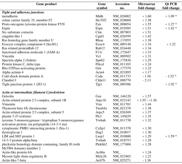
Chronology of the Claudin11-/- phenotype
210
To characterize the beginning of the testicular phenotype in Claudin11-/- mice, we first 211
analyzed testis histological sections throughout postnatal testicular development (Fig. 1). At 212
two weeks of age, seminiferous tubes contain Sertoli cells and early pachytene spermatocytes, 213
which are the most mature cells of the germ cell lineage. One week later, early spermatids 214
largely populate the tubes, and differentiating elongated spermatids are first detected at P28. 215
At P60, when the animals are adults, spermatogenesis is cyclic and can be divided into 12 216
stages (I-XII) based on the morphological transformation of spermatids into spermatozoa in a 217
process referred as spermiogenesis (39). 218
219
The first signs of disorganization in Claudin11-/- testes appear at P20 (Fig. 1A-B, D-E) 220
and are obvious by P28. Indeed, when compared to the well-organized epithelium in the first 221
wave of spermatogenesis from control testes (Fig. 1G), germ cells in Claudin11-/- testes 222
appear abnormally localized. Round clusters of cells are observed closely apposed to the basal 223
side of the seminiferous epithelium and the testis tubular lumen is poorly defined and is filled 224
with round spermatids or cell clusters (Fig. 1H). Moreover, while elongated spermatids 225
appear in some tubes of P28 control testes, only scarce ectopically localized round spermatids 226
are present in Claudin11-/- testes (Fig. 1 G-H). In addition, elongated spermatids are never 227
observed in Claudin11-/- testes at P28 or P60, and spermatogenesis does not proceed beyond 228
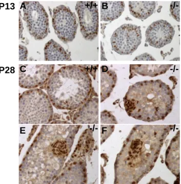
meiosis, which is consistent with published findings (18). The phenotype is accentuated at 229
P60, and PAS-positive material is observed inside the cell clusters indicative of the presence 230
of glycoproteins (Fig. 1J-K). 231
232
Because Claudin11 expression has been reported during fetal testis development (23), we 233
compared the temporal appearance of the Claudin11-/- testes phenotype with that of protein 234
expression in control testes (Fig. 1C, E, I). Although claudin11 is not detected by 235
immunohistochemistry at P10 (data not shown), it is obvious in some tubes at P13, with 236
labeling extending from the basal membrane to the lumen (Fig. 2C). Such staining is 237
consistent with the localization of claudin11 to plasma membrane and the polarization of 238
Sertoli cells from P20 onwards (Fig. 1F, I). Thus, the appearance of the first histological 239
defects in Claudin11-/- testes closely parallels the kinetics of claudin11 expression in control 240
testes. Testis weight in Claudin11-/- mice is normal up to P28 (Fig. 1L), which suggests 241
defects at the organ weight level are delayed compared to the histological level. 242
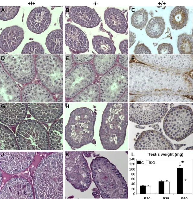
243
Cell clusters are comprised of Sertoli cells
244
Histological sections reveal that nuclei from the vast majority of cells in adluminal cell 245
clusters are clear and contain 1-3 nucleoli, which is reminiscent of normal Sertoli cell 246
morphology (data not shown). However, in P60 Claudin11-/- testes, mixed cell nuclei are 247
observed in the clusters (Fig. 1K). To identify the cells comprising these clusters, we 248
performed immunohistochemistry for several germ cell markers (Fig. 2): the Ddx4 protein 249
(Mouse Vasa Homolog) is expressed in primary spermatocyte and more differentiated cells 250
(54) (Fig. 2A), Clgn is expressed in early pachytene spermatocytes to step 14 spermatids (D) 251
(63), vitronectin (30) and claudin1 (data not shown) are expressed in the acrosomes of 252
spermatids. The absence of these markers demonstrates that cell clusters sloughing from the 253
epithelium of Claudin11-/- testes do not contain germ cells (Fig. 2B-C, E, F and data not 254
shown), although a few germ cells are found in some cell clusters from P60 – P180 mice (data 255
not shown and Fig. 7I). By contrast, immunolabeling of Gata4, which identifies Sertoli cells 256
inside the seminiferous epithelium (Fig 2G) (27, 57) clearly indicates that clusters are 257
comprised of Sertoli cells (Fig. 2H-I). 258
259
Increased apoptosis in testis of Claudin11-/- mice
260
Although claudin11 expression in testis is Sertoli cell-specific, the primary defect in 261
Claudin11-/- mice is that of a failure of spermatogenesis. Apoptosis is the dominant pathway 262
for eliminating germ cells whenever the supporting Sertoli cells are unable to provide a 263
supportive environment for their development (41); thus, we characterized the fate of germ 264
cells using TUNEL labeling (Fig. 3). Quantification of TUNEL-labeled cells in Claudin11 -/-265
testis shows a significant (p < 0.05) increase in the incidence of apoptosis at P20 and P28 266
compared to age-matched controls (Fig. 3A-E). The proportion of round tubes containing one 267
or more TUNEL-positive cell is increased 1.5-fold (p < 0.05) in Claudin11-/- testes at P15 268
(Fig. 3E), suggesting an early impact of the absence of claudin11 on germ cell development. 269
Apoptosis reaches a peak at P20 and is maintained through P28, but remains elevated at P60 270
and is not statistically different from levels at P20. In addition, abundant vesicles of 271
fragmented TUNEL positive-DNA are detected in the cytoplasm of sloughing Sertoli cells 272
(Fig. 3D), which likely indicates that Sertoli cells actively phagocytose degenerating germ 273
cells. These kinetics are consistent with the notion that germ cell differentiation, not stem cell 274
renewal, is perturbed by the absence of Sertoli cell TJs. 275
276
To further dissect the fate of germ cells in Claudin11-/- testes, sections were 277
immunostained with phosphorylated histone H3 (Ph-H3) (Fig. 4). Ph-H3 is normally 278
expressed in mitotic spermatogonia and in diplotene spermatocytes approaching cell division 279
at stages XI and XII (10). Immunolabeling of Ph-H3 at P15, P28 and P60 show no obvious 280
changes in mitotic spermatogonia or meiotic spermatocytes (small magnifications not shown; 281
Fig. 4A-F) from the proportions of round tubes containing one or more Ph-H3 positive cells 282
(Fig. 4G). By contrast, the number of Ph-H3 positive cells per round tube increase from P28 283
to P60 in control testes but remains constant in Claudin11-/- testes (Fig. 4C-F, H). 284
Consequently, the total number of Ph-H3 positive cells decreases in Claudin11-/- testes (Fig. 285
4H) at P28 and P60. Together, these data suggest that amplification of the germ cell 286
population resulting from active spermatogenesis does not occur in absence of claudin11 at 287
the inter-Sertoli BTB. 288
289
Dynamics of Sertoli cell sloughing in Claudin11-/- testes
290
To determine how a lack of claudin11 might impact Sertoli cell topography within the 291
seminiferous epithelium, sections were immunolabeled with Gata4 (Fig. 5). In control testes, 292
Sertoli cell nuclei are positioned at the basement membrane of the seminiferous epithelium as 293
expected. In Claudin-/- testes, the nuclei are localized toward the centre of the tube as early as 294
P13 (Fig. 5A, B), which coincides with the beginning of claudin11 expression in control 295
testes (Fig. 1C). At P28 (Fig. 5) - also seen at P20 (data not shown) - basally located Sertoli 296
cells are observed as well as several abnormal Sertoli cell arrangements (Fig. 5C-F). These 297
include groupings of Sertoli cells adjacent to the base of the tube (Fig. 5D), round clusters 298
with few cells attached to the basement membrane (Fig. 5E) and completely detached Sertoli 299
cell clusters filling the tube lumen (Fig. 5F). These data are indicative of dynamic Sertoli cell 300
sloughing from their basal sites into the lumen of the tube. 301
302
In this light, we hypothesize that Sertoli cells may migrate along the basement 303
membrane to form small groups which then detach, or they may form clusters by cell division 304
and thereafter detach. To distinguish between these possibilities, we examined changes in the 305
number of Gata4 labeled Sertoli cells from P15 to P60 (Fig. 6A-C). Consistent with the 306
temporal development of the phenotype, the number of Sertoli cells per cluster rises from P15 307
to P28 and is maintained at this level through P60 in Claudin11-/- testes (Fig. 6A). In addition, 308
the proportion of round tubes containing detached Sertoli cells almost doubles between P15 309
and P60 (p < 0.05). Indeed, more than 80% of the tubes contain Sertoli cell clusters at P60 310
(Fig. 6B). 311
312
While the number of Sertoli cells per round tube decreases from P15 to P60 in control 313
testes (probably as a result of dilution due to massive increase of the germ cell population 314
within the tubules as the animals mature), the size of this population remains almost constant 315
in Claudin11-/- testes (Fig. 6C). The number of Sertoli cells per round tube in control and 316
Claudin11-/- testes is similar at P15 and at P28 if both peripherally- and cluster-located Sertoli 317
cells are considered. At P60, the number of peripherally-located Sertoli cells is similar in 318
control and Claudin11-/- testes, but higher in Claudin11-/- testes when considering total Sertoli 319
cell numbers. Together, these data suggest a continuous renewal of Sertoli cells to 320
compensate for their losses from detachment and shedding into the lumen. 321
322
Sertoli cells undergo cell division in Claudin11-/- testes
323
Sertoli cells cease dividing in immature animals during the first 2 weeks after birth, 324
which is concomitant with the appearance of meiotic germ cells and formation of the lumen 325
(28, 56). To determine the cell cycle status of Sertoli cells, we performed an in-depth survey 326
of Gata4-labeled Sertoli cells (Fig. 6E-J). In control testes, Gata4 labeling of Sertoli cell 327
nuclei is homogenous, with the exception of nucleoli (Fig. 6E, H) and similarly labeled 328
Sertoli cells are found in P28 (Fig. 6D-F) and P60 (not shown) Claudin11-/- testes. In addition, 329
compacted chromatin in Gata4-positive nuclei is also observed, together with the 330
characteristic meiotic figures of germ cells (Fig. 6F). 331
332
Juxtaposed Sertoli cells in the plane parallel to the basement membrane in Claudin11 -/-333
testes (Fig. 6E), suggest that this cell population may be dividing. To unambiguously identify 334
dividing Sertoli cells, we used in vivo BrdU-incorporation prior to the fixation and tissue 335
processing (Fig. 6G-H). Comparison of the Gata4-positive Sertoli cell and BrdU-positive 336
dividing cell populations in wild type testes reveals the canonical complementary pattern of 337
Sertoli cell and spermatogonia (Fig. 6G). This is a hallmark of stage VIII tubes, wherein 338
spermatogonia proliferate and leptotene spermatocytes traverse the BTB into the adluminal 339
compartment for further development (12, 27, 29, 29, 32). In Claudin11-/- testes, stage VIII 340
tubes contain Gata4-positive Sertoli cells and BrdU-labeled spermatogonia, indicating that the 341
lack of claudin11 expression does not hamper the spermatogenic cycle, or at least the cyclical 342
entry of spermatogonia into mitosis at stage VIII (39). However, double labeled cells are also 343
present at the periphery of some tubes (Fig. 6H), which demonstrates that Sertoli cells are 344
proliferating in Claudin11-/- testes. Importantly, we do not observe BrdU labeling of cell 345
clusters, indicating that Sertoli cell division occurs prior to cluster formation and shedding 346
into the lumen. 347
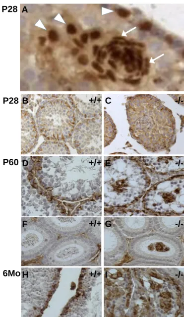
Loss of Sertoli cell polarity but maintenance of differentiation markers
349
In Fig. 5, we observe that Sertoli cells in Claudin11-/- testes change shape in the course 350
of cell cluster sloughing. Thus, the nuclei of basal Gata4-labelled Sertoli cells are round or 351
triangular, while Sertoli cells located at the periphery of clusters have round nuclei and those 352
within clusters have smaller and elongated or comma-shaped nuclei. Such shapes are 353
reminiscent of fibroblasts (Fig. 7A), and indicate a loss of cell polarity and possibly 354
differentiation status. To examine these possibilities, we analyzed several Sertoli cell 355
differentiation markers. Vimentin, androgen receptor and N-cadherin labeling (Fig. 7B-E, H-I 356
and data not shown) demonstrates that Claudin11-/- Sertoli cells retain mature markers, even 357
in luminal cell clusters from P180 testes when the number of germ cells and, consequently, 358
testis weight is in decline. We also observe vimentin-positive Sertoli cell clusters in the 359
epididymides of Claudin11-/- mice (Fig. 7F-G), suggesting that they are cleared from the 360
testes in similar fashion to sperm cells. Consistent with the absence of sperm, the lumen of 361
most epididymides are empty (Fig. 7G). By contrast, epididymides from control animals are 362
filled with sperm cells (Fig. 7F). 363
364
Molecular characterization of Claudin11-/- testes using microarrays
365
To better understand the molecular consequences of the absence of claudin11 on 366
Sertoli cells, we performed a microarray analysis of whole testis mRNA from P20 Claudin11 -367
mice. Littermate Claudin11+/- mice were used as controls, which are fertile and otherwise 368
indistinguishable from wild type mice in all studies we have performed. The choice of P20 369
stems from two main considerations; testis weight and germ cell composition are similar in 370
Claudin11-/- and control groups; Sertoli cells had begun to detach from the basement 371
membrane. The strategy was based on a substractive approach following a dye swap 372
experimental procedure to catch the most differentially up and down-regulated genes. 373
Microarray analysis identified 108 genes significantly up- and 98 down-regulated by > 1.3-374
fold in Claudin11+/- compared with Claudin11-/- testes (SAM procedure with FDR < 5%). 375
Importantly, Claudin11 is the most down-regulated gene (Table 2), which accords with the 376
genotype of Claudin11-/- mice. In addition, we selected 6 up- and 1 down-regulated genes on 377
the basis of the amount of expression and their possible biological significance and confirmed 378
differences in the expression levels by quantitative PCR analysis (Table 2). In agreement with 379
the histological data (Fig. 1D-E), amongst the up-regulated genes, 22 (20.4 %) were inferred 380
to Sertoli cells and 4 to germ cells (3.7 %); amongst the down regulated genes, 28 (29.6 %) 381
were inferred to germ cells, most often spermatocytes (Supplemental Table 1). Also 382
consistent with the immunohistological data, the Vimentin gene was already up-regulated in 383
P20 Claudin11-/- testes. Interestingly, typical Sertoli cell markers such as Etv5 (also known as 384
Erm), Sox8, Sox9, Desert hedgehog, transferrin or Kit ligand displayed a similar expression 385
in control and Claudin11-/- testes (data not shown). Notably, WebGestalt analysis of 386
microarray data revealed a statistical enrichment of TJ (p = 1.36 10-6), regulation of actin 387
cytoskeleton (p = 2.72 10-3), cell cycle (p = 1.14 10-4), gluthatione metabolism (p = 2.75 10-7), 388
adherens junction (p = 1.84 10-3) KEGG pathways. Interestingly, expression of genes of the 389
tight and adherens junctions and actin and intermediate filament cytoskeleton pathways were 390
coordinately up-regulated (16 of 18 genes and 14 of 15 genes, respectively). 391
392 393
DISCUSSION
394
The present data highlight the critical role of claudin11 in the maintenance of Sertoli 395
cell epithelial differentiation. Indeed, in the absence of claudin11, Sertoli cells differentiate 396
but tend to re-enter the cell cycle rather than permanently arresting as non-mitotic quiescent 397
cells. They form homogeneous clusters, lose their contact with basement membrane, detach 398
from the seminiferous epithelium and acquire a fibroblast-like cell shape. At the molecular 399
level, the absence of claudin11 expression induces selective changes in several cell-junction 400
related genes, specifically TJ genes, as well as genes associated with the cytoskeleton. 401
402
A growing number of genes associated with male infertility have been generated using 403
homologous recombination in embryonic stem cells (11, 52). A frequent feature of mouse 404
models of infertility is germ cell defect consecutive to either depletion of stem cells, meiosis 405
arrest, or spermiogenesis default. Disruption of meiosis notably induces massive germ cell 406
apoptosis and elimination by their phagocytosis by Sertoli cells, resulting in histological 407
characteristics such as vacuoles in Sertoli cell cytoplasm and multinucleated spermatids. As 408
well, the primary impact of somatic cell physiology disruption (very often through 409
impairment of hormonal action) is germ cell development damage. By comparison, gene 410
ablation of genes encoding TJ proteins such as Occludin, ZO-1 and JAM1 display variable 411
phenotypes ranging from Sertoli cell only syndrome of the old adult to the apparent absence 412
of a testicular phenotype (32, 45, 53). Together, our results demonstrate that claudin11 plays a 413
key role in both Sertoli cell physiology and spermatogenesis. 414
415
Amongst mouse models of infertility, very few involve a primary defect in the Sertoli 416
cell maturation process, i.e., proliferation and differentiation, and none of them results in the 417
uncoupling between these two states. For example, delayed Sertoli cell cluster formation has 418
been reported in adult Dazl-/- mice, where the primary defect is a failure of stem 419
spermatogonia to differentiate into spermatogonia committed to spermatogenesis (43), and in 420
rats treated with busulphan, which interferes with germ cell renewal (29, 48). In mice with a 421
Sertoli cell-specific gene ablation of Connexin43, continued Sertoli cell proliferation in 422
adulthood and clusters of sloughed cells comprised of Sertoli cells have been observed. 423
However, Sertoli cell sloughing is concurrent with a dramatic early depletion of germ cells 424
and Sertoli cell differentiation is inhibited (17, 45). By contrast, Sertoli cell sloughing in 425
Claudin11-/- testes is conspicuous from P13, when there is little evidence of germ cell 426
pathology or apoptosis. At P20, tubes are filled with germ cells and germ cell depletion is not 427
obvious until germ cells fail to progress beyond meiosis. Consequently, we speculate that 428
Sertoli cell detachment is a primary defect of Claudin11-/- testes, and that the lack of 429
Claudin11 does not preclude maturation of Sertoli cells as shown in the microarray study.
430 431
Several lines of evidence indicate that claudin11 expression and TJ permeability are 432
directly regulated by androgen signaling. Adult mice that are deficient for androgen receptor 433
only in Sertoli cells (S-AR-/y) display Sertoli cell disorganization similar to that observed in 434
Claudin11-/- mice. This is associated with a sharp decrease in Claudin11, Occludin and 435
Gelsolin mRNA levels as well as an increase in Vimentin expression (61). Mice expressing a
436
mutant form of the androgen receptor display abnormal expression of TJ proteins and Sertoli 437
cell cytoskeleton genes (66). Detachment of Sertoli cells has been reported in anti-androgen 438
cimetidin-treated rats (46). Collectively, these in vivo mouse models support the physiological 439
relevance of in vitro demonstrations that Claudin11 expression is regulated by androgens 440
(26). 441
442
In contrast to cimetidine-treated rats, in which Sertoli cell detachment from the 443
basement membrane parallels Sertoli cell apoptosis (46), claudin11 deficiency is associated 444
with Sertoli cell proliferation and germ cell apoptosis and phagocytosis. Microarray data from 445
these animals reveal an induction of detoxification machinery in Sertoli cells [up-regulation of 446
Gstm1 and Gstm6, (5, 37)] and a suppression of detoxification machinery in germ cells
447
[down-regulation of Gstm3 (18)]. It is tempting to speculate that these changes constitute an 448
initial trigger for germ cell apoptosis. Tight junctions are critical regulators of the 449
microenvironment in epithelia and determine paracellular barrier properties. Because 450
claudin11 forms TJs by itself (24, 40, 51), its loss disrupts Sertoli cell TJs and perturbs the 451
seminiferous epithelium micro-environment. This is analogous to previous studies showing 452
that germ cell apoptosis is an indirect effect of a leaky BTB caused by the absence of a 453
protease inhibitor (30). 454
455
Alternatively, germ cell apoptosis from the loss of TJs may occur concurrently with an 456
intrinsic Sertoli cell defect. This is similar to a loss of fetal androgen, which disrupts Sertoli 457
cell maturation and impedes the ability of these cells to protect germ cells (4). Phagocytosing 458
Sertoli cells generate large amounts of free-radicals (2), which is compensated by induction of 459
antioxidation pathways. However, germ cells are poorly equipped to combat free radical 460
attack and are particularly vulnerable because their plasma membrane is rich in 461
polyunsaturated fatty acids (2, 3). Oxidative stress has been linked to apoptosis (16); thus, it 462
could be hypothesized that the germ cell apoptosis system may run off like a snowball effect. 463
However, the two hypotheses do not exclude each other. 464
465
In light of the possibility that the Sertoli cell phenotype stems directly from claudin11 466
deficiency, we hypothesize that the underlying mechanism involves Sertoli cell re-entry into 467
the cell cycle. During development, mouse Sertoli cells normally stop dividing commensurate 468
with assembly of the BTB and the beginning of spermatogenesis. This leads to an almost 469
quiescent state during the second week of life in mice (8, 28, 56). In vitro, recent studies show 470
that P7-P8 and adult mouse Sertoli cells can resume mitosis, and that this phenomenon is 471
accentuated by the absence of connexin43, suggesting that Sertoli cells may be arrested 472
proliferative cells rather than terminally differentiated somatic cells (1, 19). In the current 473
study, we show that Sertoli cells lacking claudin11 in vivo incorporate BrdU at P28 and P60, 474
thereby demonstrating that they have re-entered the cell cycle, albeit at a low proliferation 475
rate. Nevertheless, Sertoli cell number in Claudin11-/- mice is comparable to controls at P28 476
and slightly increased at P60. Additionally, Sertoli cells are still present in 6 month old 477
Claudin11-/- testes despite their continuous sloughing from the epithelium. 478
479
Interestingly, functional TJs assemble when Sertoli cells cease to divide, which 480
suggests that BTB formation could regulate Sertoli cell cycle. Consistent with this notion, the 481
up-regulation of cell cycle genes observed in the microarray data are partially attributable to 482
Sertoli cell proliferation rather than only to germ cells committed to apoptosis. Indeed, 483
amongst cell cycle up-regulated genes, Rab12 is expressed by Sertoli cells while Cdk5 is 484
germ cell-specific (25, 49). Down-regulated cell cycle genes in Claudin11-/- testes including 485
Ccnb2 and Ccna1 have mostly been described in meiotic germ cells (9, 31). Thus, we
486
hypothesize that the assembly of claudin 11 TJs helps to ensure that Sertoli cells are contact 487
inhibited and remain quiescent. 488
489
In the current study, we find that the Sertoli cell phenotype in the seminiferous tubules 490
of Claudin11-/- mice includes the acquisition of a fibroblastic cell shape in cell clusters. 491
Changes in cell shape are often associated with altered cytoskeletal gene expression and it is 492
satisfying that our microarray analysis reveals a number of changes in the expression of 493
Sertoli cell-specific genes including Vinculin, Actinin4, Spectrin 2 and Vimentin.
494
Conceivably, the induction of these genes may be related to a germ cell apoptosis-related shift 495
in the composition of the epithelium; however, several canonical Sertoli cell markers are 496
unchanged, which argues against a significant change in the Sertoli cell:germ cell ratio. While 497
de-differentiation of Sertoli cells might be an expected outcome of persistent proliferation and 498
reorganization of the vimentin network, our microarray data show that these cells maintain the 499
expression of several mature Sertoli cell markers, including the androgen-receptor, and 500
express high levels of Gata1 and Fshr, (50) at P20. Therefore, we find no evidence that 501
Sertoli cells are committed to a dedifferentiation process in the absence of TJs. 502
503
Alternatively, Sertoli cells could become committed to a pre-neoplasic process in the 504
absence of TJs. In addition to Sertoli cell proliferation, Claudin11-/- testes express high levels 505
of Metadherin, which is a known tumor cell marker (6). However, dividing Sertoli cells are 506
only observed at the periphery of tubules, and not in luminal cell clusters, indicating that these 507
cells become quiescent before they slough. Moreover, instead of invading the entire testis, 508
Sertoli cell clusters are cleared via the normal conducts and are found in the epididymis. At 6 509
months, testis histology is not significantly different from that at 2 months, but is dramatically 510
different from typical Sertoli cell tumors (64, 65). Finally, testicular tumors have not been 511
observed in our mouse colony in more than 10 years and despite maintaining mice beyond 12 512
months of age (A.G., unpublished). Although several studies have demonstrated the 513
induction or suppression of various CLAUDIN genes in different cancers, in support of a 514
relationship between TJ-based barrier function and cell proliferation (24, 40, 51), our data 515
strongly suggest that the absence of claudin11 in Sertoli cells does not commit these cells to a 516
neoplastic transformation. 517
518
A surprising finding in Claudin11-/- mice involves the ability of mature Sertoli cells to 519
re-aggregate after sloughing. Aggregation of these cells is the first sign of fetal testis 520
differentiation (33) but this behavior is probably independent of Claudin11 expression 521
because testis development and morphology at P10 are normal in Claudin11-/- mice. Although 522
mechanisms regulating homophilic recognition and cell aggregation in Sertoli cells are 523
unclear, changes in expression of intercellular junction and cytoskeletal genes from our 524
microarrays suggest a role for claudin11 in intracellular signaling, including a feedback loop 525
for the cell to compensate for excessive TJ permeability. Regulation of adherens junction 526
proteins such as N-cadherin (data not shown) may be sufficient for Sertoli cell aggregation, 527
with claudin11 mediating polarization. Alternatively, high levels of clusterin expression may 528
contribute to Sertoli cell aggregation. Clusterin is the most highly stimulated gene in our 529
microarrays study and its encoded protein is known to cause aggregation of Sertoli cells from 530
immature rats and TM-4 cells from mouse testis (17). In addition, induction of the actin and 531
intermediate filament genes may be related to the Sertoli cell shape changes and their 532
sloughing into the lumen. 533
534
Among mouse models of male infertility, Claudin11-/- mice display a unique testis 535
phenotype in which the primary defect is epithelial disorganization, mitosis, and detachment 536
of Sertoli cells from the basement membrane in the face of adult differentiation marker 537
expression. These data notwithstanding, in the testis, the absence of TJs and subsequent 538
epithelial-to-mesenchymal transition of Sertoli cells does not reflect a transformation toward 539
neoplasia and tumor formation. Furthermore, although more than one claudin family member 540
has been shown at the BTB (35), our demonstrate a lack of functional redundancy at the BTB. 541
ACKNOWLEDEMENTS
543
We thank Drs. T. Noce and H. Tanaka for providing for Ddx4 and Cgln antibodies, 544
respectively. We thank Mr Kevin Olson, Center for Molecular Medicine and Genetics, WSU, 545
for technical assistance in the collection of tissues from the Claudin11 mouse colony. This 546
work was supported by Inserm, Inra, University Lyon I, and partly from grants to BLMB by 547
ANR (ANR-06-PNRA-006) and AFSSET (EST-2006/1/33), and to A.G. by NIDCD, NIH 548 (DC006262). 549 550 FIGURE LEGENDS 551
Fig. 1. Chronology of the testis phenotype in Claudin11-/- mice. Testicular sections of P13
552
(A-C), P20 (D-F), P28 (G-I) and P60 (J, K) from control (A, D, G, J, C, F, I) and Claudin11 -/-553
(B, E, H, K) mice were processed for PAS histological staining (A-B, D-E, G-H, J-K) and 554
claudin11 immunohistochemistry (C, F, I). Control (black) and Claudin11-/- (white) testis 555
weight (n = 3) (L). Asterisks indicate statistical significance (p < 0.001) by One-way ANOVA 556
followed by Tukey’s post hoc testing. Bars : 100 µm. 557
558
Fig. 2. Nature of cell clusters. Immunolabeling of germ cell markers Ddx4 (A-C), calmegin
559
(Clgn) recognized by the TRA369 antibody (D-F) and Sertoli cell marker Gata4 (G-I) of P28 560
control (A, D, G) and Claudin11-/- (B-C, E-F, H-I) testes show that cell cluster found in the 561
lumen of Claudin11-/- testes are Sertoli cells. Bars : 100 µm.
562 563
Fig. 3 High incidence of germ cell apoptosis in Claudin11-/- testes. TUNEL analysis of
564
control (A) and Claudin11-/- (B, D) testes at P20 (A-B) show increased germ cell apoptosis in 565
Claudin11-/- testes. In (C), Counts of round seminiferous tubes containing at least one
566
TUNEL-positive cell. Data show that first significant increase was seen at P15. Values are 567
mean +/- S.E.M. of 3-6 animals. Asterisks indicate statistical significance (p < 0.001) by One-568
way ANOVA followed by Holm-Sidak’s post hoc testing. In addition to TUNEL-positive 569
germ cells localized in the seminiferous epithelium, small vesicles of fragmented TUNEL 570
positive-DNA reminiscent of phagocytosis are abundant in cytoplasm of sloughing Sertoli 571
cells (arrows in D). Asterisks indicate statistical significance (p < 0.001) by One-way 572
ANOVA followed by Holm-Sidak’s post hoc testing. Bars : 100 µm. 573
574
Fig. 4. Exhaustion of germ cells. Immunolabeling of Phospho-H3 in primary spermatocytes
575
of P15 (A, B), P28 (C, D) and P60 (E-F) control (A, C, E) and Claudin11-/- testes (B, D, F) 576
show germ cells in the division phases. Analysis of the percentage of round tubes that contain 577
at least one Phospho-H3-positive cell (G) together with the number of Phospho-H3 positive 578
cell per round tube (H) shows the progressive loss of germ cells. Asterisks indicate statistical 579
significance (p < 0.001) by Mann-Whitney rank test (G) or Kruskal-Wallis One-way ANOVA 580
followed by Dunn’s pairwise multiple comparison testing (H). Bars : 100 µm. 581
582
Fig. 5. Dynamics of Sertoli cell sloughing. Immunolabeling of Gata4 of P13 (A-B) and P28
583
(C-F) control (A, C) and Claudin11-/- (B, D-F) testes shows the progressive sloughing of 584
Sertoli cells from P13 onward and from the basal pole (D) to the lumen (E-F) of the 585
seminiferous tube. Bars : 100 µm. 586
587
Fig. 6. Sertoli cell number evolution. Counts of Gata4-positive cells in round tubes show
588
that both the number of Sertoli cells per cluster (A) and the percentage of seminiferous tubes 589
that contain clusters (B) increase with age in Claudin11-/- testes. The comparison of the 590
number of Sertoli cells located at the periphery of control round tubes with the number of 591
Sertoli cells located at the periphery of round tubes and with the total number of Sertoli cell 592
per round tube in Claudin11-/- testes show the relative maintenance of Sertoli cells at P28 and 593
the relatively high total number of Sertoli cells at P60 (C). Asterisks indicate statistical 594
significance (P<0.001) by One-way ANOVA followed by Tukey’s post hoc testing. Close 595
examination of Gata4 labeling of control (D) and Claudin11-/- (E-F) testis sections at P28 (D-596
F) show the chromatin aspect of Gata4-labeled Sertoli cell nuclei, from dense compact 597
(arrowheads) to compacted (arrows), compared with the compacted chromatin of meiotic 598
spermatocytes (large arrows). Double labeling of Gata4-positive Sertoli cells (green, 599
arrowheads) and BrdU-positive dividing cells (red) in control (G) and Claudin11-/- (H) P28 600
testes show the presence of double labeled dividing Sertoli cells (large arrows), compared to 601
dividing germ cells (arrows). Asterisks depict Gata4-positive Sertoli cell clusters. Bars: 50 µm 602
in D-F and 100 µm in G-H. 603
604
Fig. 7. Sertoli cell loss of polarity. Immunolabeling of Gata4 in Sertoli cell nuclei of P28 (A)
605
Claudin11-/- testes shows the progressive change in Sertoli nuclei shape, from round 606
(arrowhead) to fibroblastic-like elongated (arrows). Immunolabeling of vimentin in Sertoli 607
cells of P28 (B, C), P60 (D-G) and 6 months (H, I) control (B, D, F, H) and Claudin11-/- (C, 608
E, G, I) testes (B-E, H-I) and epididymes (F-G) shows the maintenance of vimentin 609
expression in detaching Sertoli cell clusters from the seminiferous tube to the epididimys 610
lumen in Claudin11-/- animals. Bars : 20µm in A and 100µm in B-I. 611
612 613
REFERENCES
614 615
1. Ahmed, E. A., A. D. Barten-van Rijbroek, H. B. Kal, H. Sadri-Ardekani, S. C.
616
Mizrak, A. M. van Pelt, and D. G. de Rooij. 2009. Proliferative Activity In Vitro
617
and DNA Repair Indicate that Adult Mouse and Human Sertoli Cells Are Not 618
Terminally Differentiated, Quiescent Cells. Biol Reprod. 619
2. Bauche, F., M. H. Fouchard, and B. Jegou. 1994. Antioxidant system in rat
620
testicular cells. FEBS Lett 349:392-6. 621
3. Beckman, J. K., and J. G. Coniglio. 1979. A comparative study of the lipid
622
composition of isolated rat Sertoli and germinal cells. Lipids 14:262-7. 623
4. Benbrahim-Tallaa, L., B. Siddeek, A. Bozec, V. Tronchon, A. Florin, C. Friry, E.
624
Tabone, C. Mauduit, and M. Benahmed. 2008. Alterations of Sertoli cell activity in
625
the long-term testicular germ cell death process induced by fetal androgen disruption. 626
J Endocrinol 196:21-31. 627
5. Beverdam, A., T. Svingen, S. Bagheri-Fam, P. Bernard, P. McClive, M. Robson,
628
M. B. Khojasteh, M. Salehi, A. H. Sinclair, V. R. Harley, and P. Koopman. 2009.
629
Sox9-dependent expression of Gstm6 in Sertoli cells during testis development in 630
mice. Reproduction 137:481-6. 631
6. Britt, D. E., D. F. Yang, D. Q. Yang, D. Flanagan, H. Callanan, Y. P. Lim, S. H.
632
Lin, and D. C. Hixson. 2004. Identification of a novel protein, LYRIC, localized to
633
tight junctions of polarized epithelial cells. Exp Cell Res 300:134-48. 634
7. Brokelmann, J. 1963. Fine structure of germ cells and Sertoli cells during the cycle
635
of the seminiferous epithelium in the rat. Z Zellforsch Mikrosk Anat 59:820-50. 636
8. Byers, S., R. Graham, H. N. Dai, and B. Hoxter. 1991. Development of Sertoli cell
637
junctional specializations and the distribution of the tight-junction-associated protein 638
ZO-1 in the mouse testis. Am J Anat 191:35-47. 639
9. Chapman, D. L., and D. J. Wolgemuth. 1993. Isolation of the murine cyclin B2
640
cDNA and characterization of the lineage and temporal specificity of expression of the 641
B1 and B2 cyclins during oogenesis, spermatogenesis and early embryogenesis. 642
Development 118:229-40. 643
10. Cobb, J., M. Miyaike, A. Kikuchi, and M. A. Handel. 1999. Meiotic events at the
644
centromeric heterochromatin: histone H3 phosphorylation, topoisomerase II alpha 645
localization and chromosome condensation. Chromosoma 108:412-25. 646
11. Cooke, H. J., and P. T. Saunders. 2002. Mouse models of male infertility. Nat Rev
647
Genet 3:790-801. 648
12. Cooke, V. G., M. U. Naik, and U. P. Naik. 2006. Fibroblast growth factor-2 failed to
649
induce angiogenesis in junctional adhesion molecule-A-deficient mice. Arterioscler 650
Thromb Vasc Biol 26:2005-11. 651
13. Devaux, J., and A. Gow. 2008. Tight junctions potentiate the insulative properties of
652
small CNS myelinated axons. J Cell Biol 183:909-21. 653
14. Dym, M., and D. W. Fawcett. 1970. The blood-testis barrier in the rat and the
654
physiological compartmentation of the seminiferous epithelium. Biol Reprod 3:308-655
26. 656
15. Flickinger, C., and D. W. Fawcett. 1967. The junctional specializations of Sertoli
657
cells in the seminiferous epithelium. Anat Rec 158:207-21. 658
16. Franco, R., R. Sanchez-Olea, E. M. Reyes-Reyes, and M. I. Panayiotidis. 2008.
659
Environmental toxicity, oxidative stress and apoptosis: Menage a Trois. Mutat Res. 660
17. Fritz, I. B., K. Burdzy, B. Setchell, and O. Blaschuk. 1983. Ram rete testis fluid
661
contains a protein (clusterin) which influences cell-cell interactions in vitro. Biol 662
Reprod 28:1173-88. 663
18. Fulcher, K. D., J. E. Welch, D. G. Klapper, D. A. O'Brien, and E. M. Eddy. 1995.
664
Identification of a unique mu-class glutathione S-transferase in mouse spermatogenic 665
cells. Mol Reprod Dev 42:415-24. 666
19. Gilleron, J., D. Carette, P. Durand, G. Pointis, and D. Segretain. 2009. Connexin
667
43 a potential regulator of cell proliferation and apoptosis within the seminiferous 668
epithelium. Int J Biochem Cell Biol 41:1381-90. 669
20. Gow, A., C. Davies, C. M. Southwood, G. Frolenkov, M. Chrustowski, L. Ng, D.
670
Yamauchi, D. C. Marcus, and B. Kachar. 2004. Deafness in Claudin 11-null mice
671
reveals the critical contribution of basal cell tight junctions to stria vascularis function. 672
J Neurosci 24:7051-62. 673
21. Gow, A., C. M. Southwood, J. S. Li, M. Pariali, G. P. Riordan, S. E. Brodie, J.
674
Danias, J. M. Bronstein, B. Kachar, and R. A. Lazzarini. 1999. CNS myelin and
675
sertoli cell tight junction strands are absent in Osp/claudin-11 null mice. Cell 99:649-676
59. 677
22. Gumbiner, B. M. 1993. Breaking through the tight junction barrier. J Cell Biol
678
123:1631-3.
679
23. Hellani, A., J. Ji, C. Mauduit, C. Deschildre, E. Tabone, and M. Benahmed. 2000.
680
Developmental and hormonal regulation of the expression of oligodendrocyte-specific 681
protein/claudin 11 in mouse testis. Endocrinology 141:3012-9. 682
24. Hewitt, K. J., R. Agarwal, and P. J. Morin. 2006. The claudin gene family:
683
expression in normal and neoplastic tissues. BMC Cancer 6:186. 684
25. Iida, H., M. Noda, T. Kaneko, M. Doiguchi, and T. Mori. 2005. Identification of
685
rab12 as a vesicle-associated small GTPase highly expressed in Sertoli cells of rat 686
testis. Mol Reprod Dev 71:178-85. 687
26. Kaitu'u-Lino, T. J., P. Sluka, C. F. Foo, and P. G. Stanton. 2007. Claudin-11
688
expression and localisation is regulated by androgens in rat Sertoli cells in vitro. 689
Reproduction 133:1169-79. 690
27. Ketola, I., N. Rahman, J. Toppari, M. Bielinska, S. B. Porter-Tinge, J. S.
691
Tapanainen, I. T. Huhtaniemi, D. B. Wilson, and M. Heikinheimo. 1999.
692
Expression and regulation of transcription factors GATA-4 and GATA-6 in 693
developing mouse testis. Endocrinology 140:1470-80. 694
28. Kluin, P. M., M. F. Kramer, and D. G. de Rooij. 1984. Proliferation of
695
spermatogonia and Sertoli cells in maturing mice. Anat Embryol (Berl) 169:73-8. 696
29. Kopecky, M., V. Semecky, and P. Nachtigal. 2005. Vimentin expression during
697
altered spermatogenesis in rats. Acta Histochem 107:279-89. 698
30. Le Magueresse-Battistoni, B. 2007. Serine proteases and serine protease inhibitors in
699
testicular physiology: the plasminogen activation system. Reproduction 134:721-9. 700
31. Liu, D., M. M. Matzuk, W. K. Sung, Q. Guo, P. Wang, and D. J. Wolgemuth.
701
1998. Cyclin A1 is required for meiosis in the male mouse. Nat Genet 20:377-80. 702
32. Lui, W. Y., and C. Y. Cheng. 2007. Regulation of cell junction dynamics by
703
cytokines in the testis: a molecular and biochemical perspective. Cytokine Growth 704
Factor Rev 18:299-311. 705
33. Magre, S., and A. Jost. 1980. The initial phases of testicular organogenesis in the rat.
706
An electron microscopy study. Arch Anat Microsc Morphol Exp 69:297-318. 707
34. Masri, B. A., L. D. Russell, and A. W. Vogl. 1987. Distribution of actin in
708
spermatids and adjacent Sertoli cell regions of the rat. Anat Rec 218:20-6. 709
35. Meng, J., R. W. Holdcraft, J. E. Shima, M. D. Griswold, and R. E. Braun. 2005.
710
Androgens regulate the permeability of the blood-testis barrier. Proc Natl Acad Sci U 711
S A 102:16696-700. 712
36. Mirza, M., C. Petersen, K. Nordqvist, and K. Sollerbrant. 2007. Coxsackievirus
713
and adenovirus receptor is up-regulated in migratory germ cells during passage of the 714
blood-testis barrier. Endocrinology 148:5459-69. 715
37. Mukherjee, S. B., S. Aravinda, B. Gopalakrishnan, S. Nagpal, D. M. Salunke, and
716
C. Shaha. 1999. Secretion of glutathione S-transferase isoforms in the seminiferous
717
tubular fluid, tissue distribution and sex steroid binding by rat GSTM1. Biochem J 340 718
( Pt 1):309-20.
719
38. Nicander, L. 1967. An electron microscopical study of cell contacts in the
720
seminiferous tubules of some mammals. Z Zellforsch Mikrosk Anat 83:375-97. 721
39. Oakberg, E. F. 1956. Duration of spermatogenesis in the mouse and timing of stages
722
of the cycle of the seminiferous epithelium. Am J Anat 99:507-16. 723
40. Oliveira, S. S., and J. A. Morgado-Diaz. 2007. Claudins: multifunctional players in
724
epithelial tight junctions and their role in cancer. Cell Mol Life Sci 64:17-28. 725
41. Print, C. G., and K. L. Loveland. 2000. Germ cell suicide: new insights into
726
apoptosis during spermatogenesis. Bioessays 22:423-30. 727
42. Romrell, L. J., and M. H. Ross. 1979. Characterization of Sertoli cell-germ cell
728
junctional specializations in dissociated testicular cells. Anat Rec 193:23-41. 729
43. Russell, L. 1977. Observations on rat Sertoli ectoplasmic ('junctional') specializations
730
in their association with germ cells of the rat testis. Tissue Cell 9:475-98. 731
44. Russell, L. D., J. C. Goh, R. M. Rashed, and A. W. Vogl. 1988. The consequences
732
of actin disruption at Sertoli ectoplasmic specialization sites facing spermatids after in 733
vivo exposure of rat testis to cytochalasin D. Biol Reprod 39:105-18. 734
45. Saitou, M., M. Furuse, H. Sasaki, J. D. Schulzke, M. Fromm, H. Takano, T.
735
Noda, and S. Tsukita. 2000. Complex phenotype of mice lacking occludin, a
736
component of tight junction strands. Mol Biol Cell 11:4131-42. 737
46. Sasso-Cerri, E., and P. S. Cerri. 2008. Morphological evidences indicate that the
738
interference of cimetidine on the peritubular components is responsible for detachment 739
and apoptosis of Sertoli cells. Reprod Biol Endocrinol 6:18. 740
47. Schneeberger, E. E., and R. D. Lynch. 2004. The tight junction: a multifunctional
741
complex. Am J Physiol Cell Physiol 286:C1213-28. 742
48. Schrans-Stassen, B. H., P. T. Saunders, H. J. Cooke, and D. G. de Rooij. 2001.
743
Nature of the spermatogenic arrest in Dazl -/- mice. Biol Reprod 65:771-6. 744
49. Session, D. R., M. P. Fautsch, R. Avula, W. R. Jones, A. Nehra, and E. D.
745
Wieben. 2001. Cyclin-dependent kinase 5 is expressed in both Sertoli cells and
746
metaphase spermatocytes. Fertil Steril 75:669-73. 747
50. Sharpe, R. M., C. McKinnell, C. Kivlin, and J. S. Fisher. 2003. Proliferation and
748
functional maturation of Sertoli cells, and their relevance to disorders of testis function 749
in adulthood. Reproduction 125:769-84. 750
51. Swisshelm, K., R. Macek, and M. Kubbies. 2005. Role of claudins in tumorigenesis.
751
Adv Drug Deliv Rev 57:919-28. 752
52. Toshimori, K., C. Ito, M. Maekawa, Y. Toyama, F. Suzuki-Toyota, and D. K.
753
Saxena. 2004. Impairment of spermatogenesis leading to infertility. Anat Sci Int
754
79:101-11.
755
53. Toyama, Y., M. Maekawa, and S. Yuasa. 2003. Ectoplasmic specializations in the
756
Sertoli cell: new vistas based on genetic defects and testicular toxicology. Anat Sci Int 757
78:1-16.
758
54. Toyooka, Y., N. Tsunekawa, Y. Takahashi, Y. Matsui, M. Satoh, and T. Noce.
759
2000. Expression and intracellular localization of mouse Vasa-homologue protein 760
during germ cell development. Mech Dev 93:139-49. 761
55. Tusher, V. G., R. Tibshirani, and G. Chu. 2001. Significance analysis of
762
microarrays applied to the ionizing radiation response. Proc Natl Acad Sci U S A 763
98:5116-21.
764
56. Vergouwen, R. P., S. G. Jacobs, R. Huiskamp, J. A. Davids, and D. G. de Rooij.
765
1991. Proliferative activity of gonocytes, Sertoli cells and interstitial cells during 766
testicular development in mice. J Reprod Fertil 93:233-43. 767
57. Viger, R. S., C. Mertineit, J. M. Trasler, and M. Nemer. 1998. Transcription factor
768
GATA-4 is expressed in a sexually dimorphic pattern during mouse gonadal 769
development and is a potent activator of the Mullerian inhibiting substance promoter. 770
Development 125:2665-75. 771
58. Vogl, A. W., B. D. Grove, and G. J. Lew. 1986. Distribution of actin in Sertoli cell
772
ectoplasmic specializations and associated spermatids in the ground squirrel testis. 773
Anat Rec 215:331-41. 774
59. Vogl, A. W., D. C. Pfeiffer, D. Mulholland, G. Kimel, and J. Guttman. 2000.
775
Unique and multifunctional adhesion junctions in the testis: ectoplasmic 776
specializations. Arch Histol Cytol 63:1-15. 777
60. Vogl, A. W., and L. J. Soucy. 1985. Arrangement and possible function of actin
778
filament bundles in ectoplasmic specializations of ground squirrel Sertoli cells. J Cell 779
Biol 100:814-25. 780
61. Wang, R. S., S. Yeh, L. M. Chen, H. Y. Lin, C. Zhang, J. Ni, C. C. Wu, P. A. di
781
Sant'Agnese, K. L. deMesy-Bentley, C. R. Tzeng, and C. Chang. 2006. Androgen
782
receptor in sertoli cell is essential for germ cell nursery and junctional complex 783
formation in mouse testes. Endocrinology 147:5624-33. 784
62. Weber, J. E., T. T. Turner, K. S. Tung, and L. D. Russell. 1988. Effects of
785
cytochalasin D on the integrity of the Sertoli cell (blood-testis) barrier. Am J Anat 786
182:130-47.
787
63. Yoshinaga, K., I. Tanii, and K. Toshimori. 1999. Molecular chaperone calmegin
788
localization to the endoplasmic reticulum of meiotic and post-meiotic germ cells in the 789
mouse testis. Arch Histol Cytol 62:283-93. 790
64. Young, R. H. 2005. Sex cord-stromal tumors of the ovary and testis: their similarities
791
and differences with consideration of selected problems. Mod Pathol 18 Suppl 2:S81-792
98. 793
65. Young, R. H. 2008. Testicular tumors--some new and a few perennial problems. Arch
794
Pathol Lab Med 132:548-64. 795
66. Yu, Z., N. Dadgar, M. Albertelli, A. Scheller, R. L. Albin, D. M. Robins, and A. P.
796
Lieberman. 2006. Abnormalities of germ cell maturation and sertoli cell cytoskeleton
797
in androgen receptor 113 CAG knock-in mice reveal toxic effects of the mutant 798
protein. Am J Pathol 168:195-204. 799
67. Zhang, B., S. Kirov, and J. Snoddy. 2005. WebGestalt: an integrated system for
800
exploring gene sets in various biological contexts. Nucleic Acids Res 33:W741-8. 801
802 803 804