HAL Id: hal-02317741
https://hal.sorbonne-universite.fr/hal-02317741
Submitted on 16 Oct 2019
HAL is a multi-disciplinary open access
archive for the deposit and dissemination of
sci-entific research documents, whether they are
pub-lished or not. The documents may come from
teaching and research institutions in France or
abroad, or from public or private research centers.
L’archive ouverte pluridisciplinaire HAL, est
destinée au dépôt et à la diffusion de documents
scientifiques de niveau recherche, publiés ou non,
émanant des établissements d’enseignement et de
recherche français ou étrangers, des laboratoires
publics ou privés.
lymphatic network in mice
Laurent Jacob, Ligia Simoes Braga Boisserand, Luiz Henrique Medeiros
Geraldo, Jose de Brito Neto, Thomas Mathivet, Salli Antila, Besma Barka,
Yunling Xu, Jean-Mickael Thomas, Juliette Pestel, et al.
To cite this version:
Laurent Jacob, Ligia Simoes Braga Boisserand, Luiz Henrique Medeiros Geraldo, Jose de Brito Neto,
Thomas Mathivet, et al.. Anatomy and function of the vertebral column lymphatic network in mice.
Nature Communications, Nature Publishing Group, 2019, 10, pp.4594. �10.1038/s41467-019-12568-w�.
�hal-02317741�
Anatomy and function of the vertebral column
lymphatic network in mice
Laurent Jacob
1
, Ligia Simoes Braga Boisserand
2
, Luiz Henrique Medeiros Geraldo
3,4
, Jose de Brito Neto
1,4
,
Thomas Mathivet
3
, Salli Antila
5
, Besma Barka
1
, Yunling Xu
3
, Jean-Mickael Thomas
6
, Juliette Pestel
1
,
Marie-Stéphane Aigrot
1
, Eric Song
7
, Harri Nurmi
5
, Seyoung Lee
2
, Kari Alitalo
5
, Nicolas Renier
1
,
Anne Eichmann
3,8
& Jean-Leon Thomas
1,2
*
Cranial lymphatic vessels (LVs) are involved in the transport of
fluids, macromolecules and
central nervous system (CNS) immune responses. Little information about spinal LVs is
available, because these delicate structures are embedded within vertebral tissues and
dif-ficult to visualize using traditional histology. Here we show an extended vertebral column LV
network using three-dimensional imaging of decalci
fied iDISCO
+-clari
fied spine segments.
Vertebral LVs connect to peripheral sensory and sympathetic ganglia and form metameric
vertebral circuits connecting to lymph nodes and the thoracic duct. They drain the epidural
space and the dura mater around the spinal cord and associate with leukocytes. Vertebral
LVs remodel extensively after spinal cord injury and VEGF-C-induced vertebral
lym-phangiogenesis exacerbates the in
flammatory responses, T cell infiltration and demyelination
following focal spinal cord lesion. Therefore, vertebral LVs add to skull meningeal LVs as
gatekeepers of CNS immunity and may be potential targets to improve the maintenance and
repair of spinal tissues.
https://doi.org/10.1038/s41467-019-12568-w
OPEN
1Université Pierre et Marie Curie Paris 06 UMRS1127, Sorbonne Université, Institut du Cerveau et de la Moelle Epinière, Paris, France.2Department of
Neurology, Yale University School of Medicine, New Haven, CT 06511, USA.3INSERM U970, Paris Cardiovascular Research Center, 56 Rue Leblanc, 75015 Paris, France.4Institute of Biomedical Sciences, Federal University of Rio de Janeiro, Rio de Janeiro, Brazil.5Wihuri Research Institute and Translational Cancer Medicine
Program, Faculty of Medicine, University of Helsinki, Helsinki, Finland.6Ecole Nationale Supérieure d’Art de la Villa Arson, 06100 Nice, France.7Department of
Immunology, Yale University School of Medicine, New Haven, CT 06510-3221, USA.8Cardiovascular Research Center and the Department of Cellular and
Molecular Physiology, Yale University School of Medicine, New Haven, CT 06510-3221, USA. *email:[email protected]
123456789
T
he lymphatic vasculature controls
fluid homeostasis,
macromolecular clearance, and immune responses in
peripheral tissues
1,2. The brain was long considered to lack
lymphatic vasculature, which has raised questions about how the
cerebral interstitial
fluid is cleared of waste products
3,4and how
immune surveillance of the brain is maintained
5–7. This
fluid is
formed by water and small solutes that are exchanged through the
capillary walls between the blood vessels and the brain. It has a
similar composition to the cerebrospinal
fluid (CSF) which drains
the brain ventricles and meninges and is mainly produced in the
choroid plexus
8. The CSF has been proposed to dynamically
exchange with interstitial
fluid along glial lymphatic (glymphatic)
non-vascular periarterial routes, without crossing the endothelial
cell layer, and subsequently to be cleared from the brain into the
subarachnoid space via similar perivenous routes
6,9. The CSF
outflow system involves specific extracranial lymphatic
vascu-lature beds
10,11. The recent identification of cranial meningeal
LVs (mLVs) established another pathway for CSF outflow into
deep cervical lymph nodes (dcLNs)
12–15. In mice, cranial mLVs
are mainly aligned alongside large dural venous sinuses,
menin-geal arteries and cranial nerves. Along the sagittal suture, the
cranial lymphatic vasculature is valveless with small-diameter
LVs, while it forms a larger network with valves and capillaries
located adjacent to the subarachnoid space toward the basal
aspects of the skull
12–16. Cranial mLVs in the basal parts of the
skull were initially shown to transport
fluorescent tracers toward
dcLNs via foramina at the base of the skull
12. The basal mLVs
include capillaries located adjacent to the subarachnoid space that
have button-like junctions, allowing CSF uptake for the clearance
of CSF macromolecules
15. Using multiphoton microscopy,
mac-romolecule and cell transport was reported also in mLVs
along-side the superior sagittal and transverse sinuses
17, and consistent
results were obtained by MRI imaging of primate and human
mLs
16. Meningeal lymphatic vasculature also exists in the skull
of primates, including common marmoset monkeys and
humans
14,16.
VEGF-C expression in vascular smooth muscle cells and
VEGFR3 in lymphatic endothelial cells (LECs) are essential for
the development of cranial mLVs
14,18. The meningeal lymphatic
vasculature develops later than the rest of the lymphatic network,
first appearing at birth in the basal parts of the skull, then
expanding during the neonatal period along dural blood vessels
whose vascular smooth muscle cells supply the VEGF-C
14.
Immuno-histology on whole-mount preparations or cryosections
showed that, during the
first weeks after birth, LVs also developed
a large network closely attached to the vertebral column
14. These
vertebral lymphatic vessels (vLVs) occur mainly in intervertebral
spaces, having different morphology ventrally and dorsally, as
well as along spinal nerve rami when exciting the spinal canal.
Cranial mLVs located dorsally around the cisterna magna and
ventrally around the foramen magnum appeared to be connected
to vertebral LVs
14. Further characterization confirmed that
lym-phatic vasculature extended caudally into into the whole vertebral
canal and connected from there to the peripheral lymphatic
vessels, as proposed by seminal papers
19,20. We wanted to
pro-duce a three-dimensional (3D)-map of the vertebral lymphatic
system that respects structural interactions between the spinal
cord and meninges, the surrounding bone and mesenchymal
environment and the neighboring peripheral nervous system
(PNS). This required us to preserve the overall bone structures
around the CNS while simultaneously accessing and labeling the
LVs of meninges contained within the protective layers of
mus-cular and skeletal tissues. To do so, we used the iDISCO
+tech-nique, which enables volume imaging of immunolabeled
structures in complex tissues
21,22. Imaging of iDISCO
+treated
vertebral segments with a light-sheet
fluorescent microscope
(LSFM) revealed an extensive lymphatic vasculature inside the
vertebral canal.
Here, we report that vertebral lymphatics are predominantly
localized in the epidural space above the dura mater and drain
tracers injected into the thoraco-lumbar spinal cord toward
thoracic mediastinal lymph nodes. In addition, we show that
VEGF-C-induced
vertebral
lymphangiogenesis
exacerbates
immune-cell infiltration and cytotoxic demyelination of spinal
cord lesions in the lysolecithin (LPC)-induced focal
demyelina-tion model
21. In the CNS, photoablation of the skull meningeal
lymphatic vasculature has been reported to reduce the
inflam-matory response of brain-reactive T cells around demyelinated
lesions in the EAE (experimental autoimmune encephalomyelitis)
model of multiple sclerosis
22. Therefore, the vertebral lymphatic
system conveys an additional remote control of immune
sur-veillance to the CNS.
Results
Lymphatic vasculature pattern in the thoracic spine. To label
vascular, immune and neural cell compartments within the intact
vertebral column, segments of 2–4 vertebrae were dissected
together with the surrounding muscle tissue and decalcified in
Morse’s solution
23. iDISCO
+tissue clearing and
immunolabel-ing followed by light-sheet
fluorescence microscope (LSFM)
imaging were then used for 3D-reconstruction of the spinal LV
network.
The iDISCO
+protocol was
first applied to the thoracic spine,
with the goal to characterize the 3D anatomy of vLVs. Figure
1
a,
b illustrates a lateral view of Alizarin red staining of bones within
a cleared spinal column segment to reveal the vertebrae,
intervertebral spaces and ligamentum
flavum. Figure
1
c shows
a schematic latero-frontal perspective view of a thoracic vertebral
segment. Lymphatic endothelial cells (LECs) were labeled using
polyclonal antibodies against two well-established LEC markers,
the LYVE1 cell surface receptor and the nuclear PROX1
transcription factor
24–26. PROX1-labeled LYVE1-positive LECs
and LYVE1-negative cells within the spinal cord that were
previously identified as oligodendroglial cells
27(Supplementary
Fig. 1a, b). LYVE1 labeled positive LECs and
PROX1-negative myeloid cells, as previously reported
27(Supplementary
Fig. 1a, b). LYVE1-positive LECs within the vertebral column
were negative for the blood vessel marker Podocalyxin
28in the
present
conditions
of
immunolabeling
(Supplementary
Fig. 1c–h).
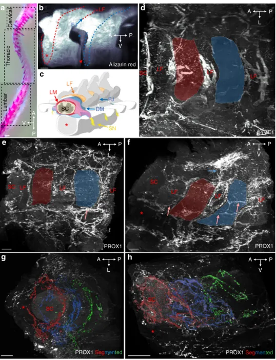
Despite labeling of some non-LECs, both markers clearly
revealed a dense lymphatic network that was present between
vertebrae and appeared mainly confined to the intervertebral
spaces (Fig.
1
d, e and Supplementary Movies 1, 2)
14. A few
longitudinal vessels linked adjacent intervertebral lymphatic
circuits together along the spinal cord (salmon arrows in Fig.
1
e,
f). Vertebral LVs (vLVs) were also connected to the peripheral
lymphatic system surrounding the vertebrae, dorsally through the
ligamentum
flavum (Fig.
1
f), dorsolaterally along the dorsal facet
joint and ventrolaterally through the intervertebral foramen along
ventral nerve rami (Fig.
1
e, f).
We next used Imaris-3D software to illustrate the anatomy of
vLV circuits. We used global image acquisitions of the thoracic
spine (see Supplementary Movie 2), showing a succession of
vertebral lymphatic units along the rostro-caudal axis. Images
were then segmented to generate a color-coded map of vLV
circuits. In Fig.
1
g, h and Supplementary Movies 3, 4, each color
defines the PROX1
+pattern of one vertebra along three
successive thoracic vertebrae (red, blue, green) as well as the
peripheral lymphatic vasculature (white). This confirms a
metameric organization of vLVs
14.
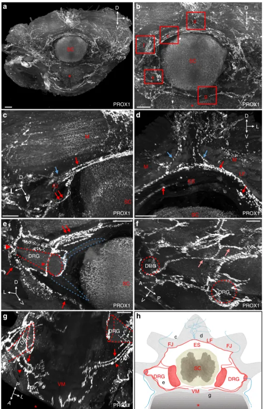
Modular architecture of vertebral lymphatic vasculature. We
next mapped the vLV network in the vertebral canal from the
dorsal to the ventral part of a vertebra. Supplementary Movie 2
and the corresponding view in Fig.
2
a, b show vLVs around one
segment of the thoracic spinal cord, and areas where higher
magnification views were taken. Dorsally, semicircular lymphatic
vessels navigate around the spinal cord (Fig.
2
c). At the ventral
border of the ligamentum
flavum, located at the dorsal midline
between two spinous processes, these vLVs contact lymphatic
branches entering the epidural space from the overlying dense
peripheral lymphatic vasculature (Fig.
2
d). Laterally, at the level
of the transverse facet joints, semicircular vessels including
per-ipheral lymphatic vessels from the dorsal plexus converge toward
a lymphatic circle (blue arrow in Fig.
2
c). From this point, vLVs
distribute either radially toward the periphery, or ventrally toward
the emergence of the dorsal spinal nerve roots (red double arrow
in Fig.
2
c). Supplementary Movie 2 allows to follow peripheral
lymphatic vessels from the dorsal midline of intervertebral
liga-ments to the lateral exit points of the vertebral canal. At the
intervertebral foramen, DRGs are covered by vLVs that converge
SC LF LF LF LYVE- 1 LYVE1 SC SC PROX1
g
h
e
SCd
Alizarin red*
b
a
A V P A V P A V P A L P A L P L P APROX1 Segmented PROX1 Segmented
Thoracic Lumbar Cervical
c
LF LF LF PROX1f
SC LF LF LF LF SC LF DM LM SN P V A FJ FJFig. 1 Segmental pattern of the vertebral lymphatic vasculature in the thoracic spine. a Alcian blue/Alizarin red staining of the mouse vertebral column with boxes indicating position of images shown in Figs.1–4(thoracic vertebrae) and 5 (cervical and lumbar vertebrae), spatial orientation (A: anterior, P: posterior, L: lateral, V: ventral).b Alizarin red staining of two successive thoracic vertebrae (delimited by red/blue dots, lateral view). LF: ligamentum flavum, red asterisk: ventral vertebral body, blue arrow: facet joint (FJ), red arrowhead: ventral intervertebral disk, blue asterisk: intervertebral foramen. c latero-frontal schematic drawing corresponding to (b). DM: dura mater, LM: leptomeninges (pia mater and arachnoid), SC: spinal cord, SN: spinal nerve. d Dorsal view of LYVE1 staining. Red and blue areas correspond to two successive vertebrae. Note LVs lining ligamentumflavum. e, f Dorsal (e) and lateral (f) views of the PROX1 expression pattern. Red and blue areas correspond to two successive vertebrae. Salmon arrows: intervertebral LVs, blue arrow: dorsal LVs.g, h Segmented images of the PROX1 LV network (fronto-dorsal (g) and lateral (h) views) highlighting three successive vertebral LV units (red, blue, green). Scale bars: 2 mm (a); 300µm (b, e, f); 200 µm (d, g, h)
DRG DRG PROX1 A V P SC PROX1 DRG PROX1 SC
a
PROX1 VM SC PROX1 SC PROX1c
d
e
f
g
h
D D V L D V L D V Lb
c f d g e SC M M M FJ DRG DRG PROX1 SC DRG DRG FJ FJ d c e g LF ES LF ES VM D V L A L PFig. 2 Modular architecture of vertebral lymphatic vasculature. a Frontal view of a cleared thoracic vertebra stained with an anti-PROX1 antibody. Red asterisk: vertebral ventral body, SC: spinal cord spatial orientation (D: dorsal, L: lateral, V: ventral).b Magnifications of red boxed areas are shown in (c–g). c Semicircular dorsal LVs (red arrow) surround the spinal cord, exit dorsolaterally (blue arrow) and also extend a latero-ventral connection to the dorsal nerve root (double red arrows in (c) and (e)). Note PROX1+cells in SC and perivertebral muscles (M), FJ: facet joint.d At the ventral face of the ligamentumflavum (LF) located between two spinous processes, dorsal LVs (blue arrows) enter the vertebral canal and join semicircular LVs (red arrows). Note circles of LVs bordering the upper side of the epidural space (ES).e Ventrolateral LV circuitry around DRG (red arrowheads). Blue dotted-lines: spinal nerve roots, red dotted-lines: DRG.f Lateral view with intervertebral LVs (salmon arrows). g Two ventral branches (red arrows and arrowheads) run between each side of the ventral midline (VM) and the DRG.h Schematic representation of a frontal view of a thoracic vertebral LV unit. Longitudinal connecting vessels between vertebral units are not represented. FJ: facet joint; LF: ligamentumflavum; ES: epidural space; DRG: dorsal root ganglia; VM: ventral midline; SC: spinal cord. Black letters refer to images in (c–g). Scale bars: 300 µm (a–g)
from the ventral and dorsolateral circuits at their proximal and
distal end, respectively (arrowheads in Fig.
2
e). In addition to
these two circuits, a few longitudinal connecting vessels link
vertebral lymphatic units together (Fig.
2
f). Ventrally, a second
circuit of semicircular lymphatic vessels converges toward the
ventral spinal nerve rami exit, while no lymphatic vessels are
observed at the ventral midline (Fig.
2
g)
14. We observed a similar
vLV pattern in other thoracic segments (n
= 5) allowing us to
generate a schematic representation of the different
compart-ments of the vLV circuitry with a specific color code for the
intervertebral circuits (red) and vertebral branches of the
per-ipheral LV (blue) (Fig.
2
h).
To verify that the iDISCO
+protocol preserved the integrity of
tissue structure and anatomy
29, we performed additional
immunostainings on decalcified EDTA-treated vertebral
seg-ments that were either cryoprotected and frozen, or dehydrated
and embedded in paraffin. Confocal and conventional
micro-scopy imaging of sections perfectly reproduced the 3D-images
collected with a LSFM on IDISCO
+treated samples
(Supple-mentary Fig. 2) and thus substantiated the iDISCO
+based-model
described above.
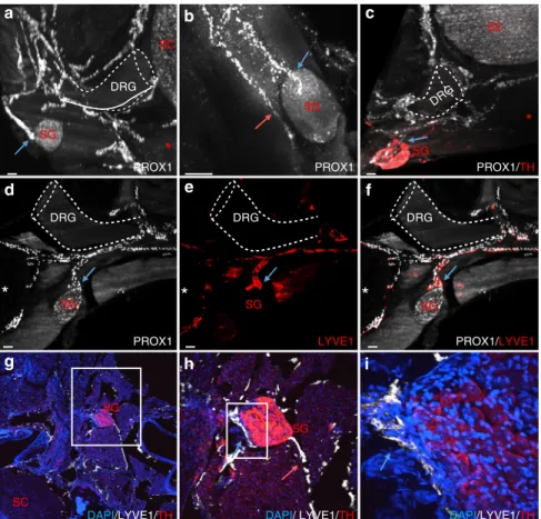
Vertebral lymphatic vessels contact sympathetic ganglia. vLVs
covering the dura mater of DRGs (Fig.
2
e) extend collateral
branches bilaterally along the spine, which contact paravertebral
PROX1
lowganglia (Fig.
3
a, b). PROX1 is known to be expressed
in the sympathetic neuronal lineage
30and double labeling with
antibodies against PROX1 and tyrosine hydroxylase (TH), a
specific marker of adrenergic nerves and ganglia, confirmed that
specific branches emerging from vLVs connected to TH
+/
PROX1
lowsympathetic ganglia (Fig.
3
c). On each side of the
spinal cord, PROX1
+/LYVE1
+LVs contacted one sympathetic
ganglion per spinal level (Fig.
3
d–f). Complementary analyses by
high resolution confocal imaging on vertebral column
cryosec-tions showed that the connection between LVs and sympathetic
ganglia (Fig.
3
g, h) occured at the surface of the ganglion cortical
layer (Fig.
3
i). These data reveal a hitherto unknown anatomical
interaction between the autonomous nervous system and
lym-phatic vessels derived from vLVs.
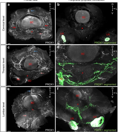
vLV patterning differs between vertebral column levels. We
next examined the lymphatic vasculature at the cervical and
thoracic level of the vertebral column. Stereomicroscopic imaging
of whole-mount preparations revealed LVs around the cisterna
magna and within the vertebral canal, where they located at the
level of intervertebral ligaments and surrounding spinal nerve
rami (Supplementary Fig. 3a–d). The LV patterning in cervical
vertebrae and along the vertebral column was then analyzed in
further detail by volume imaging.
In the cervical region, we observed a dorsal extravertebral
lymphatic plexus (blue arrow, Fig.
4
a) as well as intravertebral
vLVs that exited ventrally and bilaterally (red arrows) through the
intervertebral foramen to ventrally (salmon arrow) connect to
dcLNs (green in Fig.
4
b; Supplementary Fig. 3e, f). Thoracic vLVs
were defined by a large dorsal extravertebral plexus (blue arrow,
Fig.
4
c) and a direct connection from ventrolateral DRG LVs (red
h
DAPI/ LYVE1/TH DAPI/LYVE1/TH
i
DRG DRG DRG DRG*
*
*
PROX1/LYVE1 LYVE1 PROX1d
e
f
PROX1 PROX1a
SC SC DRG SG SG SGc
SG SG SG PROX1/TH DAPI/LYVE1/THg
SC SG SGb
Fig. 3 Vertebral lymphatic vessel connections with sympathetic ganglia. a, b Thoracic PROX1+LVs contact paravertebral sympathetic ganglia (SG) (blue arrows). White dotted-lines: DRG.c PROX1 (white) and tyrosine hydroxylase (TH, red) double labeling shows a ventral LV branch contacting a paravertebral TH+sympathetic ganglion (blue arrow).d–f PROX1 (white)/LYVE1 (red) double labeling of the LV-SG connection (blue arrow). g–i 2D-confocal images of cervical cryosections labeled with LYVE1 (white), TH (red), and DAPI (blue). White box: area magnified in (h) and (i). A LV contacts a TH+SG (blue arrow). Note a second ventral LV branch running along the SG, without entering its cortical layer (h, salmon arrow). This branch is also seen in panel (b) (salmon arrow). White dotted-lines: DRG; Red asterisk: vertebral ventral body; SC: spinal cord. Scale bars: 100µm (a–h); 200 µm (i)
arrow) to the thoracic lymphatic duct (green in Fig.
4
d). The
thoracic and lumbar regions displayed similar extensions and
patterns of extravertebral and intravertebral LVs (Fig.
4
c, e). In
lumbar vertebrae, the ventrolateral circuits that exited on each
side of the vertebral canal connected to lymph nodes. As shown
in green in Fig.
4
f, lymphatic vessels circumvented the ventral
body of the lumbar vertebra (salmon arrow), converged on the
ventral midline and split into two branches running toward the
pair of renal lymph nodes. Therefore, the vLV architecture is
conserved along the vertebral column, but the extension of
extravertebral and intravertebral vessels around the spinal cord
and their connection to the peripheral lymphatic system differs
between the cervical, thoracic and lumbar vertebral levels.
Epidural and dural vertebral lymphatic vessels. To obtain a 3D
annotation of vLV localization in the spinal canal and meninges,
PROX1-labeled vertebral volumetric images were used to
gen-erate segmented images of membranes and the epidural space
around the spinal cord. We manually annotated in 3D the
meninges, the epidural space and ligamentum
flavum (Fig.
5
a).
Figure
5
a shows one image slice with a color code for meningeal
layers (purple area) and the epidural space (green area). We also
present color-coded layer masks for the arachnoid and dura
mater together (purple area in Fig.
5
b), or the dura mater and the
epidural space together (green area in Fig.
5
c). The overlay of
both masks revealed that PROX1
+vLVs mainly localized in the
epidural space (green), while the underlying dura mater layer
(gray) includes ventral vLVs (white) around DRGs (Fig.
5
d and
Supplementary Movie 5). As shown on a lateral view (Fig.
5
e),
dura mater vLVs localized most extensively at bilateral DRGs.
Interestingly, connecting vessels between two successive
verteb-rae (salmon arrows in Fig.
5
e) navigated in the epidural space
and appeared to join vLVs of the dura mater close to the DRGs
(white arrows in Fig.
5
e), suggesting a possible confluence of
peripheral lymph and CSF at this level. Scheme representing the
anatomy of vLVs in the thoracic vertebral canal is shown in
Fig.
5
f.
Complementary examination of whole-mounted spinal cord
meninges (Supplementary Fig. 4a) and cryo/paraffin sections
(Supplementary Fig. 4b–g) confirmed that the dura mater
lymphatic vessels were restricted around the DRGs and spinal
nerve rami and located on the dorsal surface of the dura mater
(Supplementary Fig. 4c, f), which is not in direct contact with
the CSF.
C PROX1 SCe
Lumbar level Frontal view SCf
Peripheral lymphatic connection
PROX1 SC Thoracic level SC PROX1 segmented
c
d
PROX1 SCa
Cervical level SC*
b
PROX1 segmented PROX1 segmented D L V D L V D L V D L V D L V L V LN LN TD LN LN Ao TD DFig. 4 Variations of vLV pattern along the vertebral column. a–f Pattern of PROX1+LVs in the cervical (a, b), thoracic (c, d), and lumbar (e, f) vertebral column, spatial orientation (D: dorsal, L: lateral, V: ventral). Left panels show frontal views, right panels show connection to peripheral lymph nodes (LN) and thoracic duct (TD).a, c, e Note fewer LVs in the dorsal plexus between intervertebral spinous processes of cervical and lumbar vertebrae compared to thoracic ones (blue arrows).a LVs exit bilaterally (red arrows) through the intervertebral foramen. a, c, e Also note differences in ventral root exit circuits between regions (salmon arrows).b, d, f LV ventral exit circuits (green) to b deep cervical LNs, d thoracic duct, or f to renal LNs. Red asterisk: vertebral ventral body; SC: spinal cord; Ao: Aorta. Scale bars: 300µm (a–f)
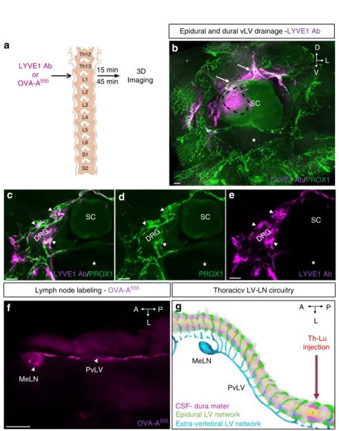
vLV-mediated drainage of the vertebral column. The function
of the vLV system was explored by testing the drainage potential
of epidural and dura mater vLVs. Molecular tracers were injected
into one side of the spinal cord parenchyme at the
thoraco-lumbar level, and their distribution around the injection site was
examined 15 and 45 min after injection (a.i.) (Fig.
6
a). We used as
molecular tracers either LYVE1 antibodies that were detected
with a secondary antibody, or
fluorescent albumin (OVA-A
555).
It is worth noting that this surgery procedure punctures the dura
mater, which allows access of injected tracer into the epidural
space located above spinal meninges, locally at the puncture site.
LSFM imaging of iDISCO
+-treated vertebral samples revealed
f
TD SG SC DRG SG SG SG DRG DRG DRG Epidural LV networksDural LV networks Extra-vertebral LV networks LF LF LF FJ FJ FJ SC
d
Dura matere
SC AV P Epidural and dura mater area
a
PROX1 ES ES PROX1 PROX1 Vx+1 Vx LF Pi A DM SCb
DRG PROX1 Arachnoid and dura mater areaDRG
Dura mater and epidural area
c
SC DRG DRG DRG DRG D V L D V L D V L PROX1Fig. 5 Epidural and dural lymphatic circuits of the spine. a 2D-single frontal image slice (2µm thick) of the cervical vertebral column with enhanced brightness to reveal PROX1-expressing nuclei and spinal cord (SC), meninges including pia mater (Pi), arachnoid (A) and dura mater (DM), the epidural space (ES), and the ligamentumflavum (LF). A color-coded segmentation of layers around the spinal cord shows the meningeal layers in purple and the dura mater plus the epidural space in green.b–d 3D-reconstruction of frontal images of the cervical vertebral column with color-coded layers: the arachnoid and dura mater in purple (b); the dura mater and epidural space in green (c); combined layer marks showing the arachnoid in purple, the dura mater in white, and the epidural space in green (d), spatial orientation (D: dorsal, L: lateral, V: ventral). A noticeable LV networkfills the epidural space (green) while dura mater LVs (white) are mainly restricted to DRGs (white arrows) and few branches on each side of the dorsal and ventral midline.e 3D-reconstruction of lateral images of the thoracic vertebral column with color-coded layers illustrated in (d). Blue dotted-lines: bilateral DRGs; salmon arrows: intervertebral LVs; Red asterisk: vertebral ventral body. Vx: vertebra x, Vx+ 1: vertebra x + 1. f Schematic representation of the lymphatic vasculature in the thoracic vertebral column. LVs are present in the epidural space (green) around the spinal cord and in the dura mater (purple). Extravertebral LVs extend dorsal processes (blue) and ventral connections with sympathetic ganglia (SG, deep blue) and the thoracic duct (TD, light blue). Blue arrowheads; exit points of vertebral lymphatic circuits; Blue dots: connections with extravertebral lymphatic networks; Black asterisk: vertebral ventral body; DRG: dorsal root ganglia; FJ: facet joint; LF: ligamentumflavum; SC: spinal cord; SG: sympathetic ganglia. Scale bars: 300 µm (a–e)
A D L V LYVE1 Ab/PROX1
b
SC SCe
LYVE1 Ab SC SCd
PROX1 LYVE1 Ab/PROX1 DRG DRG DRGc
a
Lymph node labeling - OVA-A555 Thoracicv LV-LN circuitry
PvLV Th-Lu injection
g
P MeLNf
MeLN OVA-A555 PvLV A L P L CSF- dura mater Epidural LV network Extra-vertebral LV network S2 S1 L6 L5 L4 L3 L2 L1 3D Imaging LYVE1 Ab or OVA-A555 15 min 45 min Th13 Th12Epidural and dural vLV drainage -LYVE1 Ab
Fig. 6 Epidural and dural lymphatic drainage. a Schematic representation of the procedure used to test spinal cord drainage: LYVE1 Ab or OVA-A555was
injected in the thoraco-lumbar (Th-Lb) region of the spinal cord parenchyme. Fifteen or forty-five minutes later, mice were sacrificed. LYVE1 was detected by labeling with a secondary antibody, while OVA-A555was directly identified by fluorescence detection. Schema is adapted from Fig.2c in ref.59.
b–e LYVE1 Ab uptake by epidural and dural lymphatic circuits after 45 min. b LYVE1 Ab (purple) injection area (black dotted line) and epidural LVs recapture (white arrows).c–e Colocalization of LYVE1 with LVs around DRG, in contact with dura mater (white arrowheads). Asterisk: vertebral ventral body, SC: spinal cord.f OVA-A555injection leads to labeling of ipsilateral mediastinal lymph node (MeLNs) after 15 min.g Schematic representation of the
lymphatic drainage pattern in the thoracic vertebral column. Thoracic vertebrae (gray), dura mater and CSF (purple), LVs of epidural space (green), extravertebral LVs and lymph node (blue). Blue dots: accumulation of OVA-A555tracer in LVs and lymph node. MeLN: mediastinal lymph node; PvLV:
that the injected markers localized in and around PROX1
+vLVs
of the epidural space and dura mater (white arrows, Fig.
6
b–e and
Supplementary Movie 6). Confocal imaging of decalcified and
frozen samples showed that OVA-A
555localized within the vLV
lumen (Supplementary Fig. 5), thus demonstrating the uptake
and drainage properties of vLVs.
15 min after OVA-A
555injections into the thoraco-lumbar
spinal cord, tracer accumulated in the ipsilateral paravertebral
lymphatic vessel and mediastinal lymph node, in 9 out of 12 cases
(Fig.
6
f). Therefore, vLVs provide a regional outflow for epidural
fluids toward lymph nodes. A schematic model of the
thoraco-lumbar lymphatic drainage circuitry is shown in Fig.
6
g.
Vertebral LVs respond to VEGF-C and spinal cord injury. To
assess the dependence of vLVs on VEGF-C, we generated
gain-of-VEGF-C signaling mice by either intra-cisterna magna (i.c.m.) or
lumbo-sacral (l.s.) injection of adeno-associated viral vectors
(AAVs) encoding mVEGF-C (AAV-mVEGF-C)
15(Fig.
7
a, d).
Control mice were injected with AAVs encoding soluble
mVEGFR34–7
-Ig (VEGFR3 ectodomains that do not bind
VEGF-C) (AAV-control)
31. One month later, mice were analyzed by
PROX1 immunostaining. Compared to controls, VEGF-C
injec-ted mice showed a strongly expanded vLV network, in particular
of dorsolateral lymphatic rings in the intervertebral disk of
cer-vical vertebrae after i.c.m. injection (Fig.
7
b, c and Supplementary
Movie 7), and lumbar vertebrae after l.s. injection (Fig.
7
e, f).
To determine if adult vLVs might respond to spinal cord
injury, we injected LPC (1μl) into one side of the spinal cord at
the thoraco-lumbar level (Fig.
7
g). LPC is toxic to
oligoden-drocytes and rapidly induces demyelinating spinal cord lesions
(Fig.
7
i inset)
32,33. Within a week after the surgery, a robust
extravertebral and intravertebral growth in LVs was induced in
LPC-injured mice (Fig.
7
h–i and Supplementary Movie 8). To
quantify the response, we opened the vertebral column to expose
intravertebral vLVs that were stained with LYVE1 on
whole-mount preparations, followed by vessels diameter and surface
area measurements (as red stippled area in Fig.
7
i). Pairwise
Mann–Whitney U test comparison to control mice revealed a
significant increase in vLV area after LPC injury (p = 0.0286),
however, this did not reach statistical significance in a mutliple
group comparison (Supplementary Fig. 6e). LPC-lymphatic vessel
diameter and area were not affected by control
AAV-mVEGFR34–7
-Ig (LPC
control), while they were significantly
enhanced in mice pretreated with AAV-mVEGF-C (LPC
VEGF-Cmice) for one month (Fig.
7
j, Supplementary Fig. 6e). LPC injury
in mice pretreated by lumbo-sacral (l.s.) injection of AAVs
encoding soluble mVEGFR31–3
-Ig (LPC
VEGF-C trapmice) for one
month resulted in reduction of vLV diameter that was
significantly different from LPC
controlmice (Fig.
7
j). LPC
VEGF-C trapmice showed a reduction of vLV area that was significantly
different from LPC
controlmice in a pairwise Mann–Whitney
U-test comparison (p
= 0.0286), but failed to reach statistical
significance in a multiple group comparison (Supplementary
Fig. 6e). However, LPC injury in K14-VEGFR3-Ig homozygous
mice, in which endogenous VEGF-C/VEGF-D ligands are
constitutively trapped to prevent VEGFR3 signaling
31, was
associated with a significant reduction of vLV diameter and area
compared to heterozygous littermates (Fig.
7
k, Supplementary
Fig. 6f). Taken together, these data show that vLVs respond to
VEGF-C and spinal cord injury.
Effects of vLVs on myeloid and lymphoid cells. We next asked
whether vLVs concentrated myeloid and lymphoid cells, as
pre-viously reported for skull lymphatics
13. Pre-cleared segments of
the vertebral column co-labeled with antibodies against LYVE1
and the common leukocyte antigen CD45 showed that CD45
+leukocytes inside the vertebral canal were concentrated around
LYVE1
+vLVs (Fig.
8
a–d). On cryosections we observed
leuko-cytes concentrated close to, or within, Vegfr3:YFP
+lymphatic
vessels in intervertebral ligaments (Fig.
8
e). CD45
+leukocytes
included around 40% of C11b
+macrophages, 40% of CD3
+T cells, and 20% of CD19
+B cells. Furthermore, around 40% of
CD45
+leukocytes expressed MHCII (Supplementary Fig. 6a–d).
Leukocyte numbers and ratios were similar between control,
AAV-mVEGF-C and K14-VEGFR3-Ig homozygous and
hetero-zygous mice (Supplementary Fig. 6a–d). In contrast, LPC injury
induced a strong increase in leukocyte numbers around vLVs that
was further enhanced by AAV-mVEGF-C and reduced by
mVEGF-C trap (Fig.
8
f).
The size of demyelinated lesions, identified as spinal white
mater areas devoid of MBP (Myelin Basic Protein) expression,
was significantly increased in LPC
VEGF-Cmice compared to
control AAV treated mice (Fig.
8
g, h). LPC
VEGF-C trapmice mice
showed a significantly reduced lesion size when compared to
LPC
VEGF-Cmice, and a slight but not significant reduction in
lesion area when compared to LPC
controlmice (Fig.
8
g, h).
Further quantifications were done on spinal cord sections in
the lesioned area vs. the contralateral uninjured side. As expected,
LPC injection reduced the number of NeuN-positive neurons in
the peri-lesional area compared to the uninjured side
(Supple-mentary Fig. 7a). Pre-treatment with control or with
AAV-mVEGF-C trap had no effect compared to LPC injury alone,
while AAV-mVEGF-C further reduced the number of
NeuN-positive neurons on the injured side, indicating deleterious effects
of expanded vLVs on spinal cord demyelinating lesions
(Supplementary Fig. 7a). LPC injection induced an increase in
the number of F4/80
+microglia and monocyte-derived
macro-phages, Iba1
+microglia, and CD3
+T cells in the injected side
compared with the contralateral side (Supplementary Fig. 7b–d).
AAV-control had no effect on immune-cell numbers, while the
numbers of F4/80
+, Iba1
+, and CD3
+cells were further amplified
in LPC
VEGF-Cmice (Supplementary Fig. 7b–d). LPC
VEGF-C trapmice showed significantly reduced leukocyte numbers when
compared to LPC
VEGF-Cmice, however, leukocyte infiltration
was not reduced when compared to untreated LPC
controlmice
(Supplementary Fig. 7b–d). Inflitration of F4/80
+macrophages/
microglia and CD3
+T cells was also not significantly different
between K14 homozygous and heterzozygous LPC-injured mice
(Supplementary Fig. 7e, f). Altogether, these results demonstrate
that a gain-of-vLVs amplified the cytotoxic effect resulting from
LPC-induced injury.
Discussion
We here report the 3D anatomy and the function of vLVs in the
vertebral canal (Fig.
5
f). The data reveal an extensive and complex
lymphatic vasculature in the vertebral column, surprisingly dense
in comparison to the one that covers the cranial dura mater
15.
Previous literature reported the presence of LVs on whole-mount
preparations of vertebral dura mater in monkeys
34and on
sec-tions of intact and injured vertebral tissue in humans
35. Pioneer
studies in the late 19th and early 20th centuries
36–38and later
works of Ivanow
19and Brierley and Fields
20had investigated the
flow of the lymph stream along the spine and in the spinal roots
of the cord, providing evidence that lymphatics contribute to the
propagation of infectious agents (toxins, polyomyelitis, tetanus)
39.
More recently, a continuum of metameric spinal lymphatics was
described in the cervical, thoracic and lumbar areas of the
ver-tebral column, both on dorsal and ventral sides, with lateral exits
of the vertebral canal along blood vessels and spinal nerves
14. We
vasculature organization and function in vertebral canal drainage
(Fig.
6
g).
Each vertebra is drained by semicircular dorsal and ventral
vessels, which exit the vertebral column at intervertebral
for-amina. vLVs extend along spinal nerve rami to reach lymph
nodes adjacent to the cervical, thoracic and lumbar regions of the
spine, as well as the thoracic lymphatic duct in the thoracic
region. The vertebral lymphatic network is thus organized as a
metameric network of peripheral LV-connected vertebral
lym-phatic units that are interconnected by a few thin longitudinal
vessels. The absence of large longitudinal dorsal or ventral LVs
suggests that vLVs do not drain vertebral lymph as a continuous
stream along the vertebral column axis, but rather at the level of
each vertebra. This model is supported by our
findings that
vertebral lymphatic vasculature consists mainly of an extensive
network of epidural vessels, located in the intervertebral tissue
and beneath the ligamentum
flavum, and which drain the
epi-dural lymph of the vertebral column. These observations do not
exclude that the spinal CSF may also follow a directional
down-ward
flux within the central canal and the spinal subarachnoid
L5 L4 L3 L2 L1 Th13 Th12 LPC 1 week S2 S1 L6 L5 L4 1
j
1 monthd
g
k
e
f
SC PROX1 PROX1 D L V D L V SC AAV-control AAV-mVEGF-Ci
PROX1 SC AAV-control PROX1c
SCb
AAV-mVEGF-C D L V D L V D L V Cervical Lb-Sa Cervical Lb-Sa SC Th-Lb PROX1 No injection SCh
Th-Lb LPC PROX1Control LPC LPCControlLPCVEGF-CLPCVEGF-C trap
Lymphatic vessel diameter (
µ
m)
Lymphatic vessel diameter (
µ m) *** LPC-K14 VEGFR3-Ighet LPC-K14 VEGFR3-Ighom 100 200 0 150 50 0 100 1 month AAVs AAVs Cisterna magna
a
D L Vspace, toward the caudal end of the spine, as recently
demon-strated by dynamic in vivo imaging of CSF
flow after
intraven-tricular CSF tracer injection
40. In line with our observations, the
authors report the outflow of CSF tracers from the spinal
sub-arachnoid space from intravertebral regions of the sacral spine
toward sacral and iliac LNs.
Interestingly, consistently with our earlier previous
findings
14,
we found vLVs in contact with dura mater (Supplementary
Fig. 4c, f), where molecular tracers were also detected within
vLVs, around DRGs and spinal nerve rami (Fig.
6
b–e).
Sub-sequent surgeries specifically delivering fluorescent tracers within
the spinal cord subarachnoid space will be required to determine
whether these vertebral dural vLVs may be hotspots contributing
to CSF uptake toward lumbo-sacral LNs.
We note that there is a regional variation in the LV size, which
is inversely correlated to the volume of CSF, with large cerebral
ventricular volumes associated with a discrete network of cranial
mLVs and a small vertebral ependymal volume correlated with a
large vertebral lymphatic vasculature. One possible explanation is
that vertebral LVs strongly contribute to the reuptake of the CSF
that is continuously produced by the ventricular choroid plexus
and circulates in the ependymal canal of the spinal cord
8. This
model is supported by the presence of dura mater vLVs in the
spine and their dense location around spinal nerve rami. A
sec-ond and likely possibility is that the largest part of vLVs uptakes
epidural
fluids and cells. This assumption is supported by the
presence of a large network of epidural vessels that extends from
the peripheral lymphatic system (Fig.
5
e, f) and drains the
ver-tebral canal toward lymph nodes adjacent to the spinal cord
(Fig.
6
f, g).
Vertebral LVs localize mainly at the level of intervertebral
ligaments or joints, much like cranial lymphatics that navigate in
skull commissures alongside blood vasculature and spinal
nerves
14. We and others
find that LVs avoid bone tissues
41,42.
Interestingly, the presence of LVs inside bone is observed in
patients with vanishing bone disease (also called Gorham Stout
disease GSD)
43. GSD is a sporadic disease characterized by the
presence of lymphatic vessels in bone and progressive bone loss.
GSD can affect any bone in the body, but it most frequently
affects the ribs and vertebrae, with poor prognosis
44,45.
Mechanisms preventing LV formation in bone are, however,
currently unknown.
In contrast to skull sutures between skull cap bones, which are
few, narrow and
fixed, the vertebral disks, joints and ligaments
between vertebral bones are numerous, large and mobile. They
sustain the integrity and
flexibility of the spine, which is predicted
to require extensive interstitial
fluid drainage. The large network
of epidural vLV appears to be exquisitely adapted to this extensive
drainage of non-neural peripheral tissues in the spine and to
provide each vertebra with its proper clearance system toward
collecting lymph node (Fig.
6
g). It is predictable that defective
vLVs will alter vertebral and intervertebral tissue maintenance,
leading to spine orthopedic pathologies.
The proximity of two distinct epidural and dura mater vLV
circuits raises questions about the privileged immune status of the
CNS. Like in the skull
13, a proximity between vLVs and CD45
+leukocytes is observed along the spine (Fig.
8
). The spinal cord
lymphatic vasculature thus appears as a potentially important
immune surveillance interface between the CNS and peripheral
tissues. The cervical, thoracic and lumbar regions directly drain
into cervical, mediastinal and renal/lumbar lymph nodes,
respectively, which suggests that the peri-lymphatic dendritic
immune cells may transfer to those lymph nodes and initiate
lymphocyte activation against specific pathogens or antigens. On
the other hand, epidural and dura mater vLV may facilitate the
propagation of peripheral infections through the vertebral canal
toward neural tissues. For example, epidural vLVs may provide
entry for meningitis infection into spinal meninges. The contact
zone between DRGs and vLVs appears as another potential gate
for entry into the CNS for pathogens. For example, bovine scrapie
protein is
first detected in the mesenteric lymph nodes and DRGs
of lemurs or cattles infected orally with the agent of bovine
spongiform encephalopathy (BSE)
46,47.
The spine is affected by variety of diseases including
infec-tions
47, acute spinal cord compressions
48, and degenerative spine
disorders, a common condition in the ageing Western
popula-tion
49. Vertebral LVs are potential targets for these pathologies.
The vertebral column is also a common site for skeletal
metas-tastic tumors; as many as 70% of cancer patients have spinal
metastases, and up to 10% of cancer patients develop metastatic
cord compression
50. Since lymphatics may serve as conduits for
primary tumor cells in metastatic spreading
51, specific
inter-ference with the vertebral lymphatic vasculature could reduce or
prevent spinal metastasis. Alternatively, lymphatic vessels are the
first barrier for initiation of an adaptive immune response by
antigen-presenting cells
52. Facilitating the entry of immune cells
into vLVs might thus also potentially improve the efficiency of
immune checkpoint inhibitor treatments to destroy spinal
tumor cells.
We found that adult vLVs rapidly expand in response to
VEGF-C or tissue injury (Fig.
7
c, f, i). In inflammatory conditions
such as LPC-induced spinal cord demyelination,
VEGF-C-pre-treatment resulted in a strong expansion of vLV circuits and the
epidural immune-cell pool. These extra-parenchymal
lympho-immune responses were associated with larger demyelinated
lesions and increased number of peri-lesional neuronal cells in
the parenchyme compared to LPC
controlmice (Fig.
8
g, h and
Supplementary Fig. 7a). In this setting, the expansion of vLV
coverage therefore exacerbated the cytotoxic inflammation and
impaired the recovery of local tissue damage. In contrast to the
beneficial effect of loss of skull meningeal lymphatics in EAE
mice
17, LPC
Vegf-C trapand LPC-K14-VEGFR3-Ig mice failed to
Fig. 7 vLVs are VEGF-C dependent and remodel after spinal injury. a–f VEGF-C induces epidural and dural lymphangiogenesis. a–c Cervical spine lymphangiogenesis after i.c.m. injection of AAV-mVEGF-C.a Schematic of i.c.m. injection to deliver AAVs into the CSF and toward the cervical spine. b, c LSFM coronal view of the cervical spine one month after AAV injection. Pattern of PROX1+LVs (white) in AAV-control (b) and AAV-mVEGF-C (c) mice. Note that VEGF-C induced a robust epidural and dural lymphangiogenesis.d–f Thoraco-lumbar lymphangiogenesis induced by lumbo-sacral delivery of AAV-mVEGF-C.d Schematic of AAV injection sites into the lumbo-sacral spinal cord, adapted from Fig.2c in ref.59.e, f Pattern of PROX1+LVs (white)
in AAV-control (e) and AAV-mVEGF-C (f) mice. White asterisk: vertebral ventral body; SC: Spinal cord. g–i Focal injury in the thoraco-lumbar spinal cord. g Schematic of LPC injection into the thoraco-lumbar spinal cord, adapted from Fig.2c in ref.59.h, i Pattern of PROX1+LVs (white) in control-non lesioned
(h) and LPC-injected mice (i). Inset in (i) shows the spinal cord lesion (stippled area), spatial orientation (D: dorsal, L: lateral, V: ventral). j, k Quantification of lymphatic vessel diameter (red stippled area in (i)) after LPC-spinal cord injury in gain- and loss-of-mVEGF-C signaling mice (j) and in LPC-injured K14-VEGFR3-Ighommice and -K14-VEGFR3-Ighet(control) mice (k). n= 4 biologically independent mice/independent experiment, and data show mean+/−SD
(error bar) in (j, k); one-way ANOVA with Tukey’s multiple-comparisons test (j) and Mann–Whitney U test (k); *p < 0.05, ***p < 0.001. Source data are provided as a Source Datafile. Scale bars: 300 µm (b, c, e, f, h, i)
show significantly reduced immune-cell inflitration and
demye-lination resulting from vLV blockade. This may be due to
alter-native growth factors, incomplete vLV deletion, or simply harder
to detect in a context of lower levels of inflammation in
LPC-treated mice compared with EAE mice.
Vertebral LVs never contact the spinal cord tissue, even upon
VEGF-C overexpression or acute spine lesion. In contrast, vLVs
are closely apposed around the chains of sensory and sympathetic
nervous ganglia. Although no lymphatic vascularization of
sym-pathetic ganglia was observed, lymphatic vessels may provide
LYVE1c
SC LYVE1/CD45 SC*
a
D L V CD45b
SCd
SC LYVE1/CD45 CD45 Merged VEGFR3:YFP MHCIIe
CD45 positive cells per field
150
100
50
0
Control LPC LPCControl LPCVEGF-C LPCVEGF-C trap
LPCVEGF-C LPCVEGF-C trap LPCControl
f
h
MBP/DAPIg
2*105 4*105 6*105 8*105 MBP demyelinisation area ( µ m 2) 0molecular signals to the sympathetic neurons that control
vas-cular tone of lymphatic ducts and cerebral arteries and arterioles.
Previous observations also showed that adrenergic
fibers connect
to the thoracic lymphatic duct and also innervate the wall of
lymph node arterioles
53,54. The crosstalk between spine LVs and
the sympathetic system is thus likely relevant for the regulation of
peripheral lymph and glymphatic drainage and may coordinate
them with the activity of brain and spine tissues. We speculate
that a regulatory loop may link meningeal LV, sympathetic chain
neurons and both CNS and peripheral
fluid drainage.
To conclude, this study shows that the volume imaging
tech-nique allows the description of neurovascular systems by
pre-serving the anatomy and the 3D-continuity of vascular and neural
structures. In particular, our studies characterize the
3D-anatomical organization, the remodeling and function of the
lymphatic vasculature along the spine. Our
findings identify
vertebral LVs as an important component for the maintenance
and repair of vertebral tissues as well as a gatekeeper of CNS
immunity.
Methods
Study approval and mice. All in vivo procedures used in this study complied with all relevant ethical regulations for animal testing and research, in accordance to the European Community for experimental animal use guidelines (L358-86/609EEC). The study received ethical approval by the Ethical Committee of INSERM (n° 2016110111126651) and the Institutional Animal Care and Use Committee of ICM (Institut du Cerveau et de la Moelle épinière). Male C57BL/6J mice, Vegfr3:YFP lymphatic reporter mice55, K14-VEGFR3-Ig mice31, or Prox1-eGFP mice50
between 2 and 3 months of age were used for all experiments.
Tissue preparation. Mice were given a lethal dose of Sodium Pentobarbital (Euthasol Vet) and perfusion-fixed through the left ventricle with 10 ml ice-cold PBS then 20 ml 4% paraformaldehyde (PFA) in PBS. To dissect the spine, the skin was completely removed, all the organs were eliminated and the ribs were removed to keep only the vertebral column from the cervical part until the lumbar part with the spinal cord inside. All the surrounding tissues including muscles, aorta and ligaments were maintained around the vertebral column. The spine was cut into pieces of about 0.5 cm (1–3 vertebrae) corresponding to the cervical, thoracic and lumbar regions. The different spinal segments were immediately immersed in ice-cold 4% PFA,fixed overnight at +4 °C, washed in PBS, and processed for staining. Sample pre-treatment in methanol for iDISCO+protocol. We used a clearing protocol developed by Renier and colleagues, which is based on methanol dehy-dration and called the immunolabeling-enabled three-dimensional imaging of solvent-cleared organs (iDISCO+,http://www.idisco.info)21. The steadily
increas-ing methanol concentrations result in modest tissue-shrinkage (about 10%), while the“transparency” of tissues, such as the adult mouse brain, is increased. In detail, fixed samples were dehydrated progressively in methanol/PBS, 20, 40, 60, 80, and 100% for 1 h each (all steps were done with agitation). They were then incubated overnight in a solution of methanol 33%/dichloromethane 66% (DCM) (Sigma 270997-12X100 ML). After 2 × 1 h washes with methanol 100%, samples were bleached with 5% H2O2in methanol (1 vol 30% H2O2/5 vol methanol) at 4 °C
overnight. After bleaching, samples were rehydrated in methanol for 1 h each, 80%, 60%, 40%, 20%, and PBS. To clarify vertebral bone, we here added a decalcification step using Morse solution23during 30 min at RT. A weak acid treatment with
Morse solution (1/1 tri-sodium citrate and 45% formic acid) decalcifies tissues efficiently while preserving their structure56–58. Samples were washed rapidly
with PBS then incubated 2 × 1 h in PTx2 (PBS/0.2% Triton X-100). At this step they were processed for immunostaining.
Immunolabeling iDISCO+protocol. Pretreated samples were incubated in PBS/ 0.2% Triton X-100/20% DMSO/0.3 M glycine at 37 °C for 24 h, then blocked in PBS/0.2% Triton X-100/10% DMSO/6% Donkey Serum at 37 °C for 24 h. Samples were incubated in primary antibody dilutions in PTwH (PBS/0.2% Tween-20 with 10 mg/ml heparin)/5% DMSO/3% Donkey Serum at 37 °C for 6 days. Samples were washedfive times in PTwH until the next day, and then incubated in secondary antibody dilutions in PTwH/3% Donkey Serum at 37 °C for 4 days. Samples were finally washed in PTwH five times until the next day before clearing and imaging. We used the primary antibodies listed in the Supplementary Table 1. Primary antibodies were detected with the corresponding Alexa Fluor -555, -568, or -647 conjugated secondary antibodies from Jackson ImmunoResearch at 1/1000 dilution.
iDISCO+tissue clearing. After immunolabeling, samples were dehydrated pro-gressively in methanol in PBS, 20, 40, 60, 80, and 100% each for 1 h. They were then incubated overnight in a solution of methanol 33%/DCM 66% followed by incubation in 100% DCM for 2 × 15 min to wash the methanol. Finally, samples were incubated in dibenzyl ether (DBE) (without shaking) until cleared (4 h) and then stored in DBE at room temperature before imaging.
Paraffin section immunolabeling and imaging. Vertebrae were decalcified for 3 weeks in 10% EDTA in 4% paraformaldehyde/PBS, dehydrated through ethanol, cleared in xylene and embedded in paraffin. Serial cross sections (5 µm thick) were immunostained with rabbit anti-mouse LYVE1 (1:100) polyclonal antibody (11-034, AngioBio Co). DAB (3,3′-Diaminobenzidine) staining was performed using the biotin avidin complex kit (PK-6100, Vectastain®Vector). Masson’s trichrome staining was carried out using the Masson Trichrome Kit (BioGnost®- Ref. MST-100T). Hematoxylin (5 s) was used for counter staining. HRP-labeled paraffin sections were analyzed with a Zeiss Axio Scope.A1.
Cryostat section immunolabeling. For cryosections of the vertebral canal,fixed tissues underwent decalcification with 0.5 M EDTA, pH 7.4, at 4 °C. When the bone was becoming soft, samples were washed thoroughly with PBS and immersed in PBS containing 20% sucrose and 2% polyvinylpyrrolidone for 24 h at 4 °C, embedded in OCT compound (Tissue-Tek), and frozen at−80 °C. In total, 50–100-μm-thick sections were cut using a cryostat (Microm HM 550/CryoStar NX70; Thermo Fisher Scientific), air-dried, encircled with a pap pen, permeabilized with 0.3% PBS-TX, washed with PBS, and blocked in 5% donkey serum in PBS-TX at RT. After overnight primary antibody incubation at 4 °C in the same solution, the sections were washed with PBS and incubated with the appropriate fluorophore-conjugated secondary antibodies diluted in 0.3% PBS-TX for 1–2 h at RT. After washing with PBS, the sections were mounted with Vectashield mounting medium (Vector Laboratories), sealed with Cytoseal 60, and vertebra canal cryosections were imaged with afluorescent macroscope.
For cryosections of the spinal cord, afterfixation, the vertebral canal was opened and spinal cord dissected and dehydrated in a gradient of sucrose (10, 20, and 30% sucrose in PBS overnight for each solution at 4 °C). Then samples were embedded in OCT compound (Tissue-Tek), and frozen for storage at−80 °C. In total, 50–100-μm-thick sections were cut using a cryostat (Microm HM 550/ CryoStar NX70; Thermo Fisher Scientific), then blocked and permeabilized as free floating sections in TNBT (0.1 M Tris pH 7.4; NaCl 150 mM; 0.5% blocking reagent from Perkin Elmer; 0.5% Triton X-100) for 2 h at room temperature. Samples were incubated in primary antibodies diluted in TNBT overnight at 4 °C, washedfive times in TNT (0.1 M Tris pH 7.4; NaCl 150 mM; 0.5% Triton X-100) and incubated with Alexa Fluor-conjugated secondary antibodies diluted in TNBT overnight at 4 °C. Finally, tissues were washedfive times in TNT mounted in DAKO Fluorescent Mounting Media and spinal cord cryosections were imaged with a laser confocal microscope.
Fig. 8 Interactions of spinal LVs with immune cells. a–d Double labeling of cleared cervical vertebral column segments with antibodies against LYVE1 (green) and CD45 (purple), spatial orientation (D: dorsal, L: lateral, V: ventral).b–d Magnified images of white box in (a). Merged images (a), (d) show CD45+leukocytes located along LYVE1+vLVs. White asterisk: vertebral ventral body; SC: Spinal cord.e Cryosection of a cervical vertebra from a Vegfr3: YFP mouse labeled with antibodies against MHCII (red) and CD45 (white). CD45+leukocytes including MHCII+antigen-presenting cells are located close
to and inside a YFP+vLV (green) in the ligamentflavum. f Quantification of CD45+cells in vertebral column whole-mount preparations (see stippled area in Fig.7i).g Cryosections of the lumbar spinal cord from LPC-injured mice previously injected with AAV-VEGFR34–7-Ig (LPCcontrol), AAV-mVEGF-C
(LPCVEGF-C) or AAV-mVEGFR-31–3-Ig (LPCVEGF-C trap) in the lumbo-sacral region. Images representative of the ipsilateral side showing MBP+myelin
(green) and demyelinated area (dashed lines) with Hoechst+nuclear staining (blue) in (g). h Histograms showing quantification of MBP-negative demyelinated area (dotted line in (g)) at the lesion site. Demyelinated area is increased in LPCVEGF-Cmice compared to LPCcontrolmice. n= 4 biologically
independent mice/independent experiment, and data represent mean+/−SD (error bar); one-way ANOVA with Tukey’s multiple-comparisons test; *p < 0.05, ***p < 0.001. Source data are provided as a Source Datafile. Scale bars: 300 µm (a–d); 50 µm (e); 100 µm (g)