HAL Id: hal-01663651
https://hal-amu.archives-ouvertes.fr/hal-01663651
Submitted on 14 Dec 2017
HAL is a multi-disciplinary open access
archive for the deposit and dissemination of
sci-entific research documents, whether they are
pub-lished or not. The documents may come from
teaching and research institutions in France or
abroad, or from public or private research centers.
L’archive ouverte pluridisciplinaire HAL, est
destinée au dépôt et à la diffusion de documents
scientifiques de niveau recherche, publiés ou non,
émanant des établissements d’enseignement et de
recherche français ou étrangers, des laboratoires
publics ou privés.
To cite this version:
Jérôme Robin, Frédérique Magdinier. Physiological and Pathological Aging Affects Chromatin
Dy-namics, Structure and Function at the Nuclear Edge. Frontiers in Genetics, Frontiers, 2016, 7,
�10.3389/fgene.2016.00153�. �hal-01663651�
doi: 10.3389/fgene.2016.00153
Edited by: Joanna Mary Bridger, Brunel University London, UK Reviewed by: Christopher Eskiw, University of Saskatchewan, Canada Karen Meaburn, National Institutes of Health/National Cancer Institute, USA *Correspondence: Frédérique Magdinier frederique.magdinier@univ-amu.fr Jérôme D. Robin jerome.robin@unice.fr Specialty section: This article was submitted to Epigenomics and Epigenetics, a section of the journal Frontiers in Genetics Received: 04 June 2016 Accepted: 08 August 2016 Published: 23 August 2016 Citation: Robin JD and Magdinier F (2016) Physiological and Pathological Aging Affects Chromatin Dynamics, Structure and Function at the Nuclear Edge. Front. Genet. 7:153. doi: 10.3389/fgene.2016.00153
Physiological and Pathological Aging
Affects Chromatin Dynamics,
Structure and Function at the
Nuclear Edge
Jérôme D. Robin
1* and Frédérique Magdinier
2*
1IRCAN, CNRS UMR 7284/INSERM U1081, Faculté de Médecine, Nice, France,2Aix Marseille Université, INSERM,
GMGF, UMRS_910, Marseille, France
Lamins are intermediate filaments that form a complex meshwork at the inner nuclear
membrane. Mammalian cells express two types of Lamins, Lamins A/C and Lamins
B, encoded by three different genes, LMNA, LMNB1, and LMNB2. Mutations in the
LMNA gene are associated with a group of phenotypically diverse diseases referred to
as laminopathies. Lamins interact with a large number of binding partners including
proteins of the nuclear envelope but also chromatin-associated factors. Lamins not
only constitute a scaffold for nuclear shape, rigidity and resistance to stress but also
contribute to the organization of chromatin and chromosomal domains. We will discuss
here the impact of A-type Lamins loss on alterations of chromatin organization and
formation of chromatin domains and how disorganization of the lamina contributes to
the patho-physiology of premature aging syndromes.
Keywords: Lamins, premature aging, nuclear topology, epigenetics, telomeres
INTRODUCTION
In interphase eukaryotic cells, the nucleus appears as a heterogeneous compartment filed with
chromatin. Chromatin structure can be functionnaly divided into heterochromatin (condensed
and non-active) and euchromatin (less condensed and active). Chromatin dynamics involves a
number of proteins such as DNA binding factors, enzymes involved in histone marks deposition or
reading, nucleosome sliding, eviction or replacement and molecules regulating DNA methylation.
Several nuclear substructures can be visualized using phase contrast microscopy and a highly
complex organization is observable by fluorescence microscopy. Most of the nuclear substructures,
including the nuclear pore complex (NPC), the nuclear lamina (NL), and more than 10 different
types of nuclear bodies have been characterized (
Spector, 2006
).
The nucleus is delimited by the nuclear envelope (NE) that lies at the interface between
this compartment and the cytoplasm, segregates the DNA from the cytoplasm and allows
communication between these two compartments. The NE consists of three intimately linked
substructures: the nuclear membranes, the NL and the NPCs. The outer nuclear membrane (ONM)
is continuous with the endoplasmic reticulum and shares some features with this cytoplasmic
organelle. The NE is interrupted by the nuclear pores allowing connections between the ONM and
inner nuclear menbranes (INM), but more importantly nucleocytoplasmic transport. The INM
is composed of Lamins and specific integrated proteins called nuclear envelope transmembrane
proteins (NETs). To date, more than 90 NETs have been identified, but only a small subset is fully
characterized (
Chen et al., 2006
;
Schirmer and Foisner, 2007
;
Srsen et al., 2011
).
At the INM, the lamina constitutes the major protein
fraction that resists to detergent-high salt extraction (
Aaronson
and Blobel, 1975
). Lamins, the principal components of this
fraction belong to the intermediate filament (IF) supergene
family (
Aebi et al., 1986
;
Fisher et al., 1986
;
Goldman et al.,
1986
) and form a complex meshwork at the INM. Lamins exist
in all metazoans, but are not found in plants and unicellular
eukaryotes. In
Caenorhabditis elegans, there is only one Lamin
protein (
Riemer et al., 1993
;
Lyakhovetsky and Gruenbaum,
2014
), but in mammals the different isoforms are classified
as A- and B-types according to their sequence homologies.
B-type Lamins are farnesylated and expressed at the earliest
stages of development and later on, whereas A-type Lamins are
soluble and their expression seems related to cell differentiation
(
Dechat et al., 2008
). Lamins A/C have a large number of
binding partners including other nuclear Lamins, INM proteins
as well as chromatin-associated and DNA binding proteins
(Figure 1). By providing a strong mechanical scaffold for
stable nuclear organization, Lamins play different structural
roles such as the maintenance of the nuclear shape, rigidity
and resistance to mechanical stress (
Gruenbaum and Foisner,
2015
). In addition, Lamins and associated proteins regulate
chromatin by modulating and maintaining heterochromatin and
chromosomal domains through interactions with chromatin
binding and transcription factors (Figure 1). Lamins also
contribute to the maintenance of genome integrity through the
recruitment of the DNA damage response machinery (
Gonzalo,
2014
;
Gonzalo and Kreienkamp, 2015
).
Mutations in A-type Lamins are associated with a group of
phenotypically diverse diseases referred to as laminopathies
including Hutchinson–Gilford Progeria (HGPS), Emery–
Dreifuss Muscular Dystrophy (EDMD), Limb-Girdle Muscular
Dystrophy (LGMD), Familial Partial Lipodystrophy (FPLD),
Acro-Mandibular Dysplasia (MAD), Charcot-Marie-Tooth, type
B (CMT2B) or Restrictive Dermopathy (RD) while Lamin B is
mutated in adult-onset demyelinating leukodystrophy (ADLD)
[reviewed in
Burke and Stewart (2014)
and
Camozzi et al.
(2014)
]. The broad impact of A-type Lamins loss on alterations
of chromatin organization and formation of chromatin domains
will be detailed here with a particular focus on the consequences
in progeroid syndromes (
Cau et al., 2014
).
LAMIN MUTATIONS AND PREMATURE
AGING SYNDROMES
Premature aging or progeroid syndromes are a group of
rare diseases characterized by clinical features of accelerated
FIGURE 1 | Lamins as a nuclear scaffold. (A) The nuclear lamina (NL) interacts with modified histones. Left, histones marks associated with constitutive or facultative heterochromatin and involved in chromatin condensation. Right, histone marks associated with euchromatin and open chromatin region. (B) The nucleus is delimited by the nuclear envelope (NE) that consists in three substructures, the outer and inner nuclear membranes [outer nuclear membrane (ONM); inner nuclear menbranes (INM)], the nuclear pore and the NL. The NL is a filamentous network of A- and B-type Lamins that ensures nuclear functions and involves a number of lamina-associated proteins such as Barrier to Autointegration Factor (BAF), LAP proteins, Lamin B Receptor (LBR) or Emerin. Proteins of the LINC complex such as Nesprins and Sun proteins extend through the nucleoplasm, associate with microtubules and ensure the continuity between the nucleus and the cytoskeleton. lamina components interact with chromatin and a number of chromatin-associated factors.
aging such as hair loss, skin tightness, cardiovascular diseases,
osteoporosis, lipodystrophy, and reduced lifespan. Hence,
progeroid syndromes recapitulate clinical features of normal
aging but in a precocious manner and their underlying
mechanisms may vary depending on the gene or pathways
involved. We will focus here on progeroid syndromes associated
with mutations in genes encoding components of the NE (
Lopez-Otin et al., 2013
;
Cau et al., 2014
;
Kennedy et al., 2014
).
Among them, HGPS is the most prevalent accelerated-aging
syndrome. This disease is linked to a heterozygous mutation in
the
LMNA gene (G608G) encoding A-type Lamins (
De
Sandre-Giovannoli et al., 2003
;
Eriksson et al., 2003
). Mature Lamin
A requires if the nuclear periphery (NP) is generally associated
posttranslational modification and cleavage of pre-Lamin A to
become mature. The G608G mutation activates a cryptic splicing
site that results in the deletion of 50 amino acids at the C terminus
of pre-Lamin A, leading to production and accumulation in
the nucleoplasm of a toxic protein called progerin; utlimately
disrupting the integrity of the NE detectable as nuclear shape
distorsions and blebbings (
De Sandre-Giovannoli et al., 2003
;
Eriksson et al., 2003
). Other
LMNA mutations are associated
with atypical progeroid syndromes, not systematically associated
with progerin accumulation (
Fukuchi et al., 2004
;
Moulson et al.,
2007
;
Doubaj et al., 2012
;
Barthelemy et al., 2015
) or MAD
(
Novelli et al., 2002
). Recessive mutations in the
ZMPSTE24
gene, encoding the Zinc Metallopeptidase STE24 that cleaves
the prenylated and carboxy methylated 15-amino acid tail
from the C-terminus of pre-Lamin A, are also associated with
diseases that share features of accelerated aging such as MAD
(
Agarwal et al., 2003
),or RD (
Navarro et al., 2014
). Mutation
in the
BANF1 (Barrier to Autointegration Factor 1) gene
which encodes the BAF1 Lamin-associated protein are linked
to the Nestor-Guillermo progeria syndrome (NGPS) clinically
resembling HGPS (
Puente et al., 2011
). Interestingly, progerin
also accumulates during physiological aging and correlates with
chromatin changes reinforcing the parallel between physiological
aging and progeroid syndromes (
Scaffidi and Misteli, 2006
) and
highlighting the importance of the NL in the organization and
regulation of the genome during the aging process.
CHROMATIN ORGANIZATION IN THE
NUCLEAR SPACE
In interphase, individual chromosomes cannot be visualized with
simple phase contrast microscopy and it was first supposed
that mitotic chromosomes rapidly entangle after decondensation.
However, Rabl and Boveri demonstrated that plant chromosomes
conserve polarity in interphase nuclei and hypothesized that
chromosomes occupy discrete territories within the nucleus
(
Cremer et al., 1982
;
Rabl, 1885
). This hypothesis was further
demonstrated with the work of Cremer and collaborators who
observed that UV laser irradiation of discrete regions of Chinese
hamster cell nuclei damages only a small subset of mitotic
chromosomes (
Cremer et al., 1982
). More recently, the use of
whole chromosome painting probes revealed that individual
chromosomes occupy distinct territories and that homologous
Chromosomes Territories (CTs) are not adjacent (
Cremer and
Cremer, 2001
). However, interchromosomal interactions are also
observed between adjacent CTs and can be driven through
association with nuclear substructures (
Branco and Pombo, 2006
;
Lomvardas et al., 2006
). More recently, the rise of next generation
sequencing has brought further insights into the higher-order,
organization of DNA territories and regulation of intra- or
interchromosomal interactions (
de Wit and de Laat, 2012
).
At the molecular scale, chromatin is constantly moving by
Brownian motion and chromatin flexibility allows the spatial
relocation of genomic segments (
Marshall et al., 1997
;
Edelmann
et al., 2001
;
Gerlich et al., 2003
). In many instances, the
position of a particular sequence is correlated with its degree
of compaction or transcriptional status, depends on the nature
of nuclear substructures it interacts with and
cis or trans
chromatin interactions (
Link et al., 2013
). In budding yeast and
mammalian cells, some regions can be tethered to subnuclear
sites such as the nuclear or nucleolar periphery where chromatin
motion is subsequently reduced while being more dynamic
in the inner part of the nucleus (
Heun et al., 2001
;
Chubb
et al., 2002
). Heterochromatin, gene-poor regions or
switched-off genes are mostly localized within three intranuclear regions;
the pericentromeric bodies, perinucleolar regions, and the NP
(
Lemaitre and Bickmore, 2015
;
Saksouk et al., 2015
;
Politz
et al., 2016
). Gene-poor CTs are usually found at the NP
whereas gene-rich CTs are localized internally (
Croft et al.,
1999
;
Boyle et al., 2001
), a spatial organization conserved in
primates (
Tanabe et al., 2002
). Nevertheless, even if active
regions rather lie within the inner nuclear space, positioning
of genes at the NP, in the vicinity of the NPC is associated
with active transcription in many organisms (
Blobel, 1985
;
Ibarra and Hetzer, 2015
;
Ben-Yishay et al., 2016
).
Cis-looping
of chromosomal regions represents the majority of chromatin–
chromatin interactions. Nevertheless, increasing evidence for
interchromosomal association accumulates and transcriptional
regulation seems to be a major determinant in this process
possibly through functional sequence redistribution (
Fullwood
et al., 2009
;
Lieberman-Aiden et al., 2009
;
Tang et al., 2015
). Thus,
the position of a gene within the nucleus is not random and
inter-or intra-chromosomal interactions are often required finter-or the
coordinated regulation of loci but also for other DNA processes
such as replication or DNA repair. In addition, plasticity of the
chromatin fiber can be limited by formation of higher-order
structures linked to interactions with the INM.
THE NUCLEAR ENVELOPE INFLUENCES
NUCLEAR ORGANIZATION AND
CHROMATIN REGULATION
Early studies on purified nuclear “shells” uncovered the presence
of tightly packed chromatin stably associated to nuclear Lamins
(
Bouvier et al., 1985
;
Boulikas, 1986
). Since, accumulation of
evidences supports a key role for Lamins and INM components
in chromatin organization. Among Lamins and INM proteins,
many bind directly to chromatin or recruit intermediate factors.
Lamins interact with naked DNA in a non sequence-specific
manner (
Shoeman and Traub, 1990
;
Stierle et al., 2003
) but also
to mitotic chromosomes (
Glass et al., 1993
). Amino acids 396–
430 of the human Lamins A/C tail binds core histones
in vitro
(
Taniura et al., 1995
) and
in vivo (
Taniura et al., 1995
;
Goldberg
et al., 1999
;
Mattout et al., 2007
). At the level of DNA, Lamins
bind to A-T rich sequences called scaffold/matrix attachment
regions (S/MARs)
in vitro depending on their polymerization
state (
Luderus et al., 1992, 1994
;
Zhao et al., 1996
) and
in vivo
(
Guelen et al., 2008
). In addition, interactions with specific
loci have been described in the mouse where Lamin-associated
domain at the
IgH and Cyp3a loci are enriched for repetitive
GAGA sequences (
Zullo et al., 2012
) while, in human cells, the
D4Z4 macrosatellite elements is tethered to the lamina through
interactions with a GA-rich motif (
Ottaviani et al., 2009, 2010
).
Besides direct interactions with DNA, both A- and B-type
Lamins associate with chromatin through LEM-domain proteins
(LAP2-Emerin-MAN1), particularly LAP2 isoforms (LAP2
α and
LAP2
β), which in turn bind to BAF1. BAF1 is a critical
chromatin-lamina bridging factor and expression of
BANF
mutants in human cells prevents the correct assembly of the
nuclei after mitosis (
Haraguchi et al., 2001
). Last, the Lamin
B Receptor (LBR) specifically interacts with B-type Lamins
and binds to DNA and the Heterochromatin Protein 1 (HP1;
Figure 1
;
Polioudaki et al., 2001
).
The multiple layers of association between lamina-associated
factors and chromatin thus constitute a very dense network at the
NE serving as an anchoring platform for genome organization
and chromatin interactions.
THE NUCLEAR PERIPHERY AS A
SCAFFOLD FOR THE FOLDING OF
LARGE CHROMOSOMAL DOMAINS
Evidences such as the mislocalization away from the NP of
chromosome 13 and 18 in cells from patients carrying mutations
in Emerin or A-type Lamins (
Meaburn et al., 2007
) linked the
repartition of whole CTs with the lamina and suggested that
integrity of the NL is essential for correct CTs organization
(
McCord et al., 2013
).
Recent experimental developments provided new insights
on the spatial organization of chromatin during interphase.
Three- or four-dimensional light microscopy and cellular models
using the tagging of loci
ex vivo through the targeting of
fluorescent fusion proteins allowed researchers to follow the
spatial dynamics of a single locus in a single cell throughout
cell cycle and cell differentiation. More recently, new techniques
such as Chromatin Conformation Capture (3C;
Dekker et al.,
2002, 2013
) and derivatives (4C, 5C, HiC), ChIA-PET (
de Wit
and de Laat, 2012
), RNA Tagging and Recovery of Associated
Proteins (RNA TRAP) and DamID (Dam IDentification) based
on the ectopic production of a chimerical bacterial DNA adenine
methyltransferase (Dam) fused to a chromatin protein (
van
Steensel and Henikoff, 2000
;
Guelen et al., 2008
), have been
powerful tools to detect inter- or intrachromosomal interactions
and to decipher 3-dimensional genomic organization. The use
of these different techniques has led to the identification and
description of subdomains such as LADs (Lamin Associated
Domains;
Guelen et al., 2008
) and NADs (Nucleolar Associated
Domains;
van Koningsbruggen et al., 2010
) formed by association
of chromatin with the NL and the nucleolus, respectively;
while individual chromosomes are organized in active and open
domains or inactive and condensed structures named TADs
(topologically associated domains;
Dixon et al., 2012
;
Nora et al.,
2012
;
Sexton et al., 2012
;
Phillips-Cremins et al., 2013
) (Figure 2).
TADs are separated by boundary regions that contain CTCF
binding sites or housekeeping genes able to block interactions
between adjacent TADs. These TADs are conserved between cell
types but contain smaller domains, or sub-TADs diferentially
regulated depending on the cell type or differentiation stage
(
Phillips-Cremins et al., 2013
;
Rao et al., 2014
). Recent evidences
suggest a key role for TADs and TAD boundaries in a number
of congenital diseases (
Ibn-Salem et al., 2014
). For instance, in a
case of ADLD, a deletion upstream of the
LMNB1 gene eliminates
the TAD and its boundary causing ectopic interactions between at
least three forebrain enhancers and the
LMNB1 promoter leading
to Lamin B1 overexpression, as observed in other typical ADLD
cases (
Brussino et al., 2010
;
Giorgio et al., 2015
). Nevertheless, to
date, nothing is known on the role of the NE in TADs regulation.
We will only focus here on LADs and the role of the NL in the
distribution and regulation of these chromatin domains.
Initial genome-wide mapping using the DamID technique
in human cells has revealed the existence of about 1,100–1,400
LADs defined as large genomic regions closely associated with
the lamina through interaction with Emerin or B1-type Lamin
(
Guelen et al., 2008
; Figure 2). These 0.1–10 Mb gene-poor
regions are characterized by weak expression and a repressive
chromatin signature supporting the view of the NP as a
heterochromatin-clustering compartment. Similar LADs were
identified in mammalian cells using fusions with Lamin A, LBR
and BAF (
Guelen et al., 2008
;
Meuleman et al., 2013
;
Kind
and van Steensel, 2014
) possibly organized in two different
complexes, one formed by Lamin B1 and LBR and the second
one containing A-type Lamins and LEM domain proteins such
as LAP-Emerin and MAN1 (
Solovei et al., 2013
). Interestingly,
genome-wide studies have shown that NADs overlap with LADs,
reinforcing the idea that repressed domains can shuttle between
heterochromatic compartments such as the NP or the nucleolus
((
Nemeth et al., 2001
;
van Koningsbruggen et al., 2010
). LADs
can be relocated in the absence of Lamin B1, B2 or BAF while
relocation of NADs mostly depends on BAF and Lamin A
suggesting a dynamic regulation of the two types of topological
structures by specific components of the NL (
Kind and van
Steensel, 2014
;
Padeken and Heun, 2014
).
The NL is disassembled during mitosis and reassembled at
the surface of chromosomes at the anaphase-telophase transition
(
Moir et al., 2000
). Contacts between the NL and LADs are
dynamic, established in a stochastic manner early after mitosis
and depends on the cell-to-cell or allele-to-allele variation in
the level of Histone H3 dimethylated Lysine 9 (H3K9me2) at
the surface of the different LADs with a key role for the G9a
histone methyltransferase in LADs formation and organization
(
Guelen et al., 2008
;
Peric-Hupkes and van Steensel, 2010
;
Kind
and van Steensel, 2014
). Nevertheless, little is known on how
FIGURE 2 | Chromatin is rearranged upon physiological or premature aging. (Left) In young cells, large chromatin domains [topologically associated domains(TADs), Lamin Associated Domains (LADs)] are associated to the NL and localized at the nuclear periphery (NP). These domains mainly correspond to repressed loci and are flanked by CTCF and Cohesin binding sites. (Right) In senescent cells or cells from laminopathy patients, chromatin organization is modified upon disruption of the lamina either after degradation of B-type Lamins by autophagy or production of dysfunctional A-type Lamins. LADs are relocalized away from the NP or the nucleolus and correlates with the formation of SAHF (Senescent Associated Heterochromatin Foci) enriched in repressive histone marks and chromatin remodeling factors such as HIRA, ASF1a, HP1 or MacroH2A.
LADs are directed to the NL. Using an artificially tagging system,
YY1 (Yin Yang 1) has recently been involved in LAD-tethering,
dependent on histone H3 lysine 27 trimethylation and lysine 9
di- and trimethylation (
Harr et al., 2015
). YY1 might facilitate
recruitment of PRC2 (Polycomb repressive complex 2) to LAD
borders. In addition, Lamin C seems more specifically implicated
in the repositioning of PRC2 to LADs borders compared to
Lamin A (
Harr et al., 2015
). However, the precise orchestration
of this process remains to be established.
Lamins directly interact with DNA, histones and contribute
with Lamin binding proteins to the higher-order chromatin
organization and gene regulation. It is thus not surprising that
changes in the organization of the NL associated with normal and
pathological aging profoundly impact genome topology, gene
expression and genome stability as detailed below.
LAMINS AND LAMIN-BINDING
PROTEINS IN CHROMATIN
ORGANIZATION
Lamins are required for the organization of chromatin at early
developmental stages (
Melcer et al., 2012
). Absence of Lamins
affects chromatin plasticity and the differentiation potential of
embryonic stem cells (ESCs) while ectopic
LMNA expression
restricts the dynamics of heterochromatin proteins such as
histone H1 (
Melcer et al., 2012
). LBR binds to H3K9me3 via
Heterochomatin Protein 1 (HP1;
Ye and Worman, 1996
) and to
H4K20me3 via its Tudor domain (
Hirano et al., 2012
; Figure 1).
Lamin A and the LBR protein might play similar functions
but at different differentiation stages. LBR is predominantly
expressed at early developemental stages while Lamin A is
expressed later. LBR is sufficient to tether heterochromatin
at the NP and LBR knock-down in Lamin A-deficient cells
and
vice versa induces inverted chromatin distribution with
heterochromatin accumulation at the center of the nucleus and
euchromatin at the periphery (
Solovei et al., 2013
).
The dynamics of nucleus reformation after mitosis further
supports a major role for other NE-associated proteins in
constraining and organizing the nuclear architecture. BAF and
INM proteins (LBR, LAP2
β, LAP2α, or Emerin) are associated
with the early presence of NE vesicles around chromatin at late
anaphase in HeLa cells, and segregate differently with chromatin
during nuclear reformation (
Haraguchi et al., 2000, 2001
;
Dechat
et al., 2004
). LAP2β and Emerin, two members of the LEM
protein family that interact with chromatin through the LEM
domain of the BAF protein and to DNA via its LEM-like domain
(
Cai et al., 2001
) contribute to gene repression by interacting
with the histone deacetylase 3 (HDAC3) and transcriptional
repressors (
Somech et al., 2005a,b
;
Demmerle et al., 2012
;
Zullo
et al., 2012
). Another LEM protein, LAP2
α is a non-membrane
associated isoform of the LAP2 protein family. LAP2
α shares a
common N-terminus with other LAP2 proteins but has a unique
C terminus lacking a transmembrane domain (
Dechat et al.,
2000
). LAP2
α specifically interacts with A-type Lamins (
Dechat
et al., 2004
) and the Retinoblastoma protein (
Markiewicz et al.,
2002
;
Dorner et al., 2006
). Lap2
α loss in mice is associated with
defects in different tissues such as skin, colon, hematopoietic
system, skeletal muscle and heart, and causes loss of Lamin A
(
Naetar et al., 2008
;
Gotic and Foisner, 2010
;
Gotic et al., 2010a,b
).
However,
Lap2
α knock-out Lmna
−/−rescues the muscular
phenotype and extends life of the mice (
Gotic et al., 2010b
).
LAP2 binds to chromatin and telomeres and modulates
association of the high mobility group protein N5 (HMGN5)
to chromatin (
Zhang et al., 2013
). In addition, both A-type
and B-type Lamins are required for the recruitment of factors
involved in DNA repair (
Butin-Israeli et al., 2013
). Lamin
B1 anchors proteins involved in nucleotide excision repair
(
Butin-Israeli et al., 2013
). Furthermore, as recently described
for telomeres, mobility of chromosome internal double strand
breaks (DSB) is mediated by 53BP1/SUN1/2 [p53 Binding
Protein 1/(Sad1p, UNC 84 proteins 1 and 2)] and microtubules
(
Lottersberger et al., 2015
) opening new perspectives for
understanding DNA damage response defect in laminopathies.
Lamins A/C are also found as soluble proteins in the nuclear
interior. Using euchromatin- and heterochromatin-enriched
samples, it has recently been shown that soluble Lamins A/C
associates with euchromatin and that Lamins A/C-enriched
fractions overlap with those bound by LAP2
α (
Gesson et al.,
2016
). On the other hand, Lamin B1 is mostly associated
with heterochromatin and
Lap2
α deficiency shifts binding of
Lamins A/C toward heterochromatin (
Gesson et al., 2016
).
In premature aging syndromes, expression of progerin causes
loss of nucleoplasmic Lamins A/C and LAP2
α (
Vidak et al.,
2015
).
TRANSCRIPTIONAL CONTROL AT THE
NUCLEAR PERIPHERY
If the NP is generally associated with heterochromatin and silent
chromatin modifications, causality between the repositioning
of genes away from the NP and transcriptional activation is
not always clear and a number of examples escapes this strict
definition.
The movement of immunoglobulin loci away from the
NE occurs in mouse pro-B cells, prior to recombination and
transcriptional activation (
Kosak et al., 2002
), whereas robust
expression of the murine
β-globin gene can be detected while
the gene is still associated with the NL, at stages preceding
locus displacement (
Ragoczy et al., 2006
). Upon differentiation
of mouse ES cells into neural progenitors, the
Mash1 locus is
displaced away from the NE concomitantly with
Mash1 gene
activation, however, expression of some neighboring genes is
not affected (
Williams et al., 2006
). This displacement correlates
with a low level of acetylated H3K9 and enhanced H3K27
methylation principally at the
Mash1 gene. Interestingly, ESCs
mutated for histone and DNA methyltransferases (HMTs and
DNMTs, respectively) do not exhibit changes in the association of
the locus to the NP and it was suggested that histone acetylation
rather than methylation could be functionally linked to the
control of chromatin association to the nuclear rim. During
mouse T-cell differentiation, the cell type-specific regulators of
cytokine expression are repositioned away from the NE, whereas
their target cytokine genes are not, such as the
IFN-γ gene,
constitutively associated to the NP even when transcriptionaly
active in T helper types 1 and 2 (Th1 and Th2) cells (
Hewitt
et al., 2004
). Finally, the human
CFTR gene is also delocalized
from the NP when activated. However, its delocalization after
disruption of the peripheral heterochromatin with the HDAC
inhibitor Trichostatine A (TSA) is not accompanied with
transcriptional upregulation. On the other hand, inhibition of
transcription with 5,6-dichlorobenzimidazole riboside (DRB)
resulted in downregulation of active
CFTR and a movement
toward the NP (
Zink et al., 2004
).
In order to study the transcriptional and epigenetic
modifications linked to changes in radial positioning, several
research groups have developed methodologies based on the
introduction of transgenes by artificially targeting fusion proteins
to the NP in living cells. In the first one, using the
LacO targeting
system in mouse 3T3 cells, GFP and LacI were fused to the
C-terminal end of Emerin allowing, upon removal of IPTG,
the targeting of 70% of the transgenes to Lamins and Laps
but not to NPC or pericentric heterochromatin (
Reddy et al.,
2008
). Upon peripheral tethering, transgene expression and
histone H4 acetylation are decreased. In human cells, similar
results were described with a more complex system coupling
independent targeting induction and transgene expression with
simultaneous fluorescent labeling of the integrated construct,
the transgene mRNA and protein (
Kumaran and Spector,
2008
). However, in this setting that couples LacI to Lamin B1,
transgene expression is similar in terms of levels and polymerase
recruitment whether it is targeted to the periphery or not.
Nevertheless, both studies agreed that the relocation to the NP
needs a cellular division to occur suggesting that the nucleus
has to go through very plastic phases during mitosis and early
G1 in order to efficiently achieve chromatin reorganization.
One last study, using fusion to LAP2
β for targeting and random
integrations of
LacO arrays in the human genome, reported
a downregulation of genes neighboring the transgene upon
targeting to the NE (
Finlan et al., 2008
). This silencing effect
can be reversed by untethering the locus with IPTG treatment,
and is also sensitive to TSA, even if the initial positioning
remains. However, some neighboring genes escape silencing,
suggesting that peripheral anchoring is not always sufficient to
suppress transcriptional activity. On the contrary, targeting the
transactivator domain of VP16 (Herpes Simplex Virus Protein
vmw65) results in transgene stabilization in the interior of the
nucleus and is dependent on nuclear actin and myosin suggesting
the existence of active and specialized mechanisms compared
to the passive repositioning described above (
Chuang et al.,
2006
).
In conclusion, what emerges from these different studies and
models is the possibility of peculiar situation with transcription
occuring at the NP and silencing in the nuclear interior even if
fewer cases are reported for the later.
LAMINA AND CHROMATIN
REMODELING DURING NORMAL AND
PATHOLOGICAL AGING
Lamin B1 is down-regulated in senescent cells and forced
reduction of Lamin B1 leads to premature senescence, modifies
the distribution of histone marks with a profound redistribution
in H3K27me3-enriched or depleted regions (
Shimi et al., 2011
;
Freund et al., 2012
). In addition, changes in Lamin B1 contributes
to SADS (senescence-associated distension of satellites), a
large-scale decondensation of pericentromeric satellites (
Swanson
et al., 2013
). In senescent cells, Lamin B1 decrease is mediated
by autophagy via interactions between Lamin B1 and the LC3II
autophagy marker at the NP (
Dou et al., 2015
) while Lamins
A/C and B2 poorly bind to LC3 (Figure 2). Impressively,
the lipidated form of LC3II overlaps with Lamin B1 binding
sites and corresponds to heterochromatic LAD domains linking
the autophagy machinery to the massive chromatin changes
associated with senescence (
Dou et al., 2015
).
During
replicative
or
oncogene-induced
senescence,
H3K4me3 at promoters of transcriptionally active genes
and H3K27me3 at facultative heterochromatin display an
altered distribution compared to proliferative cells with large
domains either enriched in H3K4me3 or H3K27me3 (
Sadaie
et al., 2013
;
Shah et al., 2013
). In proliferating cells, H3K4me3
enrichment is visible as sharp peaks at promoters after ChIP-Seq.
In senescent cells, this euchromatin mark decorates extremely
large domains spanning up to several hundred of kilobases
corresponding to approximately 17% of the genome (
Shah
et al., 2013
). H3K27me3 spans several hundred of kilobases in
average and overlaps in a number of cases, with large
H3K4me3-enriched loci forming large regions of bivalent chromatin
while H3K27me3 is depleted in specific regions. Large regions
enriched in H3K4me3 and H3K27me3 overlap with LADs
while H3K27me3-depleted regions overlap with enhancers
and form between LADs underlining the massive chromatin
changes occurring during senescence and the close connection
between these changes and the NL (Figure 2). Interestingly, a
high proportion of genes encoding for proteins found in the
senescence-associated secretory phenotype (SASP) are located
in these large H3K27me3-depleted regions (
Shah et al., 2013
)
strongly linking the integrity of the NE with the induction and
propagation of the senescent phenotype.
In laminopathies, mutations in A-type Lamins alter chromatin
regulation either by modifying directly the global organization,
the interactions with chromatin-associated factors or by
modifying the distribution of chromosomal domains with
gene-poor regions relocated to the nuclear interior (
Meaburn
et al., 2007
). In HGPS, progerin accumulation correlates with loss
of heterochromatin, H3K9 trimethylation and decreased HP1
but increased H4K20me3 levels (
Goldman et al., 2004
;
Misteli
and Scaffidi, 2005
;
Shumaker et al., 2006
;
McCord et al., 2013
)
and correlates with reduction in the level of factors forming the
Mi2/NURD complex that harbors both nucleosome remodeling
and histone deacetylase activities, such as RBBP4, RBBP7, and
HDAC1 (
Pegoraro et al., 2009
). Progerin expression induces
decondensation of the inactive X chromosome (
Shumaker
et al., 2006
) and satellite III transcription (
Jolly et al., 2004
;
Shumaker et al., 2006
). In HGPS, the global H3K27me3 level
is decreased but not massively modified compared to senescent
IMR90 cells while enrichment in H3K4me3 is observed over
large regions (
Shah et al., 2013
). In mouse cardiac myocytes and
embryonic fibroblasts expressing progerin, nearly all (99.5%) of
lamina-associated genes attach to the INM (
Kubben et al., 2012
).
Besides, progerin enhances interaction between the NL and a
specific subset of additional genes suggesting specific function for
this protein in the genomic dysregulation associated with aging.
Progerin also sequesters the NRF2 [Nuclear Factor
(erythroid-derived 2)-like 2] transcription factor impairing thereby the
response to oxidative stress leading to an accumulation of ROS as
well as an impairment of the DNA damage response, especially
in mesenchymal stem cells (
Kubben et al., 2016
).
Farnesylation also influences Lamin-chromatin interactions.
Non-farnesylated pre-lamin A recruits Lap2
α, HP1α, and BAF1
into nuclear foci (
Mattioli et al., 2008
;
Capanni et al., 2010
)
whereas farnesylated pre-lamin A interacts with NARF and SUN1
(
Mattioli et al., 2011
) but blocks interaction with HP1α. Thus,
disruption of the NL might be perceived as a major stress for
the cell with profound impact on global epigenetic regulation,
chromatin marks spreading along large chromosomal domains
and chromatin topology. Hence, the gradual reassociation of
A-type Lamins concomitantly to reformation of heterochromatin
domains in early G1 indicates that the NE is a major backbone for
nuclear organization and gene regulation at the NP. Molecular
evidences supporting a role for the NE in heterochromatin
tethering and regulation also come from the high number of BAF
binding partners among INM proteins and the complexes formed
by B-type Lamins, LBR LAP2β, HP1, and the HDAC3 deacetylase
(
Somech et al., 2005b
). Nevertheless, A- and B-type Lamins
might form separate meshworks with a preferential association
of B-type Lamins with silenced regions and A-type Lamins with
euchromatin (
Shimi et al., 2008
).
Another
HGPS-related
LMNA
mutation
(E145K)
in
the central rod domain of Lamins A/C is associated with
multilobulated nuclei and leads to alterations in pericentric
chromatin, abnormal central clustering of centromeres, defects
in the reassembly of the NE after mitosis or anaphase and
mislocalization of telomeres (
Taimen et al., 2009
). In cells
from a patient with atypical progeria, carrying a rare point
missense mutation p.S143F (C428T) in the
LMNA gene, blebs
enriched in A-type Lamins but devoid of the major structural
NE components have been observed (
Bercht Pfleghaar et al.,
2015
). These blebs are vacant of centromeric heterochromatin
and gene-poor regions but enriched in gene-rich chromosomal
regions in which transcription is not globally inhibited or
reduced. In MAD,
LMNA mutation is associated with loss of
heterochromatin or BAF, HP1
β, and H3K9me3 mislocalization
(
Cenni et al., 2011
;
Capanni et al., 2012
).
Similar defects were observed in mouse models of premature
aging syndromes. The
Lmna
18−11/18−11mice express a
truncated form of Lamin A lacking the domain encoded by
exons 8–11 and develops abnormalities that resemble EDMD.
MEFs from these mice present defects in non-homologous end
joining (NHEJ) and homologous recombination (HR) together
with alteration in telomere structure and function, with complete
telomere loss (
Gonzalez-Suarez et al., 2009
;
Redwood et al.,
2011a,b
) associated with a marked decrease in H4K20me3 at
telomeres and pericentromeres.
Mutations in exon 9 of the
LMNA gene are linked to HGPS
(5527C) and muscular dystrophy (L530P). Interestingly,
Lmna
19/19mice display progeria features with faster
telomere attrition compared to the
Lmna
18−11/18−11mice
and chromatin defects but not genomic instability (
Das et al.,
2013
). Interestingly, chromatin alterations mainly affect telomere
and are not observed at pericentromeric regions suggesting
that specific mutations have different impact on the epigenetic
status of different heterochromatin compartments. However,
the
19/19 mutation modifies the organization of pericentric
chromatin suggesting a role for the domain encoded by exon 9
in the nuclear compartimentalization of heterochromatin (
Das
et al., 2013
).
As observed in premature aging syndromes associated with
defects of the NE, pre-Lamin A accumulates with normal
aging and correlates with heterochromatin decondensation and
recruitment of DNA repair factors (
Scaffidi and Misteli, 2006
).
In addition, normal aging is accompanied with Lamin B1
downregulation and degradation by autophagy (
Dou et al.,
2015
) underlining the key role of the NE in the occurrence
of epigenetic changes associated with onset and progression of
cellular senescence and organismal aging.
SENESCENCE-ASSOCIATED
HETEROCHROMATIN FOCI,
CHROMATIN SIGNATURE AND
TOPOLOGY
As described above, senescence is associated with global
changes in chromatin structure spatial heterochromatin
rearrangement and decreased peripheral heterochromatin
thickness (
De Cecco et al., 2013
;
Sadaie et al., 2013
). Senescent
cells are also characterized by decreased repression of genes
regulated by H3K27me3 associated with deficiency in the
polycomb-associated EZH2 histone methyltransferase (
Bracken
et al., 2007
). During aging, many mammalian cell types
develop regions of condensed chromatin named
Senescence-Associated Heterochromatin Foci (SAHF). SAHFs do not form
in all senescent cells. Their production might depend on the
senescence induction and are mainly associated with oncogenic
senescence rather than replicative senescence (
Narita et al., 2003
;
Zhang et al., 2005
;
Funayama et al., 2006
;
Jeanblanc et al., 2012
).
Senescence-Associated Heterochromatin Focis are highly
organized structures characterized by HP1 and H3K9me3
accumulation and composed of multiple concentric layers
of chromatin where H3K9me3 forms a core surrounded by
a layer of H3K27me3 (
Chandra et al., 2012
;
Chandra and
Narita, 2013
). In oncogene-induced senescence, heterochromatin
thickness is decreased at the NP and correlates with repositioning
of heterochromatin marks to SAHF (
Chandra and Narita,
2013
; Figure 2). SAHFs control gene expression, especially of
senescence-associated cell cycle arrest genes through their spatial
repositioning to repressive regions (
Narita et al., 2003
;
Zhang
et al., 2007
) or by suppressing the DNA damage response
upon oncogene-induced senescence (
Di Micco et al., 2011
).
H3K9me3-enriched chromatin domains such as pericentromeres
or telomeres are not part of SAHF but localized at their
periphery (
Funayama et al., 2006
;
Narita et al., 2003, 2006
;
Zhang et al., 2007
). Chromosome painting analyses showed
that individual chromosomes form individual SAHF suggesting
that these structures form at specific domains (
Narita et al.,
2003
;
Funayama et al., 2006
;
Ye et al., 2007
;
Zhang et al.,
2007
).
In addition to the two well-known HP1 and H3K9me3
heterochromatin components, other proteins contribute to this
massive heterochomatinization such as MacroH2A, a histone
variant associated with gene silencing and the HIRA (histone cell
cycle defective homolog A) or ASF1 (anti-silencing function 1A)
histone chaperones (
Funayama and Ishikawa, 2007
;
Zhang and
Adams, 2007
;
Zhang et al., 2007
). When cells enter senescence,
HIRA and the other members of the HUCA complex, CABIN1
and UBN1, localizes in PML (ProMyelocytic Leukemia) nuclear
bodies (PML-NBs) and this process is required for SAHF
formation (
Zhang et al., 2005
;
Ye et al., 2007
;
Banumathy et al.,
2009
;
Rai et al., 2011
). Euchromatin marks are excluded from
SAHF, including the H3.3 histone variant deposited by the HIRA
chaperone.
HP1 proteins transiently localize to PML-NBs prior to their
incorporation into SAHF. However, H3K9me3 and HP1 appear
dispensable for the formation of the compacted SAHFs core while
the HMG1A (High Mobility Group Protein 1A) that binds the
AT-rich minor DNA groove is required for SAHF establishment
and maintenance, possibly by bending DNA and promoting
chromatin condensation (
Narita et al., 2006
). In addition, during
senescence, Lamin B1 depletion from the central regions of LADs
facilitates the repositioning of H3K9me3-rich regions toward the
inner nuclear space and SAHF formation (
Sadaie et al., 2013
;
Shah et al., 2013
).
The absence of nascent RNA from SAHF indicates that these
subnuclear domains mostly correlate with repressed regions
(
Narita et al., 2003
;
Funayama et al., 2006
). The direct link
between gene repositioning and regulation remains to be
established but formation of these compact chromatin foci are
thought to silence expression of genes such as those controlling
the E2F, p16INK4a-RB and p53-RB pathways by producing a
repressive environment that prevents transcription (
Narita et al.,
2006
).
Although cells from HGPS patients show a decrease of
heterochromatin but are devoid of SAHFs (
Scaffidi and Misteli,
2006
;
Shumaker et al., 2006
), recent findings uncovered
similarities between SAHF- and progerin-induced senescence
with the loss of contacts of GC-poor LADs prior to SAHF
formation suggesting a two-step mechanism for SAHF formation
involving also disruption of the NL (
Chandra et al., 2015
).
In agreement with an overall role for SAHF during the
aging process, increased heterochromatinization and activation
of the HIRA/ASF1A pathway have also been observed in
different tissues and species and elderly people show increased
heterochromatinization
accompanied
by
transcriptional
inactivation (
Sarg et al., 2002
;
Herbig et al., 2006
;
Jeyapalan et al.,
2007
). Hence, chromatin remodeling observed in senescent cells
likely reflects the modifications of the nuclear architecture also
observed at the organismal level during physiological aging.
REGULATION OF HUMAN TELOMERES
AT THE NUCLEAR PERIPHERY
Telomeres are nucleoproteic structures composed of repetitions
of a T
2AG
3motif combined to a protein complex called Shelterin
mainly composed of six proteins [e.g., TRF1 (Telomeric repeat
binding factor 1), TRF2 (Telomeric repeat binding factor 2),
POT1 (Protection of Telomere 1), RAP1 (Repressor/Activator
Protein 1), TPP1 (TINT1, PTOP, PIP1) and TIN2 (TRF1- and
TRF2-Interacting Nuclear Protein 2)]. More particularly, TRF1
and TRF2 are found all along the T
2AG
3repeats and are the
main actors in the recruitment of the others (
Palm and de
Lange, 2008
). The shelterin complex, located at the extremity
of all chromosomes, ensures the stability of the genome and
protects chromosome from the ectopic action of the DNA repair
machinery (
Palm and de Lange, 2008
) by allowing the formation
of a chromatin loop called T-Loop (
Doksani et al., 2013
). Due
to the non-conservative replicative machinery, telomere shortens
with each cell divisions and telomere erosion is seen in all human
tissues (
Daniali et al., 2013
) making telomere size reduction one
of the Hallmark of Aging (
Lopez-Otin et al., 2013
). Strikingly,
among telomere-deficiency syndromes (or telomeropathies) such
as Werner Syndrome (WS) and Dyskeratosis Congenita (DC)
premature aging features are observed. WS is a recessive disorder
caused by mutations in the
RECQL2 gene, causing the production
of a truncated WRN protein, a DNA helicase involved in
DNA repair (
Moser et al., 2000a,b
). Hence, WS individuals
present signs of genomic instability, DNA repair, replication
and apoptosis defects, along with susceptibility to cancer (
Goto
et al., 1996
;
Croteau et al., 2014
). At telomeres, WRN facilitates
replication by unwinding the shelterin structure and participates
in the telomeric DNA damage regulation and processing (
Crabbe
et al., 2004, 2007
;
Eller et al., 2006
). Another progeroid syndrome,
DC is characterized by a failure of tissue homeostasis due
to stem cell loss and renewal deficiency. Telomere dynamics
is profoundly affected in DC due to haploinsufficiency of
components of either the telomerase holoenzyme (DKC1, TERT,
and
TERC), the TCAB1 shuttling factor, SnRNP components
(NHP2 and NOP10 required for telomerase activity), the TIN2
shelterin component or the regulator of telomerase length
helicase,
RTEL1 (
Heiss et al., 1998
;
Knight et al., 1999
;
Armanios
et al., 2007
;
Savage, 2014
).
Beyond the clear correlation between replicative senescence,
telomere attrition and aging, an intriguing relationship also
exists between telomeres and the NP. Progeria-associated
LMNA
mutation and progerin accumulation causes proliferation defect
in primary fibrobalsts, telomere shortening and increased DNA
damage response at telomeres, which can be rescued by
hTERT
expression (
Allsopp et al., 1992
;
Kudlow et al., 2008
;
Decker
et al., 2009
). In addition, telomere erosion during replicative
senescence induces progerin expression in normal fibroblasts
(
Cao et al., 2011
). However, the direct link between telomere
stability and the lamina remains partially understood. Earlier
studies in human cells did not reveal any interaction between
telomeres and the NL but with the nuclear matrix (
de Lange,
1992
). In the same line, another report on the localization of
centromeres and telomeres described the peripheral clustering
of mouse telomeres and centromeres whereas human telomeres
localize in the nuclear interior (
Weierich et al., 2003
), except at
meiosis where the LINC complex and SUN proteins contribute to
the bouquet formation and the clustering of telomeres in a single
subnuclear area with the pairing of homologous chromosomes
(
Penkner et al., 2009
;
Ding et al., 2007
;
Lottersberger et al., 2015
).
Nevertheless over the recent years, a number of reports revealed
different links between telomere positioning or regulation and
the NL. In particular, emerging evidences suggest that loss of
A-type Lamins has an impact on telomere biology especially
in progeroid syndromes (
Decker et al., 2009
). Some insights
on the outcome of telomere positioning in cells invalidated for
A-type Lamins were also obtained in mouse cells (
Gonzalez-Suarez et al., 2009
). Loss of A-type Lamins alters the subnuclear
positioning of telomeres leading to telomere shortening, defect in
telomeric heterochromatin, increased 53BP1-dependent genomic
instability and hinders the processing of dysfunctional telomeres
by NHEJ (
Gonzalez-Suarez et al., 2009
).
In human cells, dysfunctional telomeres linked to loss of
the TRF2 or TIN2 shelterin proteins, travel greater distances
than functional telomeres (
Dimitrova et al., 2008
;
Chen et al.,
2013
). This mobility is dependent on 53BP1 and mediated by
the SUN1 and SUN2 LINC complex proteins and connected to
the cytoskeleton in particular through associations with Nesprin
4 and Kinesin 1 and 2 (
Lottersberger et al., 2015
). This 53BP1 and
LINC complex-dependent mobility of dysfunctional telomeres
promotes NHEJ and DNA repair suggesting a key role for
the lamina-associated proteins in the subnuclear positioning
of telomeres either during meiosis or in the DNA damage
repair process (
Lottersberger et al., 2015
). Lamins A/C might
also regulate telomeres directly through interactions with TRF2
while progerin do not. This interaction might contribute to
the organization of long-range chromatin loops forming at
interstitial telomeres encompassing megabases of chromatin,
which might be disrupted in Lamin A/C deficient cells (
Wood
et al., 2014
).
Telomeres are also regulated by a number of lamina-associated
proteins. LAP2
α forms discrete foci distributed throughout
the nucleoplasm with many foci colocalized with telomeres
(
Chojnowski et al., 2015
). At the end of mitosis, LAP2α and BAF
stably associate with telomeres on one side of the decondensing
chromatin during NE reformation, but then co-segregate with
telomeres in the inner nuclear space (
Dechat et al., 2004
).
LAP2
α-telomeres association is impaired in HGPS (
Chojnowski
et al., 2015
). LAP1, another type II transmembrane protein
of the inner nuclear membrane is involved in the positioning
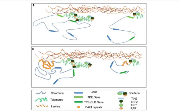
FIGURE 3 | Telomeres-Lamins interaction at the NP. (A) In cells with long telomeres, Lamins A/C interact with TRF2, thus tethering some telomeres in a peripheral localization. Telomeres might also interact with soluble Lamins in the nucleoplasm. The silencing signature of telomeres can spread in cis (TPE-sensitive genes) but also in trans (TPE-OLD-sensitive genes) due to a mechanism yet to explore. With telomeres shortening, the diminution of repressive mark (TPE) or a local change in the chromatin conformation (TPE-OLD), modulate expression of TPE and TPE-OLD genes. (B) Both TPE and TPE-OLD have been described in FSHD, a muscular dystrophy genetically linked to the subtelomeric 4q35 locus. This subtelomeric locus and its abutting telomere are located at the NP and regulated by A-type Lamins. Moreover, the number of D4Z4 repeats influences the localization of the telomeres within the nuclei. In cells with few D4Z4 repeats the transcription of DUX4, a gene encoded by D4Z4 repeats, is increased upon telomere shortening through direct TPE while expression of SORBS2, located 5 Mb upstream is up-regulated through a TPE-OLD phenomenon.
of Lamins and chromatin might also associate with telomere
through interaction with TRF2 and RIF1. However, the precise
mechanisms linking LAP1, chromatin and telomeres remains
to be uncovered (
Serrano et al., 2016
). In addition,
Lamins-TRF2 interactions might stabilize t-loops forming at interstitial
telomeric sequences (ITS) while progerin does not suggesting
a key role for TRF2-Lamins interactions in the stability of
telomeric sequences at chromosome ends and ITS (
Wood et al.,
2014
).
Interestingly, in human cells, early replicating telomere
are localized in the inner nuclear volume while peripheral
positioning of telomeres correlates with late replication
suggesting that components of the INM also control other
aspects of telomere regulation (
Arnoult et al., 2010
) but this
regulation might not be directly associated with the tip of
chromosomes but with the subterminal subtelomeric regions.
Telomeres are separated from chromosome-specific gene-rich
region by complex sequences named subtelomeres. In the human
population, these subtelomeric regions are highly polymorphic
and their recombination rate is higher than in the rest of the
genome. If the 46 human telomeres share the same structure, this
subtelomeric unique arm-specific composition concurs to the
propensity of telomeres to regulate gene expression but also to
the replication and maintenance of chromosome ends and likely
their subnuclear positioning (
Ottaviani et al., 2008
;
Arnoult et al.,
2010
). These subtelomeric regions are associated with genome
evolution and contribute to the regulation of telomeric position
effect (TPE) and production of telomeric repeat
contanning-RNA (TERRA) transcribed from the subtelomeric regions
through the adjacent telomere. The implication of TPE and
TERRA has been evoked in many diseases but rarely investigated
in depth (
Ballif et al., 2007
;
Shao et al., 2008
;
Maicher et al.,
2012
).
Overall, telomere positioning to the NP is variable between
species and might be determined by subtelomere composition,
although this remains to be determined for most human
subtelomeres. Thus, transcriptional regulation of natural
subtelomeric genes in human cells likely depends on telomere
length, the structure of the telomeric chromatin but also on the
composition of the subtelomeric regions and spatial distribution
of chromosome ends. In addition, as observed experimentally
using artificial systems (
Baur et al., 2001
;
Koering et al.,
2002
), age-dependent telomere erosion might also be a key
player in the modulation of subtelomeric genes in elders and
contribute to physiological and pathological aging. In this
model, if with long telomeres, a subtelomeric gene is captured
within the structure, hence silenced, telomere shortening might
unlock the gene from the heterochromatin signature and
trigger gene derepression. Thus, over time telomere shortening
can modify
in cis or trans expression of various genes. In
classical TPE, the degree of repression declines with distance
from the telomere and spreading of silencing marks from
the telomere might be limited to 100 kb in mammalian cells
(
Kulkarni et al., 2010
). It was recently shown that telomeres
affect gene expression over much larger distances through a
telomere-length dependent looping (
Robin et al., 2014
). This
phenomenon named TPE-OLD (TPE over long distance) can
affect expression of genes located at a distance of up to
10 Mb from the telomere (Figure 3A). Unlike the classical
TPE signature, this phenomenon is not continuous and relies
in changes of the chromatin conformation. Interestingly,
TPE-OLD also occurs in pathologies and was first described in
Facio-Scapulo-Humeral Dystrophy, an age-dependent muscular
dystrophy linked to the 4q subtelomere located at the NP
and bound by A-type Lamins (
Masny et al., 2004
;
Tam et al.,
2004
;
Ottaviani et al., 2009, 2010
;
Arnoult et al., 2010
;
Robin
et al., 2015
) (Figure 3B). The role of telomere silencing by
TPE or TPE-OLD, especially in pathologies is still in infancy
(
Ottaviani et al., 2008
;
Lou et al., 2009
;
Robin et al., 2014, 2015
).
However, converging evidence suggests that factors affecting
directly or indirectly telomere length and structure will likely
affect the topology and expression of subtelomeric regions.
In this scenario, alterations of the lamina and production
of progerin might interfere with telomere and subtelomere
homeostasis and contribute directly or indirectly to diseases
linked to these complex chromosomal regions as exemplified in
FSHD.
CONCLUSION
We highlighted here the diverse function of a number of proteins
implicated in the filamentous network forming the NE. Some
of them interact directly with chromatin, regulate chromatin
architecture and organize the topology of chromosomal domains.
In such a scenario, mutations in Lamins A/C or genes encoding
other INM-associated factors in diseases and decrease in Lamin
B1 upon senesecence contribute to physiological or pathological
aging at different levels. Moreover, even if anchoring of genes
at the NL might contribute to gene repression as suggested by
artificial tethering systems (
Finlan et al., 2008
;
Reddy et al., 2008
;
Dialynas et al., 2010
), the mechanim of NL-mediated repression
still remains to be elucidated. With a limited number of INM
proteins characterized to date and our current knowledge of
cell-specific variations in NE structures, the regulatory potential of the
NP is a promising field of investigation in particular regarding
the specificity of the phenotypes in the different types of nuclear
envelopathies.
AUTHOR CONTRIBUTIONS
Both authors have contributed equally to the writing of the
manuscript, edition and preparation of the figures.
ACKNOWLEDGMENT
We thank Pr. Pierre Cau and Pr. Nicolas Lévy for constructive
discussions during the preparation of this manuscript.
REFERENCES
Aaronson, R. P., and Blobel, G. (1975). Isolation of nuclear pore complexes in
association with a lamina.Proc. Natl. Acad. Sci. U.S.A. 72, 1007–1011. doi:
10.1073/pnas.72.3.1007
Aebi, U., Cohn, J., Buhle, L., and Gerace, L. (1986). The nuclear lamina is a
meshwork of intermediate-type filaments.Nature 323, 560–564. doi: 10.1038/
323560a0
Agarwal, A. K., Fryns, J. P., Auchus, R. J., and Garg, A. (2003). Zinc
metalloproteinase, ZMPSTE24, is mutated in mandibuloacral dysplasia.Hum.
Mol. Genet. 12, 1995–2001. doi: 10.1093/hmg/ddg213
Allsopp, R. C., Vaziri, H., Patterson, C., Goldstein, S., Younglai, E. V., Futcher, A. B., et al. (1992). Telomere length predicts replicative capacity of human fibroblasts. Proc. Natl. Acad. Sci. U.S.A. 89, 10114–10118. doi: 10.1073/pnas.89.21.10114 Armanios, M. Y., Chen, J. J., Cogan, J. D., Alder, J. K., Ingersoll, R. G., Markin, C.,
et al. (2007). Telomerase mutations in families with idiopathic pulmonary
fibrosis.N. Engl. J. Med. 356, 1317–1326. doi: 10.1056/NEJMoa066157
Arnoult, N., Schluth-Bolard, C., Letessier, A., Drascovic, I., Bouarich-Bourimi, R., Campisi, J., et al. (2010). Replication timing of human telomeres is chromosome arm-specific, influenced by subtelomeric structures and connected to nuclear localization.PLoS Genet. 6:e1000920. doi: 10.1371/journal.pgen.1000920 Ballif, B. C., Sulpizio, S. G., Lloyd, R. M., Minier, S. L., Theisen, A., Bejjani, B. A.,
et al. (2007). The clinical utility of enhanced subtelomeric coverage in array
CGH.Am. J. Med. Genet. A 143A, 1850–1857. doi: 10.1002/ajmg.a.31842
Banumathy, G., Somaiah, N., Zhang, R., Tang, Y., Hoffmann, J., Andrake, M., et al. (2009). Human UBN1 is an ortholog of yeast Hpc2p and has an essential role in
the HIRA/ASF1a chromatin-remodeling pathway in senescent cells.Mol. Cell.
Biol. 29, 758–770. doi: 10.1128/MCB.01047-08
Barthelemy, F., Navarro, C., Fayek, R., Da Silva, N., Roll, P., Sigaudy, S., et al. (2015). Truncated prelamin A expression in HGPS-like patients: a transcriptional study.Eur. J. Hum. Genet. 23, 1051–1061. doi: 10.1038/ejhg.2014.239 Baur, J. A., Zou, Y., Shay, J. W., and Wright, W. E. (2001). Telomere position effect
in human cells.Science 292, 2075–2077. doi: 10.1126/science.1062329 Ben-Yishay, R., Ashkenazy, A. J., and Shav-Tal, Y. (2016). Dynamic encounters of
genes and transcripts with the nuclear pore.Trends Genet. 32, 419–431. doi:
10.1016/j.tig.2016.04.003
Bercht Pfleghaar, K., Taimen, P., Butin-Israeli, V., Shimi, T., Langer-Freitag, S., Markaki, Y., et al. (2015). Gene-rich chromosomal regions are preferentially localized in the lamin B deficient nuclear blebs of atypical progeria cells.Nucleus 6, 66–76. doi: 10.1080/19491034.2015.1004256
Blobel, G. (1985). Gene gating: a hypothesis.Proc. Natl. Acad. Sci. U.S.A. 82,
8527–8529. doi: 10.1073/pnas.82.24.8527
Boulikas, T. (1986). Protein-protein and protein-DNA interactions in calf thymus nuclear matrix using cross-linking by ultraviolet irradiation.Biochem. Cell Biol. 64, 474–484. doi: 10.1139/o86-066
Bouvier, D., Hubert, J., Seve, A. P., and Bouteille, M. (1985). Characterization of lamina-bound chromatin in the nuclear shell isolated from HeLa cells.Exp. Cell Res. 156, 500–512. doi: 10.1016/0014-4827(85)90557-9

![FIGURE 2 | Chromatin is rearranged upon physiological or premature aging. (Left) In young cells, large chromatin domains [topologically associated domains(TADs), Lamin Associated Domains (LADs)] are associated to the NL and localized at the nuclear periphe](https://thumb-eu.123doks.com/thumbv2/123doknet/14649493.551080/6.892.70.825.90.508/chromatin-rearranged-physiological-premature-topologically-associated-associated-associated.webp)