“Candidatus Mesochlamydia elodeae”
(Chlamydiae:
Parachlamydiaceae), a novel
chlamydia parasite of free-living amoebae
Daniele Corsaro&Karl-Dieter Müller&Jost Wingender&Rolf Michel
Abstract Vannella sp. isolated from waterweed Elodea sp. was found infected by a chlamydia-like organism. This organ-ism behaves like a parasite, causing the death through burst of its host. Once the vannellae degenerated, the parasite was successfully kept in laboratory within a Saccamoeba sp. iso-lated from the same waterweed sample, which revealed in fine through electron microscopy to harbor two bacterial endo-symbionts: the chlamydial parasite we introduce and another endosymbiont initially and naturally present in the host. Herein, we provide molecular-based identification of both the amoeba host and its two endosymbionts, with special focus
on the chlamydia parasite. High sequence similarity values of the 18S rDNA permitted to assign the amoeba to the species Saccamoeba lacustris (Amoebozoa, Tubulinea). The bacterial endosymbiont naturally harbored by the host belonged to Sphingomonas koreensis (Alpha-Proteobacteria). The chla-mydial parasite showed a strict specificity for Saccamoeba spp., being unable to infect a variety of other amoebae, in-cluding Acanthamoeba, and it was itself infected by a bacte-riophage. Sequence similarity values of the 16S rDNA and phylogenetic analysis indicated that this strain is a new mem-ber of the family Parachlamydiaceae, for which we propose the name“Candidatus Mesochlamydia elodeae.”
Introduction
Chlamydiae constitute a large group of intracellular para-sites of eukaryotes, infecting amoebae and some inverte-brates and verteinverte-brates, including humans (Corsaro and Venditti 2004; Corsaro and Greub 2006). Most
endosym-bionts of amoebae form the monophyletic
family Parachlamydiaceae; the first recognized members, recovered within environmental and clinical isolates of
Acanthamoeba (Fritsche et al.
1993; M i c h e l e t a l . 1992, 1994), include Parachlamydia acanthamoebae, Protochlamydia amoebo-phila, and other unnamed strains (Amann et al. 1997; Collingro et al. 2005; F r i t s c h e e t a l . 2000).
Acanthamoeba spp. (Amoebozoa, Centramoebida) are na-ked free-living amoebae widespread in many environments and are able to cause by themselves diseases in vertebrates, forming the heterogeneous functional group of amphizoic amoebae (Visvesvara et al. 2007). Many Acanthamoeba strains harbor endosymbionts and may be vehicle for various pathogens. Due to their medical importance as well as their easy management in laboratory in terms of recovery,
D. Corsaro (*)
Chlamydia Research Association (CHLAREAS), 12 rue du Maconnais,
54500 Vandoeuvre-lès-Nancy, France e-mail: corsaro@voila.fr
D. Corsaro
Laboratory of Soil Biology, Institute of Biology, University of Neuchâtel, rue Emile Argand 11, 2000 Neuchâtel, Switzerland
K.-D. Müller
Institut für Medizinische Mikrobiologie der Universität Duisburg-Essen, Virchowstr.179, 45147 Essen, Germany
J. Wingender
Biofilm Centre, Aquatic Microbiology,
University of Duisburg-Essen, Universitätsstrasse 5, 45141 Essen, Germany
R. Michel
Central Institute of the Federal Armed Forces Medical Services, P.O. Box 7340, 56070 Koblenz, Germany
maintenance, and propagation, Acanthamoeba spp. are mostly studied as natural and experimental hosts for emerging patho-gens, including chlamydiae (Horn and Wagner 2004; C o r s a r o and Greub 2006). A wider host range for all chlamydiae was suggested from early environmental molecular surveys show-ing huge phylotype diversity (Horn and Wagner 2001; Corsaro et al. 2002) as well as from experimental studies using a large variety of amoeba species (Michel et al. 2004, 2005) and from mixed cocultures (Corsaro and Venditti 2009). This was further confirmed by finding chlamydiae in “unusu-al” hosts (e.g., Thao et al. 2003; Corsaro et al. 2007; I s r a e l s s o n 2008). Many strains and species of Parachlamydiaceae how-ever have been recovered from Acanthamoeba, e i t h e r a s n a t - ural endosymbionts or by coculture (e.g., Schmitz-Esser et al. 2008; Corsaro et al. 2009; M a t s u o e t a l . 2010). Nevertheless, Neochlamydia hartmannellae inhabits Hartmannella (Vermamoeba) vermiformis (Amoebozoa, Echinamoebida) but failed to infect Acanthamoeba (Horn et al. 2000), and Protochlamydia naegleriophila resides naturally in Naegleria spp. (Excavata, Heterolobosea) and grows easily within Acanthamoeba and several other amoebae (Michel et al. 2000). By contrast, Metachlamydia lacustris parasitizes naturally nonamphizoic Saccamoeba spp. (Amoebozoa, Euamoebida) and fails to infect amphizoic amoebae like Acanthamoeba, Vermamoeba, a n d Naegleria (Michel et al. 2006; Corsaro et al. 2010).
Recently, Michel et al.
(2010) i s o l a t e d Vannella sp. heavily infected by intracellular bacterial parasites from the waterweed
Elodea sp. (Angiospermae, Alismatales,
Hydrocharitaceae), a basal monocot generally used as aquarium vegetation. Vannellae rapidly degenerated, but a parasite was preserved by transferring it to a Saccamoeba sp. also isolated from the same Elodea sample. Electron microscopy analysis revealed that the new amoeba host harbored finally two different types of endosymbionts. The endosymbiont presumably responsible for the death of the vannellae, called KV, exhibited a chlamydia-like morpholo-gy and was infected by a bacteriophage (Michel et al. 2010).
This study focuses on the molecular phylogenetic analysis based on the small subunit rRNA genes of both the Saccamoeba host and the chlamydia-like endoparasite KV. Our results confirm amoeba to be a new strain of Saccamoeba lacustris and show that the strain KV belongs to a new genus-level lineage within the family Parachlamydiaceae, for which we propose the name“Candidatus Mesochlamydia elodeae”. Materials and methods
Samples
Original infected amoebae hosts, identified morphologically as Vannella sp., were isolated from leaves of commercial
waterweed Elodea sp. (Angiospermae, Alismatales). From the same sample, some uninfected amoebae, identified mor-phologically as Saccamoeba limax, were also recovered and used to successfully propagate the endocytobiont after the decay of the original vannellid host (Michel et al. 2010). Morphological identification of amoebae was performed according to Page (1988).
Clonal subpopulations of infected and uninfected Saccamoeba (strain SL-elo) were maintained for over 1 year at room temperature on 1.5 % nonnutritive agar (NNA) covered with Enterobacter cloacae or Escherichia coli. To allow maintenance of the endosymbionts, infected amoebae were periodically transferred to fresh NNA plates containing uninfected amoebae as new hosts.
Amoeba coculture and host range
A preliminary amoeba host range for KV, including various Saccamoeba spp. (Saccamoeba limax, Saccamoeba lucens, Saccamoeba lacustris) and other Amoebozoa (e.g., Acanthamoeba lenticulata, Sappinia spp., Thecamoeba spp.) and Heterolobosea (e.g., Naegleria, Tetramitus), was studied previously (Michel et al. 2010). In this study, further Acanthamoeba (genotype T4) and Vermamoeba cocultures were performed, as previously described (Corsaro et al. 2009, 2010). Briefly, trophozoites were prepared as host cells in six-well microplates in Page’s amoeba saline (PAS), inoculated with chlamydiae and incubated at room temperature in a humidified atmosphere in the dark. Five days postinfection, wells were screened by chlamydia-specific PCR (see below). Saccamoeba strain SL-elo was grown onto inactivated bacteria, thus inoculated with chla-mydiae and incubated at room temperature. Amoebae were inspected daily at light microscope and screened for chla-mydiae by PCR.
DNA amplification, sequencing, and phylogenetic analysis
Amoebae were harvested from the agar plates, suspended in PAS, and rinsed three times in PAS at 200 × g. Infected amoebae were freeze-thawed, and further low-speed centri-fugation steps were applied to separate amoebal cell debris from the cytoplasm containing the endosymbionts.
Whole DNA was extracted with the Wizard Genomic DNA kit (Promega) according to the manufacturer’s recom-mendations. Amoebal 18S rRNA gene was amplified by using the primers 42F (5′-CTC AAR GAY TAA GCC ATG CA-3′) and 1498R (5′-CAC CTA CGG AAA CCT TGT TA-3′) (López-García et al. 2007) and 6F (5′-CCA GCT CYA AKA GCG TAT ATT-3′) and 9R (5′-GTT GAG TCR AAT TAA GCC GC-3′) (modified from Corsaro et al. 2009), in the reaction conditions of 5 min at 94 °C, followed by 35 cycles for 1 min at 94 °C, 1 min at 56 °C, and 2 min at
72 °C, with a final extension of 5 min at 72 °C. Chlamydial 16S rRNA gene was amplified by using the pan-chlamydia primers CF1 (5′-CGT GGATGA GGC ATG CRA GTC G-3′) and CR7 (5′-TAC CTT GTT ACG ACT TMA YCC YAG-3′) (Corsaro and Venditti 2009; Corsaro and Work 2012), under the reaction conditions of 5 min at 94 °C, followed by 35 cycles for 1 min at 94 °C, 1 min at 60 °C, and 1 min 30 s at 72 °C, with a final extension of 5 min at 72 °C. Bacterial 16S rDNAs from additional endosymbionts were amplified by using the eubacterial primers EBF (5′-AGA GTT TGA TCM TGG CTC AG-3′) a n d E B R ( 5 ′-ACG GCT ACC TTG TTA CGA CTT-3′) (Corsaro and Venditti), as well as the primers alpha-F19 (5′-CCT GGC TCA GAA CGA ACG-3′) a n d alpha-R1517 (5′-TGATCC AGC CGC AGG TCC-3′) s p e c i f - ic for Alpha-Proteobacteria (Vannini et al. 2004). PCR con-ditions were 5 min at 94 °C, followed by 35 cycles for 1 min at 94 °C, 1 min at 51 °C or 56 °C, respectively, and 1 min 30 s at 72 °C, with a final extension of 5 min at 72 °C. A 600-bp 16S rDNA fragment was separately amplified with eubacterial primers 519f (5′-CAG CAG CCG CGG TAA TAC-3′) a n d 1100r (5′-GGG TTG CGC TCG TTG-3′) and cloned in Escherichia coli by using the TOPO TA Cloning System (Invitrogen). Six clones were randomly selected for sequencing.
Purified PCR products were sequenced with the same primer sets and a series of inner primers by using an auto-matic ABI DNA Sequencer (Applied Biosystems) with the BigDye Terminator Cycle Sequencing Kit. Sequences were edited by using BioEdit and analyzed through BLAST serv-er to search for closest relatives. SSU rDNA sequences retrieved from GenBank were aligned by using MUSCLE.
Phylogenetic analyses were performed by applying distance (neighbor joining, NJ) and maximum parsimony (MP) with MEGA5 (Tamura et al. 2011), and maximum likelihood (ML, GTR, G+I:4 model) with TREEFINDER (Jobb et al. 2004), with 1,000 bootstraps.
Sequence similarity values were calculated with BioEdit. An overall sequence similarity matrix, including one mem-ber for each major species of all Chlamydiae, was calculated using all the common sites and excluding indels. Genetic relatedness of KV with Parachlamydiaceae was further analyzed by considering near full 16S rDNA sequences representing the various species and clades within this family, as defined by previous studies (Corsaro and Venditti 2006, 2009; Corsaro et al. 2010).
Electron microscopy
Methods for electron microscopy were reported previously (Michel et al. 2006, 2010). Briefly, infected Saccamoeba trophozoites were harvested from NNA plates, pelleted for 15 min at 200×g, fixed for 1 h in 3 % ice-cold glutaralde-hyde in 0.1 M cacodylate buffer pH 7.2, postfixed for 1 h in
1 % osmium tetroxide and 2 % uranyl acetate, dehydrated in alcohol, and embedded in Spurr resin. Sections were stained with 1 % lead citrate.
Fluorescence in situ hybridization
Harvesting of KV-infected amoebae from agar plate cul-tures, fixation, and dehydration of the cells were performed according to the procedure described by Grimm et al. (2001). Fluorescence in situ hybridization (FISH) was per-formed on KV-infected amoebae, using the oligonucleotide probe Chls-523, 5′-labeled with the fluorescent dye Cy3, specific for Chlamydiales as described in Poppert et al. (2002). The cells were viewed under an epifluorescence microscope (Carl Zeiss Microscopy GmbH, Germany) using Filter Set 20.
Results and discussion
The putative natural amoeba host Saccamoeba strain SL-elo
The strain SL-elo was isolated from the same Elodea sam-ple, from which vannellid amoebae infected with the chla-mydia KV originated, and was successfully used for its propagation (Michel et al. 2010). By considering both its origin and high susceptibility, this strain might be a natural host for KV. We thus consider this Saccamoeba species as a putative natural host.
Morphologically, SL-elo was very similar to Saccamoeba limax sensu Page and it was described as belonging to this species in the first publication (Michel et al. 2010). No cyst formation was observed for over 1 year of cultivation onto NNA (Michel et al. 2010; this study). On the basis of near full 18S rDNA sequence, SL-elo (GenBank account no. JN112797) showed 99.5 % similarities with Saccamoeba lacustris strain SL2 (CCAP 1572/4) (Corsaro et al. 2010) and 95.7 % with Saccamoeba limax strain NTSHR, isolated from decomposing gills of aquarium fish (Dyková et al. 2008), followed by Glaeseria mira with 86.4 %. Similarity value with the strain F-13 ATCC 30942, designed as Saccamoeba limax, was only of 77.5 %. In phylogenetic reconstruction (Fig. 1), our Saccamoeba strain emerged within the newly recognized Saccamoeba lineage repre-sented by strains SL2 and NTSHR, as sister group to Glaeseria mira within the Hartmannellidae (Euamoebida, Tubulinea) (Smirnov et al. 2011). We assigned thus the strain SL-elo to the species Saccamoeba lacustris. It has been deposited at the Culture Collection of Algae and Protozoa (CCAP 1572/6). The Saccamoeba/Glaeseria is sister to the Copromyxa lineage, which includes the Hartmannella cantabrigensis/Copromyxa clade sensu Smirnov et al. (2011), and the strain 4730 MK-2011
(Dyková et al. 2011), early misidentified as Vexillifera expectata. We further confirmed that the strain F-13 ATCC 30942 is not a member of the genus Saccamoeba, emerging with other two closely related phylotypes in a distinct branch within the Tubulinea. We suggest to consider the strain NTSHR as a neotype for Saccamoeba limax.
The putative endosymbiont of Saccamoeba
Electron microscopy showed that a different type of endo-symbiont was harbored in at least some subclonal popula-tions of our Saccamoeba strain (Michel et al. 2010). The presence of endosymbionts was considered to be a typical feature of this amoeba genus by Page (1988). A unique 16S
rDNA sequence was obtained from both uninfected and KV-infected Saccamoeba plates, suggesting that this putative symbiont was originally present within Saccamoeba and did not derive from vannellae along with KV. The sequence was obtained from uninfected amoebae with eubacterial primers by direct sequencing of PCR products and after cloning, indicating that the recovered sequence likely rep-resents the dominant phylotype. The same sequence was obtained from KV-infected amoebae with primers specific for Alpha-Proteobacteria, to avoid coamplification of chlamydial DNA.
This putative endosymbiont belongs to the genus Sphingomonas (Alpha-Proteobacteria, Sphingomonadales), the obtained sequence (GenBank account no. JN112798)
Fig. 1 Maximum likelihood 18S rDNA tree of Lobosa (Tubulinea + Discosea), showing the major inner groups, and the position of the recovered Saccamoeba lacustris strain SL-elo (in bold) in the family Hartmannellidae. Acramoeba dendroida and Filamoeba sinensis (Varipodida, Variosea) were used as outgroup. Bootstrap values
after 1,000 replicates for ML/NJ/MP were indicated at nodes. Filled circle, node 100 % supported with all three methods; asterisk, node supported but BV <40 %; hyphen, node not supported. The scale bar represents substitution/site
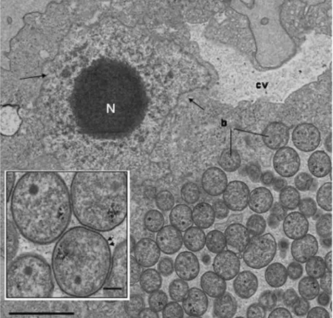
showing 99.8 % similarity with the type strain JSS-26 of Sphingomonas koreensis, isolated from natural mineral wa-ter (Lee et al. 2001). Similarity value with the Elodea epiphyte Sphingomonas elodea (GenBank account no. AF503278), an important producer of exopolysaccharide known as gellan (Fialho et al. 2008), was of only 96.1 %, thus allowing to reasonably exclude a contamination. Sphingomonas spp. are widespread in the environment and have occasionally been isolated in Acanthamoeba cocul-tures (e.g., Evstigneeva et al. 2009). However, these bacteria are rod-shaped measuring 0.5×1.5–2-μm, while our amoe-bae showed at electron microscopy smaller irregularly shaped but mostly coccoid Gram-negative endosymbionts (Fig. 2). One possibility may be that strict endosymbiosis lifestyle has led to a change from rod to coccus shape for this strain. Interestingly, shorter (0.6–0.8 μm i n l e n g t h ) Sphingomonas endosymbionts, matching 100 % with S. koreensis JSS-26 and our putative symbiont in partial 16S rDNA sequences, were found in the cytoplasm of the testate amoeba Arcella rotundata (Amoebozoa, Arcellinida) (Török et al. 2008); such shape change was supported by the find-ing of coccoid Paenibacillus as endosymbionts in the Laccaria mycelium (Bertaux et al. 2003). Since we did not explore more deeply this association in the present study, further research is now needed to confirm the real corre-spondence between molecular and microphotographic data, as well as to test the strict endosymbiotic hypothesis.
The chlamydial endoparasite KV
The chlamydial strain KV was unable to grow in amoeba coculture in microplates or NNA, using various amoebae as hosts, including both Amoebozoa (Acanthamoeba, Flamella, Sappinia) a n d
H e t e r o l o b o s e a ( Naegleria, Tetramitus, Willaertia). KV was able to grow only within strains of Saccamoeba spp. and it was thus kept within Saccamoeba lacustris strain SL-elo by periodic passages onto fresh
troph-ozoites for over 1 year (Michel et al.
2010; t h i s s t u d y ) . Presence of KV in Saccamoeba was documented by electron microscopy (see below) and FISH (Supplementary Fig. 1).
The near full-length 16S rDNA of KV was obtained from infected Saccamoeba cells by using PCR primers specific for chlamydiae. Overall, sequence similarities of the 16S rDNA from KV were 91.0 to 91.8 % with members of
Parachlamydiaceae, 8 8 . 4 –89.1 % with
Criblamydiaceae and less than 89 % with members of the other chlamydial lineages (Supplementary Table 1). Sequence similarity values of >90 and >95 % have been proposed to include strains within the same family or genus, respectively (Everett et al. 1999; C o r s a r o e t a l . 2003). KV appeared thus as a new genus-level taxon within the family Parachlamydiaceae. To better infer genetic relatedness within Parachlamydiaceae, similarity values were calculated considering only members of this family (Supplementary Table 2). KV had a maximum similarity value of 91.87 % with Metachlamydia lacustris,
Fig. 2 Coccoid endosymbionts (b) within
Saccamoeba, b e n e a t h the contractile vacuole (cv). Vesicular nucleus (N) t y p i c a l o f
Saccamoeba. Scale bar 2 μm. Inset: Enlarged view of coccoid symbionts showing Gram-negative structure (arrows). Scale bar 200 nm. The figures are reproduced from a former article (Michel et al. 2010) w i t h the kind permission of the publisher of “Endocytobiosis and Cell Research”: http://zs. thulb.uni-jena.de/content/main/journals/ ecb.xml?lang0en
followed by Parachlamydia acanthamoebae (mean value 91.7 %). Mean similarity values shared by KV with the other parachlamydiae were 88.1–91.5 %.
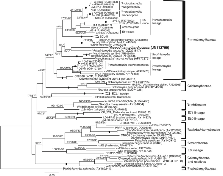
In molecular phylogenetic analyses based on 16S rRNA gene sequence (Fig. 3), Parachlamydiaceae was recognized as a holophyletic group with moderate to high bootstrap supports. Criblamydiaceae emerged as the sister group but with low support. Inner groups within Parachlamydiaceae were highly supported and corresponded to the previously identified clades (Corsaro and Venditti 2009). The strain KV emerged as a unique lineage within Parachlamydiaceae, h a v -ing a moderate ML support (70 %) for a relationship with Metachlamydia, corvenA4 group, and Neochlamydia lineage.
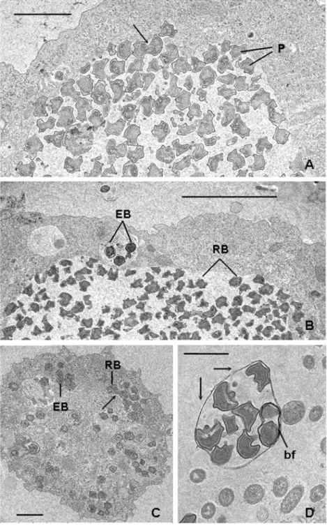
Typical chlamydial developmental cycle was documented for strain KV infecting Saccamoeba through electron micros-copy (Michel et al. 2010). Highly pleomorphic reticulate
bodies (RBs), approximately 1 μm in diameter, reside mainly within a large cytoplasmic inclusion of the amoeba host, where they divide by binary fission (Fig. 4a, c). Elementary bodies (EBs), approximately 0.5 μm in diameter, rounded and edged, with a highly condensed central nucleoid, reside in smaller vacuoles containing one to three cells (Fig. 4b, c). Intermediate stages were not only mainly observable inside inclusions containing RB but also within small vesicles orig-inated after the amoeba burst; 5 to 7 days postinfection, amoebae were completely filled and burst, releasing EB and small vesicular membranes containing chlamydiae at different stages (Fig. 4d). Some RBs contain both empty and filled hexagonal bacteriophages of about 55 nm in diameter (Fig. 5). Highly pleomorphic RB were observed in the related Metachlamydia, also infecting Saccamoeba (Michel et al. 2006; Corsaro et al. 2010), whereas other parachlamydial
0.03
CRIB44 (FJ532290)
chlam-122 (fecal sample, Larus, GU068510) p22m06ok (tall grass prairie, FJ479189)
530-2 (AY326519)
ECL-II
corvenA4 (respiratory sample, AF308693)
Waddliaceae cvE65 (JF706723) Berg17 (AM941720)
PRPR83 (poriferan, DQ903996)
LD1-PA42 (marine sediment, AY114327)
Piscichlamydia salmonis (AY462244)
CRIB32 (WTP, EU363464)
MABRDTU43 (nitrifying biofilm, FJ529996)
CN808 (respiratory sample, EU090709) cvE60 (freshwater, FJ976107)
Chlamydia trachomatis A-Har13 (D89067) cvC7 (respiratory sample, AF478463)
Neochlamydia sp. UWC22 (AF083616)
Chlamydophila psittaci NJ1 (U68419) soy123 (soybean field, FJ415740)
Rhabdochlamydia porcellionis (AY223862)
Simkania negevensis (U68460) ECL-I (wwtp) UV-7 (AJ715410) cvE71 (freshwater, FJ706724) KNic (DQ632609) 3-1 (AY326517) cvE14 (FJ976093) Neochlamydia sp. S40 (AB506678) cvE12 (FJ976092) Metachlamydia lacustris (GQ221847) cvE18 (freshwater, FJ976098) cvC15 (respiratory sample, AF478473)
Waddlia chondrophila (AF042496)
Clavichlamydia salmonicola (DQ011662)
Estrella lausannensis (EU074225)
Rhabdochlamydia crassificans (AY928092) cvE27 (FJ976101)
CRIB38 (WTP, EU683886)
Mesochlamydia elodeae (JN112799)
CK_2C2_23 (bivalve sediment, EU488135)
Renichlamydia lutjani (JN167597)
Neochlamydia hartmannellae (AF177275)
PRPR10 (poriferan, DQ903988) CRIB33 (WTP, EU683887)
cvE6 (freshwater, AF448722) Acanthamoeba symbiont UWE1 (AF083614)
Waddlia malaysiensis (AY184804) UWE25 (AF083615) Criblamydiaceae cvE70 (JF706725) cvE99 (JF513057) EI2 (AM408789) cvE4b (freshwater, JN093034) cvE5 (AY220545) CRIB35 (EU384664) OEW1 (AM412760) Criblamydia sequanensis (DQ1234300) Chlamydophila pneumoniae TW183 (L06108) CRIB39 (FJ532292) cvE26 (FJ976102)
cvE9 (freshwater, AF448723)
Fritschea bemisiae (AY140910) cvE22 (FJ976104) 98/98/92 81/70/88 87/-/-48/-/* 97/99/94 96/95/83 99/100/98 79/55/65 92/95/87 99/100/90 97/96/95 97/99/99 63/*/* 87/59/84 91/90/83 98/83/94 67/86/58 71/99/76 94/62/* 98/99/99 67/-/* 100/99/90 97/93/96 100/81/98 70/*/* 54/72/57 87/72/* 97/98/88 84/-/67 100/99/98 48/57/* 93/99/97 61/84/* 100/99/96 77/67/* 65/*/* Parachlamydia acanthamoebae Protochlamydia naegleriophila Protochlamydia amoebophila E5 clade Amazon group E14 clade Neochlamydia lineage Parachlamydia lineage Protochlamydia lineage Parachlamydiaceae Criblamydiaceae Waddliaceae Rhabdochlamydiaceae Piscichlamydiaceae Simkaniaceae Chlamydiaceae and relatives E6 lineage E60 lineage E71 lineage
Fig. 3 Maximum likelihood 16S rDNA tree of Chlamydiae, showing the major lineages and sublineages, and the position of the recovered Mesochlamydia elodeae (in bold) within the Parachlamydiaceae. Bootstrap values after 1,000 replicates for ML/NJ/MP were indicated
at nodes. Filled circle, node 100 % supported with all three methods; asterisk, node supported but BV <40 %; hyphen, node not supported. The scale bar represents substitution/site
taxa exhibit more rounded or wrinkled RB (e.g., Amann et al. 1997; Fritsche et al. 2000; Michel et al. 2000). Also, bacteriophages with similar morphology and size of 50– 70 nm have been reported in other parachlamydiae strains
infecting Vermamoeba (H.
vermiformis) a n d Naegleria (Michel et al. 2001; S c h m i d e t a l . 2001), as well as in uncharacterized “chlamydia-like organisms” infecting bivalves (Harshbarger et al. 1977). These phages how-ever appear different from the well-characterized but smaller, about 25 nm in diameter, Chlamydiamicrovirus (Microviridae: Gokushovirinae), infecting members of Chlamydophila spp. (Chlamydiaceae) (Everson et al. 2003).
It is to note that virome analyses in marine and freshwater biomes revealed unexpected high diversity and even dominant presence for chlamydiaviruses (Angly et al. 2006; D e s n u e s e t al. 2008; R o u x e t a l . 2012). Description of“Candidatus Mesochlamydia elodeae” Etymology: Candidatus, bacterial category including well-characterized but as yet uncultured organisms, among which are obligate intracellular bacteria. Mesochlamydia n. gen. Meso, Gr. prep. for middle, intermediate; Chlamydia, N.L. fem. n., a bacterial genus name; Mesochlamydia N.L. fem.
Fig. 4 Saccamoeba infected with Mesochlamydia. a Large inclusion in trophozoite showing irregular-shaped chlamydial parasites (P), mostly RBs, some in binary fission (arrow). Scale bar 2 μm. b Large inclusion with RB and a small inclusion with EB. Scale bar 5 μm. c EB with highly condensed central nucleoid and RB in binary fission (arrow). Scale bar 2 μm. d Vesicle released after amoeba burst containing Mesochlamydia cells at various stages, one of which in binary fission (bf). c was reproduced from a former article (Michel et al. 2010) w i t h the kind permission of the publisher of “Endocytobiosis and Cell Research”: http://
zs.thulb.uni-jena.de/content/ main/journals/ecb.xml?lang0en
n., an intermediate genus-level lineage (between the other ones within the family Parachlamydiaceae). Candidatus Mesochlamydia elodeae n. sp., elodeae N.L. gen. sing. n., of Elodea, genus name of the waterweed from which the new chlamydia and the host amoebae were isolated.
Gram-negative, chlamydial developmental stages occurred within amoebae of the genus Saccamoeba (Amoebozoa, Tubulinea). Pleomorphic reticulate bodies, 0.8–1.1 μm in diameter, are generally inside a large cyto-plasmic inclusion of the amoeba host, where they divide by binary fission. Some RB may contain hexagonal phages. Membrane-bounded vesicles containing single or small clusters of RB or intermediate bodies are also formed after the burst of the amoeba host. More rounded but edged elementary bodies, 0.5–0.6 μm in diameter, reside in single vacuoles or as small clusters in the cytoplasm.
The 16S rDNA (GenBank account no. JN112799) showed similarity values lower than 92 % with all the other members of Parachlamydiaceae, with a maximum value of 91.8 % with Metachlamydia lacustris. Phylogenetic analysis supported the emergence of strain KV as a unique genus-level lineage (sequence similarity with known members >90 and <95 %) within the family Parachlamydiaceae.
Natural host: Vannellid amoeba recovered from Elodea sp.; putative natural host: Saccamoeba lacustris strain SL-elo (CCAP 1572/6), recovered from Elodea sp. Observed behavior in sensitive amoebae is parasitic. Infection of other Amoebozoa and Heterolobosea was unsuccessful (Michel et al. 2010; this study).
Ecological considerations
Several strains of Amoebozoa and Heterolobosea were tested as host cells, but only strains of Saccamoeba spp. were found to successfully permit Mesochlamydia elodeae mul-tiplication. Such a strict host preference for Saccamoeba spp. was recorded previously for Metachlamydia lacustris, which was unable to infect a variety of Amoebozoa and
Heterolobosea under different growth conditions (Michel et al. 2006; Corsaro et al. 2010). Both chlamydiae seemed also to affect heterolobosean amoebae even if not able to multi-ply within them, Metachlamydia causing the formation of multinucleated cells in Naegleria clarki (Michel et al. 2006) and Mesochlamydia of empty cysts in Tetramitus horticolus (Michel et al. 2010).
An additional environmental chlamydial strain, cvE4b, recovered from a freshwater pond in France and kept in mixed protist coculture as described previously (Corsaro and Venditti 2009), also was unable to infect Acanthamoeba but rapidly killed Saccamoeba, suggesting that this amoeba is partially susceptible. This strain is closely related (98 % se-quence similarity) to corvenA4, a parachlamydia recovered from a bronchoalveolar sample from a patient with pneumonia (Corsaro et al. 2001), emerging within the corvenA4 group sister to Metachlamydia/Mesochlamydia (Fig. 3).
These data suggest the existence of a possibly related group of parasites of saccamoebae or lobose amoebae in general, whose ecology is not based on Acanthamoeba. T o this putative group also belongs the Neochlamydia clade, with Neochlamydia hartmannellae specifically infecting Vermamoeba (Hartmannella) vermiformis but not Acanthamoeba (Horn et al. 2000).
Only few other partial chlamydial sequences showed some affinity with the putative group identified here. The 1,079-bp clone SOY123, from a soybean field, showed 99 % similarity with corvenA4, 92.9 % with Metachlamydia, and 92 % with Mesochlamydia. The 770-bp clones DDC2W1u16 (GenBank account no. EU634866), from showerhead biofilms, and BDC1_E12 (GenBank account no. AY689535), from pristine mountain stream sediment, shared 93.2–93.4 % similarities with Mesochlamydia, 94.5– 94.9 % with Metachlamydia, and 93.9–96.7 % with corvenA4. Data on possible hosts for these phylotypes are lacking, as well as for their possible implication in human or animal infections. In this latter context, various recent studies employed the detection of either very small 16S
Fig. 5 Mesochlamydia infected with bacteriophages. a Pleomor-phic RBs, some in binary fission (arrows), one showing phage particles (inset). Scale bar 1 μm. b Enlarged view of panel a showing both filled and empty bacterioph-ages within RB of Mesochlamy-dia. Scale bar 200 nm. The figures are reproduced from a former article (Michel et al. 2010) with the kind permission of the publisher of “Endocytobiosis and Cell Research”: http:// zs.thulb.uni-jena.de/content/main/ journals/ecb.xml?lang0en
rDNA portions (<300 bp) and/or of highly species-specific PCR (e.g., qPCR) to recover novel chlamydiae from clinical samples. While increasing sensitivity and specificity in par-ticular situations, these approaches however also underesti-mate the real incidence of these organisms for which, by definition, data are lacking.
Among other chlamydial groups, it is to note that Waddlia chondrophila (Waddliaceae) is able to infect a variety of vertebrate cell lines, but the infection potential of Waddlia for various amoebal strains was increased only a f t e r a n a d a p t a t i o n t o
g r o w t h w i t h i n Hartmannella
(0Vermamoeba) which was highly susceptible from the start (Michel et al. 2004). Furthermore, several strains of various chlamydial lineages are unable to grow in Acanthamoeba (Corsaro and Venditti 2009). Two main chlamydiae infect-ing fish gills, Piscichlamydia and Clavichlamydia, failed to infect Acanthamoeba in vitro (Corsaro and Karslen, unpub-lished data), but it is possible that other protists and/or invertebrates can play the role of intermediate hosts. In fact, a large variety of amoebae other than Acanthamoeba may be recovered from both gills and internal organs of various fishes (Dyková and Lom 2004). Interestingly, recent studies strongly suggest environmental niche(s) for gills chlamydiae (Schmidt-Posthaus et al. 2012) and allowed to identify chla-mydiae in internal organs of fish (Corsaro and Work 2012). Thus, the use of only Acanthamoeba as amoeba host for coculture and/or the search for natural endosymbionts/endo-parasites present only in Acanthamoeba strains lead very likely to greatly underestimate both the real biodiversity and the possible host range of Chlamydiae.
Acknowledgments We thank Elke Schneider and Liane Junglas from the Electron Microscopy Department for the excellent technical assistance (Head of the department: Dr. Bärbel Hauröder). DC was partially supported by Novartis Foundation.
References
Amann R, Springer N, Schönhuber W, Ludwig W, Schmid EN, Müller K-D, Michel R (1997) Obligate intracellular bacterial parasites of acanthamoebae related to Chlamydia spp. Appl Environ Microbiol 63:115–121
Angly FE, Felts B, Breitbart M, Salamon P, Edwards RA, Carlson C, Chan AM, Haynes M, Kelley S, Liu H, Mahaffy JM, Mueller JE, Nulton J, Olson R, Parsons R, Rayhawk S, Suttle CA, Rohwer F (2006) The marine viromes of four oceanic regions. PLoS Biol 4: e368
Bertaux J, Schmid M, Chemidlin Prevost-Boure N, Churin JL, Hartmann A, Garbaye J, Frey-Klett P (2003) In situ identification of intracellular bacteria related to Paenibacillus spp. in the myce-lium of the ectomycorrhizal fungus Laccaria bicolor S238N. Appl Environ Microbiol 69:290–306
Collingro A, Toenshoff ER, Taylor MW, Fritsche TR, Wagner M, Horn M (2005)‘Candidatus Protochlamydia amoebophila’, an endo-symbiont of Acanthamoeba spp. Int J Syst Evol Microbiol 55:1863–1866
Corsaro D, Greub G (2006) Pathogenic potential of novel chlamydiae and diagnostic approaches to infections due to these obligate intracellular bacteria. Clin Microbiol Rev 19:283–297
Corsaro D, Venditti D (2004) Emerging chlamydial infection. Crit Rev Microbiol 30:75–106
Corsaro D, Venditti D (2006) Diversity of the parachlamydiae in the environment. Crit Rev Microbiol 32:185–199
Corsaro D, Venditti D (2009) Detection of Chlamydiae from freshwater environments by PCR, amoeba coculture and mixed coculture. Res Microbiol 160:547–552
Corsaro D, Work TM (2012) Candidatus Renichlamydia lutjani, a Gram-negative bacterium in internal organs of blue-striped snapper Lutjanus kasmira from Hawaii. Dis Aquat Organ 98:249–254
Corsaro D, Venditti D, Le Faou A, Guglielmetti P, Valassina M (2001) A new chlamydia-like 16S rDNA sequence from a clinical sample. Microbiology 147:515–516
Corsaro D, Venditti D, Valassina M (2002) New chlamydial lineages from freshwater samples. Microbiology 148:343–344
Corsaro D, Valassina M, Venditti D (2003) Increasing diversity within Chlamydiae. Crit Rev Microbiol 29:37–78
Corsaro D, Thomas V, Goy G, Venditti D, Radek R, Greub G (2007) ‘Candidatus Rhabdochlamydia crassificans’, an intracellular bacterial pathogen of the cockroach Blatta orientalis (Insecta: Blattodea). Syst Appl Microbiol 30:221–228
Corsaro D, Feroldi V, Saucedo G, Ribas F, Loret J-F, Greub G (2009) Novel Chlamydiales strains isolated from a water treatment plant. Environ Microbiol 11:188–200
Corsaro D, Michel R, Walochnik J, Müller K-D, Greub G (2010) Saccamoeba lacustris, sp. nov. (Amoebozoa: Lobosea: Hartmannellidae), a new lobose amoeba, parasitized by the novel chlamydia‘Candidatus Metachlamydia lacustris’ (Chlamydiae: Parachlamydiaceae). Eur J Protistol 46:86–95
Desnues C, Rodriguez-Brito B, Rayhawk S, Kelley S, Tran T, Haynes M, Liu H, Furlan M, Wegley L, Chau B, Ruan Y, Hall D, Angly FE, Edwards RA, Li L, Thurber RV, Reid RP, Siefert J, Souza V, Valentine DL, Swan BK, Breitbart M, Rohwer F (2008) Biodiversity and biogeography of phages in modern stromatolites and thrombolites. Nature 452:340–343
Dyková I, Lom J (2004) Advances in the knowledge of amphizoic amoebae infecting fish. Folia Parasitol 51:81–97
Dyková I, Kostka M, Pecková H (2008) Morphology and SSU rDNA-based phylogeny of a new strain of Saccamoeba sp. (Saccamoeba Frenzel, 1892, Amoebozoa). Acta Protozool 47:397–405 Dyková I, Kostka M, Pecková H (2011) Three new species of the
amoebozoan genus Vexillifera Schaeffer, 1926. Acta Protozool 50:55–63
Everett KDE, Bush RM, Andersen AA (1999) Emended description of the order Chlamydiales, proposal of Parachlamydiaceae fam. nov. and Simkaniaceae fam. nov., each containing one mono-typic genus, revised taxonomy of the family Chlamydiaceae, including a new genus and five new species, and standards for the identification of organisms. Int J Syst Bacteriol 49:415–440 Everson JS, Garner SA, Lambden PR, Fane BA, Clarke IN (2003) Host range of chlamydiaphages phiCPAR39 and Chp3. J Bacteriol 185:6490–6492
Evstigneeva A, Raoult D, Karpachevskiy L, La Scola B (2009) Amoeba co-culture of soil specimens recovered 33 different bac-teria, including four new species and Streptococcus pneumoniae. Microbiology 155:657–664
Fialho AM, Moreira LM, Granja AT, Popescu AO, Hoffmann K, Sá-Correia I (2008) Occurrence, production, and applications of gellan: current state and perspectives. Appl Microbiol Biotechnol 79:889–900
Fritsche TR, Gautom RK, Seyedirashti S, Bergeron DL, Lindquist TD (1993) Occurrence of bacteria endosymbionts in Acanthamoeba
spp. isolated from corneal and environmental specimens and contact lenses. J Clin Microbiol 31:1122–1126
Fritsche TR, Horn M, Wagner M, Herwig RP, Schleifer K-H, Gautom RK (2000) Phylogenetic diversity among geographycally dispersed Chlamydiales endosymbionts recovered from clinical and environmental isolates of Acanthamoeba spp. Appl Environ Microbiol 66:2613–2619
Grimm D, Ludwig W, Brandt BC, Michel R, Schleifer K-H, Hacker J, Steinert M (2001) Development of 18S rRNA-targeted oligonu-cleotide probes for specific detection of Hartmannella and Naegleria in Legionella-positive environmental samples. Syst Appl Microbiol 24:76–82
Harshbarger JC, Chang SC, Otto SV (1977) Chlamydiae (with phages), mycoplasmas, and rickettsiae in Chesapeake Bay bivalves. Science 196:666–668
Horn M, Wagner M (2001) Evidence for additional genus-level diversity of Chlamydiales in the environment. FEMS Microbiol Lett 204:71–74 Horn M, Wagner M (2004) Bacterial endosymbionts of free-living
amoebae. J Eukaryot Microbiol 51:509–514
Horn M, Wagner M, Müller K-D, Schmid EN, Fritsche TR, Schleifer KH, Michel R (2000) Neochlamydia hartmannellae gen. nov., sp. nov., (Parachlamydiaceae), an endoparasite of the amoeba Hartmannella vermiformis. Microbiology 146:1231–1239 Israelsson O (2008) Chlamydial symbionts in the enigmatic
Xenoturbella (Deuterostomia). J Invertebr Pathol 96:213–220 Jobb G, von Haeseler A, Strimmer K (2004) TREEFINDER: a
powerful ghraphical analysis environment for molecular phyloge-netics. BMC Evol Biol 4:18
Lee JS, Shin YK, Yoon JH, Takeuchi M, Pyun YR, Park YH (2001) Sphingomonas aquatilis sp. nov., Sphingomonas koreensis sp. nov. and Sphingomonas taejonensis sp. nov., yellow-pigmented bacteria isolated from natural mineral water. Int J Syst Evol Microbiol 51:1491–1498
López-García P, Vereshchaka A, Moreira D (2007) Eukaryotic diver-sity associated with carbonates and fluid-seawater interface in Lost City hydrothermal field. Environ Microbiol 9:546–554 Matsuo J, Kawaguchi K, Nakamura S, Hayashi Y, Yoshida M,
Takahashi K, Mizutani Y, Yao T, Yamaguchi H (2010) Survival and transfer ability of phylogenetically diverse bacterial endo-symbionts in environmental Acanthamoeba isolates. Environ Microbiol Rep 2:524–533
Michel R, Hauröder-Philippczyk B, Müller K-D, Weishaar I (1992) Observations on acanthamoebae from nasal mucosa infected by obligate intracellular parasites. Zbl Bakt Hyg 325:56
Michel R, Hauröder-Philippvzyk B, Müller K-D, Weishaar I (1994) Acanthamoeba from human nasal mucosa infected with an obli-gate intracellular parasite. Eur J Protistol 30:104–110
Michel R, Müller K-D, Hauröder B, Zöller L (2000) A coccoid bacte-rial parasite of Naegleria sp. (Schizopyrenida: Vahlkampfiidae) inhibits cyst formation of its host but not transformation to the flagellate stage. Acta Protozool 39:199–207
Michel R, Schmid EN, Gmeiner G, Müller K-D, Hauröder B (2001) Evidence for bacteriophages within Gram-negative cocci obligate endoparasitic bacteria of Naegleria sp. Acta Protozool 40:229–232 Michel R, Steinert M, Zöller L, Hauröder B, Hennig K (2004) Free-living amoebae may serve as hosts for the Chlamydia-like bacte-rium Waddlia chondrophila isolated from an aborted bovine foe-tus. Acta Protozool 43:37–42
Michel R, Müller K-D, Zöller L, Walochnik J, Hartmann M, Schmid EN (2005) Free-living amoebae serve as a host for the Chlamydia-like bacterium Simkania negevensis. Acta Protozool 44:113–121
Michel R, Müller K-D, Hauröder B, Zöller L (2006) Isolation of Saccamoeba limax simultaneously harboring both a Chlamydia-like endoparasite and a rod-shaped bacterium as endosymbionts. Endocytobiosis Cell Res 17:171–179
Michel R, Hauröder B, Müller K-D (2010) Saccamoeba limax (Hartmannellidae) isolated from Elodea sp. was colonized by two strains of endocytic bacteria and a bacteriophage. Endocytobiosis Cell Res 20:38–44
Page FC (1988) A new key to freshwater and soil gymnamoebae. Freshwater Biological Association, Ambleside
Poppert S, Essig A, Marre R, Wagner M, Horn M (2002) Detection and differentiation of Chlamydiae by fluorescence in situ hybridiza-tion. Appl Environ Microbiol 68:4081–4089
Roux S, Enault F, Robin A, Ravet V, Personnic S, Theil S, Colombet J, Sime-Ngando T, Debroas D (2012) Assessing the diversity and specificity of two freshwater viral communities through metage-nomics. PLoS One 7:e33641
Schmid EN, Müller K-D, Michel R (2001) Evidence for bacteriophages within Neochlamydia hartmannellae, an obligate endoparasitic bacterium of the free-living amoeba Hartmannella vermiformis. Endocytobiosis Cell Res 14:115–119
Schmidt-Posthaus H, Polkinghorne A, Nufer L, Schifferli A, Zimmermann DR, Segner H, Steiner P, Vaughan L (2012) A natural freshwater origin for two chlamydial species, Candidatus Piscichlamydia salmonis and Candidatus Clavochlamydia sal-monicola, causing mixed infections in wild brown trout (Salmo trutta). Environ Microbiol. doi:10.1111/j.1462-2920. 2011.02670.x
Schmitz-Esser S, Toenshoff ER, Haider S, Heinz E, Hoenninger VM, Wagner M, Horn M (2008) Diversity of bacterial endosymbionts of environmental Acanthamoeba isolates. Appl Environ Microbiol 74:5822–5831
Smirnov AV, Chao E, Nassonova ES, Cavalier-Smith T (2011) A revised classification of naked lobose amoebae (Amoebozoa: Lobosa). Protist 162:545–570
Tamura K, Peterson D, Peterson N, Stecher G, Nei M, Kumar S (2011) MEGA5: Molecular Evolutionary Genetics Analysis using max-imum likelihood, evolutionary distance, and maxmax-imum parsimony methods. Mol Biol Evol 28:2731–2739
Thao ML, Baumann L, Hess JM, Falk BW, Ng JCK, Gullan PJ, Baumann P (2003) Phylogenetic evidence for two new insect-associated chlamydia of the family Simkaniaceae. Curr Microbiol 47:46–50
Török JK, Pollák B, Heéger Z, Csikós G, Márialigeti K (2008) First evidence of bacterial endocytobionts in the lobose testate amoeba Arcella (Amoebozoa, Arcellinida). Protistology 5:303–312 Vannini C, Rosati G, Verni F, Petroni G (2004) Identification of the
bacterial endosymbionts of the marine ciliate Euplotes magnicir-ratus (Ciliophora, Hypotrichia) and proposal of “Candidatus Devosia euplotis. Int J Syst Evol Microbiol 54:1151–1156 Visvesvara GS, Moura H, Schuster FL (2007) Pathogenic and
oppor-tunistic free-living amoebae: Acanthamoeba spp., Balamuthia mandrillaris, Naegleria fowleri, and Sappinia diploidea. FEMS Immunol Med Microbiol 50:1–26