HAL Id: hal-03028485
https://hal.archives-ouvertes.fr/hal-03028485
Submitted on 7 Dec 2020HAL is a multi-disciplinary open access archive for the deposit and dissemination of sci-entific research documents, whether they are pub-lished or not. The documents may come from teaching and research institutions in France or abroad, or from public or private research centers.
L’archive ouverte pluridisciplinaire HAL, est destinée au dépôt et à la diffusion de documents scientifiques de niveau recherche, publiés ou non, émanant des établissements d’enseignement et de recherche français ou étrangers, des laboratoires publics ou privés.
Structural control in micelles of alkyl acrylate-acrylate
copolymers via alkyl chain length and block length
Özge Azeri, Dennis Schönfeld, Laurence Noirez, Michael Gradzielski
To cite this version:
Özge Azeri, Dennis Schönfeld, Laurence Noirez, Michael Gradzielski. Structural control in micelles of alkyl acrylate-acrylate copolymers via alkyl chain length and block length. Colloid and Polymer Science, Springer Verlag, 2020, 298, pp.829 - 840. �10.1007/s00396-020-04663-y�. �hal-03028485�
1
Author version
Structural Control in Micelles of Alkylacrylate-Acrylate
Copolymers via the Hydrophobicity of the Alkyl Chain
and the Length of the Blocks
Özge Azeri1*, Dennis Schönfeld1, Uwe Keiderling2, Laurence Noirez3, Michael Gradzielski1* 1: Stranski-Laboratorium für Physikalische und Theoretische Chemie, Institut für Chemie, Technische Universität Berlin, D-10623 Berlin, Germany
2: Helmholtz-Zentrum Berlin für Materialien and Energie, 14109 Berlin, Germany
3: Laboratoire León Brillouin CEA-CNRS, Université Paris-Saclay, F-91191 Gif-sur-Yvette Cedex, France
*corresponding authors: azeri@campus.tu-berlin.de, michael.gradzielski@tu-berlin.de
Abstract
In this work we prepared amphiphilic copolymers with alkylacrylate as hydrophobic and acrylic acid (AA) as hydrophilic block. The alkyl chain was varied from butyl to dodecyl, thereby varying systematically the polarity of the hydrophobic block and its length was between 20 and 40, while the PAA block had ~ 100 units. Such relatively short amphiphiles should equilibrate quickly and their self-assembly properties were characterized by means of critical micelle concentration(cmc) determination. Static light scattering (SLS) and small angle neutron scattering (SANS) allowed to gain detailed information about how the micellar structure depends on the molecular build-up of the copolymers and the degree of ionization of the PAA block. In particular, the extent of hydrophobicity of the alkylacrylate block and its length determine the tendency for micelle formation and the size of the formed micelles.
Keywords ATRP, Polyacrylate, Self-assembly, Amphiphiles, Small Angle Neutron Scattering 1. Introduction
Amphiphilic block copolymers with a hydrophilic and a hydrophobic block are able to form micelles in aqueous solution and such systems have been studied to quite some extent due to the fact that there is an enormous richness in terms of combining different hydrophilic and hydrophobic blocks[1] , where the hydrophilic block for instance can be a polyelectrolyte.[2] Such block copolymer micelles are interesting for a number of applications, such as drug
2
delivery[3] in nanomedicine [4] or nanolithography [5]. Via the length of the individual blocks one can control the overall size of the aggregates, while their architecture depends mostly on the length ratio of hydrophilic and hydrophobic block[6], [7] and accordingly not only micelles can be formed but also vesicular structures or nanotubes.[8] The assebly properties in general can be rationalised by the packing parameter concept according to which the ratio of the volume vh of the hydrophobic part and the product of interfacial area ah (at the interface between hydrophobic and hydrophilic part of the molecule) and the effective length L of the hydrophobic part determine the shape of the formed aggregates.[9] For p = vh/(ah·L) smaller 1/3 one expects for formation of spherical micelles, for 1/3 < p < ½ rodlike micelles, and for larger p values locally planar structures, such as vesicles.
Quite frequently such micelles are stimuli-responsive, for instance if the hydrophilic head group is a polycarboxylate or a polyamine which are switchable with respect to their charge by pH changes in the range of pH 5 to 9.[10], [11] Another requirement is that the hydrophobic blocks are not too long and must be in a fluid state, as for systems below the glass transition temperature typically static micelles are observed, as for the case of polystyrene where no response to a solvent change ws observed.[12] Such responsiveness together with the ability to incorporate payloads of drugs makes block copolymer micelles also attractive as tunable delivery vehicles for nanomedicine applications as reviewed recently.[4], [13]
Of course, when considering block copolymer micelles for solubilization and delivery purposes it is very important to have a hydrophobic domain, whose polarity can be tuned as often interesting solubilisates (e. g. drug molecules) are of intermediate polarity, and then the hydrophobic block has to be adapted to them. Accordingly, in our work we were interested in synthesising well-defined amphiphilic copolymers with a hydrophobic core of alkyl acrylate, where the extent of hydrophobicity was varied via the alkyl chain, and polyacrylic acid (PAA) as hydrophilic head group and subsequently studying their aggregation behaviour in aqueous solution. The alkyl chain was changed from butyl over hexyl to dodecyl, thereby systematically varying the extent of hydrophobicity. It might be noted that a similar system of poly(n-butyl acrylate)-b-poly(acrylic acid) has been studied before for longer PnBA chains and in this study relatively monodisperse micelles had been reported that are relatively robust agains changes of external parameters like pH or salinity.[14] In our investigation the hydrophobic block was varied from 20-40 units in order to elucidate the effect of the block length but keeping the length short in order to avoid the appearance of kinetically frozen micelles, as they have for instance been reported for longer poly(n-butyl acrylate) (PnBA) micelles.[15] However, in general it
3
was kept rather short in order to be able to form rather dynamic micellar assemblies with these polymer surfactants. These copolymer micelles formed in aqueous solution were studied in structural detail by means of static light scattering (SLS) and small angle neutron scattering (SANS) as a function of copolymer type, concentration and degree of ionization.
2. Materials and Methods 2.1. Materials and Synthesis 2.1.1. Materials
Toluene (>99.5%) from Fluka, methyl-2-bromopropionate (2-MBP, 98%) and hexane from Aldrich, N,N,N′,N′′,N′′-Pentamethyldiethylenetriamine (PMDETA, 99%), hexyl acrylate (98%), dodecyl acrylate (technical grade 90%) from Sigma-Aldrich and diethylether (>99,5%) from Carl-Roth were used as supplied. t-Butylacrylate, n-butylacrylate and dichloromethane were gifts from BASF and used as supplied. Milli-Q water was produced by a Millipore filtering system. D2O was from Eurisotop (99.5% isotopic purity, Gif-sur-Yvette, France), sodium hydroxide (99%) and sodium chloride (>99%) were obtained from Sigma-Aldrich. Silica (0.04-0.063 mm) from Merck was used for columns. Trifluoroacteic acid (>99.9%) was from Roth.
2.1.2. Synthesis
The synthetic procedure was the same for all samples (depicted in Fig.1 ), where one begins with the polymerisation of the first monomers to yield a macroinitiator for atom transfer radical polymerization (ATRP). In the next step, this macroinitiator was then used for further polymerisation of t-butyl acrylate via the functional halide group that the polymer carries due to the chosen ATRP mechanism. The formed n-alkyl acrylate-t-butyl acrylate block copolymer was an intermediate product that was transformed to the final product by selective hydrolysis of the t-butyl groups, which was done by reacting with an excess of trifluoroacetic acid (TFA).
4
b)
c)
Figure 1 Synthetic steps to prepare the poly(alkylacrylate)-b-poly(acrylic acid) block copolymers by the ATRP procedure. a) polymerization of the alkylacrylate block; b) polymerization of the t-butyl acrylate block; intermediate product; c) hydrolysis of the intermediate product to yield the final product
In particular, in a 100 ml single-neck flask with stirrer, Cu(I)Br and the n-alkyl monomer were weighted in. Subsequently, about 40 ml toluene as solvent was added, the flask was closed with a septum and degassed while stirring for 20 min under nitrogen gas. After removal of the oxygen via bubbling with inert gas, first 2-MBP and then PMDETA were added into the reaction medium. The temperature of the oil bath was adjusted to 75 °C. The conversion was checked by means of 1H-NMR. After about 24 hours, the macroinitiator was ready. The intermediate block copolymer consisting of poly(alkylacrylate)-b-poly(t-butylacrylate) was synthesized in a one-pot reaction was performed. First, tert-butyl acrylate was added into a 50 mL single-neck flask with stirrer and closed with a septum before stirring and degassing for 20 min. CuBr and toluene were placed in another 50 mL one-necked flask and degassed. PMDETA was injected into the flask with CuBr. After dissolving the CuBr owing to complexation of ligand and metal salt, the degassed tert-butyl acrylate and ligand-metal complex were transferred via cannula to the flask containing macroinitiator. The conversion of the reaction was checked via 1H NMR and when the desired conversion was reached, the reaction was stopped by cooling down to ambient temperature and opening the flask. Next, the copper complex was removed by column chromatography with silica as a stationary phase and dichloromethane as eluent.
Finally, the hydrolysis of tert-butyl group was performed with trifluoroacetic acid (TFA) for 96 h while stirring at 40 °C in dichloromethane to obtain a final product of polyacrylic acid.
5
NMR was used to control the progress of the hydrolysis through checking to vanish the peak from tert-butyl group. When the hydrolysis was complete, the reaction was stopped via cooling down the reaction medium. Excess amount of TFA was evaporated under vacuum and the residue was washed with hexane and then diethyl ether to get the final product.
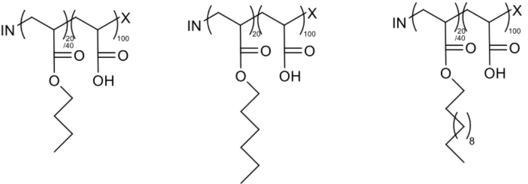
The obtained different types of block copolymers are depicted in Fig. 2.
Figure 2 The different types of polymers synthesised: poly(butyl acrylate)-b-poly(acrylic acid), poly(hexyl acrylate)-b-poly(acrylic acid), and poly(dodecyl acrylate)-b-poly(acrylic acid).
The pH-dependency of synthesized polymers was investigated via pH titration. A certain amount of polymer was dissolved in water and NaOH was added into the solution to deprotonate it completely. During the titration with 0.1 M HCl, the excess of NaOH was neutralized first and then polymer had became protonated.
The degree of deprotonation α can be defined as 𝛼 =𝑛 (𝑂𝐻
−) 𝑛(𝐴𝐴)
where 𝑛 (𝑂𝐻−) are the total moles of added NaOH and 𝑛(𝐴𝐴) the moles of acrylic acid units (not taking into consideration whether they are actually protonated or not). This value for acrylic acid/ the chargeable groups was used for the characterization of the synthesized polymers in further analysis.
Characterization of the Polymers
The above-synthesized copolymers then become analyzed with respect to their molecular build-up. In a first step, this was done by NMR on the intermediate product that still contains the t-butyl acrylate (t-Bu) units, as here the signal of the t-Bu groups yields just one sharp singlet which can be well distinguished from the signals from the alkyl groups. These measurements were done in D2-dichloromethane as solvent with a Bruker Avance II 400MHz instrument at room temperature. Taking into account the molecular formula of the corresponding building
6
blocks one can then directly calculate the relative content of hydrophobic and hydrophilic block (for details see SI). The obtained values for the six different copolymers are summarised in Table 1 and compared with the values theoretically expected from the amounts of monomer employed in the synthesis procedure. In general, we find good agreement, but especially for the case of the dodecyl acrylate containing copolymers, also a significantly higher content of this hydrophobic block compared to the expected values.
Table 1 Relative molar content of hydrophobic and hydrophilic block according to the NMR spectra compared to the theoretically expected value.
Theoretical NMR
theor. formula Hydrophobic Hydrophilic Hydrophobic Hydrophilic
nBu40-b-PAA100 29% 71% 28.77% 71,23% nBu20-b-PAA100 20% 80% 16.71% 83,29% nDo20-b-PAA100 22% 78% 16.78% 83,22% nDo40-b-PAA100 34% 66% 28.81% 71,19% nHex20-b-PAA100 18% 82% 16.82% 83,18%
The number and weight average molecular weight and weight distribution of non-hydrolysed polymers were determined via gel permeation chromatography (GPC) using THF as eluent with a flow rate of 0.5 ml per minute at 25 °C (the GPC curves are shown in the SI). The GPC is home-made with an isocratic pump and an autosampler (both from Thermo / Finnigan), a 3 μm particle diameter front & 3 μm particle diameter separation column (from PSS), as well as a refractive index detector (Wyatt Optilab DSP). The reference substance was a narrow distribution polystyrene standard.
Table 2: Theoretical molecular weight Mth, yield, number average of the molecular weight (Mn) of hydrolyzed polymer, polydispersity index (PDI) from GPC experiments, and the experimentally determined chemical formula of the produced polymers
theor. formula Mth [g/mol] Yield Mn [g/mol] PDI exp. formula
nBu40-b-tBuA100 18871 73 % 20681 1.19 nBu60-b-AA167 nBu20-b-tBuA100 15904 58 % 16896 1.15 nBu35-b-AA172 nDo20-b-tBuA100 17347 56 % 17291 1.20 nDo31-b-AA137 nDo40-b-tBuA100 23371 68 % 13497 1.28 nDo32-b-AA79 nHex20-b-tBuA100 16660 71 % 17625 1.12 nHex33-b-AA175 Further information regarding the polymer composition was obtained via pH-titration and the Mn and Mw results from GPC was shown in Table S.
7
2.2. Methods of Colloidal Characterisation
The determination of the critical micelle concentration (cmc) was done by the fluorescence method.[16] For that purpose a NaOH stock solution was used in order to adjust the degree of deprotonation as 0.2 and 1.0 (actually there was 20% excess of NaOH added for the 1.0 solutions in order to ensure complete deprotonation). The weight percentage of polymer stock solutions was 10 g/L for both the degree of deprotonation. The polymer stock solutions were then diluted to 11 different concentrations down to 0.00005 g/L using the pyrene stock solution. Steady state fluorescence spectra of pyrene were recorded with a Hitachi F-4500 Fluorescence Spectrometer at 25 °C from 350.0 to 420.0 nm after excitation at 334.0 nm. The slit width was set to 5.0 nm for both excitation and emission. For the experiments, a pyrene stock solution was prepared with a pyrene concentration of 5x10⁻⁸ mol/L.
The pH-titrations were performed at room temperature via a Titrando System with the Software tiamo™ by Metrohm.
Static light scattering (SLS) experiments were carried out with a CGS-3 (compact goniometer system) with a HeNe-Laser at 632.8 nm wavelength and using a hardware V.1.7.0 correlator ALV / LSE-5004 Light Scattering electronic and Multiple Tau Digital Correlator from ALV GmbH (Langen, Germany). Two avalanche photodiodes (APD) were used to detect the scattered light by a pseudo-cross-correlation at various angles between 40-140˚.
The SLS intensity for particles should have an angular dependence according to Guinier’s law: I(q) = I(0) • exp ( -Rg2 * q2 / 2) eq. 1
where Rg is the radius of gyration and q the modulus of the scattering vector (= 4πn0sin(θ/2)/λ; with n0 the refractive index of the solvent, θ the scattering angle, and λ the wavelength of the light). From the intensity at zero angle, I(0) one can, via the optical constant K, directly calculate the mass-averaged molecular weight Mw:
Mw = I(0) / c • K eq. 2 K = 4𝜋
2•𝑛 02•(𝑑𝑛𝑑𝑐)2
𝑁𝐴𝑣•𝜆4 eq. 3
where dn/dc is the refractive index increment and NAv the Avogadro constant.
SANS measurements were performed at PA20 from the Laboratoire Léon Brillouin (LLB, Saclay, France). Three configurations were used (1.9, 8.3 and 18.8 m sample-to-detector
8
distances) with a wavelength of 6 Å neutrons and also tested a very low q configuration at 18.8 m with 15 Å wavelength for two representative samples. In order to reach higher q, we used an off-centered detector position at the shortest detector distance, 1.9 m. Transmission values were measured in this configuration, in agreement with the existing measurement procedures (scripts) available at the instrument.
Some additional SANS measurements were performed on the V4 instrument at the Helmholtz Zentrum Berlin (HZB). The samples were measured at wavelengths of ? ̊A with sample to detector distances of ? m and collimation length of ? m respectively.
The scattering of the empty cell was subtracted as background before radial averaging and taking into account the transmissions the differential cross-sections were calculated. Subsequently the data sets obtained for the three different configurations were merged. Finally a constant background from the incoherent scattering was substracted by extrapolating the intensity at large q by Porod´s law.
3. RESULTS AND DISCUSSION
3.1 Critical Micellar Concentration (CMC)
A quantity of central importance to the self-assembly properties of amphiphilic copolymers in aqueous solution is their cmc. The cmc was determined by means of the fluorescence method where 0.05 µM pyrene was employed as a probe molecule and fluorescence spectra measured (details are described in 2.2) in the copolymer concentration range of 50 µg/L to 5 g/L are given in Fig. S. Below the cmc the pyrene has to be located within the aqueous solution, while above it will with large preference be contained in the hydrophobic core of the copolymer micelles. This leads to a change of the fluorescence spectra, as for instance seen in the changing ratio of the first and third emission maximum (I1/I3).[17] From the ratio I1/I3 as a function of concentration one can determine the cmc. An example of such a plot is given in Fig. 3 (the remaining such data sets are plotted in Figs. S). The sigmoidal shape of the data sets was described by a Boltzmann-function:
𝐼1
𝐼3= 𝐼1/3𝑓+
𝐼1/3𝑖−𝐼1/3𝑓
1+𝑒𝑥𝑝((𝑐−𝑐𝑚𝑐)/∆𝑐𝑚𝑐) eq. 4
with I1/3i and I1/3f being the initial (in pure water) and final ratio (presumably all in the micelles), c is the copolymer concentration, and Δcmc a measure for the width of the micellar transition
9
regime. The cmc then is the inflection point of these curves. The obtained cmc values are summarised in Table 2 and show only about a factor 20 difference between the most strongly hydrophobic polymer at lowest charging (40% C12, α = 0.2) and the least hydrophobic polymer at highest charging (20% C4, α = 1.0). It is interesting to note that the cmc becomes systematically lower with the increasing length of the alkyl modification but depends only little on the block length.
Figure 3 Ratio of the first and third maximum of the emission spectrum of pyrene as a function of concentration for the C4, C6, and C12 copolymer with DP = 20 and α = 0.2 (T = 25 °C). The change in Gibbs free energy (ΔGmic) is calculated from the CMC data from the following equation
𝛥𝐺𝑚𝑖𝑐 = 𝑅𝑇𝑙𝑛𝑥𝐶𝑀𝐶 eq. 5
Where 𝑥𝐶𝑀𝐶 is the critical micelle concentration expressed as mole fraction; T is the absolute temperature, and R is the gas constant.
Table 3 cmc in g/L and M, as well as the corresponding free energy of micellization ΔGmic for the different polymers at different degrees of ionization α at 25 °C.
moiety content α cmc [g/L] cmc [M] ΔGmic [kJ/mol]
C4 20 20 40 40 0.2 1.0 0.2 1.0 0.0826 0.1129 0.0260 0.0845 4.76E-06 6.51E-06 1.24E-06 4.04E-06 -40.33 -39.55 -43.66 -40.73 C6 20 20 0.2 1.0 0.0168 0.0252 9.24E-07 1.38E-06 -44.39 -43.39 C12 20 20 0.2 1.0 0.0073 0.0126 4.06E-07 7.00E-07 -46.43 -45.08
10 40 40 0.2 1.0 0.0048 0.0059 3.41E-07 4.20E-07 -46.86 -46.35
We studied polyacrylic acid-containing polymers with n-butyl, n-hexyl and n-dodecyl alkyl chain as 20% and 40%, to have a systematic variation of the hydrophobicity of polymers. Besides hydrophobicity, pH-dependency was also investigated via changing degrees of ionization α of 0.2 and 1.0 (to make sure about complete deprotonation of the polymer we worked here with an excess amount of NaOH, i.e. α was 1.2 for each fully deprotonated sample). For the structural characterization, static light scattering and small angle neutron scattering experiments were employed, which enables us to understand the mesoscopic organization of different aqueous solutions of synthesized polymer.
Static Light Scattering (SLS)
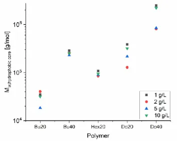
Static light scattering experiments were performed to gain a first insight into the aggregation behavior of these polymers in aqueous solution. Light scattering experiments were done for four different concentrations as 0.1 wt%, 0.2 wt%, 0.5 wt% and 1.0 wt% with fully deprotonated samples (α= 1.0) in order to investigate concentration effect on these systems. Moreover, the samples with a concentration of 0.5 wt% were also prepared for lower (a=0.2) deprotonation. SLS data was evaluated via Guinier plots for all polymers. I(0) values for all samples were obtained from these Guinier plots and Mw were calculated from these I(0) values with using refractive index increment of the polymers as 0.15 ml/g In particular, as hydrophobicity increases the calculated molecular weight of the aggregates increases as well. The obtained values are summarized in Fig. 4 and Table S1.
The obtained molecular weights for the samples with butyl (40 mol%), hexyl (20 mol%) and dodecyl (40 mol%) moieties are not affected by the concentration of the sample. On the other hand, molecular weights for the samples with butyl (20 mol%) and dodecyl (20 mol%) increase slightly with increasing concentrations. Moreover, samples with dodecyl alkyl chain modification which are the more hydrophobic ones have much higher the molecular weight values compared with other samples.
Furthermore, when the pH-dependency of the systems are compared, it can be noticed that an increase in the degree of ionization has affected on the molecular weight of those systems proportionally. Even though the molecular weights are not significantly differentiated between slightly and fully deprotonation samples, fully deprotonated samples form apparently smaller aggregates than slightly deprotonated ones.
11
Fig. 4: Molecular weight as a function of the concentration for the different polymers studied (T = 25 °C, α = 1.0; one measurement at α = 0.2, 5 g/L )
Small-Angle Neutron Scattering (SANS)
Much more detailed information regarding the structure of the micelles formed in aqueous solution can be obtained from SANS experiments, which were done for all block polymers studied and for concentrations of 0.1 wt%, 0.2 wt%, 0.5 wt%, 1.0 wt%. The degree of ionization was α = 1.0 for all concentrations and in addition α values of 0.2 and 0.5 for the 0.5 wt% sample. The experiments were performed at HZB on the instrument V4 and LLB on the instrument PA20 and the obtained scattering curves are shown in Fig. 5.
The SANS patterns for almost all samples point to the formation of self-assembled aggregates, only for Bu35-AA172 and Hex33-AA175 the scattering is so weak and from the pattern may also arise from single polymer molecules. The scattering intensity increases with increasing concentration for all samples (see Fig. S??) When comparing the scattering curves for the polymers with different length of the hydrophobic chain (Fig. ?), it is clearly seen that an increase in alkyl chain length is resulting in an increase of the scattering intensity, and the patterns at the same time are typical for globular aggregates. In other words, polymers with longer alkyl chain modification form bigger aggregates consistently. Even more obvious is the effect of the length of the hydrophobic block, where for the longer blocks much higher scattering intensities are seen. Interestingly for the shorter alkyl chain copolymers, i. e. Bu35-AA172, Bu60-AA167 and Hex33-AA175, one also sees correlation peaks from the electrostatic interaction between the aggregates. This is not seen to the same extent for the dodecyl analogues and this presumably due to the fact that the larger aggregates present there have already PAA chains of the aggregates overlapping in solution and thereby reducing the effective repulsion.
12
However, the position of the correlation peak around 0.1 nm-1 indicates a mean spacing between the aggregates of 50-60 nm, thereby corroborating the presence of aggregates with radii in the range of 20-30 nm.
Further, a comparison for fully α=1.0 and little α=0.2 deprotonated samples is also interesting to consider the pH dependency of the polymers. The similar samples at full ionization (α=1.0) show lower scattering intensities than the corresponding samples at α=0.2. The size of the aggregates is getting smaller with increasing the degrees of ionization and this is an effect well to be expected as the more charged head group of the polymeric amphiphile leads to a lower packing parameter and thereby to the formation of smaller aggregates and similar effects have been seen before.[11]
Figure 4 SANS intensity, background corrected, as a function of q for all samples. The samples signed with ** (see Table Sxx) were measured at HZB and all others were measured at LLB.
13
The results of a model free analysis were summarized in Table S2 and the key parameters molecular weight Mw is shown in Fig. 6 for the variation of the total concentration and in Fig. 7 for the variation of the degree of charging.
Fig. 6: Molecular weight as a function of the concentration for the different polymers studied
(T = 25 °C, α = 1.0).
Fig. 7: Molecular weight as a function of the degree of ionization α for the different polymers
studied (T = 25 °C, c = 5.0 g/L).
From these data it becomes evident that Bu20-PAA basically does not aggregate under these conditions and best at highest concentration some very small aggregates are formed. This is substantially changed for Bu20-PAA, where small aggregates with Nagg in the range of 7-15 are observed. Interesting the extension of the alkyl chain from butyl to hexyl does have littel effect here and for Hex20-PAA also no micellar aggregates are observed. The situation then changes
14
markedly for the dodecyl case and for Do20-PAA and Do40-PAA even for the lowest concentration and irrespective of the state of ionization globular micelles are observed. However, they are substantially larger by about a factor 8-10 in aggregation number for the longer hydrophobic block. However, this is in good agreement with the expected behaviour if the core size of the micelles is expected to scale with the length of the hydrophobic block. As a next step, the SANS data was fitted with a model of polydisperse spheres which is formed by the hydrophobic core of synthesized block copolymers. This model is described by eqs (?) and (?), ΔSLD the contrast difference in scattering length densities between the hydrocarbon block and the average of the medium (D2O), R is the radius of the aggregates and the q magnitude of the scattering vector. LogNormal distribution eq(?) was used to described the polydispersity of the scattering domains, with the width parameter s, Rm the mean radius, and the number density N(fp) which is expressed as volume fraction fp, that can defined using the eq(?) and (?). In that formula, < R3> is the third moment of the LogNorm distribution of the radii. 𝐼(𝑞, 𝑅𝑚) = 𝑁1 (𝑓𝑝) ∫ 𝐿𝑜𝑔𝑁𝑜𝑟𝑚(𝑅, 𝑁(𝑓𝑝), 𝜎, 𝑅𝑚) 𝐹(𝑞, 𝑅) 𝑑𝑅 ∞ 0 eq. 6 𝐹(𝑞, 𝑅) = (4𝜋𝑅3ΔSLD (sin(qR)−qRcos(qR)) (𝑞𝑅)3 ) 2 eq. 7 𝐿𝑜𝑔𝑁𝑜𝑟𝑚(𝑅, 𝑁(𝑓𝑝), 𝜎, 𝑅𝑚) =𝑁(𝑓𝑝) 𝑅 𝑒𝑥𝑝 ( −𝑙𝑛(𝑅/𝑅𝑚)2 2𝜎2 ) eq. 8 𝑁 1 (𝑓𝑝) = 𝑓𝑝 3 4𝜋⟨𝑅3⟩ eq. 9 ⟨𝑅3⟩ = 𝑅 𝑚3 𝑒𝑥𝑝 ( 9 2 𝜎 2) eq. 10
We assumed hydrophibilic block for the scattering length densities (SLD). Results of scattering data analysis with a model of homogenous sphere ( in Table S?) , the radius of the aggregates increases with an increasing alkyl chain length. Here it would be worth to be noted data quality is poor due to the low scattering intensities and accordingly corresponding parameters have larger errors.Also, the data with ** were measured at V4-HZB, Berlin
15
Conclusions
??
Acknowledgments
The authorsare grateful to Anja Hörrmann for data reduction, to the Helmholtz-Zentrum Berlin für Materialien und Energie GmbH (HZB, Berlin, Germany) and the Laboratoire Léon Brillouin (LLB, Saclay, France) for allocation of SANS beam time, and to Research group of Prof. Schlaad from University of Potsdam for performing the GPC measurements.
References
[1] J.-F. Gohy, “Block Copolymer Micelles BT - Block Copolymers II,” V. Abetz, Ed. Berlin, Heidelberg: Springer Berlin Heidelberg, 2005, pp. 65–136.
[2] S. Förster, V. Abetz, and A. H. E. Müller, “Polyelectrolyte Block Copolymer
Micelles,” in Polyelectrolytes with Defined Molecular Architecture II, vol. 166, 2004, pp. 173–210.
[3] K. Kataoka, A. Harada, and Y. Nagasaki, “Block copolymer micelles for drug delivery: Design, characterization and biological significance,” Adv. Drug Deliv. Rev., vol. 64, no. SUPPL., pp. 37–48, 2012.
[4] H. Cabral, K. Miyata, K. Osada, and K. Kataoka, “Block Copolymer Micelles in Nanomedicine Applications,” Chem. Rev., vol. 118, no. 14, pp. 6844–6892, 2018. [5] R. Glass, M. Möller, and J. P. Spatz, “Block copolymer micelle nanolithography,”
Nanotechnology, vol. 14, no. 10, pp. 1153–1160, 2003.
[6] H. Shen and A. Eisenberg, “Control of architecture in block-copolymer vesicles,”
Angew. Chemie - Int. Ed., vol. 39, no. 18, pp. 3310–3312, 2000.
[7] C. Fernyhough, A. J. Ryan, and G. Battaglia, “PH controlled assembly of a
polybutadiene-poly(methacrylic acid) copolymer in water: Packing considerations and kinetic limitations,” Soft Matter, vol. 5, no. 8, pp. 1674–1682, 2009.
[8] I. W. Hamley, “Nanoshells and nanotubes from block copolymers,” Soft Matter, vol. 1, no. 1, pp. 36–43, 2005.
[9] J. N. Israelachvili, D. J. Mitchell, and B. W. Ninham, “Theory of self-assembly of lipid bilayers and vesicles,” BBA - Biomembr., vol. 470, no. 2, pp. 185–201, 1977.
16
for drug delivery,” Pure Appl. Chem., vol. 76, no. 7–8, pp. 1295–1307, 2004. [11] M. Burkhardt et al., “Polyisobutylene-block-poly(methacrylic acid) diblock
copolymers: Self-assembly in aqueous media,” Langmuir, vol. 23, no. 26, pp. 12864– 12874, 2007.
[12] P. Hickl, M. Ballauff, and A. Jada, “Small-angle X-ray contrast-variation study of micelles formed by poly(styrene)-poly(ethylene oxide) block copolymers in aqueous solution,” Macromolecules, vol. 29, no. 11, pp. 4006–4014, 1996.
[13] M. E. Fox, F. C. Szoka, and J. M. J. Fréchet, “Soluble polymer carriers for the
treatment of cancer: The importance of molecular architecture,” Acc. Chem. Res., vol. 42, no. 8, pp. 1141–1151, 2009.
[14] O. Colombani, M. Ruppel, F. Schubert, H. Zettl, D. V. Pergushov, and A. H. E. Müller, “Synthesis of Poly( n -butyl acrylate)- block -poly(acrylic acid) Diblock Copolymers by ATRP and Their Micellization in Water,” Macromolecules, vol. 40, no. 12, pp. 4338–4350, Jun. 2007.
[15] P. D. Petrov, M. Drechsler, and A. H. E. Müller, “Self-assembly of asymmetric poly(ethylene oxide)-block-poly(n-butyl acrylate) diblock copolymers in aqueous media to unexpected morphologies,” J. Phys. Chem. B, vol. 113, no. 13, pp. 4218– 4225, 2009.
[16] C. Zhao, M. a Winnik, and M. D. Croucher, “Fluorescence Probe Techniques Used To Study Micelle Formation in Water-Soluble Block Copolymers,” Langmuir, vol. 6, no. 11, pp. 514–516, 1990.
[17] M. W. C. Zhao, Y. Wang, R. Xu, M. a Winnik, and M. D. Croucher, “Poly(styrene-ethylene oxide) Block Copolymer Micelle Formation in Water: A Fluorescence Probe Study’,” Macromolecules, vol. 24, no. cmc, pp. 1033–1040, 1991.
17
Supplementary Information
1. NMR Measurements
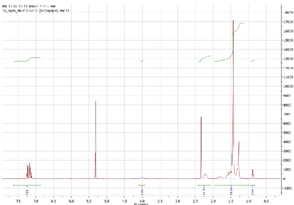
The NMR spectra for the different copolymers taken in D2-dichloro methane as solvent.
Fig S1 NMR Spectrum of nBu40-b-PAA100
18
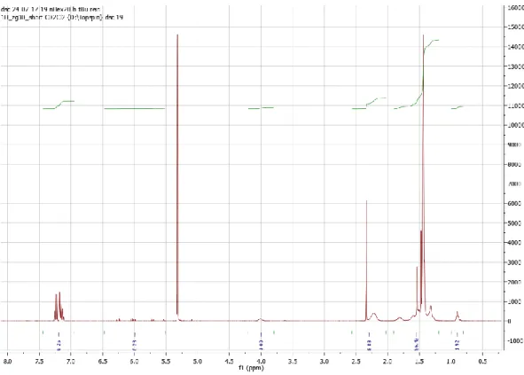
Fig S3 NMR Spectrum of nDo20-b-PAA100
19
Fig S5 NMR Spectrum of nHex20-b-PAA100
2. Gel Permeation Chromatography (GPC) Measurements
GPC measurments were done in order to determine the molecular weight of the synthesised copolymers and also to gain information regarding the pureness of the pureness of the copolymers.
Fig S6 GPC chromatograms for stat. nBu40-tBuA100 (green); nBu20-b-tBuA100 (blue); nDo20-b-tBuA100 (red); nBu40-b-nDo20-b-tBuA100 (lila); nDo40-b-nDo20-b-tBuA100 (black); nHex20-b-nDo20-b-tBuA100 (grey).
20
Table S1. GPC Results of synthesized polymers
3. pH-Titration
Fig S7 Titration curve of nBu20-b-PAA100
theor. formula Mn [g/mol] Mw [g/mol] PDI
nBu40-b-tBuA100 30200 ± 700 36000 ± 800 1.19 nBu20-b-tBuA100 26700 ± 500 30600 ± 500 1.15 nDo20-b-tBuA100 25100 ± 2400 30100 ± 2900 1.20 nDo40-b-tBuA100 18000 ± 500 23000 ± 600 1.28 nHex20-b-tBuA100 27600 ± 1700 30900 ± 1900 1.12
21
Titration curve of nBu40-b-PAA100
Fig S9. Titration curve of nHex20-b-PAA100
22
Fig S11. Titration curve of nDo40-b-PAA100
Table S2. Chain Length of the Hydrophobic and Hydrophilic part of the synthesized polymers calculated from pH-titration
theor. formula Chain Length
nBu40-b-PAA100 Hydrophobic 52 Hydrophilic 182 Total 235 nBu20-b-PAA100 Hydrophobic 30 Hydrophilic 177 Total 207 nDo20-b-PAA100 Hydrophobic 25 Hydrophilic 147 Total 172 nDo40-b-PAA100 Hydrophobic 28 Hydrophilic 87 Total 115 nHex20-b-PAA100 Hydrophobic 28 Hydrophilic 180 Total 208
23
3. Fluorescence Spectra
Obtained fluorescence spectra for the different polymers and for different degrees of ionisation are given as a function of concentration in the SI (Figs. ??)
25
Fig S12. Fluorescence spectra of pyrene in water in the presence of an increasing concentration of block copolymer samples. Concentration c of the samples given in the inset in units of g/L.
26
Fig S13. Ratio of the first and third maximum of the emission spectrum of pyrene as a function of concentration for different values of degrees of ionisation at room temperature
4. Static Light Scattering
Table S3 Parameters obtained from the static light scattering experiments. Intensity extrapolated to zero angle, I(0), the molecular weight of scattered object Mwapp, the aggregation
number , Nagg, , Mwapp,hphobic is the apperant molecular weight of the hydrophobic core, the
radius of the hydrophobic core, Rhphobic.
𝑁𝑎𝑔𝑔 = 𝑀𝑤𝑎𝑝𝑝 𝑀𝑤,𝑝𝑜𝑙𝑦𝑚𝑒𝑟 𝑀𝑤𝑎𝑝𝑝,ℎ𝑝ℎ𝑜𝑏𝑖𝑐 = 𝑁𝑎𝑔𝑔∗ 𝑀𝑤,ℎ𝑝ℎ𝑜𝑏𝑖𝑐 Rhphobic = √ 𝑉𝑝 ∗ 3 4 ∗ 𝜋 3 𝑉𝑝 =𝑀𝑤 𝑎𝑝𝑝,ℎ𝑝ℎ𝑜𝑏𝑖𝑐 𝜌 ∗ 𝑁𝑎𝑣
Static Light Scattering (SLS) c [g/L] α I(0) [1/cm] Mwapp [g/mol] Nagg Mwapp,hphobic [g/mol] Rhphobic [nm] Bu20
1 1.0 9.24E-05 5.6E+05 33.6 1.50E+05 3.78
2 1.0 1.09E-04 1.3E+05 7.9 3.53E+04 2.34
5 1.0 7.02E-04 8.6E+05 51.0 2.28E+05 4.35
10 1.0 3.50E-03 2.1E+06 127.0 5.69E+05 5.90
5 0.2 1.04E-03 1.2E+06 75.6 3.39E+05 4.96
Bu40
1 1.0 1.37E-03 8.4E+06 406.5 3.12E+06 10.40
2 1.0 7.64E-04 9.3E+05 45.4 3.48E+05 5.01
5 1.0 1.96E-03 2.4E+06 116.4 8.94E+05 6.86
10 1.0 3.74E-03 2.2E+06 110.9 8.52E+05 6.75
5 0.2 4.64E-03 5.7E+06 275.8 2.12E+06 9.14
Hex20
1 1.0 5.76E-04 3.5E+06 200.4 1.03E+06 7.19
2 1.0 3.87E-04 4.7E+05 26.9 1.39E+05 3.68
5 1.0 3.85E-04 4.7E+05 26.8 1.38E+05 3.68
10 1.0 5.69E-04 3.4E+05 19.8 1.02E+05 3.32
27
5. Small Angle Neutron Scattering (SANS)
Do20
1 1.0 1.84E-03 1.1E+07 651.5 4.85E+06 12.04
2 1.0 4.28E-04 5.2E+06 304.0 2.26E+06 9.34
5 1.0 7.51E-04 9.2E+05 53.3 3.96E+05 5.23
10 1.0 1.37E-02 8.4E+06 488.5 3.63E+06 10.94
5 0.2 8.65E-03 1.0E+07 614.5 4.57E+06 11.81
Do40
1 1.0 4.64E-03 2.8E+07 2110.3 1.62E+07 18.01
2 1.0 4.66E-03 5.7E+06 430.3 3.30E+06 10.60
5 1.0 6.70E-03 8.2E+06 609.4 4.68E+06 11.91
10 1.0 8.15E-03 5.0E+06 370.5 2.85E+06 10.09
28
Fig S14. SANS intensity, as a function of the magnitude q of the scattering vector for samples with different concentrations and α=1.0
The hydrophobic volume fraction Φ in the samples was calculated according to eq. S.. 𝜙 = 𝑥𝑚(
𝑎∗Vblock
𝑎∗Vblock + 𝑏∗Vblock) eq. (S.)
with 𝑥𝑚 the mass fraction of the polymer, a the length of the hydrophobic chain, b the length of the hydrophilic chain, Vblock the molecular volume of the corresponding block calculated from SASfit-tools.
29
Table S2 Mw, Nagg, and R were obtained from model free analysis of SANS data from LLB and HZB which is marked as **. Rg is calculated from Guinier fit.
SANS [LLB] and **[HZB] c [g/L] α Mw [g/mol] Nagg R [nm] Rg [nm] Bu20 1 1.0 3.46E+04 7.7 2.33 3.31** 2 1.0 4.03E+04 9.0 2.45 3.06 5 1.0 1.84E+04 4.1 1.89 3.22 10 1.0 3.17E+04 7.1 2.26 3.07 5 0.2 1.79E+05 39.9 4.02 4.07 5 0.5 1.17E+05 26.0 3.49 3.73** Bu40 1 1.0 2.87E+05 37.4 4.71 4.91** 2 1.0 2.53E+05 33.0 4.52 5.17 5 1.0 2.30E+05 30.0 4.38 5.23 10 1.0 2.55E+05 33.2 4.53 5.47 5 0.2 5.56E+05 72.4 5.88 6.33 5 0.5 3.35E+05 43.6 4.96 5.58 Hex20 1 1.0 1.08E+05 20.9 3.46 3.69** 2 1.0 8.48E+04 16.5 3.20 3.48 5 1.0 8.95E+04 17.4 3.25 3.86 10 1.0 9.42E+04 18.3 3.31 4.01 5 0.2 2.01E+05 39.0 4.26 4.95 5 0.5 1.27E+05 24.7 3.66 4.34 Do20 1 1.0 3.85E+05 51.8 5.48 4.39** 2 1.0 1.28E+05 17.2 3.80 4.59 5 1.0 2.16E+05 29.1 4.52 4.53 10 1.0 3.18E+05 42.8 5.14 4.73 5 0.2 3.24E+05 43.6 5.17 5.80 5 0.5 1.89E+05 25.4 4.32 4.87 Do40 1 1.0 2.48E+06 322.3 10.18 7.67** 2 1.0 8.10E+05 105.5 7.02 7.67 5 1.0 8.46E+05 110.1 7.12 7.61 10 1.0 2.21E+06 287.2 9.80 8.56 5 0.2 1.60E+06 207.7 8.79 9.27 5 0.5 1.08E+06 140.8 7.73 8.02
30
Table S?. Parameters calculated from a spherical model for the samples. Φ is the calculated volume fraction (eq S1) of the hydrophobic core, fp the volume fraction from the fit, Rm the radius of the hydrophobic core.
SANS- Simple spherical model
Moiety c [g/L ] α Φ fp/ Φ Rm Bu20 1 1.0 0.00023 0.595 1.92** Bu20 2 1.0 0.00046 0.308 3.62 Bu20 5 1.0 0.00116 0.425 2.26 Bu20 10 1.0 0.00231 0.329 2.62 Bu20 5 0.2 0.00116 0.737 3.62 Bu20 5 0.5 0.00116 0.672 3.22** Bu40 1 1.0 0.00035 0.660 4.85** Bu40 2 1.0 0.00069 0.565 4.02 Bu40 5 1.0 0.00173 0.452 5.03 Bu40 10 1.0 0.00347 0.377 5.38 Bu40 5 0.2 0.00173 0.559 5.67 Bu40 5 0.5 0.00173 0.516 4.89 Hex20 1 1.0 0.00022 0.761 3.42** Hex20 2 1.0 0.00044 0.707 3.22 Hex20 5 1.0 0.00109 0.636 3.25 Hex20 10 1.0 0.00266 0.435 3.50 Hex20 5 0.2 0.00109 0.736 3.69 Hex20 5 0.5 0.00109 0.591 3.77 Do20 1 1.0 0.00025 1.578 4.39** Do20 2 1.0 0.00050 0.643 4.85 Do20 5 1.0 0.00125 0.985 4.46 Do20 10 1.0 0.00424 0.941 4.56 Do20 5 0.2 0.00125 0.907 5.55 Do20 5 0.5 0.00125 0.861 4.75 Do40 1 1.0 0.00037 2.395 7.96** Do40 2 1.0 0.00075 0.796 7.87 Do40 5 1.0 0.00187 0.838 7.89 Do40 10 1.0 0.00568 1.229 7.56 Do40 5 0.2 0.00187 0.914 9.33 Do40 5 0.5 0.00187 0.901 8.46