RESEARCH OUTPUTS / RÉSULTATS DE RECHERCHE
Author(s) - Auteur(s) :
Publication date - Date de publication :
Permanent link - Permalien :
Rights / License - Licence de droit d’auteur :
Institutional Repository - Research Portal
Dépôt Institutionnel - Portail de la Recherche
researchportal.unamur.be
University of Namur
Brucella melitensis MucR, an orthologue of Sinorhizobium meliloti MucR, is involved in
resistance to oxidative, detergent, and saline stresses and cell envelope modifications
Mirabella, A.; Terwagne, M.; Zygmunt, M. S.; Cloeckaert, A.; De Bolle, X.; Letesson, J. J.
Published in:
Journal of Bacteriology
DOI:
10.1128/JB.01336-12
Publication date:
2013
Document Version
Publisher's PDF, also known as Version of record
Link to publication
Citation for pulished version (HARVARD):
Mirabella, A, Terwagne, M, Zygmunt, MS, Cloeckaert, A, De Bolle, X & Letesson, JJ 2013, 'Brucella melitensis
MucR, an orthologue of Sinorhizobium meliloti MucR, is involved in resistance to oxidative, detergent, and saline
stresses and cell envelope modifications', Journal of Bacteriology, vol. 195, no. 3, pp. 453-465.
https://doi.org/10.1128/JB.01336-12
General rights
Copyright and moral rights for the publications made accessible in the public portal are retained by the authors and/or other copyright owners and it is a condition of accessing publications that users recognise and abide by the legal requirements associated with these rights. • Users may download and print one copy of any publication from the public portal for the purpose of private study or research. • You may not further distribute the material or use it for any profit-making activity or commercial gain
• You may freely distribute the URL identifying the publication in the public portal ?
Take down policy
If you believe that this document breaches copyright please contact us providing details, and we will remove access to the work immediately and investigate your claim.
MucR, Is Involved in Resistance to Oxidative, Detergent, and Saline
Stresses and Cell Envelope Modifications
A. Mirabella,aM. Terwagne,aM. S. Zygmunt,b,cA. Cloeckaert,b,cX. De Bolle,aJ. J. Letessona
Unité de Recherche en Biologie Moléculaire (URBM), NARILIS, University of Namur (FUNDP), Namur, Belgiuma
; INRA, UMR1282 Infectiologie et Santé Publique, Nouzilly, Franceb
; Université François Rabelais de Tours, UMR1282 Infectiologie et Santé Publique, Tours, Francec
Brucella spp. and Sinorhizobium meliloti are alphaproteobacteria that share not only an intracellular lifestyle in their respective
hosts, but also a crucial requirement for cell envelope components and their timely regulation for a successful infectious cycle.
Here, we report the characterization of Brucella melitensis mucR, which encodes a zinc finger transcriptional regulator that has
previously been shown to be involved in cellular and mouse infections at early time points. MucR modulates the surface properties
of the bacteria and their resistance to environmental stresses (i.e., oxidative stress, cationic peptide, and detergents). We show that B.
melitensis mucR is a functional orthologue of S. meliloti mucR, because it was able to restore the production of succinoglycan in an S.
meliloti mucR mutant, as detected by calcofluor staining. Similar to S. meliloti MucR, B. melitensis MucR also represses its own
tran-scription and flagellar gene expression via the flagellar master regulator ftcR. More surprisingly, we demonstrate that MucR regulates a
lipid A core modification in B. melitensis. These changes could account for the attenuated virulence of a mucR mutant. These data
rein-force the idea that there is a common conserved circuitry between plant symbionts and animal pathogens that regulates the
relation-ship they have with their hosts.
T
he bacterial envelope is a bacterium’s first point of contact
with its challenging environment, and in the case of symbionts
or pathogens, it is the main target for antibacterial host defenses
(
1
). These multilayered structures (cytoplasmic membrane and
cell wall and possibly the outer membrane, polysaccharide
cap-sule, and proteinaceous S layer) have both protective and adaptive
functions that require tightly regulated gene expression to control
their biosynthesis and to adjust their properties in response to
changing environments (
2
–
4
).
In the alphaproteobacteria, Sinorhizobium meliloti is the
para-digmatic model for studying the crucial role surface components
play and how their production is finely tuned to respond
appro-priately to the environmental signals (e.g., carbon and nitrogen
sources, phosphate starvation, and plant signals) and various
stresses (osmolality, ionic strength, and oxidation) related to
ei-ther their free-living state or their partnership with leguminous
hosts. For example, the establishment of rhizobium-legume
symbiosis requires the timely and spatially regulated bacterial
syn-thesis (or modification) of four classes of envelope-associated
polysaccharides: outer membrane lipopolysaccharides (LPS),
periplasmic cyclic
-(1,2)-glucans and external capsular
polysac-charides (K), and the exopolysacpolysac-charides (EPS) (succinoglucan
[EPS I] and galactoglucan [EPS II]) (
5
–
7
). The regulated
produc-tion of EPS is particularly well described and involves an intricate
regulatory network (for a review, see reference
2
). Briefly, the
in-ner-membrane sensor histidine kinase ExoS and the cytoplasmic
transcriptional regulator ChvI constitute a two-component
sys-tem (TCS) that controls succinoglycan production (
8
). A third
partner, the periplasmic protein ExoR (believed to sense calcium
and ammonium), is involved in this regulatory cascade (
9
,
10
).
Both ExoR and ExoS are involved in regulating EPS I production,
and S. meliloti mutants with mutations of the genes involved in
LPS sulfatation and flagellum biosynthesis (
11
–
13
), exoR and
exoS, overproduce EPS I and are symbiotically deficient (
14
–
16
).
Finally, the zinc finger protein MucR appears to couple the two
EPS biosynthetic pathways by positively regulating succinoglycan
biosynthesis genes and repressing the synthesis of galactoglucan
(
17
–
20
). It remains to be determined how mucR becomes active,
but it has also been shown to repress flagellar-gene expression
(
21
). In addition, most of these signaling pathways are influenced
by the quorum-sensing (QS) hierarchy of S. meliloti (
22
). S.
meli-loti belongs to the order Rhizobiales in the
␣-2 subdivision of the
class Proteobacteria and, although a plant symbiont, is very closely
related to the animal pathogens belonging to the genus Brucella
(
23
). Brucella spp. are considered to be facultative intracellular
parasites that cause brucellosis, a chronic globally widespread
zoo-notic disease that affects a broad range of mammals, including
livestock and humans (
24
).
Most Brucella virulence determinants have been associated
with the bacterial surface as either permanent or transient
struc-tural components (e.g., the envelope and its appendices, the virB
type IV secretion system [
25
], the flagellum [
26
], or their
respec-tive regulators [
27
–
29
]). Sinorhizobium and Brucella not only lead
to similar intracellular chronic infections within a host-derived
membrane-bound compartment of their respective hosts (
30
),
but also share similar requirements for establishing a relationship
with their dedicated hosts (
31
). In Brucella spp., cell
envelope-associated polysaccharides and their regulated production also
Received 26 July 2012 Accepted 6 November 2012 Published ahead of print 16 November 2012
Address correspondence to J. J. Letesson, [email protected]. Supplemental material for this article may be found athttp://dx.doi.org/10.1128 /JB.01336-12.
Copyright © 2013, American Society for Microbiology. All Rights Reserved.
play a crucial role during the interaction with the host. First, the
LPS O chain is required to resist complement-mediated lysis (
32
),
avoid intracellular killing, mediate early steps in vacuolar
traffick-ing (
33
), and inhibit host cell apoptosis (
34
). Second, cyclic
glu-cans allow the bacteria to prevent phagosome-lysosome fusion
and reach their final replicative compartment (
35
). Third, the
BvrS/BvrR TCS, which is orthologous to ExoS/ChvI, is also critical
to the infectious cycle and is clearly involved in the homeostasis of
the outer membrane (OM) (
36
,
37
). Fourth, among the targets of
this TCS, which were identified by transcriptomic analysis (
38
), is
the QS regulator VjbR, which was previously demonstrated to be a
major regulator of outer-membrane organization (OM proteins,
flagellum, and type IV secretion system) (
39
). Notably, in medium
containing yeast extract, VjbR mutants have an aggregative
phe-notype that has been suggested to be linked to EPS production
(
29
). Fifth, a transcriptional regulator with 61% identity to S. meliloti
MucR was identified during a screen of Brucella melitensis transposon
mutants (
40
). The mucR transposon mutant had decreased virulence
in both cellular and mouse models of infection but was otherwise
uncharacterized. More recently, the protective efficiency of this
mu-tant was evaluated as a live attenuated vaccine (
41
).
Here, we report a detailed characterization of the
transcrip-tional regulator MucR and show that it modulates bacterial
sur-face properties and resistance to environmental stresses (i.e.,
oxi-dative stress, cationic peptide, and detergents). Using heterospecific
complementation, we show that B. melitensis mucR is a functional
orthologue of S. meliloti mucR based on its ability to restore
succi-noglycan production in the S. meliloti Rm101 mucR mutant. In
addi-tion, similar to S. meliloti, B. melitensis MucR inhibits flagellar-gene
expression via the flagellar master regulator and negatively regulates
its own transcription. More surprisingly, we demonstrate that MucR
regulates a lipid A core modification in B. melitensis. In addition to the
BvrS/BvrR TCS (
37
), this is the second transcriptional regulator of
Brucella spp. shown to modulate the lipid A core component of LPS.
Considering the strong conservation of the mucR gene in
alphapro-teobacteria and the link generally established between the gene and
altered host-bacterial interaction, it would be worthwhile to examine
LPS alterations associated with mucR mutations in other
alphapro-teobacteria.
MATERIALS AND METHODS
Strains, plasmids, and culture conditions. All strains and plasmids used
in this study are listed inTable 1.
Classically, Brucella strains were grown with shaking at 37°C in 2YT medium (10 g liter yeast extract⫺1, 16 g liter peptone⫺1, 5 g liter NaCl⫺1) containing the appropriate antibiotics from a stationary-phase overnight culture (2YT; 10 ml) back diluted to an optical density at 600 nm (OD600)
of 0.05.
For RNA extraction, 10 ml of bacteria was harvested from a 200-ml 2YT culture grown to mid-log phase (OD600⫽ 0.5). The 10-ml cultures
were used to follow GFP(ASV) (green fluorescent protein) production from B. melitensis pBBRPmucRgfp(ASV) and from B. melitensis harboring the vector pBBR-gfp(ASV). GFP(ASV) is an unstable variant of GFPmut3 and is a useful reporter gene for monitoring transient gene expression because of the reduced half-life of the reporter gene (49). Escherichia coli strains were routinely grown in Luria-Bertani (LB) medium with appro-priate antibiotics at 37°C. S. meliloti strains were cultivated in LB broth with 2.5 mM MgSO4and 2.5 mM CaCl2at 30°C. Matings were performed
by mixing E. coli S17-1 donor cells with Brucella or S. meliloti recipient strains on 2YT or LB medium, respectively, for 3 to 4 h. The mixed pop-ulation was plated on medium containing the appropriate antibiotics to select for B. melitensis and S. meliloti conjugants. Chloramphenicol,
gen-tamicin, and nalidixic acid were used at 20, 50, and 25 (8 for S. meliloti)g ml⫺1, respectively. Growth curves were monitored using a Bioscreen sys-tem (Thermo Fisher, Erembodegem-Aalst, Belgium), which continuously monitors OD600readings in a multiwell format.
Molecular techniques. DNA manipulations were performed using
standard molecular techniques (50). Restriction enzymes were purchased from Roche, and primers were purchased from Eurogentec. Primer se-quences are listed in Table S1 in the supplemental material.
Mutant construction. The B. melitensis 16M⌬mucR deletion mutant
was constructed by allelic replacement using a two-step strategy. Briefly, 500-bp upstream and downstream regions flanking the mucR gene were amplified by PCR from B. melitensis genomic DNA using the primers PmucRF and PmucRR and TmucRF and TmucRR, respectively. For each construction, a second PCR was used to join the two PCR products using the primer pairs PmucRF and TmucRR. Finally, the⌬mucR fragment was cloned into pGEM-T Easy (Promega) to generate the intermediate vector pGEMT⌬mucR. The ⌬mucR fragment was excised by NotI restriction, subcloned into the final vector pJQ200-uc1, and used to construct a
Bru-cella mutant following a previously described strategy (51). Gene replace-ment was confirmed by PCR using the following primers: mucR upstream and mucR downstream. To construct the strain⌬mucRKI, we used the same recombination strategy using a plasmid carrying the mucR gene with its upstream and downstream regions.
Construction of the complementation plasmid pBBRmucR. The
mucR gene was amplified by PCR from B. melitensis genomic DNA using
the primers (Eurogentec) mucR XhoI F and mucR ClaI R, which contain XhoI and ClaI restriction sites, respectively. The PCR product was cloned into pGEM-T Easy (Promega) to generate the intermediate vector pGEMTmucR. After sequencing, the fragment mucR was excised by a ClaI and XhoI double restriction digest and subcloned into a previously XhoI-ClaI-restricted plasmid, pBBRMCS1, to obtain pBBRmucR. The vector was then transferred to the⌬mucR strain to obtain the complemented ⌬mucR pBBRmucR strain.
Construction of the reporter plasmid pBBRPmucRgfp(ASV). The
region containing the putative mucR promoter was amplified by PCR from B. melitensis genomic DNA using the primers XhoIpmucRR and BamHIpmucRF, which contain XhoI and BamHI restriction sites, respec-tively. The PCR product was first cloned into the vector pGEM-T Easy. The fragment was then inserted in frame upstream of the promoterless
gfp(ASV) reporter gene in pBBR1MCS to generate the plasmid pBBRP-mucRgfp(ASV).
Cellular infection. Evaluation of the intracellular survival of B.
melitensis wild-type (WT) and⌬mucR strains in RAW 264.7 murine
mac-rophages was performed as previously described (52). Briefly, bacterial strains were grown overnight in 2YT medium and then inoculated at a multiplicity of infection (MOI) of 300 into cell monolayers in 24-well plates. After a 10-min centrifugation at 1,000 rpm at room temperature, the plates were placed in a 5% CO2atmosphere at 37°C for 1 h. Afterward,
the cells were washed with phosphate-buffered saline (PBS) and incu-bated in medium containing 50g ml gentamicin⫺1at 37°C under 5% CO2until the end of the infection (48 h). The cells were then washed and
lysed in sterile MilliQ water for 10 min, and serial dilutions of lysates were plated on 2YT solid medium to enumerate CFU. The data are expressed as CFU per well on a logarithmic scale.
Mouse infections. Virulence assays using BALB/c mice were
per-formed as described previously (26). Briefly, 8-week-old mice were inoc-ulated intraperitoneally with 500l of a suspension containing 4 ⫻ 104
CFU of the appropriate bacterial strain. At 1 and 4 weeks postinoculation, mice from each group were sacrificed, and spleens were collected. The spleens were homogenized in 2 ml of PBS– 0.1% Triton X-100, and serial dilutions of the homogenates were plated on 2YT solid medium to deter-mine the bacterial load. The lysis of spleen homogenates with 0.1% Triton X-100 does not affect the survival of mucR mutants, as similar results were obtained with a lysis protocol using distilled water (data not shown). The data are expressed as the log10CFU per spleen. Data were statistically
analyzed via a Mann-Whitney statistical test using the program Prism. The animal-handling and study procedures were in accordance with the current European legislation (directive 86/609/EEC) and in agreement with the corresponding Belgian law, Arrêté royal relatif à la protection des
animaux d’expérience du 6 avril 2010 publié le 14 mai 2010. The complete
protocol was reviewed and approved by the Animal Welfare Committee of the Facultés Universitaires Notre-Dame de la Paix (FUNDP), Belgium (permit number 05-558).
Oxidative-resistance assay. Oxidation resistance assays were
per-formed according to previously described protocols with some modifica-tions (53). Cells were grown overnight in 10 ml of 2YT medium with shaking and adjusted to a concentration of 5⫻ 105CFU ml⫺1in PBS.
In 96-well plates, 50l of the bacterial suspension was supplemented with 50l of H2O2(freshly diluted in PBS) at final concentrations of 1
mM, 2.5 mM, and 5 mM. A negative-control experiment was performed by adding 50l of PBS (without H2O2) to the same bacterial suspension.
After exposure for 1 h in a 37°C shaking incubator, the cells were rapidly diluted with PBS and plated onto 2YT medium. After 5 days at 37°C, CFU were enumerated, and the survival of each bacterial strain was determined as a percentage of the negative control.
Detergent sensitivity, polymyxin B sensitivity, and Congo red stain-ing. Bacteria from an overnight culture in 2YT medium were spotted on
tryptic soy broth (TSB) agar medium (Difco) containing 2% SDS, 0.1%
Triton X-100, or 0.01% Congo red in triplicate (20l per spot) and incu-bated at 37°C for 4 days. Images were captured using a Canon A430 cam-era, and the contrast and brightness of the complete image were optimized with the correction tool of PowerPoint software. Polymyxin B sensitivity was determined using an Etest containing a preformed gradient covering a continuous MIC range from 0.064 to 1,024g ml⫺1(bioMérieux).
Brucella was adjusted to an OD750of 0.109 (1 McFarland standard) in 2YT
medium. The suspension was spread onto Mueller-Hinton II (cation-adjusted) broth (BD Difco) plates using a cotton swab, and the Etest strips were then applied. The plates were incubated for 72 h at 37°C. For each strain, the MIC was determined as the concentration at which the ellipse intersects the concentration scale printed on the Etest strip. Three inde-pendent tests were performed.
Quantitative real-time reverse transcription-PCR (qRT-PCR). Total
RNA samples were prepared as previously described (39) for B. melitensis 16M, the⌬mucR mutant, and the complemented ⌬mucR pBBRmucR mu-tant. DNA was removed from the samples using DNase (Kit Fermentas), and samples were reverse transcribed with SuperScript II Reverse Trans-criptase (Invitrogen) using random oligonucleotide hexamers, as recom-mended in the manufacturer’s protocol. RNA and cDNA quantities were measured using a NanoDrop spectrophotometer (ND-1000; Thermo Fisher Scientific). The resulting cDNA samples were used as the template in real-time PCRs. Primers were designed using PrimerExpress 2.0
soft-TABLE 1 Strains and plasmids used in this study
Strain or plasmid Characteristicsa Source or reference
B. melitensis
16M WT; Nalr; parental strain A. Macmillan, Central Veterinary
Laboratory, Weybridge, UK 16M pBBR-gfp(ASV) NalrCmr; WT carrying pBBR-gfp(ASV) This study
16M pBBRPmucRgfp(ASV) NalrCmr; WT carrying pBBRPmucRgfp(ASV) This study
⌬mucR Nalr; deletion strain for mucR gene This study
⌬mucR KI Nalr; chromosomal insertion of the mucR gene in the⌬mucR strain This study
⌬mucR pBBRMCS1 NalrCmr;⌬mucR strain carrying the plasmid pBBRMCS1 This study
⌬mucR pBBRmucR NalrCmr;⌬mucR strain carrying the complementation plasmid pBBRmucR This study
⌬mucR
pBBRPmucRgfp(ASV)
NalrCmr;⌬mucR strain carrying pBBRPmucRgfp(ASV) This study
⌬mucRKI
pBBRPmucRgfp(ASV)
NalrCmr;⌬mucR KI strain carrying pBBRPmucRgfp(ASV) This study
⌬vjbR Nalr⌬vjbR::Kanr(referred to as CD100 in reference29) 27
S. meliloti
Rm1021 NalrSmr; WT 42
Rm1021::exoY Rm1021 exoY::Tn5 (formerly Rm7210); NalrSmrNeor 43
Rm2011 NalrSmr; wild type 44
Rm101 Rm2011 Spcrcassette inserted into the PmacI site of mucR 45
E. coli
DH10B F⫺mcrA⌬(mrr-hsdRMS-mcrBC) ⌽80lacZ⌬M15 ⌬lacX74 recA1 endA1 araD139
⌬(ara leu)7697 galU galK rpsL nupG ⫺(Smr)
Gibco BRL S17-1 recA thi pro hsdR⫺M⫹RP4:2-Tc:Mu:Km Tn7pir; allows plasmid mobilization 46
Plasmids
pBBR1MCS1 Broad-host-range cloning vector; high copy number; Cmr 47
pGEM-Teasy Ampr Promega
pJQ200-uc1 sacB Gtmr 48
pBBR-gfp(ASV) Cmr M. Terwagne
pJQ⌬mucR sacB Gtmrstrain containing the⌬mucR fragment; used to construct the deletion strain This study
pJQmucR sacB Gtmrstrain containing the mucR gene flanked by its upstream and downstream
regions
This study
pBBRmucR Cmr; complementation plasmid This study
pBBRPmucRgfp(ASV) Cmr; reporter plasmid bearing the transcriptional fusion PmucRgfp(ASV) This study a
Nalr
, nalidixic acid resistance; Smr
, streptomycin resistance; Neor
, neomycin resistance; Cmr
, chloramphenicol resistance; Spcr
, spectinomycin resistance; Ampr
, ampicillin resistance; Gtmr, gentamicin resistance.
ware (Applied Biosystems) to generate PCR products ranging from 80 to 100 bp and are listed in Table S2 in the supplemental material. Reactions performed without reverse transcriptase were used as negative controls to test for DNA contamination. Real-time PCRs were performed with SYBR green mix (Applied Biosystems) in 96-well optical reaction plates (Ap-plied Biosystems). Ratios were calculated using the⌬⌬CT method for each primer in an Applied Biosystems Step One Plus real-time PCR in-strument. The results for each target mRNA were normalized to 16S rRNA transcript levels and averaged.
Total extraction and SDS-proteinase K extraction of LPS. Total
ex-tracts were prepared from B. melitensis strains cultivated in 2YT medium. Bacteria were concentrated to obtain an OD600equivalent of 10 and
inac-tivated at 80°C for 1 h. Total extracts were used for SDS-proteinase K extraction of LPS as previously described (54). Samples were loaded onto a 16% or 15% polyacrylamide gel for SDS-PAGE analysis. The gels were then silver stained or transferred onto a nitrocellulose membrane (Amer-sham) for Western blotting (54). In the silver-stained gel, some contam-inating (or LPS-linked) proteins were also observed, and the most com-mon contaminants had masses of 25,000 to 27,000 Da (35).
Western blotting. Nitrocellulose membranes were blocked overnight
in PBS containing 5% nonfat dry milk. After being washed three times in PBS-0.05% Tween 20 for 10 min, the membranes were incubated for 1 h with the primary antibodies diluted in PBS (0.05% Tween 20, 1% dry milk), washed three times in PBS-0.05% Tween 20 for 10 min, incubated for 1 h with the secondary antibody diluted in PBS (0.05% Tween 20, 1% dry milk), and finally washed three times for 10 min in PBS-0.05% Tween 20. The blots were developed using an enhanced chemiluminescence (ECL) system (100 mM Tris-HCl, pH 8.5, 0.009% H2O2, 0.2 mM
cou-maric acid, and 1.25 mM luminol).
Immunodetection. Immunodetection was performed with primary
mouse monoclonal antibodies (MAbs) (undiluted hybridoma culture su-pernatant) against the O antigen of Brucella (A76/12G12/F12) (55) and against the LPS core (A68/24G12/A8 and A68/24D8/G9) (56) for LPS detection. Flagellar protein was detected using anti-FliC (diluted 1:3,000) or anti-FlgE (1:5,000) rabbit polyclonal antibodies (26). PrlR, which was used as a loading control, was detected using anti-PrlR polyclonal rabbit antibodies (1:1,000) (57). Secondary antibodies consisted of horseradish peroxidase (HRP)-conjugated goat anti-mouse IgG (Amersham catalog no. NA931; 1:10,000 dilution) and HRP-conjugated donkey anti-rabbit IgG, HRP-linked whole antibody (Amersham catalog no. NA934; 1:5,000 dilution).
Fluorescence microscopy. Bacteria were spotted onto a microscope
slide layered with a 1% agarose pad containing PBS (58). These slides were placed on a microscope stage at room temperature. The samples were observed on a Nikon i80 fluorescence microscope through a 100⫻ differ-ential interference contrast (DIC) (Nomarski) or phase-contrast objective with a Hamamatsu Orca-ER LCD camera. Image acquisition and process-ing were performed with NIS element software (Nikon).
Fluorescence-activated cell sorter (FACS) analysis. Bacterial
suspen-sions were washed in PBS and fixed in 2% paraformaldehyde (PFA), pH 7.4, at room temperature for 20 min. After an additional wash in PBS, the bacteria were used for flow cytometric analysis of GFP(ASV) production with a FACScalibur using CellQuest software (Becton Dickinson) as pre-viously described (59). Bacteria were gated according to size and scatter to eliminate debris from analysis. Then, 50,000 individual events were ex-cited with a 488-nm argon ion laser, and emission light was detected through a 530-nm bandpass filter.
Scanning electron microscopy. Bacterial aggregates of B. melitensis
overexpressing mucR (pBBRmucR) and WT B. melitensis were observed by scanning electron microscopy. The two strains were grown for 72 h in 2YT medium. Glass coverslips were treated with poly-L-lysine (0.05 mg
ml⫺1; Sigma) for 1 h at 4°C. For each strain, 1-ml cell suspensions were centrifuged onto the pretreated coverslips at 1,000 rpm for 10 min at room temperature. The medium was removed, and adherent cells were fixed for 20 min with 2% PFA in PBS. After the incubation, samples were washed
with PBS and dehydrated twice 10 min in 25, 50, 75, 85, and 100% ethanol at room temperature. Dehydrated samples were then prepared by critical-point drying (Balzer; CPD 030) and covered with a thin layer of gold (25 nm). Examinations were performed using a scanning electron microscope (Jeol 7500F) at the University of Namur, Namur, Belgium.
Statistical analysis. For the mouse experiments, we used a
Mann-Whitney test included in the program GraphPad Prism to statistically analyze our results. P values of⬍0.05 were considered to represent a significant difference.
For the cellular infections, oxidative-resistance assays, and qRT-PCR, after testing for homogeneity of variance (Bartlett test), one-way analysis of variance (ANOVA) was performed on the log10CFU per well, on the
survival percentage values, or on the⌬CT values, respectively. When needed, a Scheffe’s comparison test was performed, and statistical signif-icance at a P value of⬍0.05 was accepted.
RESULTS
MucR is required for a successful B. melitensis infection in both
RAW264.7 macrophages and BALB/c mice. To characterize the
role of mucR in virulence, we constructed a deletion mutant of
BMEI1364 (referred as
⌬mucR) in B. melitensis 16M by allelic
replacement and characterized it phenotypically. We first
ana-lyzed the ability of the newly constructed mutant to multiply
within cultured macrophages. As shown in
Fig. 1A
, the
intracel-lular bacterial load of the
⌬mucR strain was significantly reduced
at 24 h and 48 h postinfection (p.i.) in murine RAW 264.7
mac-rophages compared to the WT control. The intracellular
replica-tion of the
⌬mucR strain was almost restored to WT levels in the
complemented
⌬mucR strain. These results are consistent with
the reduced intracellular growth of a transposon mutant
de-scribed previously using the mouse macrophage-like cell line
J774.A1 (
40
). The difference between the
⌬mucR and WT strains
cannot be due to the mutant’s reduced capacity to invade the cells,
because the number of intracellular bacteria at 1 h p.i. was the
same for each strain. On the other hand, all strains displayed
the same growth rate between 24 h p.i. and 48 h p.i. (
Fig. 1A
). The
⌬mucR strain also showed reduced virulence in HeLa cells (data
not shown). Taken together, these results confirm the importance
of MucR in B. melitensis 16M intracellular infection, especially
during the first hours of infection.
To verify that, as previously described for the transposon
mu-tant (
40
), the mucR gene is involved in the successful infection
process of B. melitensis in vivo, we compared the behaviors of the
⌬mucR and WT strains in a mouse model of infection at 1 and 4
weeks p.i. (
Fig. 1B
). We observed a large reduction (more than a
2-log-unit difference) in the splenic bacterial load at just 1 week
p.i. in BALB/c mice infected with the
⌬mucR strain. Decreased
persistence of the
⌬mucR strain was also consistently observed at 4
weeks p.i. (
Fig. 1B
). Moreover, this virulence attenuation was
as-sociated with reduced splenomegaly (data not shown).
The mucR mutant enters prematurely into stationary phase
in bacterial cultures. The attenuation reported above could be
caused by an in vitro growth defect that is exacerbated in
intracel-lular environments; we therefore examined the growth rates of the
⌬mucR and WT strains in rich 2YT broth.
The
⌬mucR mutant had a growth rate similar to that of the WT
until 22 h (log phase) (
Fig. 2
). At this time point, the
⌬mucR
mutant transitioned into stationary phase earlier, more abruptly,
and at a lower bacterial density than the WT (
Fig. 2
). One possible
hypothesis is that the
⌬mucR mutant is poorly adapted to cope
rele-vant to its reduced virulence, as previously suggested for other
Brucella mutants whose adaptations upon entering stationary
phase are affected (
60
).
The mucR mutant is more sensitive to H
2O
2, detergents, and
polymyxin B. As a consequence of their intracellular lifestyle,
Bru-cella spp. have to withstand various harsh environmental
condi-tions in their phagosomal compartments within host
macro-phages, including exposure to reactive oxygen species (ROS) and
nutrient deprivation. The successful adaptation of Brucella to
these stresses has been correlated with stationary-phase
physiol-ogy (
60
). Moreover, it has been shown in other bacteria that the
limited nutrient availability associated with entry into stationary
phase triggers a general stress response that can be
cross-protec-tive against heat and oxidacross-protec-tive challenges (
61
,
62
).
We therefore evaluated the ability of the
⌬mucR mutant to
resist exposure to exogenous hydrogen peroxide (H
2O
2). After a
1-hour exposure to various concentrations of H
2O
2, the survival
rates of the
⌬mucR strain were significantly reduced compared to
the WT strain under the same conditions (
Fig. 3A
). The sensitivity
of the
⌬mucR mutant to this exogenous oxidative stress was
com-plemented in trans with the plasmid pBBRmucR (
Fig. 3A
).
We further hypothesized that a mucR mutation would generate
sensitivity to other stresses. To test this idea, we evaluated bacterial
growth on media containing detergents, including 0.1% Triton
X-100 and 2% sodium dodecyl sulfate (SDS). While these
concen-trations did not inhibit the growth of the WT strain, the
⌬mucR
strain had impaired growth/survival in the presence of both
deter-gents (
Fig. 3B
).
Furthermore, on a medium containing 0.01% Congo red, we
observed that the
⌬mucR strain retains much more of the dye,
resulting in a more intense dark-red appearance (
Fig. 3B
). All
these growth or staining phenotypes were restored by supplying
mucR either on a plasmid or via chromosomal insertion (
Fig. 3
).
Together, these observations suggest that there has been a major
cell envelope alteration that affects the susceptibility to these
com-pounds in a B. melitensis strain lacking mucR.
Because envelope defects in Brucella have been associated with
sensitivity to antimicrobial peptides, such as polymyxin B (
28
), we
used a polymyxin B Etest (bioMérieux) strip to determine the
MICs of polymyxin B for all the strains. The WT had a MIC of 64
units, whereas the MIC for
⌬mucR was 32 units. This result
con-firms the previous indications that the
⌬mucR strain has an altered
cell envelope.
The mucR mutant displays an altered LPS profile. In the
bru-cellae, LPS is a major virulence factor, and LPS alterations can
generally impede the successful infection process (
63
) and, in
par-ticular, resistance to antimicrobial compounds (
64
). Like the WT
strain, the
⌬mucR strain was smooth, as determined by crystal
violet staining (
Fig. 4
and data not shown). Nevertheless, to detect
some subtle LPS alterations, we examined the LPS migration
pat-tern and the reactivity of the
⌬mucR strain to anti-LPS MAbs
compared to the WT strain.
Figure 4A
shows the SDS-PAGE
pro-files of SDS-proteinase K-treated extracts of WT B. melitensis (lane
1) and the
⌬mucR mutant (lane 2) strains. As expected, the two
strains showed similar migration patterns for their smooth LPS
(S-LPS). Similarly, their reactivities with the anti-O-antigen MAb
A76/12G12/F12 in a Western blot were comparable (
Fig. 4B
).
FIG 1 MucR is crucial for the virulence of B. melitensis in both cellular and
mouse models of infection. (A) Intracellular replication of WT,⌬mucR, and ⌬mucR pBBRmucR B. melitensis strains in Raw 264.7 murine macrophages. The data are the average log10CFU per well. The error bars represent the
standard deviations of triplicates in one of three representative experiments. Significant differences between the strains are noted by an asterisk (P⬍ 0.05). (B) Virulence of WT and⌬mucR B. melitensis strains in the spleens of BALB/c mice. The graph represents pooled data from two independent experiments and indicates the log10CFU per spleen in each mouse. The lines indicate the
mean log10CFU for each group (n⫽ 15). Statistical analysis was performed
using the Mann-Whitney test (***, P⬍ 0.0001).
FIG 2 Growth curves of WT and⌬mucR B. melitensis strains in 2YT rich
medium. Cultures were inoculated from a preculture to an initial OD600of
0.05 in a Bioscreen plate. Bacteria were grown for 72 h with continuous shaking in 2YT medium. The optical density was measured every 30 min. The graph represents the average OD of technical triplicates for each condition from one of two representative experiments.
However, the lipid A core fraction of the
⌬mucR mutant migrated
faster than the corresponding WT fraction. This observation
sug-gests that there is a modification of the lipid A core. This
hypoth-esis was corroborated by differences in the reactivities of the
ex-tracts with anti-core MAbs A68/24G12/A8 and A68/24D8/G9
(
Fig. 4B
) in a Western blot analysis. Normal SDS profiles and
epitope detection were restored upon complementation in trans
or via chromosomal insertion of the mucR gene (
Fig. 4
).
The promoter activity of mucR is induced in 2YT medium
containing 400 mM NaCl, correlating with morphological
alter-ations of B. melitensis. We have reported previously that Brucella
spp. form bacterial aggregates over prolonged culture times when
grown in 2YT medium containing 400 mM NaCl (
57
). Because
MucR in S. meliloti regulates EPS I biosynthesis (
18
,
65
) and is
somehow involved in environmental-stress response (
Fig. 3
), we
tested whether MucR is required to form these bacterial clumps in
B. melitensis 16M. The
⌬mucR strain displayed a significant
growth defect when cultured in 2YT medium containing 400 mM
NaCl (see Fig. S2 in the supplemental material), suggesting that
MucR is required for optimal growth under hypersaline
condi-tions.
We used a B. melitensis strain carrying the reporter plasmid
pBBRPmucRgfp(ASV) (see Materials and Methods) to monitor
mucR promoter (PmucR) activity in B. melitensis at different
cul-ture times in unsupplemented 2YT medium or 2YT medium
sup-plemented with 400 mM NaCl. The B. melitensis strain harboring
the promoterless vector pBBR-gfp(ASV) was used as a negative
control. These strains displayed similar growth curves under both
conditions (
Fig. 5A
).
Figure 5C
to
H
represents the fluorescent
signal intensities measured by flow cytometry in the reporter
strain at different culture times in either 2YT medium or 2YT
medium plus 400 mM NaCl. In 2YT medium, PmucR activity is
constitutive and gives a mean fluorescent channel intensity (MFI)
of approximately 40 to 50, independent of the culture time
con-sidered (
Fig. 5
). Under hypersaline conditions (blue curve),
PmucR activity is induced beginning at 8 h p.i. (MFI
⫽ 200) and is
then strongly enhanced as the culture time increases (MFI
⫽ 300,
1,000, and 1,500 for 12, 24, and 48 h p.i., respectively). These data
were confirmed by fluorescence microscopy (
Fig. 5
, insets). The
progressive and major morphological changes of the bacteria were
most notable when grown at such salt concentrations (
Fig. 5
,
in-sets). An equatorial bulge can be distinguished after only 8 h of
growth in hypersaline 2YT (
Fig. 5D
). The subsequent increase in
the bulge in the ongoing culture results in an increase in cell size,
which is also evident in the small MFI shift for the negative control
(
Fig. 5
, compare the green curve to the violet curve). This cell
shape alteration seems to be salt specific, because it did not occur
under iso-osmolal conditions with sucrose (see Fig. S1 in the
sup-plemental material). At equivalent osmolalities, the
NaCl-supple-mented medium promotes cell shape alterations and strongly
induces PmucR activity compared to cells grown under
sucrose-supplemented conditions (see Fig. S1). However, the fluorescent
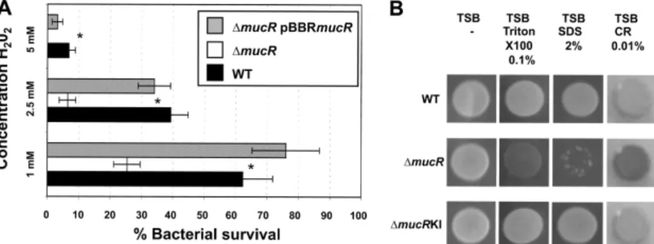
FIG 3 Sensitivity to oxidative stress and detergents. (A) Survival of WT,⌬mucR mutant, and ⌬mucR pBBRmucR B. melitensis at various concentrations of H2O2.
The data are the averages of log10CFU per well. The error bars represent the standard deviations of triplicates from one of three representative experiments.
Significant differences between the strains are indicated by asterisks (P⬍ 0.05). (B) Susceptibility of B. melitensis to surfactants and Congo red. Strains from an overnight preculture were spotted onto TSB plates containing the detergent Triton X-100 (0.1%), SDS (2%), or Congo red (CR) (0.01%) in triplicate. The plates were incubated for 4 days at 37°C. One representative spot is shown for each strain. The⌬mucR pBBRmucR strain gave the same results as the ⌬mucRKI strain (data not shown).
FIG 4 Modification of LPS in the⌬mucR strain. (A) Silver staining of SDS-proteinase K-treated extracts following electrophoresis on a 16% acrylamide gel. Lane 1, WT; lane 2,⌬mucR; lane 3, ⌬mucRKI. (B) Western blot with anti-LPS MAbs. Total extracts were separated on a 15% acrylamide gel by electrophoresis, transferred to nitrocellulose membranes, and probed using anti-O-antigen MAb A76/12G12/F12 and anti-core MAbs A68/24G12/A8 and A68/24D8/G9. Lane 1, WT; lane 2,⌬mucR; lane 3, ⌬mucRKI; lane 4, ⌬mucR pBBRMCS1; lane 5, ⌬mucR
signal for cells grown for 24 h in sucrose-supplemented 2YT
me-dium seems to be slightly higher than the signal for cells grown in
regular 2YT medium (see Fig. S1). Together, these data suggest
that the mucR promoter is induced by osmotic stress but even
more highly induced by ionic stress in B. melitensis.
MucR regulates cyclic
-glucan synthase mRNA levels. The
various susceptibilities reported above suggest that there is a
ma-jor cell envelope alteration in B. melitensis mutants lacking mucR.
Susceptibility to detergents (deoxycholic acid, SDS, and
Zwitter-gent) has been described for a cgs mutant of Brucella abortus,
FIG 5 PmucR activity is induced in B. melitensis growing in 2YT medium containing 400 mM NaCl. (A) Growth curve of the reporter [B. melitensis WT
expressing the transcriptional fusion PmucRgfp(ASV)] and the control [the WT harboring the plasmid pBBR-gfp(ASV)] strains in both 2YT medium (85.5 mM NaCl) and 2YT medium containing 400 mM NaCl. (B) Fluorescence intensity measured by flow cytometry (5⫻ 104events acquired) of WT B.
melitensis expressing the transcriptional fusion PmucRgfp(ASV) at the end of preculture in 2YT medium. The WT harboring the plasmid pBBR-gfp(ASV)
was used as a negative control. (C to H) Fluorescence intensities measured in the reporter strains growing in unsupplemented 2YT medium or 2YT medium supplemented with 314.5 mM NaCl at 2 h (C), 8 h (D), 12 h (E), 24 h (F), 48 h (G), and 72 h (H) postinoculation. The insets are phase-contrast and corresponding fluorescence images of the major morphotype of the reporter strain in 2YT medium containing 400 mM NaCl at the indicated times. Scale bar, 2m.
which was unable to produce periplasmic cyclic
-glucans (cG)
(
66
). To determine whether MucR can transcriptionally regulate
cG levels, the relative levels of cgs (the gene encoding cG
syn-thase) and cgt (encoding the c
G transporter) mRNAs were
eval-uated by qRT-PCR on RNA purified from the different strains
harvested during the exponential growth phase (OD
600⫽ 0.5) in
2YT medium. Although a mucR deletion does not affect cgt
tran-script levels, we found a nearly 2-fold reduction in cgs trantran-scripts
in the
⌬mucR mutant compared to the WT strain (
Table 2
). WT
levels of cgs mRNA were restored upon complementation with the
plasmid pBBRmucR (
Table 2
). Although additional studies are
needed, these results suggest that MucR could be the first regulator
of cG identified in B. melitensis 16M.
Restoration of EPS I production in an S. meliloti mucR
mu-tant through constitutive expression of B. melitensis mucR. The
mucR gene is well conserved within Rhizobiales, especially in S.
meliloti (61% amino acid identity), where its function has been
well studied. In S. meliloti, MucR has been described as a regulator
of both flagellar-gene expression and EPS synthesis (
21
). S.
meli-loti produces two different types of EPS: succinoglycan, which was
first described as a calcofluor-binding acidic exopolysaccharide
(EPS I), and galactoglucan (EPS II). Strains producing one versus
the other type of EPS can be rapidly discriminated on agar
me-dium containing calcofluor when placed under UV light (
65
).
Under standard laboratory conditions, both wild-type strains,
Rm1021 and Rm2011, produce EPS I and appear fluorescent on
calcofluor-containing medium, whereas non-EPS I-producing
strains do not (Rm1021 exoY:Tn5) (
Fig. 6
). An S. meliloti mucR
mutant (Rm101) forms colonies that are more mucoid and lack
the blue-green color characteristic of EPS I-producing strains (
18
,
65
) (
Fig. 6
). We observed that the constitutive expression of
mucR
Bmin an S. meliloti Rm101 background restored EPS I
pro-duction (
Fig. 6
). These data suggest that the mucR gene from B.
melitensis 16M encodes a fully functional protein that is at least
able to regulate the expression of EPS biosynthesis genes in S.
meliloti.
MucR represses flagellar-gene expression by modulating
mRNA levels. In addition to regulating EPS production, MucR
also regulates flagellar-gene expression in S. meliloti. Indeed, it has
been shown that MucR inhibits expression of rem, the flagellar
master regulator (
67
), and consequently, the expression of
rem-regulated genes (
21
). Our laboratory has previously shown that
the Brucella flagellar master regulator is orthologous to Rem (
68
).
We therefore examined the putative impact of MucR on
flagellar-gene regulation in B. melitensis 16M. Even though they are
de-scribed as nonmotile, Brucella spp. possess flagellar genes that are
expressed only in the early log phase of growth in rich medium
(
26
). Using specific antibodies against FliC (flagellin), we
exam-ined FliC protein expression at different growth phases in the
mucR mutant compared to the WT strain (
Fig. 7A
). We detected
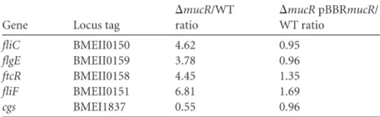
TABLE 2 MucR regulates the mRNA levels of the flagellar and cyclic
-glucan synthase genesa
Gene Locus tag
⌬mucR/WT ratio ⌬mucR pBBRmucR/ WT ratio fliC BMEII0150 4.62 0.95 flgE BMEII0159 3.78 0.96 ftcR BMEII0158 4.45 1.35 fliF BMEII0151 6.81 1.69 cgs BMEI1837 0.55 0.96 a
The relative levels of cgs, fliC, flgE, ftcR, and fliF mRNAs were determined using quantitative real-time PCR on RNA isolated from bacteria harvested at mid-exponential growth phase in rich medium (2YT). Normalization was performed using 16S rRNA. ANOVA I on⌬CT values from biological triplicates was used for statistical analysis after testing the homogeneity of variance (Bartlett test). Scheffe’s comparison test was performed, and statistical significance was obtained (at a P value of⬍0.05) between the⌬CT values of the ⌬mucR and WT or complemented strains.
FIG 6 Heterospecific complementation of an S. meliloti mucR mutant with
the mucR gene from B. melitensis on LB plates containing calcofluor. The strains Rm1021 (wild type), Rm1021 exoY:Tn5 (non-EPS I-producing strain), Rm2011 (wild type), Rm101 (mucR mutant), and Rm101 pBBRmucRBm
(mucR mutant carrying the wild-type mucR gene of B. melitensis 16M) were spotted in triplicate on LB medium containing 0.02% calcofluor and incu-bated at 30°C for 2 days before being subjected to UV light.
FIG 7 Western blot detection of the flagellar proteins FliC and FlgE. (A) Detection of
flagellin (FliC) production in B. melitensis WT,⌬fliC,and⌬mucRstrainsinbothearly log phase and stationary phase. The strains were cultivated in 2YT broth, and extracts were prepared from samples harvested at the beginning of the exponential phase of growth (OD600⫽0.2)(lanes1)andinthestationaryphase(OD600⫽1.0)(lanes2).(B
and C) FlgE (B) or FliC (C) expression in the⌬mucR mutant in stationary phase. Complementation of mucR in trans restored the WT phenotype for both FlgE and FliC. The strains were cultivated in 2YT broth, and extracts were prepared from sam-ples harvested in stationary phase (OD600⫽ 1.0). Total extracts were separated by
electrophoresis, transferred to nitrocellulose membranes, and probed with FlgE-spe-cific or FliC-speFlgE-spe-cific antiserum. A polyclonal anti-PrlR antibody was used to probe PrlR as a loading control.
FliC both at the beginning of log-phase growth, as seen in the
WT strain, and during stationary phase, when the protein is no
longer present in the WT background. Similar results were
obtained for FlgE, the hook monomer (data not shown).
Com-plementation of mucR in trans restored the WT phenotype for
both FlgE and FliC (
Fig. 7B
and
C
). This result indicates that
MucR could also be a repressor of flagellar-gene expression in
B. melitensis 16M.
To confirm this hypothesis, the relative levels of fliC (flagellin),
flgE (hook monomer), fliF (membrane and supramembrane [MS]
ring monomer), and ftcR mRNAs were determined by qRT-PCR
for RNA extracted from the different strains harvested during the
exponential growth phase (OD
600⫽ 0.5) in 2YT medium. We
found that a mucR deletion resulted in a significant increase in all
the mRNA levels tested compared to the WT stain, including
the mRNA levels corresponding to the master regulator FtcR
(
Table 2
). For each mRNA, wild-type levels were restored by
complementing the
⌬mucR deletion (
Table 2
). Together, these
results clearly indicate that B. melitensis MucR, as in S. meliloti,
is a regulator that acts upstream of the master flagellar
regula-tor FtcR, the orthologue of Rem.
MucR negatively regulates its own transcription. In S.
meli-loti, MucR negatively regulates its own transcription (
18
,
69
). To
pinpoint a potentially similar self-regulation, mucR promoter
(PmucR) activity was monitored in B. melitensis WT,
⌬mucR, and
⌬mucRKI strains. All the strains harboring the plasmid
pBBRP-mucRgfp(ASV) were grown in 2YT, and the GFP(ASV)
fluores-cence intensity was measured by flow cytometry at different time
points.
Figure 8
shows the fluorescence signals measured by flow
cytometry (left) and representative epifluorescence micrographs
(right) of the different strains in mid-log growth phase. Similar
results were obtained for each sampling time point (data not
shown). The MFI of the
⌬mucR mutant was higher (MFI ⫽ 350)
than that of the WT strain (MFI
⫽ 45) or the corresponding
com-plemented
⌬mucRKI mutant (MFI ⫽ 30) (
Fig. 8
, left). The B.
melitensis strain harboring the vector pBBR-gfp(ASV) was used as
a negative control. PmucR is strongly induced when Brucella lacks
the mucR gene, indicating that MucR negatively regulates its own
transcription in B. melitensis 16M.
DISCUSSION
In this study, we report the extensive characterization of an
in-frame deletion mutant of B. melitensis mucR. This gene was
pre-viously identified as necessary for virulence in a transposon
mu-tagenesis screen of B. melitensis but never characterized (
40
). Our
study has confirmed that a
⌬mucR mutant is attenuated in both
cellular and murine models of infection. Our study has also shown
that the most plausible explanation for its attenuation is a
defi-ciency in intracellular survival, rather than a defidefi-ciency in cellular
invasion.
In addition to reduced virulence, the
⌬mucR strain exhibits
pleiotropic complementable phenotypes, which we discuss here
and tentatively correlate with the attenuation of the
⌬mucR strain.
B. melitensis MucR is a functional orthologue of S. meliloti
MucR. The mucR gene is highly conserved in Rhizobiales (
70
) and
encodes a protein predicted to contain a C
2H
2-type zinc finger
motif. Zinc finger-containing proteins include DNA binding
pro-teins that are able to bind a zinc ion via a conserved structure (e.g.,
through cysteine and histidine amino acids) (
71
). In all the
bacte-ria in which MucR orthologues have been characterized, this
regulator controls various cell envelope modifications with a
com-mon theme of exopolysaccharide synthesis and altered
host-bac-terial interaction (
72
–
74
). In S. meliloti, where MucR (61%
iden-tity with B. melitensis MucR) has been the most extensively
studied, the regulator couples motility regulation with EPS
pro-duction (
21
). The S. meliloti MucR protein appears to be highly
specific for its own DNA recognition sequence, because it does not
bind to sites recognized by Ros, the orthologous regulator in
Agro-bacterium tumefaciens (61% identity to B. melitensis MucR) (
69
,
75
). In contrast, as shown on calcofluor plates (
Fig. 6
), the
consti-tutive expression of the B. melitensis mucR gene in a mucR mutant
of S. meliloti (Rm101) was able to restore EPS I (succinoglycan)
production. This heterospecific-complementation experiment
suggests that the mucR gene of B. melitensis 16M encodes a
func-tional protein able to recognize MucR-specific
promoter-target-ing sequences, or at least the promoter(s) regulatpromoter-target-ing the
expres-sion of EPS biosynthesis genes in S. meliloti.
B. melitensis MucR also controls flagellar-gene expression
and the formation of the aggregative phenotype. Our results
FIG 8 MucR negatively autoregulates its own transcription. (Left) Fluorescence intensity measured by flow cytometry (5⫻ 104events acquired) on B. melitensis
WT,⌬mucR, and ⌬mucRKI strains expressing the transcriptional fusion PmucRgfp(ASV) in mid-log-phase culture in 2YT medium. The WT harboring the plasmid pBBR-gfp(ASV) was used as a negative control. (Right) DIC (above) and corresponding fluorescence (bottom) images. (A) WT pBBR-gfp(ASV). (B) WT pBBRPmucRgfp(ASV). (C)⌬mucR pBBRPmucRgfp(ASV). (D) ⌬mucRKI pBBRPmucRgfp(ASV). Scale bars, 3 m.