HAL Id: tel-03163854
https://tel.archives-ouvertes.fr/tel-03163854
Submitted on 9 Mar 2021HAL is a multi-disciplinary open access archive for the deposit and dissemination of sci-entific research documents, whether they are pub-lished or not. The documents may come from teaching and research institutions in France or abroad, or from public or private research centers.
L’archive ouverte pluridisciplinaire HAL, est destinée au dépôt et à la diffusion de documents scientifiques de niveau recherche, publiés ou non, émanant des établissements d’enseignement et de recherche français ou étrangers, des laboratoires publics ou privés.
Tomosynthesis
Maissa Sghaier
To cite this version:
Maissa Sghaier. Clinical-task based reconstruction in Digital Breast Tomosynthesis. Signal and Image processing. Université Paris-Saclay, 2020. English. �NNT : 2020UPASG040�. �tel-03163854�
❞✉ s❡✐♥ ▼♦ts ❝❧és✿ ❉ét❡❝t❛❜✐❧✐té✱ ❖♣t✐♠✐s❛t✐♦♥✱ ❘❡❝♦♥str✉❝t✐♦♥ ❞✬✐♠❛❣❡s ✸❉✱ Pr♦❜❧è♠❡ ✐♥✈❡rs❡✱ ❚♦✲ ♠♦s②♥t❤ès❡ ♥✉♠ér✐q✉❡ ❞✉ s❡✐♥✱ ❱❛r✐❛t✐♦♥ ❚♦t❛❧❡ ❘és✉♠é✿ ▲❛ r❡❝♦♥str✉❝t✐♦♥ ❡♥ t♦♠♦s②♥t❤ès❡ ♥✉♠ér✐q✉❡ ❞✉ s❡✐♥ ❡st ❝♦♥s✐❞éré❡ ❝♦♠♠❡ ✉♥ ♣r♦❜❧è♠❡ ✐♥✈❡rs❡✱ ♣♦✉r ❧❡q✉❡❧ ❧❡s ♠ét❤♦❞❡s ✐tér❛t✐✈❡s ré❣✉❧❛r✐sé❡s ♣❡r♠❡tt❡♥t ❞❡ ❢♦✉r♥✐r ✉♥❡ ❜♦♥♥❡ q✉❛❧✐té ❞✬✐♠❛❣❡✳ ❇✐❡♥ q✉❡ ❧❛ tâ❝❤❡ ❝❧✐♥✲ ✐q✉❡ ❥♦✉❡ ✉♥ rô❧❡ ❝r✉❝✐❛❧ ❧♦rs ❞❡ ❧✬❡①❛♠❡♥ ❞❡s ✐♠❛❣❡s ♣❛r ❧❡ r❛❞✐♦❧♦❣✉❡✱ ❡❧❧❡ ♥✬❛ ♣❛s été ❥✉sq✉✬à ♣rés❡♥t ❞✐r❡❝t❡♠❡♥t ♣r✐s❡ ❡♥ ❝♦♠♣t❡ ❞❛♥s ❧❡ ♣r♦❝❡ss✉s ❞❡ r❡❝♦♥str✉❝t✐♦♥ ❞❡s ✐♠❛❣❡s ❞❡ t♦✲ ♠♦s②♥t❤ès❡✳ ❉❛♥s ❝❡tt❡ t❤ès❡✱ ♥♦✉s ✐♥tr♦✲ ❞✉✐s♦♥s ✉♥❡ ♥♦✉✈❡❧❧❡ ❢♦r♠✉❧❛t✐♦♥ ✈❛r✐❛t✐♦♥❡❧❧❡ ❞❡ ❧❛ r❡❝♦♥str✉❝t✐♦♥ ❞❡s ✐♠❛❣❡s ❡♥ t♦♠♦s②♥t❤ès❡ ♥✉♠ér✐q✉❡ ❞✉ s❡✐♥ q✉✐ ✐♥tè❣r❡ ❧❛ tâ❝❤❡ ❝❧✐♥✐q✉❡ ❞✉ r❛❞✐♦❧♦❣✉❡✱ ♥♦t❛♠♠❡♥t ❧❛ ❞ét❡❝t✐♦♥ ❞❡s ♠✐✲ ❝r♦❝❛❧❝✐✜❝❛t✐♦♥s✳ ▲❡ ❜✉t ❞❡ ❝❡tt❡ ❛♣♣r♦❝❤❡ ❡st ❞❡ ♣❡r♠❡ttr❡ à ❧❛ ❢♦✐s ❧❡ r❡❤❛✉ss❡♠❡♥t ❞❡ ❧❛ ❞é✲ t❡❝t❛❜✐❧✐té ❞❡s ♠✐❝r♦❝❛❧❝✐✜❝❛t✐♦♥s ❡t ✉♥❡ r❡st❛✉✲ r❛t✐♦♥ ❞❡ ❜♦♥♥❡ q✉❛❧✐té ❞❡s t✐ss✉s ♠❛♠♠❛✐r❡s✳ ❚♦✉t ❞✬❛❜♦r❞✱ ♥♦✉s ♣r♦♣♦s♦♥s ✉♥❡ ♥♦✉✈❡❧❧❡ ❛♣♣r♦❝❤❡ q✉✐ ✈✐s❡ à r❡❤❛✉ss❡r ❧❛ ❞ét❡❝t❛❜✐❧✲ ✐té ❞❡s ♠✐❝r♦❝❛❧❝✐✜❝❛t✐♦♥s✳ ◆♦✉s ❢♦r♠✉✲ ❧♦♥s ✉♥❡ ♥♦✉✈❡❧❧❡ ❢♦♥❝t✐♦♥ ❞❡ ❞ét❡❝t❛❜✐❧✐té ✐♥✲ s♣✐ré❡ ❞✬♦❜s❡r✈❛t❡✉rs ♠❛t❤é♠❛t✐q✉❡s✳ ◆♦✉s ❧✬✐♥té❣r♦♥s✱ ♣❛r ❧❛ s✉✐t❡✱ ❞❛♥s ✉♥❡ ❢♦♥❝t✐♦♥ ♦❜❥❡❝✲ t✐❢ ♠✐♥✐♠✐sé❡ ♣❛r ✉♥ ❛❧❣♦r✐t❤♠❡ ❞❡ r❡❝♦♥str✉❝✲ t✐♦♥ ❞é❞✐é✳ ◆♦✉s ♠♦♥tr♦♥s ✜♥❛❧❡♠❡♥t ❧✬✐♥térêt ❞❡ ♥♦tr❡ ❛♣♣r♦❝❤❡ à ❧✬é❣❛r❞ ❞❡s ♠ét❤♦❞❡s st❛♥✲ ❞❛r❞s ❞❡ r❡❝♦♥str✉❝t✐♦♥✳ ❉❛♥s ✉♥❡ ❞❡✉①✐è♠❡ ♣❛rt✐❡✱ ♥♦✉s ✐♥tr♦❞✉✐s♦♥s ✉♥❡ ♥♦✉✈❡❧❧❡ ré❣✉❧❛r✐s❛t✐♦♥✱ ❙♣❛t✐❛❧❧② ❆❞❛♣t✐✈❡ ❚♦t❛❧ ❱❛r✐❛t✐♦♥ ✭❙❆❚❱✮✱ ❡♥ ❝♦♠♣❧é♠❡♥t ❞❡ ❧❛ ❢♦♥❝t✐♦♥ ❞❡ ❞ét❡❝t❛❜✐❧✐té ❞❛♥s ❧❡ ♣r♦❜❧è♠❡ ❞❡ r❡✲ ❝♦♥str✉❝t✐♦♥ ❡♥ t♦♠♦s②♥t❤ès❡✳ ◆♦✉s ♣r♦♣♦s♦♥s ✉♥❡ ❢♦r♠✉❧❛t✐♦♥ ♦r✐❣✐♥❛❧❡ ♦ù ❧✬♦♣ér❛t❡✉r ❞❡ ❣r❛✲ ❞✐❡♥t ❡st r❡♠♣❧❛❝é ♣❛r ✉♥ ♦♣ér❛t❡✉r ❛❞❛♣t❛t✐❢ ❛♣♣❧✐q✉é à ❧✬✐♠❛❣❡ q✉✐ ✐♥❝♦r♣♦r❡ ❡✣❝❛❝❡♠❡♥t ❧❛ ❝♦♥♥❛✐ss❛♥❝❡ ❛ ♣r✐♦r✐ r❡❧❛t✐✈❡ à ❧❛ ❧♦❝❛❧✐s❛✲ t✐♦♥ ❞❡ ♣❡t✐ts ♦❜❥❡ts✳ ❊♥s✉✐t❡✱ ♥♦✉s ❞ér✐✈♦♥s ♥♦tr❡ ré❣✉❧❛r✐s❛t✐♦♥ ❙❆❚❱ ❡t ❧✬✐♥té❣r♦♥s ❞❛♥s ✉♥❡ ♥♦✉✈❡❧❧❡ ❛♣♣r♦❝❤❡ ❞❡ r❡❝♦♥str✉❝t✐♦♥✳ ▲❡s ré✲ s✉❧t❛ts ❡①♣ér✐♠❡♥t❛✉① ♠♦♥tr❡♥t q✉❡ ❙❆❚❱ ❡st ✉♥❡ ♣✐st❡ ♣r♦♠❡tt❡✉s❡ ♣♦✉r ❛♠é❧✐♦r❡r ❧❡s ♠ét❤✲ ♦❞❡s ❞❡ ré❣✉❧❛r✐s❛t✐♦♥ ❞❡ ❧✬ét❛t ❞❡ ❧✬❛rt✳ ❉❛♥s ✉♥❡ tr♦✐s✐è♠❡ ♣❛rt✐❡✱ ♥♦✉s ét✉❞✐♦♥s ❧✬❛♣♣❧✐❝❛t✐♦♥ ❞❡ ❧✬❛❧❣♦r✐t❤♠❡ ❞❡ ▼❛❥♦r❛t✐♦♥✲ ▼✐♥✐♠✐s❛t✐♦♥ à ▼é♠♦✐r❡ ❞❡ ●r❛❞✐❡♥t ✭✸▼●✮ à ♥♦tr❡ ♣r♦❜❧è♠❡ ❞❡ r❡❝♦♥str✉❝t✐♦♥✳ ❉❛♥s ❧❡ ❜✉t ❞✬❛❝❝r♦îtr❡ s❛ ✈✐t❡ss❡ ❞❡ ❝♦♥✈❡r❣❡♥❝❡✱ ♥♦✉s ♣r♦♣♦s♦♥s ❞❡✉① ❛♠é❧✐♦r❛t✐♦♥s ♥✉♠ér✐q✉❡s✳ ❉ès ❧♦rs✱ ❧❡s ♣❡r❢♦r♠❛♥❝❡s ♥✉♠ér✐q✉❡s s♦♥t é✈❛❧✲ ✉é❡s ❡♥ ❝♦♠♣❛r❛♥t ❧❛ ✈✐t❡ss❡ ❞❡ ❝♦♥✈❡r❣❡♥❝❡ ❞❡ ❧❛ ♠ét❤♦❞❡ ♣r♦♣♦sé❡ ❛✈❡❝ ❝❡❧❧❡s ❞✬❛❧❣♦r✐t❤♠❡s ❞✬♦♣t✐♠✐s❛t✐♦♥ ❝♦♥✈❡①❡ ❝♦♥❝✉rr❡♥ts✳ ▲❛ ❞❡r♥✐èr❡ ♣❛rt✐❡ ❞❡ ❧❛ t❤ès❡ ♣♦rt❡ s✉r ❧✬é✈❛❧✉❛t✐♦♥ q✉❛♥t✐t❛t✐✈❡ ❞❡s ❝♦♥tr✐❜✉t✐♦♥s ❞❡ ❧✬❛♣♣r♦❝❤❡ ❞❡ r❡❝♦♥str✉❝t✐♦♥ ♣r♦♣♦sé❡ ❡♥ t♦✲ ♠♦s②♥t❤ès❡ ♥✉♠ér✐q✉❡ ❞✉ s❡✐♥✳ ◆♦✉s ♠❡♥♦♥s ✉♥❡ ét✉❞❡ ❞❡ ❧❡❝t✉r❡ ❞✬✐♠❛❣❡s ✐♠♣❧✐q✉❛♥t q✉❛✲ t♦r③❡ ❧❡❝t❡✉rs ❞♦♥t ♥❡✉❢ r❛❞✐♦❧♦❣✉❡s ❛✈❡❝ ❞✐❢✲ ❢ér❡♥ts ♥✐✈❡❛✉① ❞✬❡①♣❡rt✐s❡ ❡t ❝✐♥q ❡①♣❡rts ❡♥ ♠❛♠♠♦❣r❛♣❤✐❡ ❞❡ ●❊ ❍❡❛❧t❤❝❛r❡✳ ▲❡s rés✉❧t❛ts ❞é♠♦♥tr❡♥t ❧✬✐♥térêt ❞❡ ♥♦tr❡ ❛♣♣r♦❝❤❡ ❞❡ r❡✲ ❝♦♥str✉❝t✐♦♥ ♣❛r r❛♣♣♦rt à ❧✬❛♣♣r♦❝❤❡ st❛♥❞❛r❞ ♥♦♥✲ré❣✉❧❛r✐sé❡ s❡❧♦♥ ❞❡s ❝r✐tèr❡s ✈✐s✉❡❧s s♣é❝✐✲ ✜q✉❡s✳
❚✐t❧❡✿ ❈❧✐♥✐❝❛❧✲❚❛s❦ ❜❛s❡❞ r❡❝♦♥str✉❝t✐♦♥ ✐♥ ❉✐❣✐t❛❧ ❇r❡❛st ❚♦♠♦s②♥t❤❡s✐s ❑❡②✇♦r❞s✿ ✸❉ ✐♠❛❣❡ r❡❝♦♥str✉❝t✐♦♥✱ ❉❡t❡❝t❛❜✐❧✐t②✱ ❉✐❣✐t❛❧ ❇r❡❛st ❚♦♠♦s②♥t❤❡s✐s✱ ■♥✈❡rs❡ ♣r♦❜✲ ❧❡♠✱ ❖♣t✐♠✐③❛t✐♦♥✱ ❚♦t❛❧ ❱❛r✐❛t✐♦♥ ❆❜str❛❝t✿ ❚❤❡ r❡❝♦♥str✉❝t✐♦♥ ♦❢ ❛ ✈♦❧✉♠❡t✲ r✐❝ ✐♠❛❣❡ ❢r♦♠ ❉✐❣✐t❛❧ ❇r❡❛st ❚♦♠♦s②♥t❤❡s✐s ✭❉❇❚✮ ♠❡❛s✉r❡♠❡♥ts ✐s ❛♥ ✐❧❧✲♣♦s❡❞ ✐♥✈❡rs❡ ♣r♦❜❧❡♠✱ ❢♦r ✇❤✐❝❤ ❡①✐st✐♥❣ ✐t❡r❛t✐✈❡ r❡❣✉❧❛r✐③❡❞ ❛♣♣r♦❛❝❤❡s ❝❛♥ ♣r♦✈✐❞❡ ❛ ❣♦♦❞ s♦❧✉t✐♦♥✳ ❍♦✇✲ ❡✈❡r✱ t❤❡ ❝❧✐♥✐❝❛❧ t❛s❦ ✐s s♦♠❡❤♦✇ ♦♠✐tt❡❞ ✐♥ t❤❡ ❞❡r✐✈❛t✐♦♥ ♦❢ t❤♦s❡ t❡❝❤♥✐q✉❡s✱ ❛❧t❤♦✉❣❤ ✐t ♣❧❛②s ❛ ♣r✐♠❛r② r♦❧❡ ✐♥ t❤❡ r❛❞✐♦❧♦❣✐st ❞✐❛❣♥♦s✐s✳ ■♥ t❤✐s ✇♦r❦✱ ✇❡ ❛❞❞r❡ss t❤✐s ✐ss✉❡ ❜② ✐♥tr♦❞✉❝✐♥❣ ❛ ♥♦✈❡❧ ✈❛r✐❛t✐♦♥❛❧ ❢♦r♠✉❧❛t✐♦♥ ❢♦r ❉❇❚ r❡❝♦♥✲ str✉❝t✐♦♥✳ ❖✉r ❛♣♣r♦❛❝❤ ✐s t❛✐❧♦r❡❞ ❢♦r ❛ s♣❡✲ ❝✐✜❝ ❝❧✐♥✐❝❛❧ t❛s❦✱ ♥❛♠❡❧② t❤❡ ❞❡t❡❝t✐♦♥ ♦❢ ♠✐❝r♦✲ ❝❛❧❝✐✜❝❛t✐♦♥s✳ ❖✉r ♠❡t❤♦❞ ❛✐♠s ❛t s✐♠✉❧t❛♥❡✲ ♦✉s❧② ❡♥❤❛♥❝✐♥❣ t❤❡ ❞❡t❡❝t✐♦♥ ♣❡r❢♦r♠❛♥❝❡ ❛♥❞ ❡♥❛❜❧✐♥❣ ❛ ❤✐❣❤✲q✉❛❧✐t② r❡st♦r❛t✐♦♥ ♦❢ t❤❡ ❜❛❝❦✲ ❣r♦✉♥❞ ❜r❡❛st t✐ss✉❡s✳ ❋✐rst✱ ✇❡ ♣r♦♣♦s❡ ❛♥ ♦r✐❣✐♥❛❧ ❛♣♣r♦❛❝❤ ❛✐♠✐♥❣ ❛t ❡♥❤❛♥❝✐♥❣ t❤❡ ❞❡t❡❝t❛❜✐❧✐t② ♦❢ ♠✐❝r♦❝❛❧❝✐✜❝❛✲ t✐♦♥s ✐♥ ❉❇❚ r❡❝♦♥str✉❝t✐♦♥✳ ❚❤✉s✱ ✇❡ ❢♦r♠✉✲ ❧❛t❡ ❛ ❞❡t❡❝t❛❜✐❧✐t② ❢✉♥❝t✐♦♥ ✐♥s♣✐r❡❞ ❢r♦♠ ♠❛t❤✲ ❡♠❛t✐❝❛❧ ♠♦❞❡❧ ♦❜s❡r✈❡rs✳ ❚❤❡♥✱ ✇❡ ✐♥t❡❣r❛t❡ ✐t ✐♥ ❛ ❝♦st ❢✉♥❝t✐♦♥ ✇❤✐❝❤ ✐s ♠✐♥✐♠✐③❡❞ ❢♦r ✸❉ r❡❝♦♥str✉❝t✐♦♥ ♦❢ ❉❇❚ ✈♦❧✉♠❡s✳ ❊①♣❡r✐♠❡♥t❛❧ r❡s✉❧ts ❞❡♠♦♥str❛t❡ t❤❡ ✐♥t❡r❡st ♦❢ ♦✉r ❛♣♣r♦❛❝❤ ✐♥ t❡r♠s ♦❢ ♠✐❝r♦❝❛❧❝✐✜❝❛t✐♦♥ ❞❡t❡❝t❛❜✐❧✐t②✳ ■♥ ❛ s❡❝♦♥❞ ♣❛rt✱ ✇❡ ✐♥tr♦❞✉❝❡ t❤❡ ❙♣❛t✐❛❧❧② ❆❞❛♣t✐✈❡ ❚♦t❛❧ ❱❛r✐❛t✐♦♥ ✭❙❆❚❱✮ ❛s ❛ ♥❡✇ r❡❣✲ ✉❧❛r✐③❛t✐♦♥ str❛t❡❣② ❛♣♣❧✐❡❞ t♦ ❉❇❚ r❡❝♦♥str✉❝✲ t✐♦♥✱ ✐♥ ❛❞❞✐t✐♦♥ t♦ t❤❡ ❞❡t❡❝t❛❜✐❧✐t② ❢✉♥❝t✐♦♥✳ ❍❡♥❝❡✱ ❛♥ ♦r✐❣✐♥❛❧ ❢♦r♠✉❧❛t✐♦♥ ❢♦r t❤❡ ✇❡✐❣❤t❡❞ ❣r❛❞✐❡♥t ✜❡❧❞ ✐s ✐♥tr♦❞✉❝❡❞✱ t❤❛t ❡✣❝✐❡♥t❧② ✐♥✲ ❝♦r♣♦r❛t❡s ♣r✐♦r ❦♥♦✇❧❡❞❣❡ ♦♥ t❤❡ ❧♦❝❛t✐♦♥ ♦❢ s♠❛❧❧ ♦❜❥❡❝ts✳ ❚❤❡♥✱ ✇❡ ❞❡r✐✈❡ ♦✉r ❙❆❚❱ r❡❣✉✲ ❧❛r✐③❛t✐♦♥✱ ❛♥❞ ✐♥❝♦r♣♦r❛t❡ ✐t ✐♥ ♦✉r ♣r♦♣♦s❡❞ ✸❉ r❡❝♦♥str✉❝t✐♦♥ ❛♣♣r♦❛❝❤ ❢♦r ❉❇❚✳ ❲❡ ❝❛rr② ♦✉t s❡✈❡r❛❧ ❡①♣❡r✐♠❡♥ts✱ ✐♥ ✇❤✐❝❤ ❙❆❚❱ r❡❣✉❧❛r✐③❡r s❤♦✇s ❛ ♣r♦♠✐s✐♥❣ ✐♠♣r♦✈❡♠❡♥t ✇✐t❤ r❡s♣❡❝t t♦ st❛t❡✲♦❢✲t❤❡✲❛rt r❡❣✉❧❛r✐③❛t✐♦♥ ♠❡t❤♦❞s✳ ❚❤✐r❞✱ ✇❡ ✐♥✈❡st✐❣❛t❡ t❤❡ ❛♣♣❧✐❝❛t✐♦♥ ♦❢ ▼❛✲ ❥♦r✐③❡ ▼✐♥✐♠✐③❡ ▼❡♠♦r② ●r❛❞✐❡♥t ✭✸▼●✮ ❛❧❣♦✲ r✐t❤♠ t♦ ♦✉r ♣r♦♣♦s❡❞ r❡❝♦♥str✉❝t✐♦♥ ❛♣♣r♦❛❝❤✳ ❚❤✉s✱ ✇❡ s✉❣❣❡st t✇♦ ♥✉♠❡r✐❝❛❧ ✐♠♣r♦✈❡♠❡♥ts t♦ ❜♦♦st t❤❡ s♣❡❡❞ ♦❢ t❤❡ r❡❝♦♥str✉❝t✐♦♥ s❝❤❡♠❡✳ ❚❤❡♥✱ ✇❡ ❛ss❡ss t❤❡ ♥✉♠❡r✐❝❛❧ ♣❡r❢♦r♠❛♥❝❡ ♦❢ ✸▼● ❜② ❝♦♠♣❛r✐♥❣ t❤❡ ❝♦♥✈❡r❣❡♥❝❡ s♣❡❡❞ ♦❢ t❤❡ ♣r♦♣♦s❡❞ ♠❡t❤♦❞ ✇✐t❤ st❛t❡✲♦❢✲t❤❡✲❛rt ❝♦♥✲ ✈❡① ♦♣t✐♠✐③❛t✐♦♥ ❛❧❣♦r✐t❤♠s✳ ❚❤❡ ❧❛st ♣❛rt ♦❢ t❤✐s t❤❡s✐s ✐s ❢♦❝✉s❡❞ ♦♥ t❤❡ q✉❛♥t✐t❛t✐✈❡ ❛ss❡ss♠❡♥t ♦❢ t❤❡ ❝♦♥tr✐❜✉t✐♦♥ ♦❢ ♦✉r ♣r♦♣♦s❡❞ ❉❇❚ r❡❝♦♥str✉❝t✐♦♥✳ ❚❤✉s✱ ✇❡ ❝♦♥❞✉❝t ❛ ✈✐s✉❛❧ ❡①♣❡r✐♠❡♥t tr✐❛❧ ✐♥✈♦❧✈✐♥❣ ❢♦✉r✲ t❡❡♥ r❡❛❞❡rs ✐♥❝❧✉❞✐♥❣ ♥✐♥❡ r❛❞✐♦❧♦❣✐sts ✇✐t❤ ❞✐❢✲ ❢❡r❡♥t ❧❡✈❡❧s ♦❢ ❡①♣❡rt✐s❡ ❛♥❞ ✜✈❡ ●❊ ❍❡❛❧t❤❝❛r❡ ❡①♣❡rts ✐♥ ♠❛♠♠♦❣r❛♣❤②✳ ❆❝❝♦r❞✐♥❣ t♦ s♣❡❝✐✜❝ ✈✐s✉❛❧ ❝r✐t❡r✐❛✱ t❤❡ r❡s✉❧ts s❤♦✇ t❤❡ s✉♣❡r✐♦r✐t② ♦❢ ♦✉r ♣r♦♣♦s❡❞ r❡❝♦♥str✉❝t✐♦♥ ❛♣♣r♦❛❝❤ ♦✈❡r t❤❡ st❛♥❞❛r❞ ♥♦♥✲r❡❣✉❧❛r✐③❡❞ ❧❡❛st sq✉❛r❡s s♦✲ ❧✉t✐♦♥✳ Université Paris-Saclay
Espace Technologique / Immeuble Discovery
Muqaddimah: An Introduction to History.
“We see with our brains, not with our eyes,” Norman
Doidge, The Brain That Changes Itself: Stories of Personal Triumph from the Frontiers of Brain Science
“If you hear a voice within you say ‘you cannot paint,’ then by all means paint, and that voice will be si-lenced,” Vincent Van Gogh
“Happy is the man who can with vigorous wing Mount to those luminous serene fields,
The man whose thoughts, like larks,
Take liberated flight toward the morning skies
–Who hovers over life and understands without effort The language of flowers and voiceless things!”
Charles Beaudelaire, Élévation (les fleurs du mal, 1857)
This PhD thesis research is a collaboration between GE Healthcare France and laboratory Center for Visual Computing - OPIS Inria group, Centrale-Supelec, University Paris-Saclay. This work was partially founded by the Association Nationale de la Recherche Technique (ANRT) under CIFRE grant n°2017/0106.
Undertaking this PhD has been a truly life-changing experience for me and it would not have been possible to do without the encouragement, the help and the support that I have received from many people.
First, I would like to express my sincere gratitude to my thesis advisors Prof. Jean-Christophe Pesquet, Dr. Emilie Chouzenoux and Dr. Serge Muller. Jean-Christophe and Emilie, your guidance and your insights on optimization and algorithms pushed me to sharpen my thinking and brought my work to a higher level. Serge, your commitment, continuous support and our uncountable inspiring scientific discussions have provided me with the tools that I needed to make my work a potential clinical reality. Besides, I have particularly enjoyed our philosophical discussions about life and arts that were stimulating and happy moments during my PhD. I would also like to thank Dr. Giovanni Palma for his supervision during my first year PhD and for sharing his knowledge on practical apsects of tomosynthesis reconstruction and C++ coding. I would like to convey my deepest gratitude to Dr. Ann-Katherine Carton for her expert support in the implementation and statistical analysis that provided the perfect ”Finale” to my symphony. I would like to thank all the members of my jury, who accepted to spend their precious time evaluating my thesis work. I would like to thank the president of the jury Prof. Denis Kouamé, Prof. Amel Benazza and Dr. Ioannis Sechopoulos who reviewed this manuscript and Prof. Laruent Na-jman and Dr. Aurelia Fraysse for their valuable questions. I would like to extend my gratitude to Dr. Laurent Levy for the wonderful collaboration in the preference trial and for his expert feedback.
I am also grateful for the time spent reading our images in the preference trial and their valuable comments to Dr. Hegar Bouchoucha, Dr. Stéphanie Cohen-Zarade, Dr. Sophie Grivaud, Dr. Gabrielle Journo, Dr. Hélène
Kokotek, Dr. Michael Suissa (Institut de radiologie de Paris, Paris France), Dr. Philippe Benilouche and Dr. Patrick Toubiana (CSE imagerie médicale numérique, Paris, France).
To all my WHARe-mates: Laurnece, Viviane, Clément, Pablo, Zhijin, Andrei, Ruben and Dr. to be Karine, thank you for your encouragements, stimulating conversations, food...and especially your machines to run my endless DBT reconstructions. I also wish to thank a number of GE col-leagues including Jorge Corsinoespino, Vincent Bismuth, Charlotte Delams, Cyril Riddell and Giang-Chau NGO for many inspiring discussions about DBT reconstruction challenges. A special mention to office fellows and freinds: Anna, Aymeric, Ketan, Emmanuelle, Ludovic, Thuy, Bianca, Mar-ion, Gwladys and the list never ends.
I would like to thank my fellow CVN-mates, old and new: Sagar, Kadir, Kavya, Yunshi, Yingping, Mathieu, Arthur, Marie-Caroline, Mihir, Maria V. and Maria P. It was wonderful to have the opportunity to do reseacrh by your sides. I will always cherish the memories and the fun we have had inside and outside the lab.
Finally, I could not have completed this dissertation without the un-conditional love and support of my family and my freinds. Words are just worthless to express my deepest gratitude and love especially to my parents who raised me with a love of Science and Arts and supported me in all my pursuits. To my dearest neko-punchy, riri and rourou who are always there for me, thank you my priceless gift of life.
Le cancer du sein est le cancer le plus fréquemment diagnostiqué et la deux-ième cause de décès, chez les femmes. Selon l’organisation mondiale de la santé, cette maladie touche environ 2, 1 millions de femmes chaque année. Les campagnes de dépistage à partir d’un certain âge jouent un rôle fon-damental pour réduire la mortalité causée par le cancer du sein. Dans ce contexte, la mammographie est la modalité d’imagerie actuellement utilisée en première intention pour le dépistage et le diagnostic du cancer du sein. Cette technique d’imagerie permet l’observation du sein radiographié par rayons X sur un plan 2D. Bien que cette modalité ait prouvé son efficacité, elle présente des limitations inhérentes à la superposition des tissus lors de la projection du sein sur le plan image, ce qui a pour conséquence de réduire la visibilité des lésions, voire de les occulter complétement. Afin de pallier ces défauts, une alternative est de considérer l’information tridimensionnelle du sein. L’introduction de la tomosynthèse numérique du sein semble une voie prometteuse d’imagerie tridimensionnelle utilisée dans le dépistage et le diagnostic du cancer du sein. Elle s’appuie sur l’acquisition d’un ensemble de projections 2D sur une ouverture angulaire limitée. Par la suite, un al-gorithme de reconstruction permet de retrouver un volume 3D composé de coupes parallèles au détecteur permettant la réduction de la superposition des structures. Cette modalité favorise l’exploration volumique des seins denses. En outre, elle permet une meilleure visibilité et identification des lésions potentiellement présentes dans le sein et donc de réduire le taux de femmes rappelées pour des examens complémentaires.
La précision du diagnostic lors du dépistage du cancer du sein dépend considérablement de la capacité du radiologue à détecter facilement les lé-sions et plus particulièrement les microcalcifications dans les images analysées. Bien que la tomosynthèse ait le potentiel de réduire le problème de super-positions des tissus posé en mammographie standard, la détectabilité des microcalcifications n’a pas encore atteint un niveau de qualité faisant con-sensus au sein de la communauté médicale. Premièrement, la détection des microcalcifications est une tâche difficile à cause de leur petite taille et de leur contraste parfois faible dans les seins denses. Deuxièmement, l’apport
de la représentation tridimensionnelle du sein par la tomosynthèse se fait au prix d’une plus grande quantité d’images à analyser pour le radiologue. En effet, un volume 3D peut compter dix à cinquante fois plus d’images à examiner qu’un cliché 2D issu de la mammographie standard ce qui aug-mente la durée et la complexité de l’interprétation pour le radiologue. Ses performances de détection en sont ainsi affectées. Finalement, étant don-née l’ouverture angulaire limitée lors de l’acquisition des projections en to-mosynthèse, les volumes reconstruits sont caractérisés par une résolution anisotrope, avec une haute résolution au niveau des plans parallèles au dé-tecteur et une résolution très inférieure dans la direction perpendiculaire, ce qui peut avoir un impact néfaste sur la visibilité des microcalcifications.
Afin d’améliorer la performance des radiologues en termes de détectabil-ité des microcalcifications en tomosynthèse numérique du sein, une solution est d’investiguer l’algorithme de reconstruction qui joue un rôle crucial sur l’apparence des données reconstruites. D’un point de vue mathématique, la reconstruction en tomosynthèse numérique du sein est un problème inverse mal posé, pour lequel les méthodes itératives régularisées ont démontré leur supériorité sur les méthodes analytiques. En effet, l’avantage primordial des méthodes itératives consiste à incorporer de la connaissance a priori pour pallier au problème de données manquantes. D’un point de vue pra-tique, ces termes a priori sont généralement réglés de manière à optimiser des métriques de qualité d’image. Ces dernières, bien que garantissant une certaine intégrité de la restauration des données, ne modélisent pas la tâche clinique du radiologue, notamment la détection des microcalcifications. De plus, le sein est constitué de plusieurs composantes anatomiques avec des propriétés d’atténuation différentes, par rapport à celles des microcalcifica-tions. Dans ce contexte, avoir recours à des termes de régularisation stan-dard, comme par exemple la variation totale classique, s’avère insuffisant pour gérer l’hétérogénéité des images du sein.
Dans cette thèse, nous proposons de reformuler le problème de recon-struction en tomosynthèse de manière à maximiser la détection des mi-crocalcifications par le radiologue, en prenant en compte les spécificités anatomiques du sein. Plus précisément, nous proposons la minimisation d’une fonction coût intégrant des termes de régularisation qui encodent ef-ficacement des connaissances a priori cliniquement significatives. Dès lors, nous introduisons une nouvelle formulation variationelle de la reconstruc-tion des images en tomosynthèse numérique du sein qui permet à la fois le rehaussement de la détectabilité des microcalcifications et une restauration de bonne qualité des tissus mammaires.
Les contributions de la thèse comportent quatre volets :
1. Nous proposons un nouveau terme a priori, qui vise à réhausser la détectabilité des microcalcifications. La stratégie de détection optée par les observateurs mathématiques CHO ajoutée à la reconstruction
itérative régularisée en tomosynthèse seront présentées dans le chapitre 3.
2. Nous introduisons, dans le chapitre4, une nouvelle régularisation spa-tiale, Spatially Adaptive Total Variation (SATV), en complément de la fonction de détectabilité dans le problème de reconstruction en to-mosynthèse.
3. Nous étudions l’application de l’algorithme de Majoration-Minimisation à Mémoire de Gradient (3MG) à notre problème de reconstruction et en proposons une version accélérée, dans le chapitre 5.
4. Nous menons dans le chapitre 6 une étude d’analyse d’images impli-quant quatorze lecteurs dont neuf radiologues avec différents niveaux d’expertise et cinq experts en mammographie de GE Healthcare afin d’évaluer quantitativement les contributions de l’approche de recon-struction proposée en tomosynthèse numérique du sein.
Nous commençons par une introduction générale dans le chapitre1, en détaillant les objectives de la thèse, les contributions ainsi que les publica-tions relatives à nos travaux de recherche.
Le chapitre 2 est consacré à expliquer le contexte clinique relatif à nos travaux de recherche. Tout d’abord, nous décrivons l’épidémiologie du can-cer du sein et nous présentons brièvement les différentes modalités d’imagerie actuellement utilisées pour le dépistage et le diagnostic du cancer du sein. Nous introduisons par la suite la tomosynthèse numérique du sein en mettant en relief les avantages par rapport à la mammographie standard. Dès lors, nous nous focalisons sur les principes généraux afin de mieux comprendre les enjeux et les points d’amélioration potentiels dans le cadre de détection des microcalcifications. Nous finissons ce chapitre par l’étude du processus de reconstruction en tomosynthèse. Cette dernière s’avère une voie promet-teuse et cruciale pour améliorer la détectabilité des microcalcifications par le radiologue.
Dans le chapitre3, nous introduisons la nouvelle approche proposée qui vise à rehausser la détectabilité des microcalcifications. Nous commençons par présenter les limites des approches standard de reconstruction en to-mosynthèse qui ne prennent pas en compte la tâche clinique du radiologue, notamment la détection des microcalcifications. Dès lors, nous proposons notre approche en formulant tout d’abord une nouvelle fonction de dé-tectabilité inspirée des observateurs mathématiques. Nous expliquons, par la suite, la construction et l’implémentation de ce nouveau terme a priori qui sera intégré dans une fonction objectif minimisée par un algorithme de reconstruction dédié. Nous montrons finalement l’intérêt de notre approche à l’égard des méthodes standard de reconstruction sur des données synthé-tiques et cliniques.
Le chapitre 4 est dédié à la nouvelle régularisation spatiale SATV. En premier lieu, nous présentons les régularisations spatiales de l’état de l’art basées sur la variation totale. Ensuite, nous expliquons le besoin d’introduire une régularisation spatiale plus sophistiquée et adaptée au contenu mor-phologique des images du sein. Nous suggérons alors une formulation orig-inale où l’opérateur de gradient est remplacé par un opérateur adaptatif appliqué à l’image qui incorpore efficacement la connaissance a priori rela-tive à la localisation de petits objets. Enfin, nous déduisons notre régular-isation SATV et l’intégrons dans une nouvelle approche de reconstruction. Les résultats obtenus sur des données synthétiques et cliniques démontrent que SATV présente une piste prometteuse pour améliorer les méthodes de régularisation de l’état de l’art.
Dans le chapitre5, nous étudions l’application de l’algorithme de Majora-tion MinimisaMajora-tion à Mémoire de Gradient (3MG) à notre problème de recon-struction. Dans le but d’accroître sa vitesse de convergence, nous proposons deux améliorations numériques : premièrement, nous considérons la formu-lation d’une majorante locale de la fonction objectif à minimiser dans le voisi-nage de l’itération actuelle. Cette amélioration revient à relaxer l’hypothèse de majoration dans le principe du Majoration-Minimisation (MM). Deux-ièmement, nous formulons une nouvelle approche qui vise à augmenter pro-gressivement le poids de la fonction de distance. Nous évaluons finalement les performances numériques de l’algorithme 3MG amélioré en comparant sa vitesse de convergence avec celles d’algorithmes d’optimisation convexe concurrents.
Le chapitre 6 porte sur l’évaluation quantitative des contributions de l’approche de reconstruction proposée en tomosynthèse numérique du sein. Nous décrivons tout d’abord la méthodologie adoptée dans notre étude de lecture d’images en expliquant la construction de la base de données utilisée, les lecteurs impliqués et le protocole de lecture des images. Nous présen-tons finalement les résultats qui démontrent l’intérêt de notre méthode de reconstruction par rapport à l’approche standard non-régularisée selon des critères visuels spécifiques.
Dans le chapitre7, nous résumons nos principales contributions et nous proposons plusieurs pistes pour de futurs travaux.
R : set of real numbers
Rm : set of vectors with m entries
Rm×n : set of matrices with m rows and n columns N : set of positive integers
v : scalars and vectors will be denoted by lowercase letters M : matrices will be denoted by uppercase letters
M⊤, M−1 : transpose and inverse of M, respectively Im : square identity matrix Rm
vi : ith coefficient of v kvk1 : ℓ1 norm of v kvk : ℓ2 norm of v kvk1,2 : ℓ1,2 norm of v kMkS : spectral norm of M
Diag{v} : diagonal matrix whose elements are given by v ∇f(v) : gradient function f evaluated at v
∆ : 3D discretized gradient operator in R3m×m ιC : indicator function of set C
PC : porjection onto set C
p(x) : prior probability density function of x p(x | z) : probability density function of x given z
Acronyms
2D : Two-Dimensional
3D : Three-Dimensional
3MG : Majorize-Minimize Memory Gradient ACR : American College of Radiology
ADMM : Alternating Direction Method of Multipliers AIP : Average Intensity Projection
BI-RADS : Breast Imaging Reporting and Database System CAD : Computer-Aided Detection
CC : Cranio-Caudal
CESM : Contrast Enhanced Spectral Mammography CE-MRI : Contrast Enhanced Magnetic Resonance Imaging CHO : Channelized Hotelling Observer
CTBR : Clinical Task-Based Reconstruction DBT : Digital Breast Tomosynthesis
DL : Deep Learning
dNRLS : Non-Regularized Least Squares solution using a de-tectability function dSATV : DBT reconstruction with detectability function and Spa-tially Adaptive Total Variation regularization dTV : DBT reconstruction with detectability function and TotalVariation FBP : Filtered Back-Projection
FDA : Food and Drug Administration FFDM : Full Field Digital Mammography
FISTA : Fast Iterative Shrinkage-Thresholding Algorithm FN : False Negative FP : False Positive HE : High Energy HO : Hotelling Observer IO : Ideal Observer LE : Low Energy
MAP : Maximum A Posteriori
MIP : Maximum Intensity Projection ML : Maximum Likelihood
MLO : Mediolateral Oblique MM : Majoration-Minimization MRI : Magnetic Resonance Imaging NLTV : Non-local Total Variation NRLS : Non-Regularized Least Squares PGD : Projected Gradient Descent ROI : Region Of Interest
SAA : Shift-And-Add
SART : Simultaneous Algebraic Reconstruction Technique SATV : Spatially Adaptive Total Variation
SDNR : Signal Difference to Noise Ratio
SIRT : Simultaneous Iterative Reconstruction Technique SNR : Signal to Noise Ratio
SOOT-TV : Smoothed One Over Two-Total Variation TGV : Total Generalized Variation
TV : Total Variation
TVSG : Total Variation on a Staggered Grid U/S : Breast Ultrasound
Welsch-TV : Welsch-Total Variation WHO : World Health Organization
Acknowledgment vii Résumé ix Notation xiii Acronyms xvi 1 General introduction 1 1.1 Context . . . 1 1.2 Main contributions . . . 2 1.3 Related publications . . . 4 1.4 Outline . . . 4
2 Tomographic reconstruction and its application in DBT 7 2.1 Introduction. . . 7
2.2 Clinical context . . . 8
2.2.1 Breast cancer epidemiology . . . 8
2.2.2 Imaging modalities for breast cancer screening . . . . 8
2.2.3 Radiological findings in mammography images . . . . 11
2.2.4 Impact of breast density on mammography performance 15 2.3 Digital Breast Tomosynthesis (DBT) . . . 16
2.3.1 Limitations of x-ray mammography . . . 16
2.3.2 Advantages of DBT over x-ray mammography . . . . 17
2.3.3 Image review in DBT and its impact on microcalcifi-cation detectability performance . . . 20
2.4 Inverse problem approaches in image reconstruction . . . 23
2.4.1 Analytical methods : Filtered Back-Projection (FBP) 23 2.4.2 Algebraic Reconstruction Technique (ART)-based it-erative methods. . . 25
2.4.3 Least square estimation . . . 26
2.4.4 Regularized reconstruction approach . . . 27 xix
2.5 Summary . . . 28
3 A new approach for microcalcification enhancement in DBT reconstruction 31 3.1 Introduction. . . 31
3.2 Reconstruction problem in DBT . . . 32
3.2.1 Problem statement . . . 32
3.2.2 Proposed clinical-task based reconstruction in DBT . 33 3.3 Construction of the detectability function . . . 34
3.3.1 Anthropomorphic mathematical observer as a model of the clinical task . . . 34
3.3.2 Detection map based on a Computer-Aided Detection approach . . . 37
3.3.3 Proposed reconstruction algorithm . . . 41
3.4 Assessment of microcalcification enhancement in different datasets 42 3.4.1 Experimental settings . . . 42
3.4.2 Physical phantom data. . . 45
3.4.3 Clinical data . . . 52
3.5 Summary . . . 56
4 A new Spatially Adaptive TV (SATV) regularization func-tion : Applicafunc-tion in DBT 59 4.1 Introduction. . . 59
4.2 Problem statement and motivation . . . 60
4.3 SATV regularization . . . 62
4.3.1 Mathematical formulation . . . 62
4.3.2 Proposed reconstruction algorithm . . . 64
4.4 Shape restoration of microcalcifications in different datasets . 65 4.4.1 Experimental settings . . . 65
4.4.2 Physical phantom data. . . 65
4.4.3 Clinical data . . . 68
4.5 Summary . . . 74
5 Application of Majorize-Minimize Memory Gradient (3MG) algorithm to the proposed DBT reconstruction 77 5.1 Introduction. . . 77
5.2 Majorize-Minimize Memory Gradient method . . . 78
5.2.1 Subspace algorithm . . . 78
5.2.2 Majorize-Minimize framework . . . 79
5.2.3 Construction of the majorizing approximation . . . 80
5.2.4 Formulation of the algorithm . . . 81
5.3 Numerical improvements of 3MG algorithm . . . 81
5.3.1 Construction of a local majoration . . . 82
5.4 Numerical performance of 3MG algorithm . . . 83 5.4.1 Projected Gradient Descent . . . 83 5.4.2 Fast Iterative Shrinkage-Thresholding Algorithm . . . 84 5.4.3 Experimental results . . . 84 5.5 Summary . . . 87 6 Quantitative assessment of the proposed DBT
reconstruc-tion approach 89
6.1 Introduction. . . 89 6.2 Visual Experiment settings . . . 89 6.2.1 Image data set . . . 89 6.2.2 Image readers . . . 92 6.2.3 Image review protocol . . . 92 6.3 Results. . . 95 6.3.1 Descriptive analysis . . . 95 6.3.2 Inter-reader agreements . . . 102 6.4 Summary . . . 105 7 Conclusion 107 7.1 Summary . . . 107 7.2 Perspectives . . . 109 List of figures 113 List of tables 119 List of Algorithms 121 Bibliography 123
General introduction
§ 1.1 Context
In a will to reduce the mortality linked to breast cancer, heavy demands on improving medical imaging modalities for breast cancer detection led to many developments conducted in the last few years. Full Field Digital Mam-mograph (FFDM) has for a long time been considered as the gold standard for screening and early detection of breast cancer. Recently, Digital Breast Tomosynthesis (DBT) has nonetheless demonstrated its clinical superiority over FFDM for detecting lesions with comparable X-ray dose.
One of the main features of DBT is to foster a quasi-3D image of the breast. Indeed, it relies on the acquisition of a set of low-dose 2D projections on a limited angular aperture. The reconstruction of a 3D volume of the imaged object from these projections is conducted thanks to a reconstruc-tion algorithm. This 3D nature of the reconstructed image is of a crucial importance, in particular for women with dense and heterogeneous breasts. Indeed, in dense breasts cancer detection can lead to lower clinical perfor-mance when using FFDM due to the superimposition of tissues inherent to the projection process. In analogy to looking for a needle in a haystack, detecting a cancer lesion by visual inspection of a standard 2D mammogra-phy is very challenging. Therefore, three-dimensional DBT offers a piercing view through the haystack, i.e., the breast, by decreasing masking effect of tissue superimposition. Thus, it helps to reduce recall rates, to improve the accuracy of breast cancer detection, and consequently to improve the clinical performance especially for dense breasts.
The diagnosis accuracy in breast cancer screening highly depends on the facility of the radiologist to detect lesions and more particularly micro-calcifications within the analyzed images. Nevertheless, microcalcification detectability in the context of DBT has not yet reached consensus in the
medical community. First, microcalcification detection is a very challeng-ing task due to their small size and their potential low contrast in dense areas of the breast. Second, the high number of images to be reviewed in DBT, due to its three dimensional nature, increases the time and complexity of the interpretation for the radiologist, that may negatively affect her/his performance in terms of visual detection accuracy. Third, due to inherent geometric limitations of DBT, the reconstructed DBT volumes are charac-terized by anisotropic spatial resolution, with high resolution in the planes parallel to the detector and much lower resolution in the perpendicular di-rection.
One leading research direction to improve microcalcification detectability performance in DBT is to investigate on the reconstruction process of DBT volumes. DBT image reconstruction is considered as an ill-posed problem where iterative algorithms demonstrated their superiority over the analyt-ical ones. The main advantage of such methods is their ability to incor-porate prior knowledge aiming at mitigating the missing information issue. Although these regularized approaches seem to provide a certain trade-off in terms of image quality in DBT, they do not account for the aforemen-tioned clinical task, i.e. microcalcification detection. In addition, breasts are composed of several anatomical components with different attenuation properties compared with the ones of microcalcifications. Employing clas-sical regularization approaches, such as total-variation (TV), may not be adapted to handle such heterogeneity within the DBT images.
In this work, we propose to reformulate the reconstruction method so that it maximizes the clinical task (detection of microcalcification) taking into consideration the anatomical specificities within the breast. More pre-cisely, we investigate the minimization of a penalized least squares cost function encoding some clinically meaningful prior knowledge. Then, we introduce a novel variational reconstruction framework in DBT which is tai-lored to enhance the microcalcification detectability and to enable a high quality restoration of the breast tissue background. The outcomes of our work have both mathematical and applicative components.
§ 1.2 Main contributions
Chapter3focuses on introducing a new a priori term in the DBT reconstruc-tion process by adding to classical regularized iterative DBT reconstrucreconstruc-tion a task-based assessement strategy implemented in CHO observer. Our con-tributions are:
(i) A new approach is proposed aiming at enhancing the detectability of microcalcifications in DBT reconstruction as the targeted clinical task. (ii) A detectability penalty function is defined which is computed following
an approach similar to the mathematical model observer CHO and integrated as an a priori term in the proposed optimization approach. (iii) Experimental results obtained on both synthetic and clinical datasets demonstrate the potential interest of our method with respect to stan-dard DBT reconstruction in terms of microcalcification visibility. Chapter4introduces a new Spatially Adaptive Total Variation (SATV) reg-ularization in DBT reconstruction, in addition to the detectability function. Our contributions are:
(i) We provide a new definition for the weighted gradient field by inte-grating a priori knowledge on microcalcification spatial location. (ii) SATV is incorporated as a spatial regularization term with the
de-tectability function in DBT reconstruction, leading to the dSATV re-construction approach.
(iii) Experimental results conducted on several datasets highlight the po-tential interest of our proposed dSATV approach which constitutes a promising way of improving state-of-the-art regularization methods. In Chapter 5, we investigated the application of the Majorize-Minimize Memory Gradient (3MG) algorithm to our proposed dSATV approach. Our contributions are:
(i) Two numerical improvements are proposed for 3MG algorithm when applied to our reconstruction approach. These improvements com-prise a definition of a local majoration of the objective function in the neighbourhood of the current iterate and a new scheme of the weight of the distance penalization function of each voxel of the volume to the plausible hypercube values.
(ii) We conduct a numerical comparison of the convergence speed of the proposed method with those of standard convex optimization algo-rithms and we showed the interest of the proposed numerical improve-ments of 3MG in terms of convergence speed.
In Chapter 6, a visual experiment trial is conducted with fourteen read-ers including nine radiologists with different levels of expertise and five GE Healthcare experts in mammography to quantitatively evaluate the contri-bution of our proposed dSATV approach. Our contricontri-butions in this chapter are:
(i) We present a clinical protocol to carry out a visual study assessing the potential benefits of our proposed 3D reconstruction algorithm.
(ii) We provide a comparison of dSATV and the standard non-regularized least squares solution NRLS according to four criteria: microcalcifica-tion conspicuity, rendering of breast structures, presence of potential artifacts and overall visual preference.
(iii) An inter-reader agreement analysis was conducted to highlight the im-age preference triggers from the different reader populations.
§ 1.3 Related publications
Journal papers• M. Sghaier, E. Chouzenoux, J.C. Pesquet and S. Muller, “A Novel Task-Based Reconstruction Approach for Digital Breast Tomosynthe-sis”, Submitted to Medical image analysis.
Conference papers
• M. Sghaier, E. Chouzenoux, J.C. Pesquet and S. Muller, “A New Spatially Adaptive TV Regularization for Digital Breast Tomosynthe-sis,” 2020 IEEE 17th International Symposium on Biomedical Imaging
(ISBI 2020), Iowa City, IA, USA, 2020, pp. 629-633.
• M. Sghaier, E. Chouzenoux, G. Palma, J.C. Pesquet and S. Muller, “A New Approach For Microcalcification Enhancement In Digital Breast Tomosynthesis Reconstruction,” 2019 IEEE 16th International
Sym-posium on Biomedical Imaging (ISBI 2019), Venice, Italy, 2019, pp.
1450-1454.
§ 1.4 Outline
The manuscript is organized as follows.
In Chapter 2, we introduce the context of our research work. First, we provide the clinical context in Section 2.2 by giving an overview of breast cancer epidemiology and the main diagnosis imaging modalities currently used for breast cancer screening. Then, we focus on DBT by spotlighting its main benefits over standard x-ray mammography, which is the current standard for screening and early detection of breast cancer. In Section 2.3, we foster an exhaustive presentation of DBT to understand the challenges and identify some potential improvements in microcalcification detectability task. Then, in Section2.4we investigate the reconstruction process of DBT volumes as a promising research direction to enhance microcalcification de-tectability performance in DBT.
Chapter3 is dedicated to introducing a novel approach for microcalcifi-cation enhancement in DBT reconstruction. First, in Section3.2we explain the motivation and the mathematical formalism of our approach. Then, in Section 3.3, we present the construction of the clinical task based a priori term introduced in our approach. Finally, by integrating the developed clin-ical term in an optimization framework, we illustrate on both synthetic and clinical datasets the potential interest of our method with respect to stan-dard DBT reconstruction in terms of microcalcification visibility, in Section 3.4.
In Chapter 4, we introduce a new Spatially Adaptive TV (SATV) reg-ularization function and its application in DBT. In Section 4.2, we present state-of-the-art TV-based regularization strategies. Then, in Section 4.3we provide a novel definition of the weighted gradient field by integrating a priori knowledge on microcalcification spatial location. Subsequently, we deduce the SATV regularization and integrate it in the clinical task-based reconstruction approach for DBT. Finally, we discuss in Section4.4the qual-itative results conducted on both physical phantom and clinical datasets.
In Chapter5, we investigate the application of Majorize-Minimize Mem-ory Gradient (3MG) algorithm to our proposed DBT reconstruction. There-fore, we formulate 3MG algorithm when applied to the clinical-task based reconstruction in Section5.2. We then suggest two numerical improvements of the algorithm aiming at improving the speed of the reconstruction process in Section5.3. Finally, in Section5.4we evaluate the numerical performance of 3MG by comparing the convergence speed of the proposed method with those of standard convex optimization algorithms.
Chapter 6 presents the quantitative assessment of the proposed DBT reconstruction approach through a visual evaluation study. First, we present in Section6.2, the methodology of the visual experiment study by describing the construction of the image data set, the image readers and the image review protocol. We then show and discuss the results of our experiment in Section 6.3.
Tomographic reconstruction and its application in
DBT
§ 2.1 Introduction
The mortality rate of breast cancer is one of the primary clinical motiva-tion behind developing new and improved breast imaging techniques. As technology evolves, breast cancer imaging and screening, which enables to detect the tumor at the early stage, provides guidance to more effective treatments. Therefore, the need for breast images that maximise the lesion detectability performance becomes increasingly important. In this thesis, we are interested in Digital Breast Tomosynthesis (DBT) as an emerging imaging technique that plays an important role in breast cancer detection.
The objective of this introductory chapter is to present the clinical con-text of our research. First, we introduce in Section 2.2breast cancer basics starting from the scope of epidemiology and we describe the main diagno-sis imaging modalities currently used to detect suspicious lesions. Then, we focus on DBT by highlighting its main advantages over standard x-ray mammography, which is the current standard for breast cancer screening and diagnosis. A thorough description of DBT will be provided in Sec-tion 2.3to better seize the challenges and identify some potential prospects for its improvement. Thus, in Section 2.4 we delve into one of its main features, which is the reconstruction algorithm, in order to bring insight to the contribution of our research work.
§ 2.2 Clinical context
2.2.1 Breast cancer epidemiologyCancer is one of the main causes of mortality worldwide. In 2008, 8 million deaths were recorded as a result of malignant cancer diseases. This rate is estimated to reach 11 million by 2030 [99]. Today, breast cancer is the most common cancer among women and the second-leading cause of death among them. According to the World Health Organization (WHO), it im-pacts 2.1 million women each year. Even though it is often thought of as something which only affects women, breast cancer can harm men as well but accounting for less than 1% of all cases of breast cancers [142].
Several factors contribute to the occurrence of this disease [92]. They mainly include the gender, the age, the genetic mutations (BRCA1 and BRCA2), the family history of breast cancer, the high breast tissue density, the late age at first full-term pregnancy (> 30 years), the early menar-che and/or late menopause, the smoking and the alcohol consumption, the lifestyle and the environment. Although breast cancer exists anywhere throughout the world, its incidence, mortality and survival rates vary con-siderably among different regions. For instance, the incidence rate of breast varies from 27 per 100, 000 people in Middle Africa and East Asia to 96 per 100, 000 people in Western Europe [59]. The incidence rate of breast cancer is estimated to reach 3.2 million by 2050 [70]. While the prevalence of new cases of breast cancer seems to be higher in developed countries, the less developed ones have the higher mortality rates in breast cancer [64]. In fact, the mortality rate of breast cancer in the world is estimated to be 12.9 per 100, 000people. It varies from 6 cases per 100, 000 people in East Asia while presenting 20 cases per 100, 000 people in Western Africa [76].
Despite those daunting numbers of the mortality rate by breast cancer, early screening and diagnosis can make a life-changing shift in these statis-tics. While screening consists of identifying the disease before any symptoms appear, diagnosis focuses on providing access to treating the disease. Thus, the goal is to increase the proportions of breast cancers identified at an early stage to allow more effective treatment to be deployed and to reduce drasti-cally the risks of death by breast cancer. In this context, imaging techniques have a crucial role to play, for both screening and diagnosis. Actually, about 90% of cases can be cured, as soon as the breast cancer is diagnosed at an early stage [125].
2.2.2 Imaging modalities for breast cancer screening
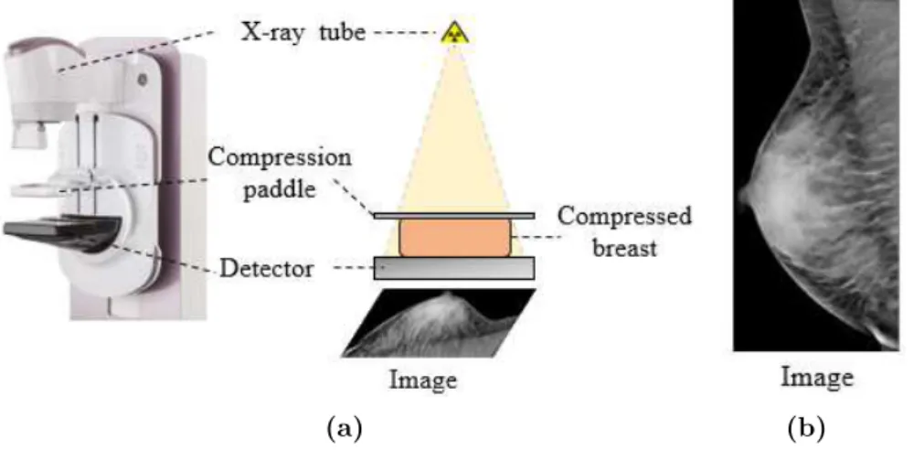
Today, x-ray mammography is recognized as the most effective technique for breast cancer detection at an early stage. It has demonstrated a decrease in mortality among screened women up to 40% [65]. This modality, also
known as Full Field Digital Mammography (FFDM), is a two-dimensional (2D) x-ray imaging technique. As depicted in Figure 2.1(a), during the image acquisition, the breast is compressed between the compression paddle and the detector. The compression is important for several reasons [80] :
• It evens out the breast thickness so all of the tissue can be visualized using a detector presenting a limited dynamic in exposure.
• It allows a lower x-ray dose delivered to the breast since a thinner amount of breast tissue is being imaged.
• It reduces motion-based blur as well as x-ray scatter which increases sharpness of the image.
Once the breast is well positioned, x-rays are emitted from the x-ray tube to be transmitted through or absorbed by the breast tissue. The detector then converts the reached x-rays to digital information.
(a) (b)
Figure 2.1: (a) A schematic of a mammography image acquisition. (b)
Example of an acquired image from the imaging system.
The pixels values of the obtained projection image, as shown in Fig-ure2.1(b), follow the Beer-Lambert law which denotes the link between the attenuation of x-ray photons, the thickness and the linear attenuation coef-ficient of the breast tissue. Assuming a monochromatic x-ray spectrum, the mathematical formulation of Beer-Lambert law is
I(x, y) = I0e − Z r∈L(x,y) µ(r) dr (2.1) where L(x, y) denotes the x-ray beam path between the x-ray source and the 2D position (x, y) on the detector, I0 is the x-ray photon intensity incident
upon the breast, I(x, y) is the x-ray photon intensity at the position (x, y). Finally, µ(r) represents the linear attenuation coefficient of the breast tis-sue material along the x-ray path. Typically, the breast is composed by fibroglandular tissue and adipose tissue with different attenuation proper-ties each. Indeed, the fibroglandular one tends to absorb more x-ray photons and translate to white areas in the image. Meanwhile, the adipose tissue permits more of the transmission of x-ray photons which translate to darker areas in the mammographic image as illustrated in Figure2.1(b).
Although FFDM has for long been accepted as the gold standard for screening and early detection of breast cancer, its sensitivity is far from perfect. It ranges from 47.8% to 98% with highly dense fibroglandular tis-sue presenting the main encumbrance to detection [77]. In this context, supplemental modalities are considered in the clinical routine in order to alleviate this limitation. Breast ultrasound (U/S) has proved its utility for women with dense breast tissue [128]. In fact, the accuracy of detecting breast cancers occulted on mammography using U/S is unaffected by breast tissue density [15,11]. Furthermore, U/S has been proven to be effective in distinguishing cystic from solid masses. It allows also determining the suspi-cion of the solid masses and whether a biopsy is needed or not [121,143,7]. Unlike x-rays imaging, ultrasound is an imaging technique based on the ap-plication of sound waves, i.e. not radiation, for producing images of the internal breast structures. In particular, this technique uses waves which frequencies are above the upper limit of human hearing, called ultrasound, and are able to penetrate biological tissues (at least 1 MHz) [57]. To obtain the image, first a gel is interposed between the U/S probe and the part of the skin of the breast to be diagnosed. The probe is then moved in multiple directions, with an adequate pressure that makes the U/S usually painless. The waves are variably reflected as echoes by the breast tissues. These re-flected echoes are received by the probe and forwarded as electronic signals to a computer system that finally generates the images. Even though U/S is more accessible and cost-effective compared to mammography, it is dis-couraged to perform screening with U/S alone. Indeed, it may not detect efficiently clusters of microcalcifications some of which being an early sign of breast cancer. Furthermore, it generates relatively higher number of false positives compared to FFDM [48, 14, 15] yielding to undesirable negative biopsies. Overall, it has not been shown to reduce mortality from breast cancer when used as stand-alone modality. Yet, it may be considered as an adjunct to mammography in asymptotic women for a specific category, i.e. women with dense breasts, despite the high false positive rate.
Other additional modalities to mammography for the screening of risk women are the functional-based techniques. They are based on high-lighting the blood vessels developed around the tumor. These blood vessels supply the tumor with oxygen and nutrients to sustain its growth. Thus, defined as tumor angiogenesis, it begins at the earliest stages of tumor
de-velopment. Therefore, imaging this proliferation would enable the detection and the characterization of breast cancer early. In this context, contrast-enhanced magnetic resonance imaging (CE-MRI) is one of the functional imaging techniques. The injection of a gadolinium-based contrast product enables the tracking of the uptake of the contrast agent in the lesions. In particular, the morphology and the kinetic profile of the contrast uptake allows the distinction between malignant lesions from the benign ones [82]. Therefore, it has been shown in several studies that MRI allowed to iden-tify earlier stage disease than mammography and that combining MRI and mammography is linked to decreased mortality rate [89]. Yet, MRI is af-fected by its high cost, reduced accessibility and a significant rate of false positives.
An additional functional-based modality worth mentioning is Contrast Enhanced Spectral Mammography (CESM). As is the case with FFDM, CESM is a 2D x-ray imaging technique. Yet, its routine is very different from FFDM [83]. First, the patient is injected with iodinated contrast agent. After letting the iodine diffuse for some time, the breast is compressed. Then, two images of the breast are acquired with different x-ray spectra. One of them is located below the discontinuity K of iodine denoted as low-energy (LE) spectrum and the other one is located above it, defined as high-energy (HE) spectrum. Finally, the iodine-enhanced image is obtained by recombining the acquired dual-energy images. The feasibility of CESM was shown in 2003 [83] and became commercially available in 2010 through the introduction of SenoBright application (GE Healthcare; Chicago, IL, USA). Several studies have shown the superiority of CESM over FFDM [133] especially for patients with dense breasts. [74].
In a nutshell, supplemental modalities may be recommended in addition to FFDM, depending on the patient case and the radiologist decision. We recall that the first task of the radiologist when reading mammograms in the context of a breast cancer screening exam is to detect signs that may indicate the presence of a breast cancer. Hence, when reading mammogra-phy images, the radiologist searches for specific radiological findings which will be detailed in the next section.
2.2.3 Radiological findings in mammography images
The mammographic images allow to spotlight anomalies which, depending on their morphology, their number, their distribution and their evolution in time, imply a diagnosis of benign pathology or cancer. Hence, their detection and characterization are of great importance to decide recalling or not the woman during the screening phase, or deciding for a biopsy and providing the right treatment during the diagnostic phase. Radiologists are commonly using the BI-RADS (Breast Imaging Reporting and Database System) ter-minology established by the ACR (American College of Radiology) to
de-scribe the main characteristics of the detected findings, as summarized in Table2.1.
Table 2.1: Mammography lexicon of the significant findings.
Radiological findings Description
Mass Shape oval-round-irregular
Margin circumscribed-obscured-microlobulated-indistinct-spiculated
Density fat-low-equal-high
Asymmetry asymmetry-global-focal-developing
Architectural distorsion distorted parenchyma with no visible mass
Calcification Morphology typically benign
suspicion 1. amorphous 2. coarse heterogeneous 3. fine pleiomorphic
4. fine linear or fine linear branching
Distribution diffuse-regional-grouped-linear-segmental
In BI-RADS terminology, a mass is a lesion occupying a 3D space seen in two different projections. If a potential mass is seen in a single projection, it should be called an ’asymmetry’ until its three-dimensionality is confirmed. The shape of a mass correlates with the level of malignancy of the lesion. The more irregular is the mass, the higher is the probability of a cancer. A mass with well-circumscribed edges has a low probability being related to a cancer, while spiculated edges are strongly indicative of a cancer. Furthermore, the density of the mass is also an indicator of the level of malignancy of the lesion.
Contrary to masses, asymmetries lack convex outward borders and con-spicuity as found in masses, according to BI-RADS third edition [144]. When such potential abnormality is detected, it is important to determine first whether it is really three dimensionally or just a projection of superimposed normal structures. Then, additional incidence with comparison with the opposite side of the breast are typically required for a better evaluation.
Architectural distortions correspond to a deviation, disharmony or rup-ture in the distribution of the conjunctivo-glandular dense tissue. They are associated to fibrous lesions which are generally difficult to detect (not visible enough and of small size). They may be detected by comparing the content in left and right breast images, or between current and prior images. On histological examination, 50 to 80% of breast cancers have calcifi-cations [72]. The latter ones are due to central necrosis, defined as a form of cell injury which results in the premature death of cells in living tissue, or secretions of malignant cells. Therefore, the detection of calcifications in mammography is of crucial importance for the early detection of breast cancers. Despite the relative high contrast of calcifications compared to the breast tissue, due to their high x-ray attenuation properties, their detection is difficult when they are of small size. In order to improve detectability, the radiologist resorts to different practices such as zooming or adjusting brightness and contrast of the images, acquiring magnified views to increase contrast and spatial resolution in order to visualize more calcifications and to better characterize their shape. Three important features are considered
to determine the malignancy of calcifications: • The size
• The morphology
• The distribution in the breast
Based on the size, calcifications are either denoted macrocalcifications if their diameter is larger than 0.5 cm or microcalcifications if their diameter is less than 0.5 cm. Typically, macrocalcifications are benign while microcal-cifications can be either benign or malignant according to their morphology as well as their distribution. Concerning the morphology, it remains the most important factor in differentiation between benign and malignant cal-cifications as illustrated in Figure 2.2and 2.3 respectively.
(a) (b) (c) (d)
(e) (f) (g) (h)
Figure 2.2: Examples of typically benign calcifications [105].(a) Round. (b) Skin. (c) Vascular. (d) Coarse or popcorn-like. (e) Rim. (f) Dystrophic. (g) Milk of calcium. (h) Suture.
(a) (b) (c) (d)
Figure 2.3: Examples of calcifications with suspicious morphology [105].(a) Coarse heterogeneous. (b) Amorphous. (c) Fine pleomorphic. (d) Fine linear or fine linear-branching.
Moreover, based on their distribution and number, the clustered ar-rangements are more or less suspicious. In the BI-RADS atlas, different
distributions are provided, as shown in Figure 2.4. Hereunder, the descrip-tion of the arrangements are depicted, according to their increased risk of malignancy:
1. Diffuse : distributed randomly throughout the breast.
2. Regional : occupying a large portion of breast tissue > 2 cm. 3. Grouped (historically cluster) : few calcifications occupying a
small portion of breast tissue; lower limit 5 calcifications within 1 cm and upper limit a larger number of calcifications within 2 cm.
4. Linear : arranged in a line, which suggests deposits in a duct. 5. Segmental : suggests deposits in a duct or in ducts and their
branches.
Figure 2.4: Examples of different microcalcification distributions
(BI-RADS for mammography and ultrasound 2013).
One can thus understand that detection is a difficult task in mammog-raphy due to the large variability of radiological findings in size, shape and contrast. In particular, detection of microcalcifications is challenging due to their small size but also because they can be obscured by the breast tissues
especially in women with dense breasts. In this context, Section 2.2.4 is dedicated to discussing the impact of breast density on microcalcification detectability.
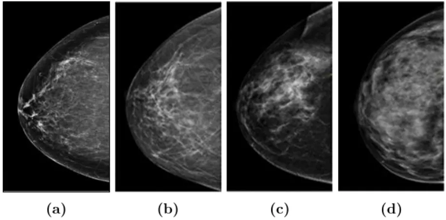
2.2.4 Impact of breast density on mammography performance Besides the fact that detecting the radiological findings remains in itself a sensitive task for the radiologist, the breast density can impact the mam-mography performance, especially when it comes to microcalcification de-tectability. In the BI-RADS terminology, the breast density is classified into four categories as illustrated in Figure2.5, according to their increased density [105].
• BI-RADS a: almost entirely fatty.
• BI-RADS b: there are scattered areas of fibroglandular density. • BI-RADS c: the breasts are heterogeneously dense, which may obscure
small masses.
• BI-RADS d: the breasts are extremely dense, which lowers the sensi-tivity of mammography.
(a) (b) (c) (d)
Figure 2.5: BI-RADS classification of breast density in mammography
images : (a) almost entirely fatty. (b) scattered fibroglandular. (c) hetero-geneously dense. (d) extremely dense.
Breast density describes the amount of the fibroglandular tissue compared to the amount of the fatty tissue in the breast on a mammogram. Typ-ically, the breast density is measured through a visual assessment by the radiologist when reviewing the mammogram [6]. The radiologist relies on the BI-RADS categorization, where she/he assigns mammograms to one of the four aforementioned classes.
So, the denser the breast, the greater the amount of fibroglandular breast tissue in the breast. This is illustrated in Figure 2.5 ranging from (a) to (d) with respect to the increased amount of fibroglandular breast tissue. Furthermore, the denser the breast, the poorer the sensitivity in FFDM. This is primarily explained by potentially occulting the lesions, especially microcalcifications, by the fibroglandular tissue [63]. One can infer that it is harder to detect microcalcifications in dense and textured breasts (i.e., BI-RADS c and d, illustrated in Figure2.5(c) and (d) respectively).
Henceforth, microcalcification detection task is simultaneously impor-tant and very challenging when breast cancer screening is performed. This task is crucial since microcalcifications might be a sign of breast cancer when grouped and presenting some specific morphological and densiometric characteristic. Yet, it is very challenging due to the small size of microcal-cifications and their low contrast when superimposed to dense areas of the breast. Our research work will be particularly focused in the enhancement of the detectability of microcalcifications. More precisely, we aim to im-prove this task in the recent modality called Digital Breast Tomosynthesis (DBT) which was introduced to curb the limitations of FFDM. Section2.3 will be dedicated to thoroughly present DBT, explain its main features and advantages over FFDM thereby discussing its impact on microcalcification detectability. Finally, a presentation of the main contributions of this re-search work will be provided.
§ 2.3 Digital Breast Tomosynthesis (DBT)
2.3.1 Limitations of x-ray mammographyEven though x-ray mammography remains the current modality of choice for breast cancer screening and diagnosis, the detection may not be accurate enough, particularly for women with highly dense breasts. As explained in section2.2.2, in a conventional FFDM exam of the breast, two x-ray images, from top-to-bottom (which corresponds to Cranio-Caudal (CC) view as in Figure 2.6(a)) and from angled side-to-side (which corresponds to Medio-lateral oblique (MLO) as in Figure 2.6(b)), are acquired while the breast is compressed between the compression paddle and the detector. The ac-quisition of a projected view of the breast, as in FFDM, implies the su-perimposition of breast tissues which is the primary limitation of FFDM senstivity. The superimposition of tissues can reduce the visibility of le-sions present in the breast or even completely occult them. This effect leads to a false negative (FN) error, that is the mammogram looks normal even though breast cancer is present [23]. This results in a lower detection rate in DBT than in FFDM [77]. The superimpostion of breast tissues may also generate structures that can mislead the radiological interpretations. For
instance, the overlapping of normal fibrosis tissue may mimic radiological findings and may lead to a false positive (FP) diagnosis [26]. This can result in undesirable recalls of women for further diagnostic tests, impelling to an increased patient anxiety [96].
Figure 2.6: (a) Cranio-caudal (CC) view (from top-to-bottom). (b)
Medi-olateral oblique (MLO) view (from angled side-to-side).
2.3.2 Advantages of DBT over x-ray mammography
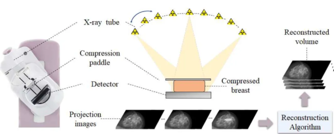
Digital Breast tomosynthesis (DBT) was designed to partially alleviate the aforementioned major limitation of FFDM, induced by the tissue overlap. The acquisition geometry is very similar to that used in mammography, but with DBT, several projections of the breast are acquired enabling the reconstruction of a stack of images parallel to the detector plane correspond-ing to breast slices. Durcorrespond-ing the acquisition process, the x-ray tube moves along an arc around the breast with a limited angular aperture, typically from 11° to 50°, whilst the detector remains stationary. A limited num-ber of low-dose projection images are acquired so that the total dose to the patient is still comparable to the one in FFDM. In DBT, the number of pro-jection images varies from 9 to 25 depending on the imaging system [120]. These two-dimensional projection images are then reconstructed, using a reconstruction algorithm, into a three-dimensional volume. Herein, the re-construction algorithm is tailored so that it estimates the 3D distribution of the breast tissues using the projection images as the input data. Thus, the resulting volume consists of slices that present less superimposition of tissues compared to a standard x-ray mammography. A schematic of a DBT acqui-sition and an example of a reconstructed tomosynthesis volume is provided in Figure 2.7.
Figure 2.7: A schematic of DBT acquisition process and reconstruction.
Since DBT projections are acquired over a limited angular aperture, the DBT volume has a high isotropic resolution in the planes parallel to the detector and a much lower resolution in the perpendicular direction, i.e. the depth direction. It is worth mentioning that the in-plane resolution of the DBT slices is limited by the detector resolution. Due to the 3D nature of the DBT reconstructed volume, DBT has the potential to help reducing recall rates, improve the accuracy of breast cancer detection, and therefore improve the clinical performance particularly in dense breasts.
During the last decade, the current DBT systems have achieved sig-nificantly improved results towards greater acceptance of DBT as a viable alternative to conventional mammography. Several clinical studies have demonstrated that using DBT in stand-alone or DBT combined with FFDM for breast cancer screening results in an improvement of cancer detection rate and a decrease in false positive recall rate compared to FFDM alone. Thanks to the reduction of breast tissue overlapping, a better visibility and depiction of radiological findings are enabled. In this context, some studies have shown that DBT should enable a better differentiation between benign and malignant lesions [107]. Other comparative studies have demonstrated that lesions are better depicted in DBT which should enable more affirma-tive interpretations. This holds in particular for masses and architectural distortions in dense breasts [90, 24]. Furthermore, DBT provides a more accurate lesion localization [9].
![Figure 2.2: Examples of typically benign calcifications [105].(a) Round. (b) Skin. (c) Vascular](https://thumb-eu.123doks.com/thumbv2/123doknet/14545514.725325/36.892.224.680.492.723/figure-examples-typically-benign-calcifications-round-skin-vascular.webp)