HAL Id: tel-01423712
https://tel.archives-ouvertes.fr/tel-01423712
Submitted on 31 Dec 2016
HAL is a multi-disciplinary open access
archive for the deposit and dissemination of sci-entific research documents, whether they are pub-lished or not. The documents may come from teaching and research institutions in France or abroad, or from public or private research centers.
L’archive ouverte pluridisciplinaire HAL, est destinée au dépôt et à la diffusion de documents scientifiques de niveau recherche, publiés ou non, émanant des établissements d’enseignement et de recherche français ou étrangers, des laboratoires publics ou privés.
Evaluation of the anticarcinogenic effects of the
synthetic flavagline FL3 on cancer stem cells :
characterization of the molecular mechanisms involved
Fathi Emhemmed
To cite this version:
Fathi Emhemmed. Evaluation of the anticarcinogenic effects of the synthetic flavagline FL3 on cancer stem cells : characterization of the molecular mechanisms involved. Cancer. Université de Strasbourg, 2015. English. �NNT : 2015STRAJ042�. �tel-01423712�
ÉCOLE DOCTORALE DES SCIENCES DE LA VIE ET DE LA SANTE
Laboratoire d’Innovation Thérapeutique
Thése
présentée par:EMHEMMED Fathi
Soutenue le :18 Septembre 2015
Pour obtenir le grade de : Docteur de l’université de Strasbourg Discipline/ Spécialité: Biologie Moléculaire et Cellulaire
Rapporteurs externes
Prof.
Jean-Yves JOUZEAU Biopôle de l’Université de Lorraine (Nancy-France)Dr. Céline HUSELSTEIN Biopôle de l’Université de Lorraine (Nancy-France)
Rapporteur interne
Prof.
Jean-Marie REIMUND INSERM UMR_S1113, Université de StrasbourgDirecteur de thèse
Dr.
Christian MULLER UMR 7200 CNRS, Université de StrasbourgCo-Encadrant de thèse / Membre invité
Dr. Guy FUHRMANN UMR 7213 CNRS, Université de Strasbourg
UNIVERSITÉ DE STRASBOURG
Evaluation des effets anticarcinogéniques de la flavagline synthétique FL3
sur les cellules souches cancéreuses; caractérisation des mécanismes
Acknowledgment
First of all, I would like to extend the warmest thanks to the Libyan ministry of higher education and scientific research for the generous financial support to my family and me during all my study, and many thanks to the biotechnology research center for giving me the opportunity to continue my studies in biology science. A massive thanks to the ‘‘Ligue contre le cancer’’ (Comité du Grand Est, France and the ‘‘Association pour la recherche sur le cancer ’’ for supporting and funding cancer research.
I would like to thank my committee members Prof. Jean-Yves JOUZEAU, Prof. Jean-Marie REIMUND and Dr. Céline HUSELSTEIN for accepting to be examiners for my PhD thesis defense and for evaluating my report.
I would like to sincerely thank my supervisor, Dr. Christian MULLER for giving me the opportunity to joint your lab, for your guidance when using flow cytometry technique and for your valuable assistance during my thesis.
A very special thanks goes out to my Co- supervisor, Dr. Guy FUHRMANN for your guidance, advices, and your help during the writing of this manuscript and constant help throughout my master doctoral studies.
I would like to thank prof. Marcel HIBERT for welcoming me in the (Laboratoire
d’Innovation Thérapeutique).
I would like to express my appreciation to Prof. Dr. Valérie SCHINI-KERTH for providing me the opportunity to perform my master degree in her lab and for her collaboration during my thesis.
I would like to express my gratitude to Dr. Laurent DÉSAUBRY for your collaboration during my thesis.
I would like to thank all the staff of the Laboratoire de Biophotonique et pharmacologie and
technical help and kindness. I would like to thank all students in our lab and especial thanks also to Sarah Ali Azouaou for your help and your gentleness and big thanks to my friends for the good time-shared.
Finally, I would like to thank my family especially my parents for your support and encouragement during all my life and big thanks to my spouse who has help and supported me over the past five years in my pursuit of completing my PhD study.
Table of contents
List of figures ... 6!
List of tables ... 7!
ABBREVIATIONS ... 8!
Thesis abstract in english ... 12!
Résumé de la thése de doctorant en français ... 15!
Introduction
... 19!1. Cancer ... 19!
2. Stem cell biology ... 20!
!"#"$%&'()*$+,-($.-**+$"""""""""""""""""""""""""""""""""""""""""""""""""""""""""""""""""""""""""""""""""""""""""""""""""""""""""""""""""""""""""""$!/! 2.1.1. Definition and classification!""""""""""""""""""""""""""""""""""""""""""""""""""""""""""""""""""""""""""""""""""""""""""""""""""""""""""""""""""""!#$! 2.1.2. Stem cell organization and niche concept!"""""""""""""""""""""""""""""""""""""""""""""""""""""""""""""""""""""""""""""""""""""""""""!#%! 2.1.3. The mode of division!""""""""""""""""""""""""""""""""""""""""""""""""""""""""""""""""""""""""""""""""""""""""""""""""""""""""""""""""""""""""""""""""""!##! !"!"$0)1.-'$+,-($.-**+$""""""""""""""""""""""""""""""""""""""""""""""""""""""""""""""""""""""""""""""""""""""""""""""""""""""""""""""""""""""""""""$!2! 2.2.1.Generalities!"""""""""""""""""""""""""""""""""""""""""""""""""""""""""""""""""""""""""""""""""""""""""""""""""""""""""""""""""""""""""""""""""""""""""""""""""""""!#&! 2.2.1.1. Historical overview!"""""""""""""""""""""""""""""""""""""""""""""""""""""""""""""""""""""""""""""""""""""""""""""""""""""""""""""""""""""""""!#&! 2.2.1.2. Evidence of the existence of CSCs!""""""""""""""""""""""""""""""""""""""""""""""""""""""""""""""""""""""""""""""""""""""""""""!#&! 2.2.1.3. Origin of the tumor heterogeneity!""""""""""""""""""""""""""""""""""""""""""""""""""""""""""""""""""""""""""""""""""""""""""""""!#'! 2.2.2. Carcinogenesis and its consequence!"""""""""""""""""""""""""""""""""""""""""""""""""""""""""""""""""""""""""""""""""""""""""""""""""""""!#(! 2.2.3. Cancer stem cell biomarkers!""""""""""""""""""""""""""""""""""""""""""""""""""""""""""""""""""""""""""""""""""""""""""""""""""""""""""""""""""""!#)! 2.2.4. Control of self-renewal and differentiation!"""""""""""""""""""""""""""""""""""""""""""""""""""""""""""""""""""""""""""""""""""""""""!#)! 2.2.4.1. The role of signaling pathways!"""""""""""""""""""""""""""""""""""""""""""""""""""""""""""""""""""""""""""""""""""""""""""""""""""!&$! 2.2.4.2. The role of transcription factors!""""""""""""""""""""""""""""""""""""""""""""""""""""""""""""""""""""""""""""""""""""""""""""""""""!&&! 3. Human embryonal carcinoma cells (hECCs) ... 40!
2"#"$3'45416$7-'48),4&1$)17$.9*,9'-$+:+,-($"""""""""""""""""""""""""""""""""""""""""""""""""""""""""""""""""""""""""""""""""""""$;/! 2"!"$<(=':&1)*$.)'.41&()$)+$)$(&7-*$,&$+,97:$,>-$=4&*&5:$&?$0@0$""""""""""""""""""""""""""""""""""""""""$;#! 4. Therapeutic strategies against CSCs ... 41!
;"#"$A)'5-,415$.)1.-'$+,-($.-**+$=:$)B&B,&+4+$4179.,4&1$""""""""""""""""""""""""""""""""""""""""""""""""""""""""""$;#! 4.1.1. The intrinsic pathway!""""""""""""""""""""""""""""""""""""""""""""""""""""""""""""""""""""""""""""""""""""""""""""""""""""""""""""""""""""""""""""""""!'%! 4.1.1.1. Generalities!""""""""""""""""""""""""""""""""""""""""""""""""""""""""""""""""""""""""""""""""""""""""""""""""""""""""""""""""""""""""""""""""""""""""!'%! 4.1.1.2. Oxidative stress and tumor suppressors!"""""""""""""""""""""""""""""""""""""""""""""""""""""""""""""""""""""""""""""""""""!'#! 4.1.1.3. p38 MAPK!"""""""""""""""""""""""""""""""""""""""""""""""""""""""""""""""""""""""""""""""""""""""""""""""""""""""""""""""""""""""""""""""""""""""""!'&! 4.1.2. The extrinsic pathway!""""""""""""""""""""""""""""""""""""""""""""""""""""""""""""""""""""""""""""""""""""""""""""""""""""""""""""""""""""""""""""""""!''!
;"!"$A)'5-,415$0@0+$=:$74??-'-1,4),4&1$4179.,4&1$"""""""""""""""""""""""""""""""""""""""""""""""""""""""""""""""""""""""""$;;! 4.2.1. Targeting self-renewal signaling pathways!""""""""""""""""""""""""""""""""""""""""""""""""""""""""""""""""""""""""""""""""""""""""!''! 4.2.2. Targeting Oct4 function in CSCs!"""""""""""""""""""""""""""""""""""""""""""""""""""""""""""""""""""""""""""""""""""""""""""""""""""""""""""!'*!
;"2"$A)'5-,415$.-**$+9'?).-$=4&()'C-'+$&?$0@0+$"""""""""""""""""""""""""""""""""""""""""""""""""""""""""""""""""""""""""""""$;D!
;";"$A)'5-,415$(4E%F+$-GB'-++4&1$&?$0@0+$"""""""""""""""""""""""""""""""""""""""""""""""""""""""""""""""""""""""""""""""""""""$;H!
;"I"$A)'5-,415$0@0$>:B&G4.$14.>-$"""""""""""""""""""""""""""""""""""""""""""""""""""""""""""""""""""""""""""""""""""""""""""""""""""""""$;H!
5. Flavaglines and cancer therapy ... 49! I"#"$P-1-')*4,4-+$""""""""""""""""""""""""""""""""""""""""""""""""""""""""""""""""""""""""""""""""""""""""""""""""""""""""""""""""""""""""""""""""""""""$;Q! I"!"$R4&*&54.)*$).,484,:$"""""""""""""""""""""""""""""""""""""""""""""""""""""""""""""""""""""""""""""""""""""""""""""""""""""""""""""""""""""""""""$I/! 5.2.1. Anticancer activity!""""""""""""""""""""""""""""""""""""""""""""""""""""""""""""""""""""""""""""""""""""""""""""""""""""""""""""""""""""""""""""""""""""""!*$! 5.2.1.1. Inhibiting translation!"""""""""""""""""""""""""""""""""""""""""""""""""""""""""""""""""""""""""""""""""""""""""""""""""""""""""""""""""""""""!*$! 5.2.1.2. Targeting prohibitins!""""""""""""""""""""""""""""""""""""""""""""""""""""""""""""""""""""""""""""""""""""""""""""""""""""""""""""""""""""""!*%! 5.2.1.3. Targeting apoptosis!"""""""""""""""""""""""""""""""""""""""""""""""""""""""""""""""""""""""""""""""""""""""""""""""""""""""""""""""""""""""""!*%! 5.2.2. Cytoprotective activity!"""""""""""""""""""""""""""""""""""""""""""""""""""""""""""""""""""""""""""""""""""""""""""""""""""""""""""""""""""""""""""""""!*#! Aims and objectives ... 54!
Results
... 55!Article1 ... 56!
Article 2 ... 69!
Article 3 ... 89!
Discussion and perspective
... 115!#"$J'&S)B&B,&,4.$).,484,:$&?$TU2$)5)41+,$0@0+$)17$917-'*:415$(&*-.9*)'$"""""""""""""""""""""""""""""""""$! (-.>)14+(+$418&*8-7$""""""""""""""""""""""""""""""""""""""""""""""""""""""""""""""""""""""""""""""""""""""""""""""""""""""""""""""""""""""""""$##I! !"$V&*-.9*)'$(-.>)14+(+$&?$,>-$B'&,-.,4&1$&?$1&'()*$.-**+$)5)41+,$,>-$""""""""""""""""""""""""""""""""""$! .:,&,&G4.$-??-.,$&?$TU2$"""""""""""""""""""""""""""""""""""""""""""""""""""""""""""""""""""""""""""""""""""""""""""""""""""""""""""""""""""""""""$##H! 2"$J'&S74??-'-1,4),4&1$).,484,:$&?$TU2$&1$0@0+$"""""""""""""""""""""""""""""""""""""""""""""""""""""""""""""""""""""""""""""$##O!
Discussion et perspective en français
... 120!H"#"$F.,484,W$B'&S)B&B,&,4X9-$7-$TU2$.&1,'-$*-+$0@0$Y$(W.)14+(-+$(&*W.9*)4'-+$+&9+S Z).-1,+"$"""""""""""""""""""""""""""""""""""""""""""""""""""""""""""""""""""""""""""""""""""""""""""""""""""""""""""""""""""""""""""""""""""""""""""""""""""""$#!#! H"!"$VW.)14+(-+$(&*W.9*)4'-+$7-$*)$B'&,-.,4&1$7-+$.-**9*-+$1&'()*-+$.&1,'-$*[-??-,$ .:,&,&G4X9-$7-$TU2$"""""""""""""""""""""""""""""""""""""""""""""""""""""""""""""""""""""""""""""""""""""""""""""""""""""""""""""""""""""""""""""""$#!!! H"2"$TU2$+,4(9*-$*)$74??W'-1.4),4&1$7-+$0@0+$""""""""""""""""""""""""""""""""""""""""""""""""""""""""""""""""""""""""""""""""$#!;! Bibliography ... 126! Annexes ... 142!
List of figures
Figure 1. Differentiation potential and classes of stem cells………. 21
Figure 2. Cellular and molecular components of three fast-cycling adult stem
cell niches………22
Figure 3. Models of tumor heterogeneity……….. 25
Figure 4. Carcinogenesis,tumorigenesis and resistance in CSCs…………... 26
Figure 5. Wnt/ß-catenin signaling pathway... 31
Figure 6. Notch signaling pathway……….... 32
Figure 7. Sonic hedgehog signaling pathway……… 33
Figure 8. In vivo and in vitro expression of Oct4 in human normal and cancer
SCs……….. 34
Figure 9. Sox2 network in the regulation of self-renewal and
differentiation………. 39
Figure 10. Proapoptotic and prosurvival pathway that can be targeted by
anticancer agents……… 42
Figure 11. Therapeutic strategy to eliminate CSCs by anticancer agents……. 45
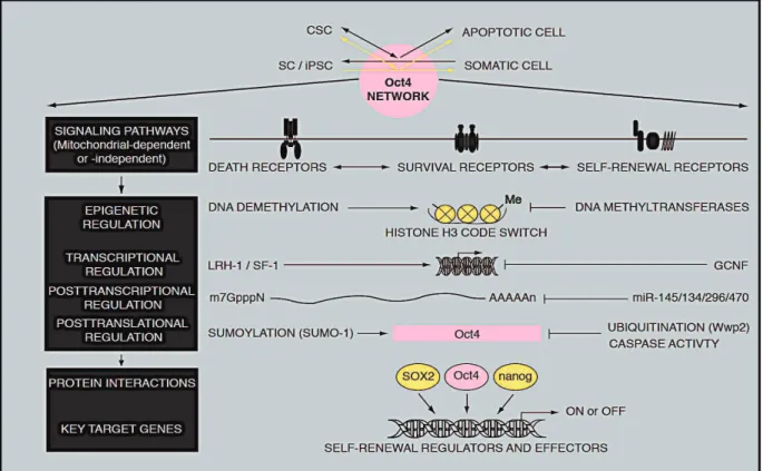
Figure 12. Potential targets of the Oct4 network by anticancer agents ………. 46
List of tables
Table 1. Summary of some chemoresistance-related signaling pathways……28
Table 2. Cell surface markers of CSCs……….29
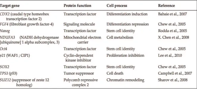
Table 3. Example of some genes involved in different cellular processes
Abbreviations
A
APC: adenomatous polyposis coli AML: acute myeloid leukemia AXIN: Axis inhibition protein
ATP: ATP-binding cassette transporters
Apaf-1: Apoptotic Protease-Activating Fctor-1 AP-1: Activator Protein-1
APO-1/Fas: Apoptosis Antigen1/FAS ATRA: All-trans retinoic acid
AIF: Apoptosis Inducing Factor AhR: aryl hydrocarbon receptor Akt: kinase protein kinase B
B
Bmi-1: Polycomb complex protein BAK1: BCL2-Antagonist/Killer 1 Bad: Bcl-2-associated death promoter Bax: Bcl-2–associated X protein Bid: BH3 interacting-domain death Bcl-xL: B-cell lymphoma-extra large
C
CSCs: Cancer stem cells CK1α: casein-kinase 1α
Cdk1: cycline-dependent kinase1 Cdx2: Caudal type homeobox
ChIP- on chip: Chromatin immunoprecipitation with DNA microarray CSL: CBF1, Suppressor of Hairless, Lag-1
D
Dnmt1: DNA methyltransferase 1 DDR: DNA damage response Dsh: disheveled
Dhh: Desert Hedgehog
E
eIF4: eukaryotic initiation translation factor-4 ECCs: Embryonal carcinoma cells
ERK: Extracellular-Signal-Regulated Kinases EMT: Epithelial to mesenchymal transition ESCs: embryonic stem cells
F
FOXD3: forkhead box 3
FADD: Fas-associated death domain FLIP: FLICE-like inhibitory protein
G
GLI: Glioma-associated oncogene homolog GSK3β: Glycogen synthase kinase
GADD: Growth arrest and DNA damage GPCRs: G protein-coupled receptors GCNF: Germ Cell Nuclear Factor
H
HDACs: Histone deacetylases)
HIF-1α: Hypoxia inducible factor-1 alpha HIF-2α: Hypoxia inducible factor-2 alpha HSP27: Heat shock protein- 27
I
Ihh: Indian Hedgehog
iPSCs : induced pluripotent stem cells
ITE: 2-(1 H-indole-3 -carbonyl)-thiazole- 4-carboxylic acid methyl ester
J
JAK: Janus Kinase
JNK: c-Jun N-terminal Kinases
K
KIF17: Kinesin Family Member 17
L
Lef: lymphoid enhancer-binding factor LRH-1: Liver Receptor Homolog-1
LRP: low-density lipoprotein receptor- related protein
M
MAML: co-factor Mastermind-like mTOR: mammalian target of rapamycin mRNA: Messenger ribonucleic acid miRNAs: microRNAs
Mcl-1: myeloid cell leukemia 1 Myc :v-Myc myelocytomatosis viral
N
NICD: Notch Intercellular Domain NF-kB: Nuclear factor –Kappa B
NADPH: Nicotinamide adenine dinucleotide phosphate
O
Oct4: Octamer-binding transcription factor
POU: Pituitary-specific, Octamer and neural Unc-86 transcription factors Oct-4pg1: Octamer-binding transcription factor Pseudogene, 1
P
RTKs: receptor tyrosine kinases PHBs: prohibitins
PTCH 1: patched protein 1
RT-PCR: Reverse transcription polymerase chain reaction PI3: phosphatidylinositol-3
PARP: ADP-ribose polymerase
p38 MAPK: p38 mitogen-activated Protein kinase
R
RARs: the retinoic acid receptor
S
SCs: stem cells Shh: Sonic Hedgehog SMO: smoothened
SUFU: Suppressor of Fused SAHA: Suberoylanilide hydroxamic acid
SUMO-1: Small Ubiquitin-related Modifier, 1 SF-1: Steroidogenic Factor 1
STAT3: Signal Transducer and Activator of Transcription Sox2: SRY (SEX determining Region Y) –box2
SSEA-3: Stage-Specific Embryonic Antigen 3
Smac /DIABLO: Second Mitochondria- derived Activation of Caspase /direct IAP binding protein with low pI
T
Tcf: T-cell-specific transcription factor TNF: Tumor necrosis factor
TRAIL: TNF-Related Apoptosis-Inducing Ligand TNFRI: TNF receptor 1
V
VEGF: Vascular Epithelial Growth Factor
W
Thesis abstract in english
The aim of my thesis is based on the evaluation of the anticancer properties of flavaglines on cancer-initiating cells also known as cancer stem cells (CSCs).
Flavaglines are a family of natural cyclopenta [b] benzofurans compounds extracted from the plant genus Aglaia. The anticancer activity of lead bioactive compound, rocaglamide has been well characterized. More than 100 flavaglines are now identified, including rocaglaol and silvestrol. The team of Dr Laurent DESAUBRY had recently synthetized the flavagline derivative FL3 with enhanced cytotoxicity against several cancer cell lines, Our purpose was then to examine the anticancer activity of this FL3 on CSCs and to identify the underlying mechanisms involved.
Accordingly, CSCs have been defined by different molecular markers, which allow to isolate them from various tissues or organs. These cells are highly suspected to be responsible for tumor recurrence and resistance to both chemo- and radio-therapy. We are focusing our study on highly aggressive and poorly differentiated CSCs, which are known to express the stemness marker Oct4. This transcription factor is the key regulator of the self-renewal of a highly pluripotent state of normal stem cells (NSCs). Recent studies demonstrated that Oct4 is expressed in many cancers and contributes to tumor growth and inhibition of apoptosis. Downregulation or suppression of Oct4 expression, following administration of specific pharmacological agents, then assumed that the cells undergo apoptosis or differentiation. Such differentiation therapy represents a novel attractive tool for the reduction of CSC aggressiveness.
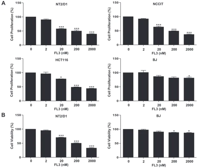
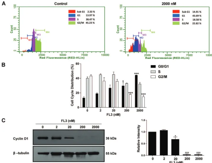
The first study aimed to examine whether there is a selective cytotoxic effect of FL3 on CSCs.The work was conducted on several cancer stem-like cell models, namely the human pluripotent embryonal carcinoma stem cell line NT2/D1 and NCCIT or the normal fibroblastic cell line BJ which have limited lineage competences. We found that FL3 selectively inhibited cell proliferation and cell viability of NT2/D1and NCCIT cells as measured by MTS or by trypan blue exclusion assays respectively. Cell growth inhibition was accompanied by cell cycle arrest at G1 phase, as measured by flow cytometry, and this arrest was associated with a downregulation of G1-phase marker cyclin D1, as detected by western
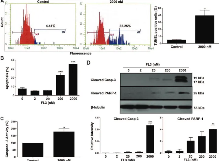
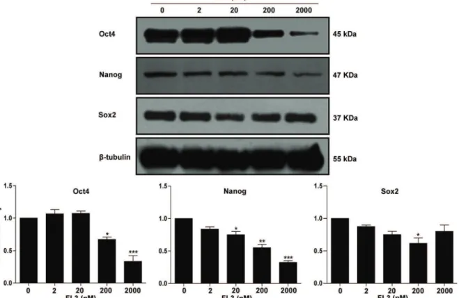
blot. Furthermore, we showed that the cytotoxic activity of FL3 involved a proapoptotic process, as shown by both TUNEL and annexin V-FITC/propidium iodide staining assays. By using immunoblot analysis, we found that FL3 triggered apoptosis in Oct4-expressing CSCs via a pathway dependent on the activation of p38 MAPK and caspase 3, followed by a significant decrease in the expression of the stemness regulator. Indeed use of pan caspase inhibitor (z-VAD-fmk), as well as inhibitor of p38MAPK activity by (SB203580) or expression by (siRNA) reduced FL3-induced cell death,this effect was accompanied by a significant accumulation of Oct4. Finally, these results revealed that FL3 is a strong anticancer drug that kills cancer stem-like cells.
Since the first study showed that, synthetic flavagline FL3 had no effect on NSCs, we aimed to identify the underlying molecular mechanisms involved in the protection of these cells against the cytotoxic effects of the drug. First we provided additional evidence for the absence of cytotoxic effect of FL3 on the normal fibroblasts by using annexin V-FITC and mitochondrial membrane potential ∆Ψm assays. We found that FL3 failed to induce apoptosis
in NSCs and was unable to induce mitochondrial depolarization and appearance of cytosolic cytochrome C. Most importantly, FL3 selectively phosphorylated Akt (Ser473), which is known to promote cell survival, FL3 also phosphorylated Bad at Ser112 and Ser136 in normal stem-like cells but not in cancer stem-like cells. The fact that its phosphorylated form serves as antiapoptotic protein could explain the resistance of NSCs. Indeed, forced inhibition of p-Akt by the specific inhibitor LY-294002 or gene knockdown of Bad by siRNA sensitized normal stem-like cells to FL3, by undergoing a caspase-3 dependent proapoptotic process, as measured by annexin V-FITC and western blot. These findings, therefore, provide new insights into the signaling pathway that could underly the resistance of normal stem-like cells against the potent anticancer agent FL3.
It is believed that the subpopulations of CSCs which reside within the bulk tumor, are responsible for tumor development and malignancy due to the deregulation of their self-renewal process. As a consequence it expected that pharmacological agents which are able to differentiate these cells, will lead to a loss of their self-renewal capacities and aggressiveness. We therefore aimed in the third study to evaluate whether FL3 administration at low concentration and for long period could be able to trigger cell differentiation of cancer stem - like cells. Our results indicated that FL3 downregulated in teratocarcinomal cell the expression of the stemness factor Oct4, at both the transcriptional and translational levels, as
early as 4 days of treatment. This effect coincided with a reduction of tumorsphere growth. The significant upregulation of the expression of specific neural markers such as GFAP (Glial fibrillary-acidic protein) and βTUBB3 (Tubulin beta-3 chain) further supported the evidence of the differentiation capacity of FL3. Interestingly, decreased expression levels of Oct4 was correlated with the appearance of cleaved caspase-3, followed by increased expression levels of GCNF (Germ cell-nuclear factor). This suggests that the degradation and gene repression of Oct4 could involve interconnected unknown mechanisms. Finally all these results showed that FL3 has similar effects than the common differentiating agent retinoic acid. Taken as a whole, my thesis has clearly demonstrated that the synthetic flavagline FL3 is a powerful anticancer compound, since it acts as a selective proapoptotic and pro-differentiating agent on cancer stem-like cells, without having any effect on normal stem-like cells.
Résumé de la thése de doctorant en français
L’objectif de ma thèse concerne l’évaluation des propriétés anticancéreuses des flavaglines sur les cellules initiatrices du cancer, plus connues sous le nom de cellules souches cancéreuses (CSCs).
Les flavaglines appartiennent à une famille de composés cyclopenta-benzofuranes naturels extraits de la plante du genre Aglaia. Le chef de file de cette famille, la rocaglamide, avait été identifié dès 1982 pour sa forte activité anticancéreuse. Depuis, plus de 100 flavaglines ont été décrites, dont le rocaglaol et le silvestrol. L’équipe du Dr. Laurent Désaubry avait récemment synthétisée la flavagline FL3 qui présentait une cytotoxicité accrue vis-à-vis de plusieurs lignées de cellules cancéreuses. Notre but était donc d’examiner l’activité anticancéreuse de la flavagline synthétique FL3 sur les CSCs et d’en caractériser les mécanismes moléculaires qui la sous-tendent.
Les CSCs se définissent par different marqueurs moléculaires qui permettent leur isolement de nombreux tissus ou organes. Ces cellules sont suspectées d’être responsables de la résurgence du processus cancéreux, ainsi que de la résistance à la chimio- et la radio-thérapie. Nous avons centré nos efforts sur des modèles de CSCs hautement agressives et peu différenciées qui sont connues pour exprimer un facteur central de la souchitude, à savoir Oct4. Ce facteur de transcription est un régulateur clé de l’auto-renouvellement de la pluripotence à spectre large des cellules souches normales (CSNs). Des données récentes ont montré qu’Oct4 est exprimé dans de nombreux cancers et contribue au développement tumoral et à l’inhibition de l’apoptose. Une répression de l’expression d’Oct4, suite à l’administration d’agents pharmacologiques, laisse donc présager que les cellules concernées rentrent en apoptose ou se différencient. Une telle thérapie de différenciation représente depuis très récemment une nouvelle arme pour réduire l’agressivité des CSCs.
La première étude que nous avons entreprise cherchait à examiner si FL3 a une activité cytotoxique sélectivement dirigée contre les CSCs. Ce travail a été conduit sur plusieurs lignées de cellules pluripotentes tératocarcinomales simili-souches NT2/D1 et NCCIT, et sur une lignée de cellules souches fibroblastiques qui répresente un modèle de CSNs ayant des potentialités restreintes de différenciation. Nous avons constaté, grâce aux tests de MTS et du trypan bleu respectivement, que FL3 inhibe sélectivement la prolifération et la viabilité des cellules NT2/D1 et
NCCIT. Cette inhibition de la croissance cellulaire s’accompagne d’un arrêt du cycle cellulaire en G1 et d’une répression de l’expression de la cycline D, une protéine majeure de régulation de la phase G1. De plus, l’activité cytotoxique de FL3 implique une induction d’un processus ayant les caractéristiques de l’apoptose, comme le montrent les tests de marquage par effet TUNEL et annexine V-FITC/propidium d’iodure. Par des analyses d’immuno-empreinte, nous avons observé que FL3 induit l’apoptose dans les CSCs exprimant Oct4, via une activation de la voie de signalisation impliquant la MAPK p38 et la caspase-3; Ceci conduit d’ailleurs à une chute significative des taux d’expression du régulateur de la souchitude. En confirmation de nos résultats, l’utilisation d’un inhibiteur universel de la caspase (z-VAD-fmk), ainsi que celle d’un inhibiteur de l’activité ou de l’expression de la MAPK p38 (respectivement par l’agent pharmacologique SB203580 ou des ARN interférents dirigés) a permis de contrecarrer les effets pro-apoptotiques de FL3 et de provoquer une accumulation significative d’Oct4 dans les cellules simili-souches cancéreuses. Finalement cette première étude a démontré que FL3 est un puissant agent anticancéreux qui tue spécifiquement les CSCs.
Comme nous avions démontré que la flavagline synthétique FL3 n’avait pas d’effets cytotoxiques sur les CSNs, nous avons cherché à identifier les mécanismes moléculaires qui pourraient rendre compte de cette résistance. Dans un premier temps, des expériences complémentaires ont permis de confirmer l’absence d’une quelconque activité délétère de FL3 sur les fibroblastes. En effet, la drogue ne modifie pas le pourcentage de cellules annexine V positives et n’induit pas de dépolarisation membranaire mitochondriale et de relargage du cytochrome C nucléaire dans le cytoplasme. De plus, FL3 phosphoryle la protéine de survie cellulaire Akt (au niveau de sa sérine 473) et la protéine régulatrice de l’apoptose Bad (au niveau de la sérine 112 et 136) dans les CSNs, et pas dans les CSCs. En fait, Bad, dans sa forme phosphorylée, assure une fonction anti-apoptotique; ceci expliquerait l’insensibilité des cellules fibroblastiques lorsqu’elles sont mises en présence de l’agent anticancéreux FL3. En effet, une inhibition forcée de la phosphorylation d’Akt (par un traitement avec le composé LY-294002), ainsi qu’une invalidation de l’expression génique de Bad (par un traitement avec des ARNs interférents), déclenche un processus pro-apoptotique caspase-3 dépendant dans les CSNs, lorsque celles-ci sont traitées par FL3. Ces résultats mettent donc en avant les principaux mécanismes moléculaires qui rendent compte de la résistance des cellules saines au puissant agent anticancéreux FL3.
Il est maintenant reconnu que la sous-population de CSCs, qui réside dans la masse tumorale, est responsable du développement tumoral, car celle-ci ne peut réguler son activité
d’auto-renouvellement. Il est dès lors postulé que les agents pharmacologiques qui sont capables d’induire la différenciation, vont affecter les capacités d’auto-renouvellement et donc l’aggressivité des CSCs. Aussi avons-nous cherché à évaluer, dans une troisième étude, si un traitement par FL3, à doses faibles et pendant une longue période, est susceptible d’entraîner une différenciation des cellules souches tératocarcinomales. Nos résultats indiquent que l’agent pharmacologique réprimait, après 4 jours d’administration, l’expression d’Oct4, tant au niveau transcriptionnel que traductionnel. Cet effet s’accompagne d’une réduction de la croissance de sphères tumorales. Une augmentation significative de l’expression de marqueurs neuraux, telle la GFAP (“Glial fibrillary-acidic protein”) et la βTUBB3 (“Tubulin beta-3 chain”) conforte l’hypothèse que FL3 pourrait agir, dans certaines conditions de traitement, comme un agent pharmacologique de différenciation cellulaire. Il est intéressant de noter également que la répression de l’expression d’Oct4 est corrélable avec l’apparition de la caspase-3 clivée, suivie d’une augmentation significative de l’expression du répresseur transcriptionnel d’Oct4, à savoir GCNF (“Germ cell-nuclear factor”). Ceci suggère que la dégradation d’Oct4, ainsi que sa répression génique, implique des mécanismes interconnectés qu’il s’agira ultérieurement d’identifier. Finalement nos résultats démontrent que FL3 présente des propriétés identiques à l’acide rétinoïque tout-trans qui est à ce jour un des plus puissants agents pharmacologiques de la différenciation cellulaire.
En conclusion, ma thèse a clairement mis en évidence que la flavagline synthétique FL3 est un puissant composé anticancéreux qui induit sélectivement l’apoptose et la différenciation des CSCs, tout en épargnant les CSNs.
Introduction
1. Cancer
Cancer disease has been identified as an anarchic cell proliferation resulting from gene mutations and epigenetic modifications in a single cell or subset of cells that will impair the ability to control its division. These mutations occur by chance during cell division or are induced by specific intrinsic factors (i.e. metabolite products) or exogenous agents (i.e. chemicals). In general, mutated genes in cancer are classified into two categories: genes with gain-of-function mutation promote cell toward a cancer phenotype; this category corresponds to oncogenes. The second class includes genes with loss-of-function mutation which block cell to progress and form tumor; this category corresponds to tumor suppressors.(Hanahan and Weinberg 2000) (Knudson 2001).
So far, there are at least 100 different human cancers and according to the world health organization, death correlated to cancer is estimated of about 8.2 million people per year. In the next two decades, the expected new cases will be estimated to be 22 million per year (world health organization).
At the beginning of the 20th century, the overall cause of cancer had been identified and was mainly related to chemical agents, but the molecular mechanisms and cellular targets remain unknown. At the end of the 20th century, due to different new technologies and tools improvements, many molecular pathways that promote carcinogenesis have been discovered (Loeb and Harris 2008). However, cancer cure remains a big challenge facing the world today. Currently, a great advance has been achieved by understanding the origin of the carcinogenesis process and the resistance properties of tumor cells to existing therapies. The concept of stem cells (SCs) and the theory of cancer stem cells (CSCs) has recently attracted most attention in cancer research to design a new therapeutics that selectively target some aspects of the cancer process.
2. Stem cell biology
2.1. Normal stem cells
SCs are known to give rise to all the specific cell types of the body with distinct biological properties. It is expected that the acquirement of multiple genetic mutations in these cells will activate the carcinogenesis process. This has led to the hypothesis of the existence of CSC. In this point of view, it is worth to briefly recall the biology of the SC.
2.1.1. Definition and classification
SCs are responsible for the maintenance of homeostasis and the repair of injured organs. These cells have been identified in nearly all tissues (e.g., skeletal muscle tissue, nervous tissue, skin’s tissue ,etc). SCs are characterized by their ability to long-term self-renew, a process by which one SC divides and copies itself, remaining in an undifferentiated state. This therefore maintains their number and thus their persistence throughout life. The second characteristic of SCs is their ability to differentiate and to give rise to one or more specialized cell types with specific function (Beck and Blanpain 2013).
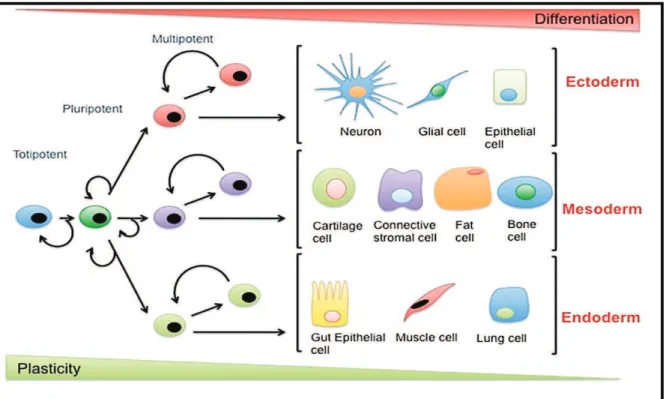
The first stem cell is initiated from the fertilization of an egg by sperm. This unique cell is a totipotent stem cell, which is able to give rise to the precursor cells of all the embryonic and extra-embryonic tissues. After several divisions, the totipotent cells themselves will lose their high proliferative potential and begin to specialize into pluripotent stem cells.
Pluripotent stem cells are capable of generating all the tissue types from the three germ layers (endoderm, mesoderm, ectoderm) necessary for the embryogenesis. Divided pluripotent stem cells give rise to adult stem cells (defined as pluripotent stem cells with restricted capacities, also called multipotent stem cells), able to generate various stem cell types and later on colony forming units and monopotent stem cells. Finally these cells will give rise to mature cells which are in the terminal stage of differentiation and have lost their self-renewal properties. As an example, the multipotent hematopoietic stem cell will produce either common myeloid or lymphoid progenitors, which will then produce colony forming
units of the myeloid or lymphoid lineage and finally all the different blood cell types (erythrocytes, platelets and white blood cells) (Kuldip S. Sidhu 2012).
We thus observe different degrees of plasticity or differentiation potential of normal SCs, as shown in Fig. 1. Alba de Luca.(2015,July 6).
Fig. 1. Differentiation potential and classes of stem cells . ( Retrieved from
http://www.fastbleep.com/biology- notes/32/158/852 . )
2.1.2. Stem cell organization and niche concept
SCs reside and are protected in a specific place called “niche”. This is a specialized microenvironment which includes SCs themselves, neighboring cells and extracellular matrix (see Fig. 2). The niche provides various molecular signals enabling the SCs 1) to undergo self-renew, 2) to differentiate to repair damaged tissues or 3) to go in a quiescent state in terms of cell cycle in order to maintain their potential throughout life (Frank, Schatton et al. 2010, Rezza, Sennett et al. 2014).
Fig. 2. Cellular and molecular components of three fast-cycling adult stem cell niches. (Rezza, Sennett et al. 2014).
2.1.3. The mode of division
SCs have the ability to divide by two different mechanisms. By symmetrical division, the SC gives rise to two identical daughter cells having the same properties as their mother; this allows to expand the pool of SCs (self-renew), as found during the embryonic development. The asymmetric division allows to generate one daughter cell identical to the mother cell (self-renew), while the other daughter cell will be differentiated into a more specialized cell. The balance between asymmetric and symmetric divisions are controlled by the niche that ensures the necessary balance between undifferentiated and differentiated cells (Morrison and Kimble 2006).
2.2. Cancer stem cells
!2.2.1.Generalities
2.2.1.1. Historical overview
The earliest proof of the existence of the CSC is announced in 1855. Actually it was suspected that the cancer arises from embryonic residues present in adults. This hypothesis is based on the similarities between teratocarcinoma and embryo development and can be summarized as follows: “The existence of teratocarcinomas which contain cells of all germ layers and afflict young adults along midline migration pathways between gonads to brain endorses this embryonal rest theory” . This theory then developed and postulated that adult tissues could include embryonic remnants that are quiescent with the possibility to become cancerous if stimulated. The residues of SCs or their descendants in tissues may therefore acquire cancer features after exposure to specific carcinogenic agents (R.Cogle 2011) .
2.2.1.2. Evidence of the existence of CSCs
After improvements in cell sorting techniques and increasing knowledge in immunology, the first conclusive evidence of the existence of CSCs could be published in 1997 by the group of John Dick (Bonnet and Dick 1997). They isolated and identified a subpopulation of human acute myeloid leukemia (AML) cells that express the specific surface marker CD34, but do not express the surface marker CD38. These CD34+/CD38- cells
showed two properties that define normal SCs, namely their ability to proliferate and differentiate after multiple transplantation in non-obese diabetic mice with severe combined immunodeficiency disease (NOD/SCID mice). Furthermore, Dick’s team showed a hierarchical organization of the blast population of AML cells, having a CD34+/CD38 -phenotype similar to normal hematopoietic stem cells. It was therefore hypothesized that mutations of these SCs could have been the cause of the appearance of cancer-initiating cells that were responsible for the emergence of the disease.
The hypothesis of leukemic stem cells encourages the research to investigate whether CSCs exist in solid tumors. This research however progressed slowly because the knowledge of the biology of other normal stem cells, the identification of specific markers and their link
with the occurrence of cancer was much less advanced compared to that related to hematopoietic stem cell and leukemia. Again cell-sorting techniques allow the identification of specific cell surface markers that characterize specific CSCs in variety of solid tumors. The first CSCs were identified in breast cancers (Al-Hajj, Wicha et al. 2003), brain tumors (Singh, Hawkins et al. 2004), prostate cancers (Collins, Berry et al. 2005), colon cancers (O'Brien, Pollett et al. 2007), pancreatic cancers (Li, Heidt et al. 2007) , liver cancers (Ma, Chan et al. 2007), ovarian cancers (Zhang, Balch et al. 2008), Melanoma (Schatton, Murphy et al. 2008) and lung cancers (Eramo, Lotti et al. 2008). In all these cases, the "tumor stem cells" share features of those present in normal SCs (immature phenotype, self-renewal capacity and differentiation potential). As an example, patient-derived brain tumor cells can form spheres having the same phenotypic heterogeneity than that found in the parental tumor.
!
2.2.1.3. Origin of the tumor heterogeneity
Since 150 years, it has been postulated that the genetic background of cells forming the bulk tumor is multi-faceted, which explains the heterogeneity of a tumor (Paget 1989). These variations include differences in proliferation and differentiation potential, in cell surface markers, in invasive capacity, and in responses to therapy (Pietras 2011). Regarding to this heterogeneity, two models for cellular origin of cancer have been proposed: the stochastic and the hierarchy models (Dick 2008).
2.2.1.3.1. Stochastic model
In the stochastic model (Fig. 3), it is predicted that tumor cells are biologically homogenous and have equal likelihood for their contribution in the tumor growth, with a possibility to become tumor-initiating cells (via a dedifferentiation process); this will generate a heterogeneous tumor, due to the influence of intrinsic signals (i.e. transcription factors) or extrinsic signals (i.e. interactive proteins from the microenvironment) (Nowell 1976) (Chandler and Lagasse 2010) .
2.2.1.3.2. Hierarchy model
Thehierarchy model (Fig. 3)postulates that the tumor is hierarchically organized and consists of heterogeneous cells. However only subpopulations of cells have the tumorigenic capacity and contribute to long-term tumor growth. These cells originate from transformed SCs and are considered as cancer-initiating cells, namely also CSCs. In the hierarchical model, CSCs reside at the top, the proliferating progenitors with a limited proliferation and differentiation capacities in the middle and the terminally differentiated cancer cells at the bottom (Nowell 1976). Recent observations revealed that both models can exist together depending on the microenvironment signals. For instance, tumor-initiating cells can originate from both transformed SCs or more differentiated cells which are affected by additional mutations or are epigenetically reprogrammed (Campbell and Polyak 2007).
Fig. 3. Models of tumor heterogeneity a) Tumor cells are stochastically self-renewed or differentiated and form an heterogeneous tumor. b) Only subset CSCs give hierarchically rise to committed progenitors with limited proliferation and differentiation potentials. c, d) In both models, new somatic mutations can increase tumor heterogeneity (Beck and Blanpain 2013).
2.2.2. Carcinogenesis and its consequence
It is well known that tumorigensis is a multistep process initiated by a single normal cell or SC that is affected by irreversible mutations, epigenetic alterations, errors in DNA repair or inappropriate signals from its microenvironment (Brenda Loaiza 2012). In solid tumors, twenty mutations seem be required to induce a cancer phenotype. Actually, to gain this number of mutations, cells should have long lifespan; therefore SCs, which possess this property, are suitable for the initiation of the carcinogenesis process (Jilkine and Gutenkunst 2014). When the initiated cells escape from apoptosis or become undetected by the immune system, they can expand in number and finally can produce malignant tumors. The fact that differentiated descendants might acquire CSC properties by the dedifferentiation process due to additional mutations, can increase the malignity (Trosko, Chang et al. 2004) (Frank, Schatton et al. 2010) (Fig. 4).
Fig. 4. Carcinogenesis, tumorigensis and resistance in CSCs . (Frank, Schatton et al. 2010).
Many evidences have proven the role of CSCs in the metastasis process. For instance, metastatic cancers such as prostate cancers (Collins, Berry et al. 2005), oligodendroglia tumors (Beier, Wischhusen et al. 2008), rectal cancers (Wang, Chen et al. 2009), high grade
of gastric adenocarcinoma (Zhao, Li et al. 2010), colon cancers (Zhang, Liu et al. 2013) , lung cancers (Wu, Qi et al. 2014) and pancreatic cancers (Nomura, Banerjee et al. 2015), correlate with the expression of CD133,a cell surface marker which is known to be expressed in normal and cancer SCs.
Epithelial to mesenchymal transition (EMT) is a key program that is activated during tumor invasion and metastasis. Immortalized human epithelial cells ectopically expressing specific transcription factors acquire a mesenchymal phenotype, with increasing migratory properties. Furthermore, isolated cells from highly invasive mouse or human mammary carcinoma show high expression of EMT markers (Mani, Guo et al. 2008) (Li and Li 2014). As it will be discussed later, Notch signaling pathway, known to regulate stem cell self-renewal, plays essential role in cell migration due to its participation in EMT (Yuan, Wu et al. 2014). Furthermore, Wnt/β-catenin signaling regulates the expression of the angiogenesis factor VEGF (Vascular Epithelial Growth Factor) in colon cancer and hepatocellular carcinoma and consequently increases vessel density (Easwaran, Lee et al. 2003) (Qu, Liu et al. 2014). On the other hand, knockdown of Oct4, un key regulator for the maintenance of the self-renewal process, inhibits cell migration and invasion of lung or colon cancer cells (Chiou, Wang et al. 2010) (Dai, Ge et al. 2013). All these examples finally demonstrate that cells with stemness properties have migratory and invasive potential.
The mechanisms involved in the resistance of CSCs to chemo- and radio-therapies have now been identified. The resistance to chemotherapy involves ATP-binding cassette transporters (ABC transporters), such as the multi drug resistance protein MDR1, which acts as efflux pump to reject drugs. Interestingly, hematopoietic stem cells show high levels of MDR1 (Chaudhary and Roninson 1991) , as well as hepatic or melanoma CSCs (Chow, Fan et al. 2012) (Keshet, Goldstein et al. 2008). On the other hand, CSCs have shown high potential to chemo- and radio-resistance by their strong ability to repair DNA damage and then to escape from apoptosis. For example, glioblastoma stem cells activate protein kinases ATM (Ataxia-Telangiectasia Mutated) and Chk1 (Checkpoint Kinase 1) more immediately after radiations than non-glioma stem cells, which then enable them to survive (Bao, Wu et al. 2006). Furthermore BRCA1 (Breast Cancer 1) and RAD51, known to be involved in the repair of damaged DNA, show increased expression levels in prostate CSCs. In other models of CSCs from colon and lung cancers, an efficient activity of Chk1 is observed, in contrast to their differentiated descendants, when treated with standard chemotherapeutic agents
(Maugeri-Saccà, Bartucci et al. 2012) . Resistance to chemo- or radio-therapies also involve other parameters, always linked with the possibility to induce a programmed cell death or inhibit the prosurvival pathway. For instance, breast cancer stem cells are considered resistant to apoptosis because they show, in comparison to non-CSCs, low levels of ROS (reactive oxygen species), which are known to induce DNA damage and to activate tumor suppressors (Diehn, Cho et al. 2009). Aldehyde dehydrogenase (ALDH), found to be highly expressed in many solid tumors (Abdullah and Chow 2013) and used as a marker to isolate CSCs, is implicated in chemo-resistance by protecting the cells against oxidative damage (Januchowski, Wojtowicz et al. 2013). Several studies demonstrate the role of the prosurvival protein BCL-2 (B-Cell Lymphoma-2) in drug resistance and these studies reveal that this protein is overexpressed in various CSCs lines (He, Zhou et al. 2014). EMT is also able to contribute to the resistance process of CSCs to the chemotherapy (Singh and Settleman 2010). Moreover CSC niche, which provides hypoxic conditions leads to increasing expression levels of hypoxia inducible factors (HIFs) which are correlated with CSCs resistance to the DNA-targeting agents through an activation of DNA repair enzymes (Vinogradov and Wei 2012). It has also been reported that inhibition of HIF-1α overcomes the resistance of lung cancer cells to topotecan and etoposide (Choi, Rho et al. 2009).
The chemoresistance has been also associated with the activity of the signaling pathways that play crucial role in the regulation of normal SCs or CSCs, especially those that promote self-renewal (see Table 1).
Table1. Summary of some chemoresistance-related signaling pathways (Abdullah and Chow 2013).
2.2.3. Cancer stem cell biomarkers
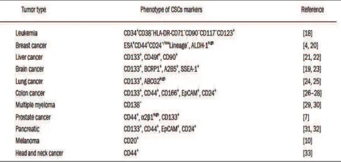
Since CSCs are implicated in the initiation and growth of tumors, in the metastasis and resistance to current therapies, it is crucial to identify them in order to develop appropriate and efficient anticancer treatments. Several cell surface antigens have been characterized as candidate markers for CSCs. For instance, CD133 has been recommended for a prospective isolation of CSCs. However different studies reveal that CD133 is also found in differentiated normal cells of various organs and cancer cells lacking CD133 can also initiate tumors. In glioblastoma tumors, subset of cancer cells with self-renewal capacity have been identified which negatively expressed CD133. In fact, a unique marker is not sufficiently specific for the isolation of one type of CSC (Chen, Nishimura et al. 2010). In general, markers of CSCs are also not steady in a single tumor or between patients, and therefore other tools, such as sphere forming, are required for the isolation of these cells (Wang, Chen et al. 2015) . Different biomarkers have been found in variety of tumors, as shown in Table 2 (Chen, Huang et al. 2013).
Table 2. Cell surface markers of CSCs.
2.2.4. Control of self-renewal and differentiation
As described above, self-renewal is a process achieved by asymmetric or symmetric division to produce one or two daughter cells that are identical to the mother cell. Stem cells undergo a limited number of divisions under physiological conditions in adult tissues whereas
they undergo indefinite division in early embryonic development to generate embryo body. Self-renewal is controlled by extrinsic and/or intrinsic regulatory networks that suppress the expression of differentiation promoting genes and activate the expression of mitosis promoting genes (He, Nakada et al. 2009). Actually, self-renewal is expected to be extended to CSCs when it occurs through different deregulated processes.
2.2.4.1. The role of signaling pathways
The major extrinsic pathways that regulate stem cell self-renewal involve the Wnt/β-catenin, Notch and Hedgehog signaling networks.
2.2.4.1.1. Wnt/ β-catenin signaling pathway
Wnt signaling has been shown to regulate subset of processes in animal development and in several adult tissues. When Wnt ligands bind their receptors (namely frizzled) on the cytoplasmic membrane, they recruit a co-receptor, namely LRP (low-density lipoprotein receptor-related protein). Frizzled can then bind Dsh (disheveled) and induce the phosphorylation of LRP which in turn allows AXIN (alternative name of Axis inhibition protein) to be relocated to the cell membrane; as a consequence, an accumulation of β-catenin occurs which fosters its translocation to the nucleus, its binding to Tcf (T-cell-specific transcription factor) and Lef (lymphoid enhancer-binding factor) and finally the activation of certain transcription factor genes necessary for the maintenance of self renewal. At the opposite, the absence of Wnt ligands leads to the capture of β-catenin by APC (adenomatous polyposis coli) and AXIN; this fosters its phosphorylation by GSK3β (glycogen synthase kinase) or CK1α (casein-kinase 1α), thereby inducing its degradation by the proteasome (Fig. 5) (Clevers and Nusse 2012) (Blank, Karlsson et al. 2008).
The biological role of Wnt/β-catenin in the self-renewal process has been characterized only in some models of stem cells. For example, β-catenin is essential for maintenance of intestinal stem cells (Fevr, Robine et al. 2007), in normal hematopoietic stem cells (Reya, Duncan et al. 2003), neural stem cells (Gong and Huang 2012) and embryonic stem cells (ESCs) (Serio 2014). On the other hand, mutations of Wnt/β-catenin signaling components are known to cause an overexpression of β-catenin. This aberrant expression has been found in prostate cancer stem cell (Chen, Shukeir et al. 2004), in colorectal cancer stem