HAL Id: tel-02177497
https://tel.archives-ouvertes.fr/tel-02177497
Submitted on 9 Jul 2019
HAL is a multi-disciplinary open access
archive for the deposit and dissemination of
sci-entific research documents, whether they are
pub-lished or not. The documents may come from
teaching and research institutions in France or
abroad, or from public or private research centers.
L’archive ouverte pluridisciplinaire HAL, est
destinée au dépôt et à la diffusion de documents
scientifiques de niveau recherche, publiés ou non,
émanant des établissements d’enseignement et de
recherche français ou étrangers, des laboratoires
publics ou privés.
Functional analysis of the TRANSPARENT TESTA 10
gene encoding a polyphenoloxidase of the laccase type.
Przemyslaw Bidzinski
To cite this version:
Przemyslaw Bidzinski. Flavonoid oxidation in Arabidopsis thaliana seeds. Functional analysis of
the TRANSPARENT TESTA 10 gene encoding a polyphenoloxidase of the laccase type.. Molecular
biology. Universite Paris XI Orsay, 2009. English. �tel-02177497�
UNIVERSITE DE PARIS-SUD
U.F.R. SCIENTIFIQUE D’ORSAY
THESE
présentée
pour obtenir le grade de
DOCTEUR EN SCIENCES
DE L’UNIVERSITE PARIS 11, ORSAY
Discipline: Sciences de la Vie
par
Przemyslaw BIDZINSKI
Oxydation des flavonoïdes dans les graines d'Arabidopsis thaliana.
Analyse fonctionnelle du gène TRANSPARENT TESTA 10 codant
une polyphénoloxydase de type laccase
Soutenue le 7 Juillet 2009 devant la commission d’examen composée de :
Graham NOCTOR
Professeur, Université Paris 11, Orsay Président
Alain GOOSSENS
Chercheur, Université Gand/VIB
Rapporteur
Wim SOPPE
Chercheur, MPI Cologne
Rapporteur
Lise JOUANIN
Directrice de Recherche CNRS
Examinatrice
Nancy TERRIER
Chargée de Recherche INRA
Examinatrice
Life, sometimes so wearying
Is worth its weight in gold
The experience of traveling
Lends a wisdom that is old
Beyond our 'living memory'
A softly spoken prayer:
"It's the journey that's important,
Not the getting there!"
[…]
By John McLeod
i
Acknowledgments
After more than three years of the journey through the PhD, the time has come to look back and acknowledge all the people I have shared this part of the the life with.
First, I would like to thank my supervisor Isabelle Debeaujon. Thank you for all your help, discussions, critics and advice. Thank you for patiently answering all my questions (some of them more than once) and sharing your research experience with me.
I would like to thank Annie Marion-Poll and all the members of the Seed Biology Laboratory for excellent work environment.
I would like to acknowledge Adeline Berger for enormous help with the cytology, Anne Frey for guiding and help for the MUG experiment, Nathalie Berger for sharing the secrets of ’boiling’ and ’dirty cloning’, Bertrand Dubreucq for explaining the qRT-PCR and all the members of the laboratory for sharing their experience.
I would like to thank the FLAVO team: Loïc Lepiniec for leading the group and all the support I got from him during my PhD. Lucille Pourcel who shared her first hand experience on work with TT10 and gave the idea to run Paris-Versailles. Jean-Marc Routaboul for all the metabolomic analyses, discussions and comments as well as the best cassoulet one can imagine. Special thanks to Christian Dubos for all the advice and help, especially when it was needed the most.
Special thanks to Martine Miquel for support, encouragement and explaining how to make stroudel dough very thin.
Managing the French administration would not be possible without Thomas Goujon, who also spread good mood everywhere he was passing and shared expert advice on rollerblading.
Thanks to Marie Lacruz and Stéphane Raude who took care of the administrative part during the whole PhD.
One must not forget Michaël Anjuere who was looking after the plants in the greenhouse.
Big thanks to Anne Plessis who explained me how to use LYX and Joseph Tran who helped to put this manuscript in the final shape.
I would like to thank the members of the jury for reading and evaluating this manuscript as well as members of my thesis committees, Lise Jouanin, Olivier Loudet and Alain Goossens who shared their time and experience. Thanks to Gregory Mouille for being the reporter at my first Monday seminar in the Versailles amphitheater.
Thanks to Wim Soppe for hosting me in his lab and Christina Philip for technical assistance in BAC library screening.
I would like to thank all my friends who were patiently listening when I was sharing my last experiments with them. Big thanks to all the VERT students. Special thanks to Laura who shared not only passion for research but also long running kilometers and a climbing rope. Thank you Theodoros for all long discussions, Greek dances and all great moments. Big thanks to Tino. Thanks to all great people I have met living in France and especially in Grignon for the multi-cultural exchange.
Thanks to my family and especially my sister Joanna, who was supporting me from far away. Thank you for always being there.
iii
Abstract
Title:
Flavonoid oxidation in Arabidopsis thaliana seeds. Functional analysis of the TRANSPARENT TESTA 10 gene encoding a polyphenoloxidase of the laccase type.
Abstract: Arabidopsis seeds accumulate flavonoids (proanthocyanidins and flavonols) during their de-velopment. A previous study has shown that a laccase (AtLAC15) encoded by the TRANSPARENT TESTA 10 (TT10 ) gene could trigger flavonoid oxidation in the seed coat. If both proanthocyanidins (PAs) and flavonols appear to be TT10 protein substrates, only PA oxidation leads to brown pigments responsible for the mature seed coat color. An important consequence of TT10 activity on seed flavonoid metabolism is an increased ratio of insoluble to soluble PAs. The physiological functions of TT10 are still unknown, however defense against biotic and abiotic stresses, either constitutive or induced, may be predicted on the basis of present knowledge on polyphenoloxidases.
The purpose of this thesis was to perform a functional characterization of the TT10 gene. A part of the work was devoted to the analysis of the regulatory mechanisms controlling the developmental pattern of TT10 gene expression in seeds and vegetative plant parts. The functional 5’-dissection of a 2.0-kb promoter realized with the uidA reporter gene encoding β-glucuronidase (GUS) was performed to identify regions responsible for activation in seed and other plant organs. TT10 promoter happens to be activated exclusively in seed coat and siliques. Directed mutagenesis was undertaken to precise the regulatory role of in silico-detected cis-acting regulatory elements (CAREs) located in a 194-bp region necessary for expression in seed coat. TT10 gene expression assessed in different tissues at various stages of development using qRT-PCR matched promoter activity pattern. Natural variation for TT10 expression among Arabidopsis accessions was also detected, with the levels of TT10 mRNA in Cvi, Ler and Sha being strongly reduced compared to the ones in Ws, Col and Bay. The impact of this molecular polymorphism on seed flavonoid composition, as analyzed on mature seeds with LC-MS, is discussed. In silico analysis of the TT10 promoter revealed the presence of putative CAREs potentially involved in signaling and response to biotic and abiotic stresses. However
histochemical analysis of GUS activity in transgenic Arabidopsis plantlets expressing pT T 102.0−kb : GU S
failed to detect any ectopic activity when submitted to a variety of stresses. This result suggests that transcriptional response to environmental stimuli is highly constrained by developmental parameters. TT10 appeared to be the only member of the laccase gene family to be strongly expressed in seeds. TT10 function may have evolved towards flavonoid oxidation by co-localization with these substrates, which is ensured by tissue-specific gene expression.
Key words: Arabidopsis, flavonoids, gene, laccase, mutant, promoter, seed, transcriptional regulation Resident laboratory:
Seed Biology Laboratory – INRA, Jean-Pierre Bourgin Institute UMR 204 INRA-AgroParisTech
Résumé
Titre:
Oxydation des flavonoïdes dans les graines d’Arabidopsis thaliana. Analyse fonctionnelle du gèneăTRANSPARENT TESTA 10 codant une polyphénoloxydase de type laccase.
Résumé:
Les graines d’Arabidopsis accumulent des flavonoïdes (proanthocyanidines et flavonols) durant leur développement. Une étude précédente a montré qu’une laccase (AtLAC15) codée par le gène TRANS-PARENT TESTA 10 (TT10 ) induisait l’oxydation des flavonoïdes au niveau des téguments. Si les pro-anthocyanidines (PAs) et les flavonols sont des substrats de la protéine TT10, seule l’oxydation des PAs conduit à la formation de pigments bruns responsables de la couleur de la graine mature. Une importante conséquence de l’activité de TT10 sur les flavonoïdes de la graine est l’augmentation du rapport PAs in-solubles / PAs in-solubles. Les fonctions physiologiques de TT10 sont toujours inconnues, mais un rôle dans la défense contre des stress biotiques et abiotiques constitutifs ou induits peut être prédit sur la base des connaissances actuelles sur les polyphénoloxydases.
L’objectif de cette thèse était de réaliser une analyse fonctionnelle du gène TT10. Une partie de l’étude était consacrée à l’analyse des mécanismes de régulation contrôlant le pattern développemental d’expression du gène TT10 dans les graines et les parties végétatives. Une dissection fonctionnelle en 5’ d’un promoteur de 2.0-kb réalisée à l’aide du gène rapporteur uidA codant la β-glucuronidase (GUS) a été réalisée pour identifier les régions responsables de l’activation du promoteur dans les graines et les autres parties de la plante. Le promoteur de TT10 est activé exclusivement dans les téguments de la graine et les siliques. Une mutagénèse dirigée a été réalisée dans une région de 194 pb nécessaire à l’expression dans les téguments, pour préciser la fonctionnalité des éléments régulateurs agissant en cis (ERACs) identifiés in silico. L’expression du gène TT10 mesurée par qRT-PCR dans différents tissus et à différents stades de développement est en accord avec le pattern d’activité du promoteur. Une variation naturelle pour l’expression de TT10 a aussi été détectée parmi plusieurs accessions d’Arabidopsis, avec des niveaux d’ARNm mesurés chez Cvi, Ler et Sha fortement réduits par rapport à ceux relevés chez Ws, Col et Bay. L’impact de ce polymorphisme moléculaire sur la composition en flavonoïdes de la graine, analysée sur graines matures par LC-MS, est discuté. L’analyse in silico du promoteur de TT10 a révélé la présence d’ERACs potentiellement impliqués dans la signalisation et la réponse aux stress biotiques et abiotiques. Cependant l’analyse histochimique de l’activité GUS de
plantes transgéniques exprimant pT T 102.0−kb : GU S n’a pas permis de détecter d’activité ectopique en
présence de stress variés. Ce résultat suggère que la réponse transcriptionnelle aux stimuli environnementaux est fortement conditionnée par les paramètres développementaux. TT10 est le seul membre de la famille des laccases à être exprimé fortement dans les graines. La fonction de TT10 a probablement évolué vers l’oxydation des flavonoïdes par la co-localisation avec ces substrats, qui est assurée par la spécificité tissulaire d’expression du gène.
Mots clés: Arabidopsis, flavonoïdes, gène, graine, laccase, mutant, promoteur, régulation transcription-nelle
Laboratoire d’accueil:
Laboratoire de Biologie des Semences – INRA, Institut Jean-Pierre Bourgin UMR 204 INRA-AgroParisTech
Contents
Acknowledgments . . . i Abstract . . . iii Résumé . . . iv Table of contents . . . v List of Figures . . . ixList of Tables . . . xii
Abbreviations . . . xv
1 Introduction 1 1.1 Seed biology . . . 1
1.1.1 Seed coat development in Arabidopsis . . . 1
1.2 Flavonoids in Arabidopsis seed . . . 2
1.2.1 Biosynthesis . . . 3
1.2.2 Biological roles of flavonoids . . . 5
1.2.3 Oxidation of end-products . . . 5
1.2.3.1 Laccases . . . 7
1.2.3.2 Catechol oxidases . . . 8
1.3 Regulation of gene expression . . . 8
1.3.1 Regulation of BAN and MYBL2 expression . . . 8
1.3.2 Two-cell layer expression pattern of TT10 . . . 9
1.3.3 One layer, two layers, which layers? . . . 9
1.4 Objectives of the thesis . . . 9
2 Results 11 2.1 Transcriptional regulation of TT10 expression . . . 11
2.1.1 TT10 transcript accumulation during plant development . . . 11
2.1.1.1 Transcript accumulation - RT-PCR . . . 11
2.1.1.2 Transcript accumulation - Transcriptomics . . . 12
2.1.1.3 Analysis of publicly available transcriptomic data for TT10 expression during plant development - Summary . . . 14
2.1.2 In silico analysis of TT10 promoter sequence . . . 15
2.1.2.1 Putative cis-acting regulatory elements found in the TT10 promoter . . . . 16
2.1.2.2 Putative CAREs associated with seed development, stress response and/or protection against various biotic and abiotic stresses . . . 17
2.1.2.3 CAREs found in promoters of flavonoid biosynthetic genes . . . 18 v
2.1.2.4 Additional comments about known transcription factors involved in flavonoid
biosynthesis . . . 19
2.1.2.5 Other CAREs . . . 19
2.1.2.6 TATA box . . . 20
2.1.2.7 In silico analysis of TT10 promoter sequence - Summary . . . 20
2.1.3 Studies of TT10 promoter activity during plant development . . . 22
2.1.3.1 5’ dissection of the TT10 promoter . . . 22
2.1.3.2 In planta analysis of GUS activity . . . 23
2.1.3.3 Quantitative GUS assay - MUG . . . 25
2.1.3.4 Sub-dissection and site-directed mutagenesis - introduction . . . 25
2.1.3.5 Patterns of GUS activities after TT10 promoter sub-dissection and site-directed mutagenesis . . . 28
2.1.4 Promoter GUS activity in protoplasts . . . 30
2.2 Finding transcription factors . . . 31
2.2.1 TT10 promoter activity in candidate regulatory mutant backgrounds . . . 31
2.2.2 Confirmation of TTG2 promoter activity in developing seeds . . . 32
2.2.3 Analysis of public micro-array data - searching for co-expressed genes . . . 33
2.2.3.1 TT10 expression in candidate regulatory mutant backgrounds obtained from co-expression analysis - seed set . . . 33
2.2.3.2 Results for regulatory mutants chosen from co-expression analysis . . . 35
2.2.3.3 Co-expression analysis - developmental set . . . 37
2.2.4 Analysis of TT10 expression in mutants involved in flavonoid biosynthesis . . . 38
2.2.5 Candidate genes related to stress response which could be involved in the regulation of TT10 expression . . . 39
2.2.6 Yeast one hybrid screening - fishing out transcription factors binding TT10 promoter 42 2.3 Insights from natural variation . . . 44
2.3.1 Differences in TT10 transcript accumulation . . . 44
2.3.2 Analysis of promoter sequences . . . 45
2.3.2.1 Focus on the proximal part of the promoter . . . 46
2.3.2.2 SNP vs. TT10 transcript accumulation . . . 48
2.3.2.3 Analysis of long promoter fragments . . . 48
2.3.2.4 BAC library screening . . . 48
2.3.2.5 Going further in the promoter: amplification of longer fragments . . . 48
2.3.3 Flavonoid composition in matures seeds of various Arabidopsis accessions . . . 49
2.3.4 Functional complementation of Ler accession with Col-0 TT10 genomic region . . . . 52
2.3.5 Functional analysis of the Columbia promoter in other backgrounds . . . 53
2.4 Stress induction of the TT10 promoter activity . . . 54
2.4.1 Abiotic stresses . . . 54
2.4.2 Wounding and biotic stresses . . . 55
2.5 Identification of laccases expressed in seeds . . . 57
2.5.1 Expression data . . . 57
2.5.2 Flavonoid composition of mature seeds of the laccase mutants . . . 58
2.5.3 Promoter GUS activities . . . 59
2.5.3.1 LAC3 . . . 60
CONTENTS vii
2.5.3.3 LAC12 . . . 60
2.5.3.4 Summary . . . 60
2.6 Cell suspension as a tool to study flavonoid biosynthesis . . . 65
3 Materials and Methods 67 3.1 Materials . . . 67
3.1.1 Plant Material . . . 67
3.1.1.1 Arabidopsis thaliana - wild type accessions . . . 67
3.1.1.2 Arabidopsis thaliana - mutants . . . 67
3.1.1.3 Arabidopsis thaliana - transgenic plants . . . 71
3.1.1.4 Arabidopsis thaliana - cell suspension . . . 72
3.1.2 Bacterial Strains . . . 72
3.1.2.1 Escherichia coli, strain DH10B . . . 72
3.1.2.2 Escherichia coli, One shot® TOP 10 . . . 72
3.1.2.3 Escherichia coli, DB3.1 . . . 72
3.1.2.4 Agrobacterium tumefasciens, strain 58C1pMP90 . . . 72
3.1.3 Yeast strain . . . 72
3.1.3.1 Saccharomyces cerevisiae, strain EGY48 . . . 72
3.1.3.2 Yeast strains in the REGIA transcription factor library . . . 72
3.1.4 Plasmids and expression vectors . . . 73
3.1.5 Cvi and Ler BAC library at MPI, Cologne . . . 74
3.1.6 Transcription factor library . . . 75
3.2 Methods . . . 75
3.2.1 Handling of bacteria . . . 75
3.2.1.1 Culture . . . 75
3.2.1.2 Preparation and transformation of electro-competent bacteria . . . 75
3.2.2 Handling of yeast . . . 75
3.2.2.1 Culture . . . 75
3.2.2.2 Preparation and transformation of yeast . . . 76
3.2.3 Handling of Arabidopsis thaliana . . . 76
3.2.3.1 Seed sterilization . . . 76
3.2.3.2 Plants growth conditions in vitro . . . 76
3.2.3.3 Plants growth conditions in greenhouse . . . 76
3.2.3.4 Crosses . . . 76
3.2.3.5 Plant transformation by the floral dip method . . . 77
3.2.3.6 Maintenance of cell suspensions . . . 77
3.2.3.7 Genotyping . . . 77
3.2.3.8 Transient expression of promoter:GUS constructs in Arabidopsis protoplasts 77 3.2.4 DNA methods . . . 77
3.2.4.1 Isolation of plasmid DNA from E. coli . . . 77
3.2.4.2 DNA fragments purification . . . 77
3.2.4.3 Isolation of genomic DNA from plants . . . 77
3.2.4.4 Rapid DNA extraction for genotyping . . . 78
3.2.4.5 Polymerase chain reaction (PCR) . . . 78
3.2.4.7 Sequencing . . . 79
3.2.4.8 Site-directed mutagenesis . . . 79
3.2.5 RNA methods . . . 79
3.2.5.1 RNA isolation and cDNA synthesis . . . 79
3.2.5.2 RT-PCR . . . 79
3.2.5.3 Quantitative real-time RT-PCR (qRT-PCR) . . . 79
3.2.5.4 Normalization controls for qRT-PCR . . . 80
3.2.5.5 Primer efficiency . . . 80
3.2.6 Histochemical methods for GUS detection and quantification . . . 82
3.2.6.1 Histochemical detection of GUS activity . . . 82
3.2.6.2 Quantification of GUS activity (MUG assay) . . . 82
3.2.7 Analysis of seed flavonoids . . . 82
3.2.8 Routine techniques . . . 83
3.2.9 Internet resources and services . . . 83
3.2.9.1 The Arabidopsis Information Resource - TAIR . . . 83
3.2.9.2 The Arabidopsis Thaliana Integrated Database - ATIDB . . . 83
3.2.9.3 Integrative database around plant genomes - FLAGdb++ v.3.9 . . . 83
3.2.9.4 The Complete Arabidopsis Transcriptome Micro Array database - CATMA Database . . . 83
3.2.9.5 AtcisDB at AGRIS . . . 83
3.2.9.6 The Genevestigator - expression database and meta-analysis system . . . 84
3.2.9.7 The Bio-Array Resource for Arabidopsis Functional Genomics - BAR . . . . 84
3.2.9.8 GeneCAT - Gene Co-expression Analysis Toolbox . . . 84
3.2.9.9 DIURNAL . . . 84
3.2.9.10 PLACE and Signal Scan - A Database of Plant Cis-acting Regulatory DNA Elements . . . 84
3.2.9.11 Plant CARE . . . 84
3.2.9.12 Plant PAN - Plant Promoter Analysis Navigator . . . 85
3.2.9.13 ATHENA . . . 85
3.2.9.14 Basic Local Alignment Search Tool - BLAST . . . 85
3.2.9.15 Multiple Sequence Alignment . . . 85
3.2.9.16 PrimerX - Primer design for the site directed mutagenesis . . . 85
4 Discussion, Conclusions and Perspectives 87 4.1 Regulation of TT10 gene expression . . . 87
4.1.1 Transcript accumulation . . . 87
4.1.2 Developmental vs. stress regulated expression . . . 88
4.1.3 Regulatory components . . . 89
4.1.3.1 Promoter and Cis-acting regulatory elements . . . 90
4.1.3.2 Candidate transcription factors for developmental regulation . . . 92
4.1.3.3 Candidate transcription factors for stress regulation . . . 93
4.1.3.4 What have we learned from natural variation? . . . 94
4.1.3.5 Transcription factor library screening . . . 95
4.1.3.6 Co-expressed genes . . . 95
CONTENTS ix
4.2 TT10 and flavonoid metabolism in seeds . . . 97
4.2.1 TT10 and flavonoid metabolism . . . 97
4.2.2 Other functions? . . . 97
4.3 Regulation in the multi-gene family . . . 98
4.4 Final remarks . . . 98
5 Appendices 99 5.1 In silico analysis of the TT10 promoter . . . 99
5.1.1 Putative cis-acting regulatory DNA elements found in PLACE database using the Signal Scan . . . 99
5.1.2 CAREs found in AtcisDB . . . 103
5.2 Primers . . . 105
5.3 Seeds colors . . . 110
5.4 Map of the methylation in the TT10 gene . . . 112
5.5 Transposon in TT10 promoter . . . 113
List of Figures
1.1 Testa structure and flavonoid localization in Arabidopsis seed. . . 2
1.2 The structure of the flavonol quercetin. . . 3
1.3 Flavonoid biosynthesis pathway in Arabidopsis. . . 4
1.4 Enzymatic reactions for polyphenoloxidases (laccase, catechol oxidase). . . 6
1.5 Seed coat pigmentation in Arabidopsis: illustration of a browning process. . . 6
1.6 Seed coat sections showing the co-localization of TT10 promoter activity, mRNA expression and protein activity with flavonoid substrates (5-daf seeds). . . 7
1.7 Spatial pattern of TT10 promoter activity in WT seeds. Comparison with BAN and MYBL2 promoter activities. . . 10
2.1 Expression pattern of the TT10 gene in developing siliques and seeds of wild-type Col-0 plants. 12 2.2 TT10 expression visualized with eFP Browser. . . 13
2.3 Comparison of TT10 and BAN expression visualized with GeneCAT expression profiling tool. 14 2.4 Comparison of TT10 and CHS expression visualized with DIURNAL. . . 15
2.5 TT10 genomic region and neighboring genes. . . 16
2.6 In silico analysis of proximal (500bp) TT10 promoter showing putative cis-acting regulatory DNA elements. . . 21
2.7 Pattern of TT10 promoter activity in wild-type plants. . . 24
2.8 Comparison of GUS activity of 5’-dissected TT10 promoter in planta. . . 26
2.9 GUS activity driven by the the TT10 promoter in pCAMBIA binary vector. . . 27
2.10 Pattern of TT10 promoter activity in seeds of the candidate regulatory mutant backgrounds. 32 2.11 Pattern of TTG2 promoter activity in wild-type seeds. . . 33
2.12 Expression levels of candidate regulators of TT10 in developing seeds. . . 35
2.13 TT10 transcript accumulation in candidate regulatory mutant background - co-expressed candidates. . . 36
2.14 Flavonoid composition of mature seeds in the mutants for genes selected from co-expression analysis. . . 37
2.15 TT10 transcript accumulation in candidate regulatory mutant backgrounds. . . 39
2.16 TT10 transcript accumulation in candidate regulatory mutant backgrounds - stress related candidates. . . 41
2.17 Flavonoid composition of mature seeds of the mutants - stress related candidates. . . 41
2.18 Summary of yeast one-hybrid experiment. . . 43
2.19 Assessment of TT10 expression levels in various Arabidopsis accessions. . . 45
2.20 Promoter sequence alignment and comparison of the putative CAREs present in various ac-cessions. . . 48
2.21 Polymorphism for length of TT10 promoter amplification. . . 49
2.22 Flavonoid composition of mature seeds of various accessions and tt10 alleles. . . 51
2.23 TT10 transcript accumulation in Ler plants transformed with the Col-0 wild-type TT10 genomic region. . . 52
2.24 Flavonoid composition of mature seeds from Ler lines complemented with the Col-0 wild-type genomic region. . . 53
2.25 Stress experiments - experimental design. . . 54
2.26 Plantlets subjected to progressive dehydration. . . 56
2.27 Ectopic GUS activity of TT10 promoter. . . 56
2.28 Comparison of the expression pattern of the laccase multigene family in seed and silique. . . . 58
2.29 Flavonoid composition of mature seeds of the laccase mutants. . . 59
2.30 Pattern of LAC3 promoter activity in wild-type plants. . . 62
2.31 Pattern of LAC5 promoter activity in wild-type plants. . . 63
2.32 Pattern of LAC12 promoter activity in wild-type plants. . . 64
2.33 Total RNA and flavonoid pathway genes expressed in the cell suspension. . . 65
3.1 Maps of the vectors used for cloning. . . 74
3.2 Flower marking and silique sample collection. . . 80
3.3 Expression of APT1 and TUBULINE in siliques 7DAF. . . 81
4.1 Organization of the regulatory elements in the TT10 Promoter. . . 91
5.1 Visualization of cis-acting regulatory elements found with AtcisDB search engine. . . 103
5.2 Seed colors of various Arabidopsis accessions and tt10 mutant allels, (A). Seed color of the plants used in the Ler ’complementation’ experiment, (B). . . 110
5.3 Seed color of various mutants used in the study - Part 1. . . 111
5.4 Seed color of various mutants used in the study - Part 2. . . 111
5.5 Seed color of mutants in laccase gene. . . 111
5.6 Methylation map of the TT10 gene region. . . 112
List of Tables
2.1 Summary of TT10 promoter activity pattern in planta. . . 25
2.2 Patterns of GUS activity observed in the seed coat after sub-dissection and site-directed mutagenesis of TT10 promoter. . . 30
2.3 List of selected genes co-expressed with TT10 - Seed Set. . . 34
2.4 List of selected genes co-expressed with TT10 - developmental set. . . 38
2.5 Concentration of hormones, salts and other treatments in stress experiments. . . 55
2.6 Comparison of laccase promoter activities. . . 61
3.1 Wild type accessions of Arabidopsis thaliana used in this work. . . 68
3.2 Alleles of the tt10 mutant studied in this work. . . 68
3.3 Arabidopsis thaliana mutant lines used in this study. . . 68
3.4 Transgenic lines generated during this work. . . 71
3.5 List of vectors used in this study. . . 73
3.6 Antibiotics . . . 75
3.7 Efficiency of the 3Fw/3Rw set of primers. . . 81
5.1 Putative cis-acting regulatory elements found in TT10 promoter with Signal Scan. . . 103
5.2 Putative cis-acting regulatory elements found in TT10 promoter with AtcisDB. . . 105
5.3 Primers used for promoter dissection to clone fragments in pBS-GUS vector. . . 105
5.4 Primers used for amplification of long promoter fragments in Ler and Cvi accessions. . . 105
5.5 Primers used to amplify promoter fragments for transcription factor library screening with pHISi vector. . . 106
5.6 Primers used for site-directed mutagenesis on pTT10_3 promoter fragment in pBS-GUS vector.106 5.7 Primers used for probes preparation for BAC library screening. . . 106
5.8 Primers used for promoter dissection - GATEWAY® cloning. . . 106
5.9 Primers used to study laccase family genes expression. . . 107
5.10 Primers used for laccase promoter cloning. . . 107
5.11 Primers used in quantitative RT-PCR. . . 108
5.12 Primers used for genotyping of SALK T-DNA mutants. . . 108
5.13 Other primers used in the study. . . 108
5.14 CATMA GST primers used to study expression of flavonoid biosynthesis genes. . . 109
Abbreviations
# number, clone
A adenosine
ATP adenosine-5’-triphosphate
aa amino acid
ABA abscisic acid
AGI arabidopsis genome initiative
attB1, attB2 recombination sites in GATEWAY® cloning
BAC Bacterial Artificial Chromosome
BAR Bio-Array Resource
Bay Bayreuth
bp base pairs
BSA Bovine Serum Albumin
C cytidine
CARE(s) Cis-Acting Regulatory Element(s)
cDNA complementary DNA
CDS coding sequence
Col Columbia
Cvi Cape Verde Islands
DAF Days After Flowering
DMF N,N-dimethylformamide
DMSO dimethylsulfoxide
DNA deoxyribonucleic acid
DNase deoxyribonuclease
dNTP deoxyribonucleotide triphosphate
°C degrees Celsius
DTT dithiothreitol
EC epicatechin
EDTA ethylene diamine tetraacetic acid
EF1 ELONGATION FACTOR 1αA4
eFP electronic fluorescent pictograph
e.g. exempli gratia(for example)
EMS ethane methyl sulfonate
EST expressed sequence tag
EtOH ethanol
g gram
G guanosine
GA gibberellic acid
gcRMA GeneChip Robust Multiarray Averaging
gDNA genomic DNA
GFP green fluorescent protein
h hours
HIS histidine
ii inner integument
INRA French National Institute for Agricultural Research (fr. l’Institut National de la Recherche Agronomique)
kb kilo base
l liter
LAC laccase
LB Luria-Bertani medium
LC-MS liquid chromatography coupled with mass spectrometry detector
Ler Landsberg erecta
μ micro
m meter
M mega
M molar concentration (mole/liter)
MCS multi cloning site
mm millimeter
MPI(K) Max Planck Institute (Cologne)
mRNA messenger ribonucleic acid
MS mass spectrometry
n nano
OD optical density
oi outer integument
o/n over night
PA(s) proanthocyanidins (condensed tannins)
p promoter
PCR polymerase chain reaction
pH negative logarithm of the proton concentration
qRT-PCR quantitative real time - PCR
RIL(s) recombinant inbred line(s)
RMA Robust Multiarray Averaging
RNA ribonucleic acid
RNase H ribonuclease H
RT reverse transcription
RT room temperature
s seconds
SD minimal synthetic defined media
SDS sodium dodecyl sulphate
Sha Shakdara
T thymidine
T-DNA transferred DNA
TF(s) transcription factor(s)
Tris 2-amino-2-hydroxymethyl-propane-1,3-diol
TT/tt TRANSPARENT TESTA
uidA β-glucuronidase
UTR untranslated region
UV ultraviolet
v/v volume per volume
Ws Wassilewskija
w/v weight per volume
w/w weight per weight
WT wild-type
x · g gravity force
X-gluc 5-bromo-4-chloro-3-indolyl-β-D-glucuronide
YPD yeast peptone dextrose medium
Nomenclature:
TT10 written in capital letters for the protein
TT10 written in capital letters, italics for the gene
tt10 written in small letters, italics for the mutant in the gene
pTT10:GUS transcriptional fusion of the TT10 promoter and reporter gene uidA
T1 primary transformants
T2 second generation of the T-DNA transformed plants
In the degenerated motifs, letters stand for nucleotides: N: A, C, G or T; R: A or G; W: A or T; Y: C or T
Chapter 1
Introduction
1.1
Seed biology
Formation of the seed is crucial in the life cycle of higher plants. Seeds serve several functions, among which the keys are nourishment of the embryo, dispersal to a new location, and dormancy to survive during unfavorable conditions. A typical seed of angiosperms, including Arabidopsis is constituted by the embryo enclosed in the endosperm, which supplies nutrients and a seed coat (integuments, testa) which provides protection (see Fig. 1.1 A.).
1.1.1
Seed coat development in Arabidopsis
In spermatophytes, the ovule is formed by the embryo sac which is surrounded by the nucellus and integu-ment(s). After fertilization, the ovule develops into a seed containing a newly formed embryo immersed in the endosperm and enclosed in the testa. The seed coat is a maternal tissue derived from the differentiation of the integuments, whereas endosperm and embryo are of both maternal and paternal origin because of the double fertilization. The endosperm in Arabidopsis mature seed is reduced to one cell layer and is tightly associated with the seed coat.
The Arabidopsis testa includes two integuments in which micropyle pore is formed allowing entrance of the pollen tube, and the chalazal tissues. Three cell layers constitute the inner integument (ii), whereas the outer integument is two-layered. The five cell layers follow one of four distinct fates, e.g. subepidermal oi1 layer is accumulating flavonols, whereas the innermost cell layer (ii1, endothelium) specializes in proanthocyanidin (PA) biosynthesis. PAs are also accumulating in some cells in the chalazal region (pigment strand) creating a continuum of the tannin-producing cells. The vascular tissues of the maternal funiculus are connected with the seed in the chalazal region, which after seed detachment forms a scar called hilum. Formation of the testa involves cellular differentiation as well as programmed cell death. The cellular organization of the integuments is easily distinguishable at the heart stage of embryo development (see Fig. 1.1 A.), however further modifications are leading to the formation of the mature seed coat (see Fig. 1.1 B.). At the late stages of seed development, structure of the epidermal cells which were accumulating starch and forming mucilage is preserved. Other layers are crushed together by that time, PAs are released from the endothelial cells and impregnate the adjacent cell layers. The mature seed coat protects the embryo, contributes to seed dormancy by creation of a physicochemical barrier and helps seed dispersal. Many studies demonstrated that phenolic compounds, especially flavonoids play an important role in plant protection against environmental constraints (e.g. resistance to pathogens and herbivores, UV radiation) and also contribute to the germination-inhibiting
effects. Seed coat development has been recently reviewed in Haughn and Chaudhury (2005), and Debeaujon et al. (2007).
Figure 1.1: Testa structure and flavonoid localization in Arabidopsis seed.
(A) Anatomy of a developing seed at the heart stage of embryo development (longitudinal section). Cells accumulating either proanthocyanidins or flavonols are highlighted in black or gray, respectively. The en-dothelium corresponds to the ii1 layer. (B) Cross section of a mature testa. c, chalaza; cl, columella; cpt, chalazal proliferating tissue (nucellus); ct, cuticle; cv, central vacuole; cw, cell wall; e, embryo; h, hyaline layer; ii, inner integument; m, micropyle; mu, mucilage; oi, outer integument; pe, peripheral endosperm (aleurone layer); ps, pigment strand; s, suspensor; vb, vascular bundle.
Bar = 40 μm in A and 7 μm in B. (Adapted from Debeaujon et al., 2007).
1.2
Flavonoids in Arabidopsis seed
Flavonoids are plants secondary metabolites derived from the phenylpropanoid pathway. Most of the polyphenolic compounds, namely hydroxycinnamates, coumarins, lignins and flavonoids are derived from phenylalanine. Flavonoid structure is characterized by two aromatic cycles (A- and B- rings) linked by a heterocycle (C-ring) (see Fig. 1.2). At this moment there are more than 6500 molecules isolated and grouped in several classes according to the oxidation degree of the C-ring. These classes include flavonols, antho-cyanins and flavan-3-ols. Additional modifications, including hydroxylations, methylations, glycosylations, acylations or prenylations account for the diversity within a compound class (Pourcel, 2006; Routaboul et al., 2006; Winkel-Shirley, 2006). The condensation of flavan-3-ols results in the formation of proanthocyanidins (Dixon et al., 2002; Marles et al., 2003; Pourcel et al., 2005).
Two classes of flavonoids, namely flavonols and proanthocyanidins are synthesized and accumulate in a tissue-specific manner in Arabidopsis seeds (Routaboul et al., 2006). Before ovule fertilization, cells of the endothelium layer are characterized by a dense cytoplasm. Rapid vacuolization occurs after fertilization and colorless tannin precursors can be detected (Devic et al., 1999). PA biosynthesis starts around 1-2 days after flowering (DAF). First, they accumulate in vacuoles of the endothelium cells as colorless compounds until 6-7 DAF (Debeaujon et al., 2003). At the beginning of seed desiccation (10 DAF) PAs stored in the vacuoles migrate to the cell walls, where oxidation occurs, leading to formation of brown pigments
1.2. FLAVONOIDS IN ARABIDOPSIS SEED 3 contributing to the color of wild-type Arabidopsis seeds. Flavonols are found in the endosperm and embryo, however in the testa they are essentially accumulated in the oi1 layer. In mature seeds, flavonols are present mainly as glycoside derivatives with quercetin-3-O-rhamnoside being the most abundant (Pourcel et al., 2005; Routaboul et al., 2006). Interestingly, natural variation in the quantity of PAs and flavonols occurs among Arabidopsis accessions (Lepiniec et al., 2006; Routaboul et al., 2006), which might reflect an important trait of plant adaptation to different habitats. Flavonoid composition of mature arabidopsis seeds has been recently described in details by Routaboul et al., 2006.
Figure 1.2: The structure of the flavonol quercetin.
Quercetin is given as an example of carbon numbering. Important features influencing antioxidant potential are the di-hydroxylated B-ring, unsaturation and a 4-oxo function at the C-ring (see Williams et al., 2004). (Adapted from Pourcel et al., 2007).
1.2.1
Biosynthesis
A majority of the genes involved in flavonoid biosynthesis were identified through visual screenings of various collections of Arabidopsis mutants for altered seed coat pigmentation (Koornneef, 1990; Shirley et al., 1995; Lepiniec et al., 2006). Many mutants have been called transparent testa (glabra) for the altered pigmentation of the seed coat. One of the mutants, banyuls (ban) is peculiar, because in place of PAs it accumulates anthocyanins. The transparent testa 10 (tt10 ) mutant is also unique in the fact that it does not affect the biosynthesis of PAs but their subsequent oxidative browning (Pourcel et al., 2005). It is important to note that six of the tt mutants are encoding transcription factors which are involved in the flavonoid biosynthetic genes regulation as well as they are required for normal seed development and cell differentiation. Flavonoid metabolism in seeds and mutants in genes affecting this metabolism have been recently reviewed in Lepiniec et al. (2006) and are summarized in Figure. 1.3.
The TT10/LAC15 gene encodes a polyphenol oxidase which belongs to a laccase multigene family of 17 members (Pourcel et al., 2005). TT10/LAC15 has been shown to be involved in the formation of epicatechin quinones that spontaneously polymerize into brown derivatives. Moreover, it may catalyze the oxidative browning of colorless PAs, which is consistent with the fact that the tt10 mutant accumulates wild-type PA levels but yellow seeds at harvest. TT10/LAC15 may also be involved in the formation of biflavonols from quercetin-3-O-rhamnoside (see Fig. 1.3 and Fig. 1.5). Interestingly, natural variation in TT10 transcript accumulation occurs between Arabidopsis accessions (Pourcel et al., 2005).
Figure 1.3: Flavonoid biosynthesis pathway in Arabidopsis.
Three major flavonoid end-products are formed: colorless proanthocyanidins (condensed tannins), which brownish upon oxidation by the TT10 / LAC15 laccase and are seed-coat specific, flavonol derivatives (col-orless) which are ubiquitous and can be dimerized by TT10 to form biflavonols, and anthocyanins (purple), which are only in vegetative parts. Enzymes are presented in capital letters. Regulatory factors are in bold capital letters and circled. Corresponding mutants are in italics green. Abbreviations: ANR, anthocyanidin reductase; CE, condensing enzyme; CHI, chalcone isomerase; CHS, chalcone synthase; DFR, dihydroflavonol reductase; F3H, flavonol 3-hydroxylase; F3’H, flavonol 3’-hydroxylase; FLS, flavonol synthase; GT, glyc-osyltransferase; LAC, laccase; LDOX, leucoanthocyanidin dioxygenase; OMT, O-methyltransferase; PFG, production of flavonol glycosides; RT, rhamnosyltransferase; tt, transparent testa; ttg, tt glabra. (Adapted from Lepiniec et al., 2006; Pourcel, 2006 and Stracke et al., 2007).
1.2. FLAVONOIDS IN ARABIDOPSIS SEED 5
1.2.2
Biological roles of flavonoids
Flavonoids are important in many aspects of plant life in relation to interactions with environment. It has been associated with resistance to pathogens and herbivores (Dixon and Paiva, 1995; Dixon et al., 2002). It seems that flavonoids can also contribute to seed longevity by providing protection against various biotic and abiotic stresses (Debeaujon et al., 2000; Winkel-Shirley, 2002; Rajjou et al., 2008). They can also reinforce testa-imposed dormancy by decreasing seed coat permeability (Debeaujon et al., 2000). Flavonols have been shown to act as signaling molecules for bacteria forming nitrogen-fixing nodules. They are also required for pollen tube germination (Taylor and Grotewold, 2005). Among other functions of flavonoids it is important to point their antioxidant and antimicrobial properties which are beneficial not only for plants but also for human and animal health (Scalbert et al., 2005; Selmi et al., 2006; Aron and Kennedy, 2008). However, some can also have a negative impact on the industrial use of some crop seeds (e.g rapeseed), or on protein digestibility and absorption from seed meal (Winkel-Shirley, 2001a; Marles et al., 2003; Dixon et al., 2005).
1.2.3
Oxidation of end-products
The protective function of flavonoids against biotic and abiotic stresses can be related to their cytotoxicity and antioxidant abilities. It seems that flavonoid oxidation contributes to these physiological and chemical properties. The oxidation of colorless flavan-3-ols and proanthocyanidins reinforces their cross-linking to the cell wall and leads to the formation of brown seed coat pigments, whereas oxidation of flavonols results in formation of antifungal agents. The topic of flavonoid oxidation in plants has been recently reviewed in Pourcel et al. (2007).
Flavonoid oxidation in planta is mainly mediated enzymatically by polyphenoloxidases (PPOs) and peroxidases, however it can also occur spontaneously (see Fig. 1.4 and Fig. 1.5). PPOs and peroxidases have been shown to be expressed during normal plant development, however they can be also induced by various environmental causes, like pathogen attacks, wounding or desiccation (Mayer and Staples, 2002; Thipyapong and Steffens, 1997; Thipyapong et al., 2004; Mayer, 2006). Physiological role of those genes seems to be regulated by differential subcellular localization of enzymes and substrates as well as by transcriptional and post-transcriptional events during gene expression.
Browning of Arabidopis seed coat is mainly due to the oxidation of epicatechin and soluble PAs by TT10 laccase (Pourcel et al., 2005). The tt10 mutant displays a delay in seed coat browning, an increase in soluble (non-oxidized) PAs and a default in the formation of biflavonols from quercetin-3-O-rhamnoside. The pattern of TT10 promoter activity analyzed with the GUS (see Fig. 1.6 A.) and GFP reporter genes as well as in planta TT10 activity (see Fig. 1.6 D.) showed that TT10 colocalizes with tannin-and flavonol-accumulating cell layers in the seed coat (see Fig. 1.6 andPourcel et al. (2005). Recent results confirmed that TT10 mRNA and also protein were localized in endothelium and outer integument 1 cell layer (see Fig. 1.6 B. and C.).
Figure 1.4: Enzymatic reactions for polyphenoloxidases (laccase, catechol oxidase).
Polyphenoloxidases (PPOs) are oxidoreductase enzymes acting on diphenols in presence of molecular oxygen (O2). Laccase (LAC) is able to oxidize both ortho- and para-diphenols. Catechol oxidase (CO) is able to oxidize ortho-diphenols. (Adapted from Pourcel et al., 2007).
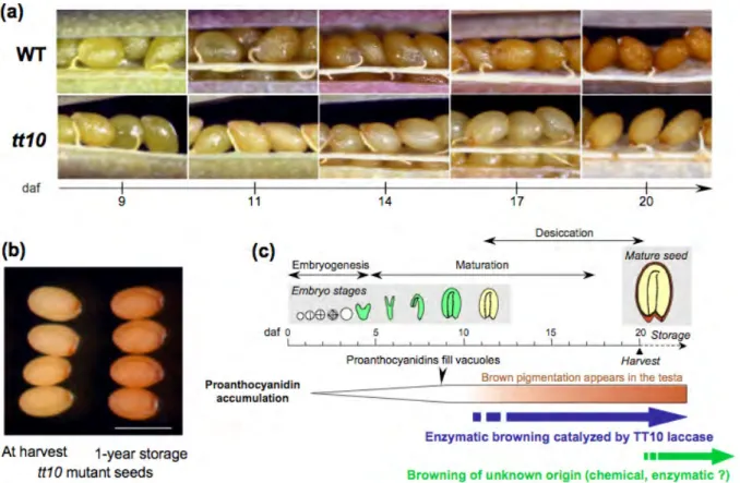
Figure 1.5: Seed coat pigmentation in Arabidopsis: illustration of a browning process.
(a) Pictures showing the apparition of a brown pigmentation in the testa of the wild-type genotype (WT) during seed desiccation. The brown pigment is absent from the transparent testa 10 (tt10 ) mutant defective for a laccase enzyme. (b) Mutant tt10 seeds slowly get brown after harvest until resembling WT seeds. Bar = 550 μm. (c) Schematical drawing pointing to the occurrence of brown pigmentation during Arabidopsis seed development. daf, days after flowering. (Adapted from Pourcel et al., 2007).
1.2. FLAVONOIDS IN ARABIDOPSIS SEED 7
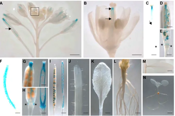
Figure 1.6: Seed coat sections showing the co-localization of TT10 promoter activity, mRNA expression and protein activity with flavonoid substrates (5-daf seeds).
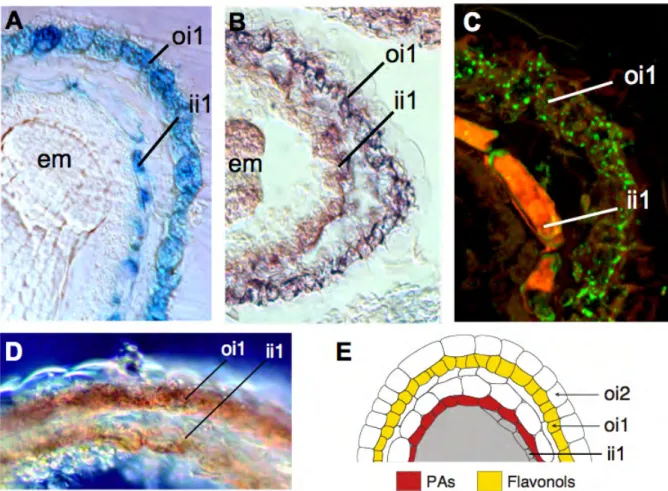
(A) Promoter activity (pTT10:uidA); (B) In situ hybridization with TT10 cDNA probe; (C) Immuno-localization of TT10 protein with anti-HA antibody (pTT10:TT10:3xHA); (D) Protein oxidative browning activity in presence of epicatechin substrate; (E) Schematic representation of the integumentary structure showing flavonoid localization.
B. and C. are unpublished data from Lucille Pourcel. A., D. and E. are from Pourcel et al., 2005.
Abbreviations: em, embryo; ii, inner integument; oi, outer integument; PAs, proanthocyanidins; HA -hemagglutinin.
1.2.3.1 Laccases
Laccases (LAC), i.e. o-and p-diphenol:dioxygen oxidoreductases (EC 1.10.3.2) belong to a larger group of enzymes called multicopper or blue copper oxidases, which includes ascorbic acid oxidase and ceruloplasmin (reviewed in Mayer and Staples, 2002). These glycoproteins are characterized by four histidin-rich copper binding domains and ability to oxidize phenolic substrates in the presence of molecular oxygen (see Fig. 1.4). Also polyphenol oxidases of the catechol oxidase type (EC 1.10.3.1) require molecular oxygen for their activity, what makes them distinct from peroxidases, which require hydrogen peroxide (Pourcel et al., 2007). Laccases have been found in eukaryotes (fungi, plants and insects) as well as in prokaryotes (Claus, 2004; Riva, 2006). The most studied are the fungal laccases, which are involved in spore pigmentation, virulence and delignification (Mayer and Staples, 2002; Pourcel et al., 2007). Plant laccases are less studied and until now were mainly associated with lignification, even if a direct activity of the enzyme on monolignol polymerization within the cell matrix has not yet been demonstrated (Mayer and Staples, 2002). Plant laccases were also shown to be involved in wound healing, iron metabolism detoxification and browning
reactions (Pourcel et al., 2007 and references therein). 1.2.3.2 Catechol oxidases
Catechol oxidases (CO) are enzymes catalyzing the oxidation of monophenols to o-diphenols as well as they can oxidize o-diphenols to the corresponding o-quinones (Marusek et al., 2006) (see Fig. 1.4). Like laccases, COs are glycosylated copper-binding proteins, that require molecular oxygen for their activity. They were found in eukaryotes and prokaryotes (Marusek et al., 2006; Mayer, 2006). Interestingly, the physiological roles of CO seem to be very close to those reported for laccases. It is not surprising as both groups of enzymes are involved in oxidative browning which in consequence can contribute to protection against UV radiations, and also senescence, wound healing and seed coat hardening (Pourcel et al., 2007 and references therein).
Interestingly, it seems that the Arabidopsis genome does not contain any typical catechol oxidase (Pourcel et al., 2005). Their lack could have been filled by laccases, in the sense of a specialization toward o-diphenol oxidation.
1.3
Regulation of gene expression
Regulation of gene expression includes all the processes leading to formation of the gene product, RNA and protein. All the steps of gene expression may be modulated, from gene transcription to the post-translational modifications of a protein. However, expression of many genes is regulated at the transcriptional level and depends on the combinatorial contribution of transcription factors, which can act as activators or repressors. Gene expression regulated at the transcriptional level results in precise spatio-temporal manner activity of their promoter. Below, expression patterns of flavonoid-related genes are given as an example.
1.3.1
Regulation of BAN and MYBL2 expression
Regulation of BAN (Debeaujon et al., 2003) and MYBL2 (Dubos et al., 2008) differs at the level of the spatial activities of their promoter in the seed coat: BAN is present in one integumentary cell layer (ii1) and MYBL2 is present in two cell layers (ii1 and oi1).
The entry step into the proanthocyanidin branch of flavonoid biosynthesis is formation of epi-flavan-3-ols. This reaction is metabolized by anthocyanidin reductase encoded by BANYULS (BAN ) (see Fig. 1.3). BAN promoter activity was detected specifically in PA-accumulating cells in the the inner integument 1 layer (endothelium) of the seed coat and pigment strand in the chalaza zone (see Fig. 1.7 G. and Debeaujon et al. (2003). The BAN promoter deletion and gain-of-function experiments led to identification of 86-bp promoter region which functions as an enhancer specific for PA-accumulating cells. Studies of the BAN promoter activity in regulatory mutant background revealed that its activity was abolished in tt2, tt8 and ttg1 (Debeaujon et al., 2003). The spatial pattern of BAN promoter activity was also modified in tt1 and tt16, but not in ttg2 (Debeaujon et al., 2003). Further studies revealed that TT2/TT8/TTG1 regulatory complex binding to the enhancer element in BAN promoter is required for its expression (Baudry et al., 2004).
Recently, a MYB family transcription factor, MYBL2 has been shown to regulate the transcription of genes involved in anthocyanin biosynthesis (Dubos et al., 2008). Although MYBL2 promoter activity is detected in vegetative tissues, in seeds its activity is restricted to the endothelium and epidermis (see Fig. 1.7 H. and Dubos et al., 2008). Cis-acting regulatory elements in promoter region and transcription factors required for MYBL2 expression are yet unknown.
1.4. OBJECTIVES OF THE THESIS 9
1.3.2
Two-cell layer expression pattern of TT10
TT10 promoter activity colocalizes first with PA-and flavonol-producing cells of the testa. Activity of TT10 promoter starts at early stages of seed development. First it is detected in the endothelium and in the pigment strand at the chalaza zone (see Fig. 1.7 A., C., and D.). Later, the activity increases and spreads to the outer integument l cell layer of the seed coat (see Fig. 1.7 E., and F.). The uidA gene was also strongly expressed in early aborted seeds (see Fig. 1.7 B.). Any difference in GUS activity compared with the wild type was observed in three regulatory mutants tt2, tt8, and ttg1 (Pourcel et al., 2005).
1.3.3
One layer, two layers, which layers?
It seems that regulation of gene expression in the seed coat is very complex. Genes can be expressed in one or more layers of the testa. Moreover they can be expressed at various developmental stages of the seed coat development. Identification of cis-acting regulatory elements required for gene expression in each of the seed coat cell layers or combination of layers would allow to target expression of other genes for crop improvement.
1.4
Objectives of the thesis
The main focus of this thesis was the functional analysis of the TRANSPARENT TESTA 10 gene. The work was following two axis, where developmental and possible stress regulation of TT10 expression was studied.
The first part of the work dealing with the developmental regulation of TT10 expression required detailed characterization of promoter activity throughout the plant to complete existing data. Functional analysis of TT10 promoter was aiming at identification of the promoter region and cis-acting regulatory elements required for seed coat specific activity. Identification of the CAREs allowing to drive gene expression sep-arately in endothelium (PA specific) or outer integument 1 (flavonol specific) cell layer, were one of the goals.
In parallel, existing natural variation for TT10 expression was exploited as it could provide an additional information about regulation of gene expression.
Several complementary approaches, namely studies of TT10 promoter activity and gene expression level in candidate regulatory mutant background as well as transcription factor library screening were aiming at identification of transcription factors which could directly regulate TT10 expression.
Second part of the work dealing with possible stress regulation was aiming at identification of the stress conditions in which TT10 would be expressed. The tools developed in the first part were planned to be used for identification of the promoter region and cis-acting regulatory elements involved in the stress regulation of TT10 expression.
The secondary objectives of this thesis were aiming at identification of other laccases expressed in seeds and manipulations of Arabidopsis cell suspensions to study flavonoid metabolism in simplified system.
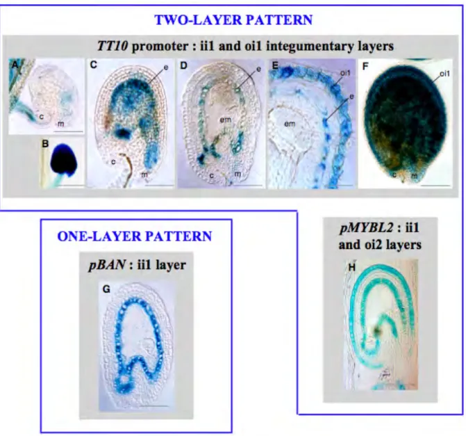
Figure 1.7: Spatial pattern of TT10 promoter activity in WT seeds. Comparison with BAN and MYBL2 promoter activities.
(A) to (F) Expression of the ProTT10:uidA cassette in developing seeds at 1 DAF A, 3 DAF C and D, 8 DAF E and F, and in an aborted seed B. GUS activity is observed with Nomarski optics on whole mounts for A to C and F, and on sections for D and E.
(G) Pattern of pBAN activity in developing seed at 4 DAF. (H) Pattern of pMYBL2 activity in developing seed at 4 DAF.
Abbreviations: c, chalaza; em, embryo; e, endothelium; ii, inner integument; m, micropyle; oi, outer integu-ment. Bars = 24 μm for F and K; 70 μm for A to D, G and H.
Chapter 2
Results
The main objective of my work was to study the regulation of TT10 gene expression. The results of experiments, techniques and approaches, concerning different aspects of gene expression, which have been used to shed light on the mechanism of TT10 expression, are described in details in following sections: TT10 expression and promoter studies (section: 2.1), techniques aiming at finding transcription factors involved (section: 2.2), insights from natural variation occurring for TT10 expression (section: 2.3) and potential stress response (section: 2.4). Each section consists of a short introduction, presentation of the results and technical discussion of the approach used.
Two secondary objectives of this work were: I) to identify other laccases expressed in seeds (section: 2.5) and II) to establish conditions and optimize techniques for the manipulation of Arabidopsis cell suspensions as a tool to study the flavonoid metabolism (section: 2.6).
2.1
Transcriptional regulation of TT10 expression
This section is focusing on TT10 expression during plant development and detailed characterization of TT10 promoter. At first, our analysis of TT10 transcript accumulation is presented and compared to publicly available expression data (2.1.1). Then the promoter activity is studied during plant development, using uidA reporter gene (2.1.3). In silico analysis (2.1.2) of the promoter sequence was used as a basis to design the 5’ dissection of the promoter (2.1.3.1), and its site-directed mutagenesis (2.1.3.4). In silico analysis of the promoter sequence is supported by literature search, documenting on the functionality of cis-acting regulatory elements (CARE).
2.1.1
TT10 transcript accumulation during plant development
2.1.1.1 Transcript accumulation - RT-PCR
The expression pattern of TT10 was previously investigated by RT-PCR (McCaig et al., 2005; Cai et al., 2006) and semiquantitative RT-PCR (Pourcel et al., 2005). In those studies TT10 transcript was detected to be: i) in all the plant organs except stem (McCaig et al., 2005), ii) predominantly in developing silique, at low level in stem, seedling and flower but not in root (Pourcel et al., 2005), iii) in silique, root, flower and stem but not in leaf (Cai et al., 2006). Here, we wanted to revisit and complete previously known and sometimes contradictory data using the very sensitive quantitative RT-PCR method, with special attention to silique development. More robust and precise data about TT10 expression were required for the analysis of its regulation. Quantitative RT-PCR had also been chosen to study differences in TT10 transcript
accumulation between accessions (see: 2.3.1) as well as in candidate regulatory mutant backgrounds (see: 2.2.3, 2.2.4 and 2.2.5).
The expression pattern was investigated in various tissues of 6-week-old Col-0 plants grown in long day conditions in the greenhouse (Fig. 2.1 A.). The TT10 mRNA was detectable as early as the open-flower stage. Detailed analysis of transcript accumulation in developing siliques, revealed a huge increase between 5 and 7 days after flowering (DAF) followed by a slight decrease at 10 DAF, and a second peak of transcript accumulation around 15 DAF. In older siliques the amount of TT10 mRNA decreases and is close to 0% of ELONGATION FACTOR 1αA4 (EF1 ) control at 21 DAF, but is still detectable. TT10 is highly expressed in developing seeds, reaching up to 350 %EF at 5 DAF and 250 %EF at 10 DAF (Fig. 2.1 B.). TT10 transcript was not detected in dry seeds, cauline and rosette leaves, stem at the lower part and terminal node.
Fig. TT10 expression has been studied during plant development with the special atention for the development of the silique. The resulats presented are for the Col-0 accesion and are representetion of three biological repetitions. (Bar = 2cm)
Developmental expression of TT10 in Col-0 accession of Arabidopsis thaliana with a special attention for the dev of the sillique: The result are the mean +- standard deviation of three biological replicates 21 17 15 13 10 7 5 3 DAF 300 200 100 Open flower 3 5 7 10 13 15 17 21 Days After Flowering (DAF)
0
%EF
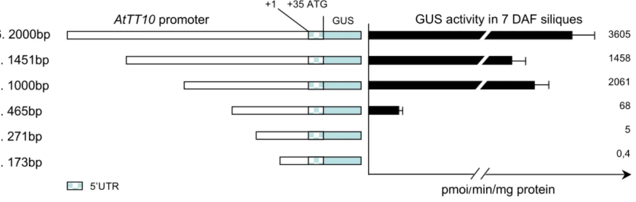
Developing siliques
A.
B.
Figure 2.1: Expression pattern of the TT10 gene in developing siliques and seeds of wild-type Col-0 plants. TT10 gene expression in wild-type Col-0 plants was detected by quantitative RT-PCR and is presented as a percentage of the expression of the reference gene ELONGATION FACTOR 1αA4 (EF 1). For details see 3.2.5.
(A) Picture illustrates siliques development and samples used for RNA extraction. Graph is representing one out of two biological repeats of the experiment.
(B) TT10 gene expression in extracted seeds.
Values represent averages ±SE of three technical replicates. Bar = 2 cm. 2.1.1.2 Transcript accumulation - Transcriptomics
Publicly available transcriptomic data are a constantly growing resource, therefore we checked (last update 7.04.2009), if new combinations of tissue/developmental stage/treatment have revealed new information about TT10 expression. Genevestigator, Bio-Array Resource (BAR), Gene Co-expression Analysis Toolbox (GeneCAT), DIURNAL and several other analysis and visualization tools were used (see section 3.2.9). For many functions those web-based tools are redundant, but for some analysis one is more adapted than the other. Below (Fig. 2.2) as an example, analysis of TT10 expression with the eFP Browser (at BAR) is presented. This tool is well adapted to gene expression analysis thanks to easily readable color code and graphic representation of plant organs/tissues for which the data are presented. Fast switch between data sets (e.g. developmental/stress), standard deviation and threshold filtering as well as easily accessible expression
2.1. TRANSCRIPTIONAL REGULATION OFTT10 EXPRESSION 13 values for more detailed analysis are making eFP Browser a very convenient tool for gene expression studies. The TT10 expression analyzed and visualized with the eFP Browser confirms that it is mainly expressed in developing seed/silique but also shows expression in pollen grain (Fig. 2.2).
Arabidopsis eFP Browser http://bar.utoronto.ca/efp/cgi-bin/efpWeb.cgi
1 of 1 17/04/09 17:14
Arabidopsis eFP Browser
Data Source Mode Primary AGI ID Secondary AGI ID Signal Threshold
Developmental Map
Developmental Map
Absolute
Absolute
This probe set reaches its maximum expression level (expression potential) of 3258.06 in the Developmental Map or Tissue Specific source.
Click Here for Table of Expression Values
248735_at was used as the probe set identifier for your primary gene, At5g48100 (TT10_LAC15__TT10 (TRANSPARENT TESTA 10); laccase )
See information about this gene at TAIR
Find co-expressed genes with Expression Angler
Perform electronic Northerns to examine this gene's response under different conditions with Expression Browser
See distribution of average expression levels for the probe sets on the ATH1 GeneChip in the samples used for the eFP Browser
determine if a given gene is a high or low expresser. The small graph that is shown on the eFP output in Compare and Absolute modes indicates the highest level of expression for the primary gene in red, and the highest level of expression of the secondary gene in blue. The grey line indicates the maximum level of expression of the first gene in any data source
At5g48100 At1g01010 100.0
Figure 2.2: TT10 expression visualized with eFP Browser.
eFP Browser is presenting gene expression as a heat map placed on the graphic representation of the Ara-bidopsis organs - Developmental Map - AtGenExpress Developmental Set (Schmid et al., 2005). Standard deviation filtering has been used to mask samples with the deviation greater than half of their expression value (grey). Signal threshold filtering has been set at 100 (expression potential) to eliminate samples where expression is considered to be below the detection level (yellow). Data are not available for separate siliques (white) from torpedo stage of embryo development onwards, where only dissected seeds were analyzed. De-veloping seed, silique and pollen where TT10 gene expression potential is > 100 (detectable) are painted in red. For details see: http://bar.utoronto.ca/efp/cgi-bin/efpWeb.cgi and Winter et al. (2007).
Figure 2.3 is presenting comparison of TT10 and BAN expression, which is nicely visualized thanks to Expression Profiling Tool available at GeneCAT. This tool allows comparing the expression of multiple genes in different plant tissues. BAN is a core enzyme of the subpathway of flavonoid biosynthesis leading to proanthocyanidins, which are later oxidized by TT10 (Debeaujon et al., 2003; Pourcel et al., 2005). The GeneCAT analysis shows two peaks of TT10 expression in developing seeds/siliques as well as weak signal in pollen and even weaker in flower and root. From the GeneCAT analysis BAN expression seems to be seed/silique specific, with a peak of expression at an earlier stages than TT10, what corresponds to the RT-PCR results presented in Debeaujon et al. (2003).
Roots
Developing
siliques
and seeds
Figure 2.3: Comparison of TT10 and BAN expression visualized with GeneCAT expression profiling tool. Graph presents expression level and tissues in which TT10 (closed squares blue) and BAN (triangles -green) are expressed. Expression profiling tool uses RMA normalized data from 121 ATH1 microarrays generated during AtGenExpress project. For details see: http://genecat.mpg.de/cgi-bin/Ainitiator.
pyand Mutwil et al. (2008).
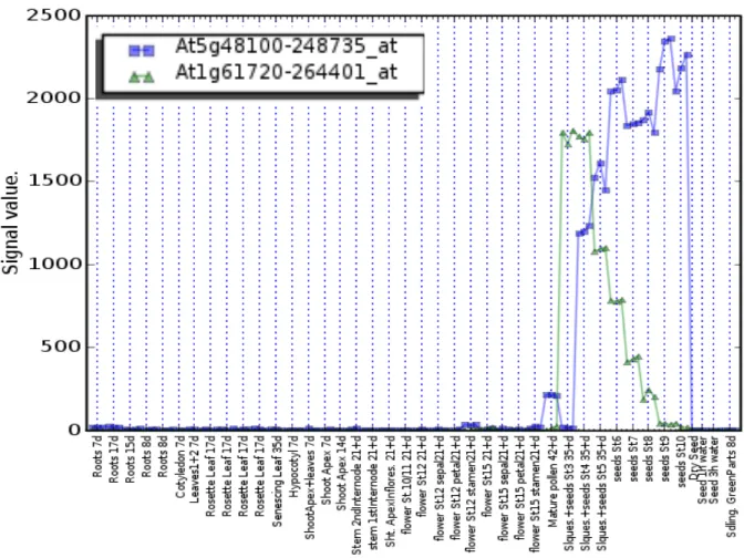
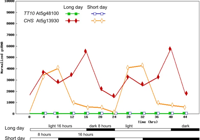
Figure 2.4 is presenting the DIURNAL tool which was used to analyze if TT10 expression could be affected by the circadian rhythm/photoperiod. Interestingly, expression of the key enzyme of flavonoid biosynthesis, encoded by CHALCONE SYNTHASE (CHS) is regulated by light and is presented as an example of gene expression affected by the circadian rhythm (Thain et al., 2002). We can clearly see that its transcript is accumulating during the day, whereas it is much lower during the night (Fig. 2.4). The signal values for TT10 in those experiments are very low, therefore it is not possible to conclude if photoperiod is affecting its expression.
2.1.1.3 Analysis of publicly available transcriptomic data for TT10 expression during plant development - Summary
Taken together all the transcriptomic data analyzed, it seems that TT10 accumulation is significant only in developing silique and seed. In some experiments, TT10 is on limit of detection level (pollen and root), however there are limited repetitions available and the standard variation is high for those data which should therefore be treated with caution. TT10 expression is below the detection level in the circadian/photoperiod experiments.
2.1. TRANSCRIPTIONAL REGULATION OFTT10 EXPRESSION 15
CHS At5g13930 TT10 At5g48100
Long day Short day
Long day Short day
light 16 hours dark 8 hours light dark
8 hours 16 hours
Figure 2.4: Comparison of TT10 and CHS expression visualized with DIURNAL.
TT10 and CHS expression in long and short day is presented as a graphical output adapted from the DIURNAL gene expression analysis tool. TT10, closed green and open blue squares for long and short days respectively. CHS, closed red and open orange diamond/rhombus for long and short days respectively. Samples, 7-day-old seedlings were grown in MS agar medium supplemented with 3% sucrose. For details see: http://diurnal.cgrb.oregonstate.edu/diurnal_details.html and Mockler et al. (2007).
2.1.2
In silico analysis of TT10 promoter sequence
Several web-based computer programs (see 3.2.9) recognizing known Cis-Acting Regulatory Elements (CAREs) have been used for the in silico analysis of the 2.0-kb TT10 promoter sequence Col-0, including the 5’ UTR (for promoter sequence used in the study see Appendix, 5.1). Detection of CAREs in the promoter by so-called ’search by signal’ methods are prone to give false positive results (Rombauts et al., 2003). For that reason the results obtained should be treated as putative cis-acting regulatory elements and their function-ality should be tested in vivo. In order to select the best candidates for further analysis, literature mining accompanied in silico analysis to find out the context and organization of the motifs in the promoter.
PLACE SignalScan server was the preferred tool as it is searching for the presence of motifs identical or similar to the previously reported cis-acting regulatory elements in the PLACE database, which presently seems to be the richest with 469 entries of CAREs deposited (PLACE 30.0, 469 entries, Jan.8, 2007). The full list of putative cis-acting regulatory elements found in the TT10 promoter is presented in the Appendix, Table 5.1. Similar results were obtained when querying PlantCARE, AtcisDB at Agris (see Appendix Tab. 5.2), ATHENA and PlantPan. In addition, in silico analysis revealed no tandem repeats regions, nor CpNpG islands in -1000bp - + 500bp of the At5g48100 locus (PlantPan) and no over-represented TF binding site has been found (ATHENA).
It is important to note that TT10 is not sharing its promoter sequence with any known or predicted gene and that the preceding gene (At5g48110) is transcribed from the same strand. The region considered as TT10 promoter is the 3’ region for that gene (Fig. 2.5). In such a case any changes arising in the promoter region on the course of the evolution would probably affect essentially TT10 expression.
Arabidopsis thaliana Chromosome 5
8.5-kb intergenic region
2.0-kb TT10 promoter studied
Figure 2.5: TT10 genomic region and neighboring genes.
The organization of the TT10 genomic region on chromosome 5 (top, blue rectangle) is presented. Tran-scription units are represented with arrows indicating the orientation of the transcripts. Bottom part is showing the gene structure where rectangles are representing exons and lines introns. UTR’s, when present are shaded orange. Figure adapted from ATIDB.
2.1.2.1 Putative cis-acting regulatory elements found in the TT10 promoter
Studies on yeast promoters suggest that regulatory elements are commonly present in the 500bp upstream region of the transcription initiation site (Caselle et al., 2002). Many plant genes have been shown to contain regulatory elements required for the tissue specific gene expression in the proximal part of their promoters (Abe et al., 2003; Debeaujon et al., 2003; Nakashima et al., 2006; Zhou et al., 2009). For that reason special attention has been taken to analyze this part of the TT10 promoter in details as well as to focus on the dissection of the proximal part of the promoter (see 2.1.3.1). Selected putative cis-regulatory elements found in that part of the promoter are presented in Figure 2.6 and described below. Several criterions have been used to limit the list of putative CAREs considered: 1.) tandem repeats and length of the motif (longer regarded as more significant) , 2.) literature evidence for being involved in seed development and/or defense/stress response (associated with putative function of laccases and flavonoids), and/or 3.) motifs similar to the one previously reported to be involved in flavonoid biosynthetic genes regulation.
Motifs are briefly described, moreover examples of transcription factors are given and briefly discussed in the context of the putative cis-acting regulatory elements.