RESEARCH OUTPUTS / RÉSULTATS DE RECHERCHE
Author(s) - Auteur(s) :
Publication date - Date de publication :
Permanent link - Permalien :
Rights / License - Licence de droit d’auteur :
Institutional Repository - Research Portal
Dépôt Institutionnel - Portail de la Recherche
researchportal.unamur.be
University of Namur
Meta-analysis and gene set analysis of archived microarrays suggest implication of the
spliceosome in metastatic and hypoxic phenotypes
De Meulder, Bertrand; Berger, Fabrice; Bareke, Eric; Depiereux, Sophie; Michiels, Carine;
Depiereux, Eric
Published in:
PLoS ONE
DOI:
10.1371/journal.pone.0086699
Publication date:
2014
Document Version
Publisher's PDF, also known as Version of record
Link to publication
Citation for pulished version (HARVARD):
De Meulder, B, Berger, F, Bareke, E, Depiereux, S, Michiels, C & Depiereux, E 2014, 'Meta-analysis and gene
set analysis of archived microarrays suggest implication of the spliceosome in metastatic and hypoxic
phenotypes', PLoS ONE, vol. 9, no. 1, e86699. https://doi.org/10.1371/journal.pone.0086699
General rights
Copyright and moral rights for the publications made accessible in the public portal are retained by the authors and/or other copyright owners and it is a condition of accessing publications that users recognise and abide by the legal requirements associated with these rights. • Users may download and print one copy of any publication from the public portal for the purpose of private study or research. • You may not further distribute the material or use it for any profit-making activity or commercial gain
• You may freely distribute the URL identifying the publication in the public portal ?
Take down policy
If you believe that this document breaches copyright please contact us providing details, and we will remove access to the work immediately and investigate your claim.
Microarrays Suggest Implication of the Spliceosome in
Metastatic and Hypoxic Phenotypes
Bertrand De Meulder1., Fabrice Berger1., Eric Bareke2, Sophie Depiereux3, Carine Michiels4, Eric Depiereux1*
1 Microorganism Biology Research Unit -NARILIS, University of Namur, Namur, Belgium, 2 Sainte Justine University Hospital Center Research Center, University of Montreal, Montreal, Canada,3 Environmental and Evolutional Research Unit, University of Namur, Namur, Belgium, 4 Cellular Biology Research Unit - NARILIS, University of Namur, Namur, Belgium
Abstract
We propose to make use of the wealth of underused DNA chip data available in public repositories to study the molecular mechanisms behind the adaptation of cancer cells to hypoxic conditions leading to the metastatic phenotype. We have developed new bioinformatics tools and adapted others to identify with maximum sensitivity those genes which are expressed differentially across several experiments. The comparison of two analytical approaches, based on either Over Representation Analysis or Functional Class Scoring, by a meta-analysis-based approach, led to the retrieval of known information about the biological situation – thus validating the model – but also more importantly to the discovery of the previously unknown implication of the spliceosome, the cellular machinery responsible for mRNA splicing, in the development of metastasis.
Citation: De Meulder B, Berger F, Bareke E, Depiereux S, Michiels C, et al. (2014) Meta-Analysis and Gene Set Analysis of Archived Microarrays Suggest Implication of the Spliceosome in Metastatic and Hypoxic Phenotypes. PLoS ONE 9(1): e86699. doi:10.1371/journal.pone.0086699
Editor: Aedı´n C. Culhane, Harvard School of Public Health, United States of America Received September 19, 2013; Accepted December 10, 2013; Published January 31, 2014
Copyright: ß 2014 De Meulder et al. This is an open-access article distributed under the terms of the Creative Commons Attribution License, which permits unrestricted use, distribution, and reproduction in any medium, provided the original author and source are credited.
Funding: This work was supported by the FRS-FNRS Te´le´vie (BDM) and FRS-FRNS(SD). FB is a volunteer for the University of Namur Biology Department. EB is funded by Prof. Daniel Sinnett (Division of Hematology-Oncology, Sainte-Justine UHC Research Center, University of Montreal). The funders had no role in study design, data collection and analysis, decision to publish, or preparation of the manuscript.
Competing Interests: The authors have declared that no competing interests exist. * E-mail: [email protected]
.These authors contributed equally to this work.
Introduction Cancer & metastasis
Despite the development of effective therapies for many cancers [1-3], the prevalence of cancer is growing alarmingly in aging populations [4]. Metastases are one of the main causes of death related to cancer [5]. It is therefore not surprising that a large number of labs and researchers focus on gaining a better understanding of the metastatic process [6–8].
Cancer is known to be a genetic disease, implying either alteration of DNA or dysregulation of gene expression [9]. In addition, the metastatic phenotype involves the combination of several factors [7], among which a hypoxic micro-environment has been reported to be a major/key parameter [10–12]. Several hypotheses have been proposed to explain this observation. First, a mechanism of adaptation is initiated, mediated by the HIF-1 transcription factor, to enhance cell survival [13]. Second, the cell response to hypoxic conditions also triggers the angiogenesis process [14]. Lastly, hypoxia has been reported to affect the selection of high potential metastatic cells [15]. As this manuscript focuses on the bioinformatics analysis of the data, we direct the reader to the following reviews for a more detailed discussion of the role of hypoxia in the development of metastasis [16–18].
Microarrays
In the last decade, the availability of microarray datasets in public repositories has grown dramatically (i.e. ArrayExpress [19], GEO [20]...). As an example, the number of datasets in the Gene Expression Omnibus (GEO) has increased from 2,000 to more than 780,000 over the last ten years (2002–2012). Previously, most researchers focused on a small handful of probe sets spotted on the arrays, ignoring thousands of other probe sets. Despite the financial cost associated with creating large collections of public datasets (millions of euros/dollars), the incomplete and/or partial analysis of the datasets consequently suggests that a large body of underexploited information could be put to use in further analyses. Many authors has also significantly improved the performance of statistical analyses by solving methodological issues [21–23], and developing the alternative chip definition file (CDF) [24]. We propose to make use of this wealth of information by including several microarray datasets, from experiments studying similar/ common biological issues, in a single analytical pipeline that makes use of the latest and best-performing algorithms, without preconceived biases.
Data preparation
Datasets must be preprocessed in preparation for statistical analysis to improve the quality of the data (background correction), to allow for a fair comparison between arrays
(standardization), and to summarize probe-level intensities to meaningful probe set values [25,26]. Several benchmarks have previously been reported to assess the performances of preproces-sing methods [27,28].
The last preprocessing step, called summarization, consists of gathering probe-level information regarding the same target. The mapping of the target definition to the probe coordinates on the chips involves a chip definition file (CDF). The annotation of the human genome has improved since the first release of CDFs by the manufacturer (Affymetrix) and several authors have thus reported the need to update the definition of chip definition files [29,30]. In 2007, Liu et al described the affyprobeminer as a tool to ease the mapping of current knowledge to probe sequences in Affymetrix arrays [24]. The authors reported discrepancies ranging from 30 to 50% between standard Affymetrix and remapped chip definition files. Affyprobeminer can also be used to build both transcript- and gene-consistent CDFs, meaning that a probe-set is defined to gather probes that specifically target only one transcript, or gene, respectively.
Single gene analysis of one dataset
Microarray data can be used to track the expression profile of the transcriptome following a hierarchical strategy that involves many levels of interpretation. The first level refers to individual analyses aimed at inferring the positive/negative regulation of transcripts and/or genes, as defined in the chip definition file (probe set definition in CDF). Wet-lab biologists mainly interpret microarray experiments based on the results of this step. Additional layers of analysis are described briefly in the next subsections (meta-analysis and gene set analysis).
In previous work, we described a relationship between the number of replicates and the selection of the best performing methods [31]. The two main results are that the best method overall is the Shrinkage t test [22], bested solely by the Window t test [32] and Regularized t test [21] when only two replicates are available; the other main result is that the overall power of such an analysis is relatively low, depending on the number of replicates available. Therefore, the authors claimed that future
methodological developments should focus on augmenting that power and on an appropriate filtering of the results.
Annotation of a list of candidate genes
After the individual analyses, the list of genes detected as differentially expressed is typically annotated using over-represen-tation analysis (ORA) methods to highlight meaningful informa-tion. In a previous work, we described the use of the DAVID webtools to perform such an analysis on the results of microarray studies [33]. The DAVID webtool analyzes the list of differentially expressed genes and returns a list of the pathways containing part of these genes, associated with an over-representation score (EASE score) [34].
Differential expression analysis of gene sets
Small datasets with only a few replicates are still a major hindrance to statistical power in conventional analyses. Gene set analysis and meta-analysis are interesting and common ways to extract more information from the data, and to test higher-level hypotheses with a power level associated with an increased number of available values.
Gene set analysis using Functional Class Scoring methodologies (FCS) has improved the understanding of differences in expression profiles, and helped unravel the biological processes underlying experimental data in several ways. First, joint analysis of multiple genes involves a higher number of values than individual analyses, hence providing the potential for a higher power level, even when conducted on small datasets (small number of replicates). Second, computation of differential expression from multiple levels of interpretation enriches the qualitative description of biological variations between experimental conditions. The criteria used to define the gene sets consequently guide interpretation of the results (i.e. regulation element/transcription factor, metabolic pathways, pathology signatures, locus, cellular components...). By extension, the comparison of the results of individual and gene set analyses allows, as with ORA methods, to refine the list of candidate genes for further testing, thanks to the criteria-based approach (i.e. if all but one gene of a set of related genes are detected as "silenced" due
Figure 1. Summary of the analytical pipeline. doi:10.1371/journal.pone.0086699.g001
to deletion, one can remove this potential false negative or screen the genome for an additional copy of the gene).
Over the last decade, various Functional Class Scoring methodologies (FCS) have been developed to analyze gene sets, including 2-step or global methods, competitive or self-contained null hypothesis and inference (gene-sampling, label-sampling...): GSEA [35,36], SAMGS [37], GlobalTest, [38]...
Method-specific biases in the detection of gene sets are associated with methodological choices, and are due to correla-tions between genes, the simultaneous presence of up/down regulated genes, the level of expression and the number of genes in
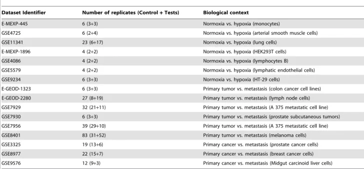
the set.... In order to detect all kinds of sets with an expression profile that differs between conditions, we developed FAERI, tailored from the two-way ANOVA [39]. Prior to analysis, FAERI applies a 2-step data reduction to avoid previously observed biases. The null distribution can then be evaluated from simulations or sample permutations. Performance comparisons conducted both on simulated and biological data illustrate that FAERI, evaluated using sample permutations, provides the most accurate results versus other methods, regardless of the composition of the gene sets (in terms of direction, level of expression, correlation and proportion of DEGs in the set). Mansmann and Meister similarly Table 1. List of the 16 datasets used in this manuscript.
Dataset Identifier Number of replicates (Control+ Tests) Biological context
E-MEXP-445 6 (3+3) Normoxia vs. hypoxia (monocytes)
GSE4725 6 (2+4) Normoxia vs. hypoxia (arterial smooth muscle cells) GSE11341 23 (6+17) Normoxia vs. hypoxia (lung cells)
E-MEXP-1896 4 (2+2) Normoxia vs. hypoxia (HEK293T cells)
GSE4086 4 (2+2) Normoxia vs. hypoxia (lymphocytes B)
GSE5579 4 (2+2) Normoxia vs. hypoxia (lymphatic endothelial cells)
GSE9234 6 (3+3) Normoxia vs. hypoxia (HT-29 cells)
E-GEOD-1323 6 (3+3) Primary tumor vs. metastasis (colon cancer cell lines) E-GEOD-2280 27 (8+19) Primary tumor vs. metastasis (lymph node cells) GSE7929 32 (21+11) Primary tumor vs. metastasis (A 375 metastatic cell line) GSE7930 6 (3+3) Primary tumor vs. metastasis (prostate subcutaneous tumors) GSE7956 39 (29+10) Primary tumor vs. metastasis (A 375 metastatic cell line) GSE8401 83 (31+52) Primary tumor vs. metastasis (melanoma cells) GSE3325 19 (13+6) Primary cancer vs. metastasis (prostate cancer cells) GSE8977 22 (15+7) Primary cancer vs. metastasis (breast cancer cells) GSE9576 12 (9+3) Primary cancer vs. metastasis (Midgut carcinoid liver cells) doi:10.1371/journal.pone.0086699.t001
Figure 2. Volcano plots for two datasets. Each volcano plot is related to a single data set, chosen among the different technologies and biological group tested. The green bars represent fold change log2values of+–2 and the blue bar represent a p-value threshold of 0.05. The red dots
are the 1156 genes selected in the meta-analysis step. doi:10.1371/journal.pone.0086699.g002
reported that sample permutations of microarray data should be preferred for evaluation of the null distribution in the GlobalAn-cova methodology, due the variability observed with real samples [40]).
Meta-analysis
Meta-analysis is a natural extension of the dataset-based analysis conducted using individual and gene set methodologies, and examines several datasets relating to similar experimental condi-tions. A meta-analysis strategy was reported previously by Simpsons et al in 1904 [41] and has been extensively used in the field of medical sciences [42–44].
To identify commonly regulated genes in multiple datasets, a higher-level analysis must be defined as opposed to the
dataset-specific strategies described above. The ideal meta-analysis design would consist of the joint analysis of multiple datasets following a higher-order multivariate analysis procedure. How-ever, post-hoc strategies require less computing time than full-on transversal analyses, which still remains a major concern in the analysis of large datasets. In a previous study, we explored an intersection-based post-hoc strategy, defined as an additional analytical step performed on results generated with several dataset-specific analyses [45].
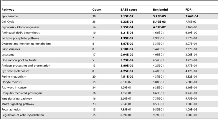
To compare the results of differential expression analyses of genes (or gene sets) across datasets, we reported use of the number of dataset-specific analyses that result in a significant detection of the gene (the number of top-lists in which each gene is present). This score, which monitors systematic differences in expression profiles across datasets, was then used as a selection criterion to define candidate genes. The reported strategy leads to three situations, depending on the strictness of the comparison across datasets: 1) the selection of genes that are detected in all (or the highest number of) datasets (intersection of all top-lists) results in a very low number of genes, which are often already well known; 2) selection of the genes detected in at least one dataset (union of all top lists) results in too many candidates for further investigation, and does not exclude false positives; 3) a balance can be reached between both situations, with an intermediate selection threshold at the number of DEGs across datasets. That intermediate situation (union of intersections between a given number of top lists) allows for inference of a workable amount of new candidates. Along these lines, several techniques have been developed to describe the intersections between lists of genes [33,45,46]. Table 2. List of pathways identified by DAVID with either a significant p-value or 14 or more genes of the 1156 DEG list detected in the map.
Pathway Count EASE score Benjamini FDR
Spliceosome 30 2.15E-07 3.75E-05 2.64E-04
Cell Cycle 25 6.33E-05 5.49E-03 7.75E-02
Glycolysis / Gluconeogenesis 14 9.55E-04 4.07E-02 1.16E+00
Aminoacyl-tRNA biosynthesis 10 5.21E-03 1.66E-01 6.19E+00
Pentose phosphate pathway 7 1.39E-02 2.93E-01 1.57E+01
Cysteine and methionine metabolism 8 1.87E-02 3.37E-01 2.07E+01
Prion diseases 8 2.18E-02 3.47E-01 2.37E+01
Lysosome 17 2.94E-02 4.05E-01 3.06E+01
One carbon pool by folate 5 3.73E-02 4.52E-01 3.72E+01
Antigen processing and presentation 13 3.80E-02 4.29E-01 3.77E+01
Pyruvate metabolism 8 4.25E-02 4.41E-01 4.12E+01
Purine metabolism 20 4.51E-02 4.37E-01 4.32E+01
Oocyte meiosis 15 6.63E-02 5.04E-01 5.68E+01
Pathways in cancer 34 1.29E-01 6.33E-01 8.16E+01
Ubiquitin mediated proteolysis 16 1.55E-01 6.63E-01 8.74E+01
Wnt signaling pathway 16 2.60E-01 7.31E-01 9.75E+01
MAPK signaling pathway 23 5.34E-01 8.58E-01 1.00E+02
Focal adhesion 15 7.85E-01 9.59E-01 1.00E+02
Regulation of actin cytoskeleton 15 8.59E-01 9.74E-01 1.00E+02
The ‘count’ column shows the number of significant genes identified within a pathway. Significant p-values (either the EASE score, the Benjamini-corrected or the FDR-corrected p-values) are shown in bold.
doi:10.1371/journal.pone.0086699.t002
Table 3. Robustness analysis of the spliceosome pathway enrichment.
DEG # threshold Count EASE score
2 (1 and 1) 30 2.15E-07
4 (2 and 2) 23 3.40E-05
6 (3 and 3) 14 4.10E-04
The strictness of the meta-analysis step was increased (selection of genes DEGs in 2 datasets out of 16 available, then 4 datasets out of 16 and 6 datasets out of 16; the number between brackets represent the minimal number of datasets for which the genes have to be DEG to be selected per biological group, i.e. hypoxia or metastasis), the count of genes highlighted in the pathway (second column) and EASE score given by DAVID (third column).
Aim of this study
We propose to use a set of statistical and bioinformatics tools to reanalyze metastasis and hypoxia-related data to gain further insight into the processes involved. The comparison of two analytical pipelines (ORA and FCS) is used to detect meaningful pathways (a diagram of the analytical pipeline is shown in figure 1). Moreover, this analysis rationale could be transposed to virtually any biological situation with microarray data available.
Results and Discussion
A major biological topic of interest in our lab is the investigation of expression profiles to describe common mechanisms between metastasis and adaptation of cells to hypoxic conditions. PathEx [47] was queried (performed on data present in PathEx in June 2012) with the keywords ‘‘hypoxia’’ and ‘‘metastasis’’ to identify datasets available from Affymetrix 133a and HGU-133Plus2 arrays. We found 7 and 9 experiments focused on hypoxia and metastasis respectively. The datasets selected (16) and are listed and described in Table 1.
Meta-analysis and over-representation in pathways
In the first analytical step, the individual analyses of differential expression for each dataset were performed using the Shrinkage t methodology, which produced 16 lists of dataset-specific p-values. Volcano plots are provided in figure 2 for two of the individual datasets, to illustrate the distribution of significant values in a separate analysis. The most interesting genes are usually identified,
in such graphs, in the upper left- and right-hand corners of the plot, depicting genes with low p-values (Y-axis) and high fold changes (X-axis). The meaning of the red dots is explained below. A meta-analysis was performed in two steps to refine the list of significant genes and to define a unique top list from the 16 lists of p-values. Significant genes were first gathered from the two categories of experiments, producing two lists of detected genes (respectively specific to hypoxia and metastasis). The intersection of both lists was then performed, as described in the materials and methods section, to identify candidate genes expected to be involved in both hypoxia and metastasis, while removing potential false detections from the large lists retrieved in the first step. The meta-analysis yielded substantially different results, as shown in figure 2 by the repartition of red dots (final DEGs detected).
Table S1 provides the list of 1156 candidates identified in the meta-analysis procedure, and figure 2 shows the scattering of the candidates in volcano plots for 2 of the 16 datasets we analyzed. The wide range of values observed in figure 2 is due to the variability of the results between the 16 dataset-specific lists (p-values), and the well-known under-estimation of fold changes in microarray experiments [48]. Meta-analysis does not select the most differentially expressed genes in single experiments. As we selected the DEGs across different biological conditions, we can hypothesize that they are representative of the common components of the cellular responses to these situations, which fits well with the purpose of this study.
The list of the identifiers for the 1156 genes obtained after the meta-analysis step was then entered into the DAVID web tool. A
Figure 3. Plot of the EASE score and number of hits in the spliceosome pathway for the 500 random selections of 1156 gene identifiers (in blue), compared with the actual result of the analysis (red). This graph plots the number of hits (X-axis) against the EASE score (Y-axis). The difference between the random selection scores and the actual result score supports the assumption that the spliceosome is over-represented in our list of genes.
total of 102 pathways containing at least 3 gene members of that list was generated (see Table S2). Among these pathways, only 12 of them have an EASE score under the threshold of 0.05. This number was further reduced to 3 pathways by applying a correction for multiple testing (Benjamini correction) and only one pathway (the spliceosome) was significant when applying a correction based on the false discovery rate (FDR) (see Table 2). However, the EASE score (and the corrected p-values derived from it) should be interpreted with caution, according to the biological relevance in the context studied, the wideness of the pathways stored in the Kegg maps and the obvious rate of false negatives induced by our screening. Many top list pathways, although characterized by low EASE scores, are well-known to be involved in metastatic processes and are therefore likely false negatives: MAPK and Wnt signaling pathways, focal adhesion pathway and the regulation of the actin cytoskeleton [49–52], which corroborates the consistency of the mapping of significant genes by our strategy. On the other hand, the robustness of the spliceosome pathway with regards to the most stringent statistical corrections supports the hypothesis of its implication in the process studied.
To further assess the significance of the spliceosome pathway in the over-representation results, we first performed 500 random selections of 1156 EntrezGeneIDs among all the identifiers present on the microarray and ran them in the DAVID tool. The EASE scores and number of hits in the spliceosome pathway were then plotted (see figure 3). The plot shows clearly the gap between the random selections (with a maximum of 19 hits and an associated
EASE score of 0.0085) and the actual result (30 hits, EASE score of 2E-7). Then, we analyzed the robustness of the discovery of the spliceosome pathway by performing a more stringent selection in the meta-analysis step (see Table 3). This table shows the EASE score obtained for the spliceosome pathway when performing a meta-analysis for genes differentially expressed in two (one in each biological group), four (two in each group) or six (three in each group) of the 16 datasets. The spliceosome pathway was largely significant even in the most stringent selection (EASE score of 4E-4). These comparisons tend to support the assumption that the spliceosome pathway is actually over-represented in our meta-analysis results.
Moreover, the spliceosome, whose implication in cancer has been reported by several authors [53–55], has never been described as specifically involved in metastasis, which is not surprising based on the red dots in our volcano plots from single analyses (figure 2). The spliceosome is a complex of RNA and many protein subunits required for the splicing of pre-mRNA. It is composed of five small nuclear RNA (snRNA) and numerous associated protein factors. Proteins and snRNA form the RNA-protein complexes (snRNP), called U1, U2, U4, U5 and U6 (see figure 4). The list of genes detected as differentially expressed contains genes coding for proteins that take part in the spliceosome pathway (see Table S4). The results of our analysis identify genes in all 5 snRNPs, reinforcing the hypothesis that this pathway plays an important role in metastatic and hypoxic processes. The list of genes detected as differentially expressed and their respective p-values per dataset are presented in the Table 4.
Figure 4. Spliceosome units. The red stars mark the genes from our list mapped on this pathway. doi:10.1371/journal.pone.0086699.g004
Table 4. This table contains the genes identified as DEG in the spliceosome pathway, with the p-values for the respective datasets. Hypoxia Metastasis Number of replicates 6 6 23 4 4 4 6 6 2 7 3 2 6 39 83 19 22 12 Datasets E-MEXP- 445 GSE4725 GSE11341 E-MEXP- 1896 GSE4086 GSE5579 GSE9234 E-GEOD- 1323 E-GEOD- 2280 GSE7929 GSE7930 GSE7956 GSE8401 GSE3325 GSE8977 GSE9576 Count DHX 15 0,67 8,98E-01 4,93E-02 1,98E-03 8,16E-03 6,72E-01 5,85E-04 1,65E-01 2 ,73E-01 2,73E-04 3,48E-01 3,80E-01 2,49E-02 3,18E-09 3,54E-01 1,71E-02 8 LSM6 1,35E-01 4,59E-02 8,95E-01 5 ,69E-01 2,06E-02 2,57E-01 6,51E-03 2,16E-01 4 ,92E-01 2,09E-03 2,43E-01 3,77E-01 1,41E-06 1,04E-02 6,51E-01 1,16E-01 6 NAA38 2,09E-02 3,13E-01 4,17E-01 8 ,58E-01 7 ,11E-01 7 ,72E-01 1,18E-04 4,19E-01 3,28E-02 6,66E-02 1 ,27E-01 5,34E-01 3,38E-10 4,34E-03 3,60E-01 7,51E-01 5 NHP2L1 5 ,89E-02 7,13E-01 2,65E-01 1,16E-02 2,23E-01 9 ,53E-01 4,75E-07 1,89E-01 2,16E-02 7,66E-01 9 ,27E-02 1,16E-01 7,92E-04 4,67E-02 5,21E-01 4,95E-01 5 PRPF19 7,29E-01 2,94E-02 1,94E-01 3,24E-02 6,95E-03 4,29E-01 3,00E-07 5,13E-02 5 ,74E-01 1,24E-01 2,27E-02 8,74E-01 3,40E-01 5,30E-01 1,12E-02 1,71E-01 6 RBM8A 2,40E-02 7,83E-01 5,74E-01 3 ,66E-01 5,26E-03 9,17E-01 4,89E-02 1,23E-02 2,35E-01 2,91E-02 2,31E-02 6,42E-01 1,89E-01 8,44E-02 4 ,33E-01 1,55E-02 7 ACIN1 4,05E-02 9,92E-01 6,55E-01 2,45E-02 9,74E-03 7,11E-01 3,20E-02 1,98E-04 7,89E-01 6,60E-05 3,58E-01 5,65E-01 5,38E-01 3,28E-02 8,78E-01 1,82E-01 7 Cdc40 9,98E-01 7,88E-01 6,80E-01 3 ,07E-01 6 ,80E-02 5 ,20E-01 1,07E-05 2,98E-01 9 ,11E-01 6,13E-03 2,26E-01 7,65E-01 1,90E-01 1,07E-07 9,77E-01 3,30E-02 4 CRNKL1 3,86E-02 7,70E-01 9,38E-03 2,73E-01 4,95E-02 8,02E-01 4,48E-09 9,23E-01 2 ,29E-01 1,55E-07 1,35E-01 4,64E-01 3,93E-08 1,09E-01 6 ,34E-01 4,83E-02 7 HSPA1A 4,37E-02 9,59E-01 4,66E-05 5,14E-03 3,13E-03 2,63E-01 1,15E-03 9,60E-01 1 ,48E-01 2,18E-08 1,34E-01 8,94E-03 2,58E-08 2,80E-06 1,05E-01 1,61E-01 9 HSPA1B 1,92E-02 4,38E-01 3,65E-01 9 ,44E-01 3 ,12E-01 8 ,72E-01 8,89E-06 1,20E-02 5,61E-01 6,93E-04 3,68E-01 7,69E-01 1,57E-01 4,47E-02 9,09E-01 1,00E-02 6 HSPA8 1,36E-03 8,33E-01 6,60E-01 3 ,76E-01 6 ,38E-02 7 ,44E-01 2,61E-02 9,19E-04 1,65E-01 5,99E-01 3 ,84E-01 3,47E-01 6,00E-08 4,91E-01 8 ,76E-01 2,63E-02 5 HNRNPA1P2 2,02E-01 4,42E-01 6,08E-01 5 ,30E-01 1 ,37E-01 8 ,63E-01 8,68E-07 9,41E-01 9 ,27E-01 1,73E-01 2 ,33E-01 4,23E-01 9,38E-02 4,20E-07 3,42E-01 4,48E-02 3 HNRNPC 1,12E-02 3,97E-02 8,77E-01 4,84E-02 2,12E-04 3,44E-01 3,52E-06 2,76E-01 1 ,99E-01 5,13E-05 1,53E-01 9,03E-01 5,45E-02 4,38E-02 1,06E-01 4,15E-02 8 HNRNPK 7,88E-01 2,29E-02 7,13E-01 1 ,84E-01 1 ,54E-01 4 ,09E-01 5,49E-01 4,84E-03 2,04E-01 1,70E-05 2,55E-01 3,03E-01 1,70E-04 4,88E-02 5,52E-05 7,26E-01 6 HNRNPM 3,61E-01 4,24E-03 8,85E-01 1 ,65E-01 6 ,57E-02 1 ,00E + 00 1,73E-02 5,15E-02 7 ,33E-01 2,36E-01 1 ,64E-01 9,32E-01 4,87E-03 4,43E-02 1,24E-01 2,42E-02 5 SNRPG 1 ,92E-01 1,26E-02 6,31E-01 1,35E-02 3,70E-02 2,88E-01 5,13E-05 8,38E-01 3,16E-02 1,82E-01 2 ,04E-01 6,29E-01 3,62E-01 6,22E-06 4,60E-03 8,42E-02 7 NCBP1 5 ,08E-01 8,32E-01 4,05E-01 6,80E-04 2,11E-01 2 ,35E-01 4,33E-02 9,02E-01 3,87E-02 4,27E-02 3,77E-01 4,40E-01 9,18E-01 4,73E-02 5,94E-05 1,52E-01 6 PUF60 4 ,94E-01 2,77E-02 3,31E-01 6 ,24E-02 2,65E-03 4,65E-01 2,20E-02 5,57E-04 2,72E-01 2,23E-01 2 ,56E-01 3,27E-01 4,63E-01 2,18E-02 1,56E-01 7,61E-01 5 SNRPD1 8,54E-03 7,39E-01 3,47E-01 7 ,28E-01 1 ,10E-01 1 ,80E-01 6,56E-03 3,67E-01 5 ,41E-01 2,50E-01 4,22E-02 3,55E-01 5,27E-03 1,57E-03 6,02E-01 3,64E-02 6 SNRPD3 7,61E-01 1,50E-01 4,52E-01 1 ,33E-01 9 ,09E-01 4 ,06E-01 1,03E-08 3,06E-02 4,87E-02 1,67E-04 7,68E-02 1,39E-01 2,09E-05 5,97E-04 4,31E-02 9,36E-01 7 SNRPEL1 8,86E-01 2,49E-02 3,98E-01 1,69E-02 3,23E-02 6,56E-01 2,37E-03 1,81E-03 9,32E-02 2,46E-01 1,78E-02 1,79E-01 4,44E-01 5,25E-03 7,15E-01 5,34E-01 7 SNRPF 8 ,75E-01 4,83E-03 6,61E-01 4 ,42E-01 2,30E-03 9,50E-01 4,19E-05 7,66E-01 5 ,57E-01 1,46E-05 7,80E-02 7,65E-01 2,10E-03 4,13E-02 2,44E-01 3,11E-02 7 SF3B3 5 ,74E-01 2,51E-01 9,73E-01 5 ,13E-01 1 ,71E-01 7 ,57E-01 6,26E-04 1,22E-01 4 ,35E-01 8,87E-01 4 ,24E-01 6,75E-01 5,96E-01 1,67E-02 6,29E-02 6,00E-02 2 SFRS1 2,00E-01 1,36E-01 9,40E-01 2 ,07E-01 1 ,37E-01 2 ,82E-01 3,64E-01 9,54E-01 7 ,99E-01 3,65E-03 3,66E-02 6,54E-01 6,22E-02 7,35E-03 5,09E-02 6,60E-02 3 SFRS3 1,87E-01 7,79E-03 2,18E-01 1 ,12E-01 4 ,32E-01 4 ,39E-01 1,94E-07 3,27E-02 3,56E-01 1,70E-05 3,53E-03 2,22E-01 1,39E-04 1,12E-02 9,77E-02 5,78E-01 7 SFRS5 4,54E-01 1,20E-01 4,66E-01 7 ,16E-02 6 ,68E-02 6 ,26E-01 5,31E-08 6,83E-03 2,61E-02 7,19E-01 7 ,70E-01 3,13E-01 7,40E-03 7,61E-06 2,99E-02 9,04E-01 6 SFRS7 3,09E-02 7,38E-01 6,83E-01 8 ,52E-01 2,31E-03 5,91E-01 4,21E-02 8,86E-01 8 ,82E-01 6,88E-01 6 ,32E-01 6,10E-01 2,92E-01 8,91E-02 8 ,23E-01 5,35E-02 3 SFRS9 4,39E-03 2,41E-01 1,02E-01 8 ,61E-02 8 ,89E-01 6 ,60E-01 9,99E-05 5,64E-04 2,77E-01 1,72E-02 2,77E-02 7,59E-01 2,93E-06 7,74E-04 5,24E-01 7,95E-03 8 TRA2B 1,00E-02 1,02E-01 4,29E-01 2 ,93E-01 1,84E-03 8,13E-01 7,30E-08 5,67E-03 2,50E-02 1,34E-01 2 ,50E-01 1,89E-01 4,05E-09 2,67E-02 1,54E-02 1,39E-01 8 USP39 1,25E-01 8,78E-01 6,01E-01 5 ,23E-01 1 ,32E-01 4 ,12E-01 6,60E-05 3,48E-01 6 ,97E-01 1,07E-02 2,63E-01 3,00E-01 2,71E-01 2,56E-05 1,65E-01 4,24E-02 4 The significant p -values are highlighted in bold. The last columns ‘count’ shows the number o f times a g iven gene is detected as DEG through all 1 6 datas ets. doi:10.1371/journal.pone. 0086699.t004
Table 5. Summary of FAERI results.
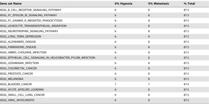
Gene set Name 5% Hypoxia 5% Metastasis % Total
KEGG_PATHWAYS_IN_CANCER 7 9 100 KEGG_GLYCOLYSIS_GLUCONEOGENESIS 7 8 93.75 KEGG_PURINE_METABOLISM 7 8 93.75 KEGG_RIBOSOME 7 8 93.75 KEGG_PPAR_SIGNALING_PATHWAY 7 8 93.75 KEGG_MAPK_SIGNALING_PATHWAY 7 8 93.75 KEGG_ERBB_SIGNALING_PATHWAY 7 8 93.75 KEGG_CYTOKINE_CYTOKINE_RECEPTOR_INTERACTION 7 8 93.75 KEGG_CHEMOKINE_SIGNALING_PATHWAY 7 8 93.75 KEGG_ENDOCYTOSIS 7 8 93.75 KEGG_APOPTOSIS 7 8 93.75 KEGG_VEGF_SIGNALING_PATHWAY 7 8 93.75 KEGG_FOCAL_ADHESION 7 8 93.75 KEGG_REGULATION_OF_ACTIN_CYTOSKELETON 7 8 93.75 KEGG_ADIPOCYTOKINE_SIGNALING_PATHWAY 7 8 93.75 KEGG_HUNTINGTONS_DISEASE 7 8 93.75 KEGG_PANCREATIC_CANCER 7 8 93.75 KEGG_CHRONIC_MYELOID_LEUKEMIA 7 8 93.75 KEGG_RENAL_CELL_CARCINOMA 7 8 93.75 KEGG_CITRATE_CYCLE_TCA_CYCLE 6 8 87.5 KEGG_FRUCTOSE_AND_MANNOSE_METABOLISM 7 7 87.5 KEGG_OXIDATIVE_PHOSPHORYLATION 6 8 87.5 KEGG_VALINE_LEUCINE_AND_ISOLEUCINE_DEGRADATION 6 8 87.5 KEGG_GLUTATHIONE_METABOLISM 6 8 87.5 KEGG_AMINO_SUGAR_AND_NUCLEOTIDE_SUGAR_METABOLISM 7 7 87.5 KEGG_PYRUVATE_METABOLISM 6 8 87.5 KEGG_RNA_DEGRADATION 6 8 87.5 KEGG_SPLICEOSOME 6 8 87.5 KEGG_CALCIUM_SIGNALING_PATHWAY 6 8 87.5 KEGG_NEUROACTIVE_LIGAND_RECEPTOR_INTERACTION 6 8 87.5 KEGG_CELL_CYCLE 6 8 87.5 KEGG_OOCYTE_MEIOSIS 6 8 87.5 KEGG_P53_SIGNALING_PATHWAY 6 8 87.5 KEGG_UBIQUITIN_MEDIATED_PROTEOLYSIS 6 8 87.5 KEGG_LYSOSOME 6 8 87.5 KEGG_MTOR_SIGNALING_PATHWAY 6 8 87.5 KEGG_VASCULAR_SMOOTH_MUSCLE_CONTRACTION 6 8 87.5 KEGG_WNT_SIGNALING_PATHWAY 6 8 87.5 KEGG_AXON_GUIDANCE 6 8 87.5 KEGG_ECM_RECEPTOR_INTERACTION 6 8 87.5 KEGG_ADHERENS_JUNCTION 6 8 87.5 KEGG_TIGHT_JUNCTION 6 8 87.5 KEGG_GAP_JUNCTION 6 8 87.5 KEGG_ANTIGEN_PROCESSING_AND_PRESENTATION 6 8 87.5 KEGG_TOLL_LIKE_RECEPTOR_SIGNALING_PATHWAY 6 8 87.5 KEGG_RIG_I_LIKE_RECEPTOR_SIGNALING_PATHWAY 6 8 87.5 KEGG_JAK_STAT_SIGNALING_PATHWAY 6 8 87.5 KEGG_NATURAL_KILLER_CELL_MEDIATED_CYTOTOXICITY 6 8 87.5 KEGG_T_CELL_RECEPTOR_SIGNALING_PATHWAY 6 8 87.5
Gene set analysis
The second part of the analytical pipeline (figure 1) relies on the inference of differentially expressed pathways in a gene set analysis procedure (functional class scoring). Here, we used FAERI, a multivariate procedure tailored from the two-way ANOVA procedure. FAERI computes a gene set statistic from the expression data of all member genes in a single step, and avoids the loss of information inherent to 2-step procedures and the risk of false negatives due to slight differences in all member genes (that would not be individually detected in the first part of the pipeline). In addition, FAERI relies on a self-contained procedure (label sampling) that only requires the expression values of the set of member genes (and not the complete dataset). Table 5 summarizes the results obtained by individual analysis of the 16 selected datasets conducted with FAERI. These results were then used to compute, for each gene set, a ratio of discovery across all the experiments (Table 5, third column). The definition of the sets was retrieved from the C2.Kegg category of MsigDB (v3.0). The full list of p-values is provided in Table S3.
Table 5 summarizes the information contained in Table S3 and highlights the high number of differentially expressed sets across both categories of experiments. The pathways identified by FAERI are involved in glycolysis, neoglucogenesis, tricarboxylic cycle, oxidative phosphorylation and other sugar metabolism pathways. These results are relevant to the cell/tissue response to hypoxic conditions. Here, only one gene set was detected across all datasets: PATHWAYS_IN_CANCER. Many other cancer-re-lated gene sets were detected in all but one experiment. Several signaling pathways were also systematically called differentially expressed, including PPAR, ERBB, MAPK, VEGF, P53, MTOR, WNT, … The pathway for the regulation of the actin cytoskeleton was also detected. The hypothesis of involvement of the
spliceosome is supported by 6 out of 7 datasets related to hypoxia and 8 out of 9 datasets related to metastasis.
Both parts of the analytical pipeline described here have detected the spliceosome pathway as involved in the hypoxic and metastatic phenotypes. Among the 31 genes detected as differen-tially expressed in this pathway, 11 have recently been shown to be involved in the metastatic process (see Table 6). The remaining 20 genes are not yet known to be involved in these processes (see Table 6, in bold). These results suggest that abnormal alternative splicing regulation can modulate the metastatic potential of cancer cells. Indeed, it is known that the recognition of splicing sites depends on the protein composition of the spliceosome [56]. Dysregulated expression of the genes coding for these proteins could therefore change the composition of the spliceosome architecture, thus affecting the splicing process. A change in the splicing process may influence the cell at all biochemical levels, from the transcriptome to the proteome and even to the genome. The 20 genes we have identified thus hold strong potential as candidates for further studies.
The results also demonstrate the potential of sensitive and specific analytical pipelines: new hypotheses can be proposed, and previously known biological features can be used as positive controls. However, comparison of the results between both parts of the analytical pipeline suggests that the two analyses behave differently: over-representation analysis of the most significant genes across datasets detects some important pathways, and the ability of gene set analysis using FAERI to detect slight cumulated differences detects more pathways. Statistical analysis with FAERI detects meaningful differences between samples, even when only small numbers of replicates are available. Nevertheless, both parts of the pipeline lead to detection of relevant information based on current knowledge, and both suggest the involvement of the spliceosome.
Table 5. Cont.
Gene set Name 5% Hypoxia 5% Metastasis % Total
KEGG_B_CELL_RECEPTOR_SIGNALING_PATHWAY 6 8 87.5 KEGG_FC_EPSILON_RI_SIGNALING_PATHWAY 6 8 87.5 KEGG_FC_GAMMA_R_MEDIATED_PHAGOCYTOSIS 6 8 87.5 KEGG_LEUKOCYTE_TRANSENDOTHELIAL_MIGRATION 6 8 87.5 KEGG_NEUROTROPHIN_SIGNALING_PATHWAY 6 8 87.5 KEGG_LONG_TERM_DEPRESSION 6 8 87.5 KEGG_ALZHEIMERS_DISEASE 6 8 87.5 KEGG_PARKINSONS_DISEASE 6 8 87.5 KEGG_VIBRIO_CHOLERAE_INFECTION 6 8 87.5 KEGG_EPITHELIAL_CELL_SIGNALING_IN_HELICOBACTER_PYLORI_INFECTION 6 8 87.5 KEGG_LEISHMANIA_INFECTION 6 8 87.5 KEGG_COLORECTAL_CANCER 6 8 87.5 KEGG_PROSTATE_CANCER 6 8 87.5 KEGG_MELANOMA 6 8 87.5 KEGG_BLADDER_CANCER 7 7 87.5 KEGG_ACUTE_MYELOID_LEUKEMIA 6 8 87.5 KEGG_SMALL_CELL_LUNG_CANCER 6 8 87.5 KEGG_VIRAL_MYOCARDITIS 6 8 87.5
The first column presents the name of the gene sets tested, the second and third columns show the number of times each gene set was detected as differentially expressed at a threshold of 5% for the p-values, for each biological group. The last column contains the discovery rate across all experiments (7 hypoxia datasets and 9 metastasis datasets).
Conclusion and Perspectives
We implemented a pipeline of bioinformatics tools to explore archived microarray data, from preprocessing to mapping of the results. We used that pipeline to examine metastasis and hypoxia data and found results in keeping with previous reports, as well as a new hypothesis. The combination of high-level analysis (Over Representation Analysis and Functional Category Scoring) with a meta-analysis step led to the discovery of involvement of the spliceosome in the hypoxic and metastatic processes, and the generation of a list of 20 new candidate genes.
Bioinformatics approaches will never replace bench validations; however we were able to form a plausible hypothesis just by re-analyzing available data. Biological investigations should therefore be performed to further refine the interpretation of the relation-ships between the pathways detected and understand how a hypoxic environment and metastasis affect both general and energetic cell metabolism. Further investigations should be
conducted to clarify the results of the statistical analyses and to discriminate between causes and consequences (mechanisms of perturbations and symptoms). However, that validation is out of the scope of this methodological paper.
We think that this analytical protocol could be used successfully in many other biological contexts, wherever several datasets are available. Indeed, we have shown that single gene analysis alone yields poor results, though this is often the only step performed by wet-lab biologists. The methodology presented here allows for improved performance, comparison with previously known information and discovery of recurrent patterns (through meta-analysis), all of which were performed using freely-available resources and software packages and without the need to perform expensive de novo microarray experiments. We think that this work will contribute to the creation of a virtual atlas for cellular biology containing the known characteristics of cells in diverse biological conditions, which is one of the major goals of the bioinformatics community.
Table 6. List of the 31 genes highlighted in the spliceosome pathway.
Gene References in literature
DHX 15 NA
LSM6 NA
NAA38 NA
NHP2L1 NA
PRPF19 NA
RBM8A Kim et al, 2008 [60] and Salicioni et al, 2000 [61] ACIN1 Lee et al, 2008 [62] and Shu et al, 2006 [63]
Cdc40 NA CRNKL1 NA HSPA1A NA HSPA1B NA HSPA8 NA HNRNPA1P2 NA HNRNPC Park et al, 2012 [64]
HNRNPK Inoue et al, 2007 [65] and Li et al, 2011 [66] HNRNPM Palermo et al, 2012 [67] and Thomas et al, 2011 [68]
SNRPG NA NCBP1 NA PUF60 NA SNRPD1 NA SNRPD3 Cunha et al, 2010 [69] SNRPEL1 NA SNRPF NA SF3B3 NA
SFRS1 Mukherji et al, 2006 [70], Hatakeyama et al, 2009 [71] and Meseguer et al, 2011 [72]
SFRS3 NA
SFRS5 Hatakeyama et al, 2009 [71]
SFRS7 Hatakeyama et al, 2009 [71]
SFRS9 Mukherji et al, 2006 [70]
TRA2B Watermann et al, 2006 [73]
USP39 NA
Eleven of those genes are previously known in the literature to be involved in metastasis (shown in grey), the 20 other are previously unknown (shown in bold) to be involved in the metastatic process.
Methods
Selection and retrieval of datasets
For the purposes of the study reported here, two sets of criteria were used to retrieve datasets with PathEx (described in [47]): technological keywords to specifically retrieve Affymetrix Gene-Chips HGU-133a and HGU-133plus2 array models; and biological keywords to retrieve datasets that met the topics of interest in this study: hypoxia or metastasis.
Entry of these technological and biological keywords into PathEx resulted in a collection of 16 distinct datasets, as listed in Table 1: 9 datasets specific to hybridizations performed on the HGU-133a chip model, including 3 experimental designs dedicated to hypoxia and 6 dedicated to metastasis; 7 datasets obtained using the HGU-133plus2 array model, including 4 hypoxia-related and 3 metastasis-related experiments. The number or replicated measurements ranged from 2 to 52 hybridizations (see Table 1). In addition, we preferred datasets reporting in vivo gene expression levels and discarded data that came from in vitro experiments.
Preprocessing and statistical analyses
The preprocessing of the data and the individual analyses reported in this paper were performed using R 2.7 and 2.10, available on the website of the R-Project (http://cran.r-project. org), and a set of packages available in the Bioconductor repository (http://www.bioconductor.org).
We used GCRMA to preprocess each of the 16 retrieved datasets, in accordance with the performances reported in previously reported benchmarks [26,57,58]. The summarization step performed by GCRMA was guided by the affyprobeminer transcript-consistent chip definition files (CDF) specific to the HGU-133a and HGU-133plus2 chip models. The probe set identifiers provided by alternative CDFs (affyprobeminer) differ from the identifiers defined by the manufacturer of the arrays (Affymetrix). Supplemental functions implemented in the affypro-beminer packages were used to convert probe set identifiers into EntrezGeneID. The identification of probe sets with EntrezGene ID identifiers allowed us to compare the gene lists between HGU-133a and HGU-133plus2 chip models, and to facilitate annotation of the results from the individual analyses.
The differential expression of individual probe sets was analyzed with the ’st’ package, which implements the Shrinkage t methodology. This procedure was conducted on each dataset, resulting in 16 dataset-specific lists of p-values, each p-value referring to a specific probe set.
Meta-analysis, annotation, and gene set analysis
For each dataset, we selected the list of genes detected as differentially expressed (p-value , 0.05). The 16 dataset-specific lists of the most significant genes were gathered into two groups, according to the experimental design (Hypoxia/Metastasis studies). In each group of datasets, a new list of genes was defined from the list of genes found to be differentially expressed in at least one dataset of the group. Lastly, the intersection of the list of genes from the two groups was performed by selecting genes that were detected in both groups, resulting in a list of 1156 unique gene identifiers (provided in Table S1, along with all the p-values
computed for the 16 datasets, p-values ranking for each dataset and mean ranking across the 16 individual ranks).
The 1156 selected EntrezGene ID identifiers were mapped to the Kegg Pathways database using the ‘‘Functional Annotation Tools’’ available on the DAVID web interface [59]. Using DAVID, 102 pathways, containing at least 3 of the 1156 candidate genes, were identified (see Table S2). To avoid biases due to potential false positives, we selected for further analysis the pathways that displayed a significant p-value (see Table 2).
Alongside the selection and annotation of the most significant genes by the meta-analysis approach, differential expression analyses of gene sets were conducted on each of the 16 datasets. Gene set analyses were performed on preprocessed data in a single step using the multivariate FAERI test. Gene set definitions were retrieved from the MSigDB database (v3.0) [36]. We evaluated the differential expression on gene sets belonging to the C.2 KEGG category, composed of 186 curated pathways. Lastly, the 16 dataset-specific lists of p-values were used to compute, for each gene set, the ratio of detection as differentially expressed across all datasets. The full code for this analysis can be found in the Table S5. For more details on the FAERI methodology, see [39].
The different steps in the analytical pipelines are summarized in figure 1. The left part of the diagram contains the single gene analysis steps (Shrinkage t test treatment, meta-analysis and over-representation analysis (ORA) in DAVID). The right part contains the gene set analysis steps (Functional Class Scoring (FCA) by FAERI and meta-analysis of the results).
Supporting Information
Table S1 Full list of p-values obtained for each dataset for each of the 1156 genes highlighted in the analysis.
(PDF)
Table S2 List of pathways highlighted in the over-representation analysis using DAVID.
(PDF)
Table S3 Full list of p-values obtained in the geneset analysis. (PDF)
Table S4 Distribution of the highlighted genes in the spliceo-some pathway.
(PDF)
Table S5 R code for the full analysis. (PDF)
Checklist S1
(PDF)
Acknowledgments
We would like to thank Jacques van Helden from the TAGC (Aix-Marseille University) for helpful comments and discussions.
Author Contributions
Conceived and designed the experiments: BDM FB CM ED. Performed the experiments: BDM FB EB. Analyzed the data: BDM FB SD. Contributed reagents/materials/analysis tools: BDM FB EB SD. Wrote the paper: BDM FB CM ED.
References
1. Lim K, Small W Jr, Portelance L, Creutzberg C, Jurgenliemk-Schulz IM, et al. (2010) Consensus guidelines for delineation of clinical target volume for intensity-modulated pelvic radiotherapy for the definitive treatment of cervix cancer. Int J Radiat Oncol Biol Phys 79: 348–355.
2. Nardella C, Clohessy JG, Alimonti A, Pandolfi PP (2011) Pro-senescence therapy for cancer treatment. Nat Rev Cancer 11: 503–511.
3. Suganuma M, Saha A, Fujiki H (2011) New cancer treatment strategy using combination of green tea catechins and anticancer drugs. Cancer Science 102: 317–323.
4. Ellison LF, Wilkins K (2009) Cancer prevalence in the Canadian population. Health Rep 20: 7–19.
5. Mehlen P, Puisieux A (2006) Metastasis: a question of life or death. Nat Rev Cancer 6: 449–458.
6. Alsarraj J, Hunter KW (2012) Bromodomain-Containing Protein 4: A Dynamic Regulator of Breast Cancer Metastasis through Modulation of the Extracellular Matrix. Int J Breast Cancer 2012: 670632.
7. Hanahan D, Weinberg RA (2011) Hallmarks of cancer: the next generation. Cell 144: 646–674.
8. Nieder C, Pawinski A, Dalhaug A (2012) Contribution of case reports to brain metastases research: systematic review and analysis of pattern of citation. PLoS One 7: e34300.
9. Hanahan D, Weinberg RA (2000) The hallmarks of cancer. Cell 100: 57–70. 10. Finger EC, Giaccia AJ (2010) Hypoxia, inflammation, and the tumor
microenvironment in metastatic disease. Cancer Metastasis Rev 29: 285–293. 11. Kingsley LA, Fournier PG, Chirgwin JM, Guise TA (2007) Molecular biology of
bone metastasis. Mol Cancer Ther 6: 2609–2617.
12. Peinado H, Cano A (2008) A hypoxic twist in metastasis. Nat Cell Biol 10: 253– 254.
13. Gordan JD, Simon MC (2007) Hypoxia-inducible factors: central regulators of the tumor phenotype. Curr Opin Genet Dev 17: 71–77.
14. Vaupel P (2004) The role of hypoxia-induced factors in tumor progression. Oncologist 9 Suppl 5: 10–17.
15. Sullivan R, Graham CH (2007) Hypoxia-driven selection of the metastatic phenotype. Cancer Metastasis Rev 26: 319–331.
16. Jiang J, Tang YL, Liang XH (2011) EMT: a new vision of hypoxia promoting cancer progression. Cancer Biol Ther 11: 714–723.
17. Lu X, Kang Y (2010) Hypoxia and hypoxia-inducible factors: master regulators of metastasis. Clin Cancer Res 16: 5928–5935.
18. Semenza GL (2001) HIF-1 and mechanisms of hypoxia sensing. Curr Opin Cell Biol 13: 167–171.
19. Parkinson H, Kapushesky M, Shojatalab M, Abeygunawardena N, Coulson R, et al. (2007) ArrayExpress—a public database of microarray experiments and gene expression profiles. Nucleic Acids Res 35: D747–750.
20. Barrett T, Troup DB, Wilhite SE, Ledoux P, Rudnev D, et al. (2007) NCBI GEO: mining tens of millions of expression profiles—database and tools update. Nucleic Acids Res 35: D760–765.
21. Baldi P, Long AD (2001) A Bayesian framework for the analysis of microarray expression data: regularized t -test and statistical inferences of gene changes. Bioinformatics 17: 509–519.
22. Opgen-Rhein R, Strimmer K (2007) Accurate ranking of differentially expressed genes by a distribution-free shrinkage approach. Stat Appl Genet Mol Biol 6: Article9.
23. Smyth GK (2004) Linear models and empirical bayes methods for assessing differential expression in microarray experiments. Stat Appl Genet Mol Biol 3: Article3.
24. Liu H, Zeeberg BR, Qu G, Koru AG, Ferrucci A, et al. (2007) AffyProbeMiner: a web resource for computing or retrieving accurately redefined Affymetrix probe sets. Bioinformatics 23: 2385–2390.
25. Bolstad BM, Irizarry RA, Astrand M, Speed TP (2003) A comparison of normalization methods for high density oligonucleotide array data based on variance and bias. Bioinformatics 19: 185–193.
26. Cope LM, Irizarry RA, Jaffee HA, Wu Z, Speed TP (2004) A benchmark for Affymetrix GeneChip expression measures. Bioinformatics 20: 323–331. 27. Irizarry R, Wu Z, Cawley S (2005) affycomp: Graphic Toolbox for Assessment
of Affymetrix Expression Measures.
28. Wu Z, Irizarry RA, Gentleman R, Murillo F, Spencer F (2004) A model-based background adjustment for oligonucleotide expression arrays. John Hopkins University, Dept of Biostatistics Working Papers Working Papers 1. 29. Dai M, Wang P, Boyd AD, Kostov G, Athey B, et al. (2005) Evolving gene/
transcript definitions significantly alter the interpretation of GeneChip data. Nucleic Acids Res 33: e175.
30. Gautier L, Moller M, Friis-Hansen L, Knudsen S (2004) Alternative mapping of probes to genes for Affymetrix chips. BMC Bioinformatics 5: 111.
31. De Hertogh B, De Meulder B, Berger F, Pierre M, Bareke E, et al. (2010) A benchmark for statistical microarray data analysis that preserves actual biological and technical variance. BMC Bioinformatics 11: 17.
32. Berger F, De Hertogh B, Pierre M, Gaigneaux A, Depiereux E (2008) The "Window-t test": a simple and powerful approach to detect differentially expressed genes in microarray datasets. Centr Eur J biol 3: 327–344. 33. Pierre M, De Hertogh B, De Meulder B, Bareke E, Depiereux S, et al. (2011)
Enhanced meta-analysis highlights genes involved in metastasis from several microarray datasets. J Proteomics Bioinform 4.
34. Huang da W, Sherman B, Lempicki R (2009) Systematic and integrative analysis of large gene lists using DAVID bioinformatics resources. Nat Protoc 4: 44–57. 35. Mootha VK, Lindgren CM, Eriksson KF, Subramanian A, Sihag S, et al. (2003) PGC-1alpha-responsive genes involved in oxidative phosphorylation are coordinately downregulated in human diabetes. Nat Genet 34: 267–273. 36. Subramanian A, Tamayo P, Mootha VK, Mukherjee S, Ebert BL, et al. (2005)
Gene set enrichment analysis: a knowledge-based approach for interpreting genome-wide expression profiles. Proc Natl Acad Sci U S A 102: 15545–15550.
37. Dinu I, Potter JD, Mueller T, Liu Q, Adewale AJ, et al. (2007) Improving gene set analysis of microarray data by SAM-GS. BMC Bioinformatics 8: 242. 38. Goeman JJ, van de Geer SA, de Kort F, van Houwelingen HC (2004) A global
test for groups of genes: testing association with a clinical outcome. Bioinformatics 20: 93–99.
39. Berger F, De Meulder B, Gaigneaux A, Depiereux S, Bareke E, et al. (2010) Functional analysis: evaluation of response intensities--tailoring ANOVA for lists of expression subsets. BMC Bioinformatics 11: 510.
40. Mansmann U, Meister R (2005) Testing differential gene expression in functional groups. Goeman’s global test versus an ANCOVA approach. Methods Inf Med 44: 449–453.
41. Simpson R, Pearson K (1904) Report on Certain Enteric Fever Inoculation Statistics. Br Med J 2: 1243–1246.
42. Palmerini T, Biondi-Zoccai G, Della Riva D, Stettler C, Sangiorgi D, et al. (2012) Stent thrombosis with drug-eluting and bare-metal stents: evidence from a comprehensive network meta-analysis. Lancet 379: 1393–1402.
43. Walton J, Ebner Y, Stewart MG, April MM (2012) Systematic review of randomized controlled trials comparing intracapsular tonsillectomy with total tonsillectomy in a pediatric population. Arch Otolaryngol Head Neck Surg 138: 243–249.
44. Zheng XY, Wei RB, Tang L, Li P, Zheng XD (2012) Meta-analysis of combined therapy for adult hepatitis B virus-associated glomerulonephritis. World J Gastroenterol 18: 821–832.
45. Pierre M, DeHertogh B, Gaigneaux A, DeMeulder B, Berger F, et al. (2010) Meta-analysis of archived DNA microarrays identifies genes regulated by hypoxia and involved in a metastatic phenotype in cancer cells. BMC Cancer 10: 176.
46. Dawany NB, Tozeren A (2010) Asymmetric microarray data produces gene lists highly predictive of research literature on multiple cancer types. BMC Bioinformatics 11: 483.
47. Bareke E, Pierre M, Gaigneaux A, De Meulder B, Depiereux S, et al. (2010) PathEx: a novel multi factors based datasets selector web tool. BMC Bioinformatics 11: 528.
48. Yuen T, Wurmbach E, Pfeffer RL, Ebersole BJ, Sealfon SC (2002) Accuracy and calibration of commercial oligonucleotide and custom cDNA microarrays. Nucleic Acids Res 30: e48.
49. Buda A, Pignatelli M (2011) E-cadherin and the cytoskeletal network in colorectal cancer development and metastasis. Cell Commun Adhes 18: 133– 143.
50. del Barco Barrantes I, Nebreda AR (2012) Roles of p38 MAPKs in invasion and metastasis. Biochem Soc Trans 40: 79–84.
51. Majid S, Saini S, Dahiya R (2012) Wnt signaling pathways in urological cancers: past decades and still growing. Mol Cancer 11: 7.
52. Mezi S, Todi L, Orsi E, Angeloni A, Mancini P (2012) Involvement of the Src-cortactin pathway in migration induced by IGF-1 and EGF in human breast cancer cells. Int J Oncol 41: 2128–2138.
53. Valles I, Pajares MJ, Segura V, Guruceaga E, Gomez-Roman J, et al. (2012) Identification of novel deregulated RNA metabolism-related genes in non-small cell lung cancer. PLoS One 7: e42086.
54. van Alphen RJ, Wiemer EA, Burger H, Eskens FA (2009) The spliceosome as target for anticancer treatment. Br J Cancer 100: 228–232.
55. Ward AJ, Cooper TA (2009) The pathobiology of splicing. J Pathol 220: 152– 163.
56. Chen M, Manley JL (2009) Mechanisms of alternative splicing regulation: insights from molecular and genomics approaches. Nat Rev Mol Cell Biol 10: 741–754.
57. Fujita A, Sato JR, Rodrigues Lde O, Ferreira CE, Sogayar MC (2006) Evaluating different methods of microarray data normalization. BMC Bioinfor-matics 7: 469.
58. Katz S, Irizarry RA, Lin X, Tripputi M, Porter MW (2006) A summarization approach for Affymetrix GeneChip data using a reference training set from a large, biologically diverse database. BMC Bioinformatics 7: 464.
59. Ogata H, Goto S, Sato K, Fujibuchi W, Bono H, et al. (1999) KEGG: Kyoto Encyclopedia of Genes and Genomes. Nucleic Acids Res 27: 29–34. 60. Kim TJ, Choi JJ, Kim WY, Choi CH, Lee JW, et al. (2008) Gene expression
profiling for the prediction of lymph node metastasis in patients with cervical cancer. Cancer Sci 99: 31–38.
61. Salicioni AM, Xi M, Vanderveer LA, Balsara B, Testa JR, et al. (2000) Identification and structural analysis of human RBM8A and RBM8B: two highly conserved RNA-binding motif proteins that interact with OVCA1, a candidate tumor suppressor. Genomics 69: 54–62.
62. Lee JH, Horak CE, Khanna C, Meng Z, Yu LR, et al. (2008) Alterations in Gemin5 expression contribute to alternative mRNA splicing patterns and tumor cell motility. Cancer Res 68: 639–644.
63. Shu Y, Iijima T, Sun W, Kano J, Ishiyama T, et al. (2006) The ACIN1 gene is hypermethylated in early stage lung adenocarcinoma. J Thorac Oncol 1: 160– 167.
64. Park YM, Hwang SJ, Masuda K, Choi KM, Jeong MR, et al. (2012) Heterogeneous nuclear ribonucleoprotein C1/C2 controls the metastatic potential of glioblastoma by regulating PDCD4. Mol Cell Biol 32: 4237–4244. 65. Inoue A, Sawata SY, Taira K, Wadhwa R (2007) Loss-of-function screening by randomized intracellular antibodies: identification of hnRNP-K as a potential target for metastasis. Proc Natl Acad Sci U S A 104: 8983–8988.
66. Li LP, Lu CH, Chen ZP, Ge F, Wang T, et al. (2011) Subcellular proteomics revealed the epithelial-mesenchymal transition phenotype in lung cancer. Proteomics 11: 429–439.
67. Palermo NY, Thomas P, Murphy RF, Lovas S (2012) Hexapeptide fragment of carcinoembryonic antigen which acts as an agonist of heterogeneous ribonucleoprotein M. J Pept Sci 18: 252–260.
68. Thomas P, Forse RA, Bajenova O (2011) Carcinoembryonic antigen (CEA) and its receptor hnRNP M are mediators of metastasis and the inflammatory response in the liver. Clin Exp Metastasis 28: 923–932.
69. Cunha IW, Carvalho KC, Martins WK, Marques SM, Muto NH, et al. (2010) Identification of genes associated with local aggressiveness and metastatic behavior in soft tissue tumors. Transl Oncol 3: 23–32.
70. Mukherji M, Brill LM, Ficarro SB, Hampton GM, Schultz PG (2006) A phosphoproteomic analysis of the ErbB2 receptor tyrosine kinase signaling pathways. Biochemistry 45: 15529–15540.
71. Hatakeyama S, Sugihara K, Nakayama J, Akama TO, Wong SM, et al. (2009) Identification of mRNA splicing factors as the endothelial receptor for carbohydrate-dependent lung colonization of cancer cells. Proc Natl Acad Sci U S A 106: 3095–3100.
72. Meseguer S, Mudduluru G, Escamilla JM, Allgayer H, Barettino D (2010) MicroRNAs-10a and -10b contribute to retinoic acid-induced differentiation of neuroblastoma cells and target the alternative splicing regulatory factor SFRS1 (SF2/ASF). J Biol Chem 286: 4150–4164.
73. Watermann DO, Tang Y, Zur Hausen A, Jager M, Stamm S, et al. (2006) Splicing factor Tra2-beta1 is specifically induced in breast cancer and regulates alternative splicing of the CD44 gene. Cancer Res 66: 4774–4780.