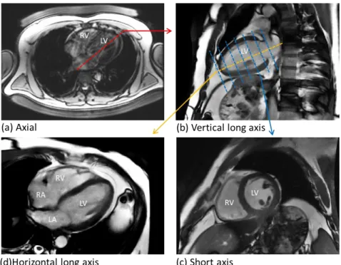
Quantitative analysis of cardiac MRI parameters in myocardial infarction
164
0
0
Texte intégral
Figure
+7

Documents relatifs