THÈSE PRÉSENTÉE À
L'UNIVERSITÉ DU QUÉBEC À TROIS-RIVIÈRES
COMME EXIGENCE PARTIELLE
DU DOCTORAT EN BIOLOGIE CELLULAIRE ET MOLÉCULAIRE
PAR
PAULINA PA WLICA
INVOL VEMENT OF MICRO TUBULES AND DYNEIN MOTOR COMPLEXES IN RIV -1 CORE DISASSEMBL y AND ITS RESTRICTION BY TRIM5a
Université du Québec à Trois-Rivières
Service de la bibliothèque
Avertissement
L’auteur de ce mémoire ou de cette thèse a autorisé l’Université du Québec
à Trois-Rivières à diffuser, à des fins non lucratives, une copie de son
mémoire ou de sa thèse.
Cette diffusion n’entraîne pas une renonciation de la part de l’auteur à ses
droits de propriété intellectuelle, incluant le droit d’auteur, sur ce mémoire
ou cette thèse. Notamment, la reproduction ou la publication de la totalité
ou d’une partie importante de ce mémoire ou de cette thèse requiert son
autorisation.
Cette thèse a été dirigée par :
Lionel Berthoux Université du Québec à Trois-Rivières
Directeur de recherche, Ph. D. Institution à laquelle se rattache l'évaluateur
Jury d'évaluation de la thèse:
Lionel Berthoux, Ph. D. Université du Québec à Trois-Rivières Prénom et nom, grade Institution à laquelle se rattache l' évaluateur
Hugo Germain, Ph. D. Université du Québec à Trois-Rivières Prénom et nom, grade Institution à laquelle se rattache l' évaluateur
Isabel Desgagné-Penix, Ph. D. Université du Québec à Trois-Rivières Prénom et nom, grade Institution à laquelle se rattache l'évaluateur
Andrew J. Mouland, Ph. D. McGill University
Prénom et nom, grade Institution à laquelle se rattache l' évaluateur
First, l would like to thank my research director Lionel Berthoux for including me in his team and introducing me to the HIV research field, in a highly simulating environment. l am grateful for the opportunities, support and advice he has offered.
Secondly, l would like to express my gratitude towards Marie-Édith Nepveu-Traversy for her constant support, advice and friendship. l would also like to thank Maxime Veilletle for his technical assistance, advice and great discussions. Additionally, l would like to acknowledge other lab members who helped with this work, Mélodie B. Plourde, Kathleen Riopel, Nolewnn Poccardi and Alan Hemandez Telavera.
l would like to thank Andrew J. Mouland and Valerie Le Sage from the McGill University for the fruitful collaboration. Additional thanks to Andrew for his support and advice.
Furthermore, l would like to acknowledge Michel J. Tremblay (Centre de Recherche du CHU de l'Université Laval) for sharing his BSLIII facility, and his staff who provided technical help: Réjean Cantin, Pascal Jalaguier and Alexandre Deshière.
Thanks to Céline Van Themsche (Université du Québec à Trois-Rivières) for sharing instruments and to Ali Saib (Université Paris, France), Trina Schroer (Johns Hopkins University) and Christopher Aiken (Vanderbilt University School of Medicine) for sharing plasmid DNA.
Finally, l would like to thank my family and friends for their great support during my studies, with special thanks to Dominic Leppla, Margaret Kucala and Maciek Puchaty-Kot.
The following thesis focuses on aspects of the replication of human immunodeficiency virus 1 (HIV -1), the causative agent of acquired immunodeficiency syndrome (AIDS). The studies described here may contribute to the development ofnew treatments to inhibit this virus.
The main body of the thesis consists of two manuscripts. The frrst article "Functional evidence for the involvement of microtubules and dynein motor complexes in TRIM5a-mediated restriction of retroviruses", presented in Chapter II, was published in Journal of virology. This study demonstrates that the full ability of the restriction factor TRIM5a (tripartite motif-containing protein 5, isoform a) to inhibit retroviruses requires functional microtubules and dynein motor complexes.
A similar study aimed at characterization of the involvement of microtubules and dynein motor complexes in owl monkey TRIMCyp-mediated restriction was also performed. We found that dependence on microtubules and dynein motor complexes in the full restriction mediated by TRIMCyp is cellular context-specific. This study, entitled "Inhibition of microtubules and dynein rescues HIV -1 from owl monkey TRIMCyp-mediated restriction in a cellular context-specific fashion" was published in Journal of General Virology and is presented here in Annex B.
The second manuscript presented in Chapter III, which was in preparation at the time of thesis submission, discusses on the role of microtubules and dynein motor complexes in HIV-l core disassembly (uncoating). We found that these two cytoskeletal components promote uncoating of the HIV -1 capsid core. A part of this study that includes additional experiments was later published in Viruses ("Cytoplasmic Dynein promotes HIV -1 Uncoating") and is presented in Annex A.
In conclusion, the studies presented in the following thesis provide novel insights into HIV -1 core uncoating and its restriction as mediated by TRIM5a.
L'infection au virus de l'immunodéficience humaine (VIH-1), l'agent causal du SIDA, reste incurable à ce jour. Par conséquent, il est essentiel de développer nos connaissances sur ses mécanismes de réplication et les stratégies d'inhibition possibles.
Le VIH -1 appartient au groupe VI de la famille de virus Retroviridae, sous-famille des Lentiviridae. Le génome des rétrovirus est retrouvé sous forme d'ARN simple brin qui, au cours de l'infection, est réverse-transcrit en ADN double brin par l'enzyme virale transcriptase inverse. Cet ADN complémentaire (ADNc) est par la suite inséré dans l'ADN génomique de l 'hôte cellulaire par l'enzyme virale intégrase. La particularité des lentivirus est leur habilité à importer activement leur ADNc dans le noyau à travers les pores nucléaires. Suivant l'entrée dans la cellule, les étapes les plus précoces de la réplication du VIH sont le déshabillage de son cœur capsidique et la transcription inverse, et ces deux processus sont probablement interdépendants. On pense que le VIH-1 est transporté en direction du noyau via les complexes de dynéine associés aux microtubules (Fig. 1).
TRIM5a. (tripartite motif-containing protein 5, isoform o.) est un facteur de restriction qui inhibe les étapes précoces de la réplication rétrovirale. TRIM5a. agit via une reconnaissance spécifique du cœur capsidique peu après l'entrée du virus et le déstabilise. TRIM5a. agit d'une façon spécifique à l'hôte et également au virus; TRIM5a. du singe Rhésus macaque inhibe le VIH-1 alors que TRIM5a. humain n'a pas d'action contre ce virus, mais inhibe d'autres rétrovirus tel que le virus de la leucémie murine N-tropique (N-tropic murine leukemia virus, N-MLV). Les protéines TRIM5a. forment des corps cytoplasmiques (CCs) qui sont associés aux microtubules (Fig. 1).
membrane
dégradation par le protéasome ~ (3) ~ reconnaissance du capside
1
de 11mmunité~
- • signalisation+
innée dé t bl d 'd transport . . . . s a lise u capsi. e . vers le noyauInhibition de la transcnptlon Inverse noyau
dynéine
Figure 1
ft
Schéma représentant les étapes précoces de la réplication du VIH-l et comment elles sont inhibées par les protéines TRIM5a.
(1) Suivant l'entrée du virus qui est médiée par des récepteurs spécifiques, la capside du VIH-l (CA) s'associe aux microtubules et utilise les complexes moteurs de dynéine pour son transport vers le noyau. (2) Pendant ou après ce transport, le VIH-l accomplit la transcription inverse (reverse transcription, R T) et subit un déshabillage, deux étapes qui sont probablement interdépendantes. (3) Dans les cellules exprimant TRIM5a, le cœur viral est intercepté peu de temps après son entrée dans le cytoplasme par ce facteur de restriction. R TC, complexe de transcription inverse (reverse transcription comp/ex); PIC, complexe de pré-intégration (pre-integration comp/ex); MTOC, centre organisateur des microtubules (microtubu/e-organizing center). CC s, corps cytoplasmiques.
Dans cette thèse, nous examinons si les microtubules et les complexes de dynéine ont un rôle dans (i) la restriction rétrovirale médiée par TRIM5a, et (ii) le déshabillage du VIH-l. Nous avons étudié ces deux processus par disruption pharmacologique ou par perturbation du transport médié par les complexes de dynéine. Les microtubules ont été inhibés par traitement au nocodazole ou au paclitaxel, et le transport dependant des
dynéines a été inhibé soit à l'aide d'un small interfering RNA (siRNA) ciblant la chaîne lourde des dynéines (dynein heavy chain, DHC), soit par sur-expression de la protéine p50/dynamitine, ou encore par un traitement pharmacologique au erythro-9-(2-hydroxy-3-nonyl)adenine (ERNA). Pour analyser l'infectivité, nous avons utilisé des vecteurs rétroviraux exprimant la GFP. Nous avons utilisé l'imagerie à immunofluorescence pour observer les CCs formés par TRIM5a ainsi que les cœurs capsidiques dans les cellules infectées. Nous avons employé un test de «fate-of-capsid assay» qui permet l'isolation de cœurs capsidiques insolubles des cellules infectées, par ultracentrifugation à travers un coussin de sucrose.
Dans la première partie, nous démontrons la dépendance de la restriction par TRIM5a aux microtubules et complexes de dynéine. La perturbation de ces deux composantes du cytosquelette résulte en un potentiel de restriction par TRIM5a significativement réduit. Cette perte de la restriction est observée soit suite à la perturbation des microtubules, ou bien suite à la perturbation de l'activité des complexes de dynéine, et n'est pas le résultat d'une diminution de la stabilité de TRIM5a ou d'une diminution de viabilité cellulaire. Les traitements au nocodazole ou la déplétion de DHC interfèrent tous deux avec la dynamique des CCs de TRIM5a, augmentant leur taille et altérant leur localisation intracellulaire, tel qu'observé par immunoflurorescence. De plus, dans le test de fate-of-capsid, les traitements au nocodazole ou au paclitaxel, et la déplétion de DHC augmentent la stabilité des cœurs viraUx de VIH-l dans les cellules infectées, fournissant une explication alternative pour la baisse de restriction.
Dans la deuxième partie, nous avons observé que les microtubules et les complexes de dynéine sont probablement importants pour le déshabillage du VIH-l. Dans des expériences d'immunofluorescence, nous avons détecté une accumulation de foci de cœurs capsidiques dans les cellules infectées par le VIH-l et où le niveau d'expression de la DHC a été diminué ou traitées de façon à perturber les microtubules. Ces foci détectés dans des conditions d'infection aigüe sont probablement des cœurs capsidiques viraux individuels. En utilisant le fate-of-capsid assay pour analyser le déshabillage du cœur capsidique dans les cellules infectées, nous avons observé une
augmentation des quantités de cœurs Vlraux insolubles suite aux traitements au nocodazole ou au paclitaxel, à la déplétion de la DHC et à la surexpression de p50/dynamitine. Nos résultats suggèrent qu'inhiber la dynamique des microtubules ou le transport médié par la dynéine interfère avec le déshabillage du VIH -1 dans le cytoplasme des cellules infectées, ce qui implique un lien fonctionnel entre le transport du VIH-1 et son déshabillage.
En conclusion, les microtubules et les complexes de dynéine sont importants pour la restriction rétrovirale médiée par TRIM5a, possiblement en permettant un contact initial entre TRIM5a et sa cible virale. Les protéines TRIM5 contiennent possiblement un PMIM qui permet l'interaction directe avec les microtubules. De plus, le cytosquelette semble réguler la dynamique des CCs de protéines TRIM5a. Enfm, le réseau de microtubules et les complexes de dynéine sont importants pour la réplication du VIH-1, en régulant son déshabillage en plus de son transport vers le noyau.
Les études présentées dans cette thèse représentent des avancées importantes dans le domaine des interactions hôte-pathogène. Elles contribuent à augmenter l'état des connaissances sur la réplication du VIH-1 et le mécanisme d'inhibition par TRIM5a. Il est à espérer que ce travail pourra contribuer au développement futur de nouvelles approches pour traiter l'infection au VIH-1.
Infection with Human immunodeficiency virus 1 (HIV -1), the causative agent of acquired immunodeficiency syndrome (AIDS), remains incurable. Thus, it is essential to gain detailed knowledge about the mechanisms of its replication and possible ways to inhibit it. Following entry to the cell, HIV -1 undergoes many processes necessary for the productive infection, of which the earliest are uncoating and reverse transcription, which are likely inter-dependent. HIV-l is thought to traffic towards the nucleus using the microtubule-associated dynein motor complex. Tripartite motif-containing prote in 5, isoform a (TRIM5a) is a cellular restriction factor, found in cytoplasmic bodies (CBs), that inhibits the early steps of retroviral replication. TRIM5a acts through specifie recognition of incoming retroviral capsid (CA) cores and destabilizes them. Interestingly, TRIM5a CBs were found to associate with microtubules. The studies in the following thesis examine whether microtubules and dynein motor complexes play a role in (i) the retroviral restriction mediated by TRIM5a and (ii) HIV -1 core disassembly (uncoating). These two processes were monitored under either pharmacological disruption of microtubule dynamics or perturbation of dynein-mediated transport. Microtubule dynamics were inhibited using nocodazole or paclitaxel, while dynein mediated-transport was interrupted by either an siRNA targeting dynein heavy chain (DHC), p50/dynamitin over-expression or a pharmacological treatment using erythro-9-(2-hydroxy-3-nonyl) adenine (EHNA). In the frrst study, we demonstrated the dependence of TRIM5a-mediated restriction on microtubules and dynein motor complexes. Disrupting these two cytoskeletal components resulted in a significantly decreased potential of TRIM5a to inhibit retroviral infection. The loss in restriction resulting from either perturbation of microtubules or disruption of dyne in motor activity was not due to differences in protein stability or cell viability. Both nocodazole treatment and DHC depletion interfered with the dynamics of TRIM5a CBs, increasing their size and altering their intracellular localization. In addition, nocodazole, paclitaxel and DHC depletion were aIl found to increase the stability of HIV -1 cores in infected cells, providing an alternative explanation for the decreased restriction. In the second study, we found that both microtubules and dynein motor complexes are likely to be important factors in HIV -1 uncoating. The immunofluorescence microscopy experiments allowed observation of an accumulation of CA/aci in HIV-l infected cells following DHC depletion or microtubule perturbation. Such CA/aci detected in infected cells are thought to be individual not-yet or partially uncoated viruses. Using a fate-of-capsid assay to monitor HIV -1 CA core disassembly in infected cells, we observed an increase in the amounts of pelletable (particulate) CA cores following treatments with nocodazole or paclitaxel, as weIl as DHC depletion and p50/dynamitin over-expression. The results suggest that inhibiting the dynamics of microtubules or dynein-mediated transport interferes with HIV -1 uncoating in the cytoplasm of infected cells, implying a functionallink between HIV -1 transport and uncoating. In conclusion, microtubules and dynein motor complexes are important for the retroviral restriction mediated by TRIM5a, possibly by allowing the initial contact between TRIM5a and its viral target. Furthermore, it is likely that the cytoskeleton regulates the dynamics of the CBs formed
by TRIM5a proteins. Finally, the microtubule network and the dynein motor complex are important in RIV -1 replication regulating, in addition to the transport of its core towards the nucleus, its proper uncoating.
ACKNOWLEDGEMENTS...
iliPREFACE ... iv
, , RESUME... v
ABSTRACT ... ... ix
LIST OF FIGURES ... xiv
LIST OF ABBREVIATIONS AND ACRONYMS ... xvi
CHAPTERI 'INTRODUCTION ... 1
1.1 HIV -1 as the causative agent of AIDS ... 1
1.1.1 The AIDS epidemic ... 2
1.1.2 Treatment and prevention HIV -1 infection... 3
1.1.3 Perspectives for a cure ... 5
1.1.4 The HIV-1 genome and its structure... 6
1.1.5 HIV -1 rep1ication cycle .... ... ... ... 8
1.1.5.1 Early phase... 8
1.1.5.2 Late phase ... 10
1.1.6 The capsid core and its uncoating... 12
1.1.6.1 Evidence supporting regulated HIV-1 uncoating ... 13
1.1.6.2 HIV-1 uncoating... 14
1.1.6.3 Cellular proteins regulating uncoating... 15
1.2 The cytoskeleton... 16
1.2.1 The microtubule network... 17
1.2.1.1 Regulation of microtubule dynamics... 18
1.2.1.2 Treatments targeting microtubules ... ... 19
1.2.2 Molecular motors and their role in cellular transport ... 19
1.2.2.1 The dynein motor complex... 21
1.2.2.2 Inhibitors of the dynein motor complex function... 22
1.2.3 Rijacking of the host cytoskeleton by viroses ... 23
1.2.3.1 RIV -1 trafficking towards the nucleus ... 23
1.3 Innate immunity and TRIM5a... 26
1.3.1 Restriction factors... 28
1.3.2 The TRIM family ofproteins... 28
1.3.3 TRIM5a ... 29
1.3.3.1 Mechanism of action ... :... 31
1.3.3.2 Cell biology ofTRIM5a ... 32
1.3.4 TRIMCyp... 33
1.4 Importance, hypotheses and objectives ... 34
1.4.1 Objective 1: To study the involvement of microtubules and dynein motor complexes in TRIM5a-mediated restriction... 34
1.4.2 Objective II: To study the involvement of microtubules and dyne in motor complexes in RIV -1 uncoating ... 35
CHAPTERII FUNCTIONAL EVIDENCE FOR THE INVOL VEMENT OF MICROTUBULES AND DYNEIN MOTOR COMPLEXES IN TRIM5a.-MEDIATED RESTRICTION OF RETROVIRUSES... 37
2.1 Contributions ... 37
2.2 Abstract... 37
2.3 Importance ... ... 38
2.4 Introduction ... 39
2.5 Materials and methods... 41
2.6 Results ... 46
2.7 Discussion... 69
2.8 Acknowledgements ... 72
2.9 References ... 73
CHAPTERIII INIDBITION OF MICROTUBULE- AND DYNE IN-DEPENDENT TRANSPORT COUNTERACTS HIV-l UNCOATING... 82
3.1 Contributions... 82
3.2 Abstract... 82
3.4 Results ... 85 3.5 Discussion... 98 3.6 Methods ... 100 3.7 Acknowledgements ... 104 3.8 References... 104 CHAPTERIV CONCLUSIONS ... 111
4.1 The involvement of microtubules and dynein motor complexes in TRIM5a-mediated restriction ... ... 111
4.1.1 How might TRlM5a use microtubules? ... ... 111
4.1.2 Future experiments ... 113
4.2 The involvement of microtubules and dynein motor complexes in IllV-l uncoating ... 115
4.2.1 Trafficking of IllV -1 and its uncoating ... ... 117
4.2.2 Future experiments ... ... 119
4.3 Perspectives: Gene therapy targeting IllV-l using TRIM5a ... 123
4.4 Perspectives: development ofnew classes of drugs ... 123
4.5 Final conclusions ... ;... 124
REFERENCES ... 125
ANNEXA CYTOPLASMIC DYNEIN PROMOTES HIV-l UNCOATING ... 154
ANNEXB INIllBITION OF MICROTUBULES AND DYNE IN RESCUES HIV-l FROM OWL MONKEY TRIMCYP-MEDIATED RESTRICTION IN A CELLULAR CONTEXT-SPECIFIC FASmON ... 179
Figure Page
1.1 Global prevalence of HIV -1 ... :... 3
1.2 Organization of HIV -1 genome and the structure ofthe virus... 7
1.3 HIV-l replication cycle... 10
1.4 Model of HIV -1 capsid core... 12
1.5 Schematic representation of cytoskeletal filaments... ... 16
1.6 The dynein motor complex ... ... ... 20
1. 7 Scpematic representation of HIV -1 trafficking inside the cell ... 25
1.8 Mechanisms of Viral Detection in dendritic cells... 27
1.9 Structure of TRIM5a and the CUITent model of its action... 30
2.1 Drug concentration-dependent enhancement of permissiveness to infection by TRIM5a-restricted retroviral vectors in human and simian cells ... 48
2.2 Pharmacological disruption of microtubules decreases endogenous TRIM5a-mediated retroviral restriction... 51
2.3 Pharmacological . disruption of dynein motor function decreases endogenous TRIM5a-mediated retroviral restriction. ... ... 53
2.4 DHC depletion decreases TRIM5a-mediated retroviral restriction and has no effect on cells treated with nocodazole or paclitaxel... 56
2.5 Inhibition of HIV-1 restriction by rhTRIM5a exogenously expressed in HeLa cells ... 59
2.6 Nocodazole and paclitaxel inhibit TRIM5a-mediated restriction of non-pseudotyped HIV -1 ... 61
2.7 Effect of treatments on TRIM5a stability and cell viability ... 63
2.8 DHC depletion, paclitaxel treatment and nocodazole treatment increase the stability ofHIV-1 cores in permissive or restrictive conditions ... 66
2.9 Influence of DHC depletion and nocodazole treatment on the localization
and dynamics ofrhTRIM5a cytoplasmic bodies (CBs) ... 68
3.1 Dynein heavy chain (DHC) depletion and microtubule perturbation cause the accumulation of HIV -1 CA cores in infected cells and alter their subcellular distribution... 88
3.2 Analysis ofHIV-1 uncoating using the fate-of-capsid assay... 91
3.3 DHC depletion and nocodazole treatment affect HIV-l uncoating... 94
3.4 Over-expression ofp50/dynamitin affects HIV-1 uncoating... 96
3.5 Nocodazole increases the relative amounts of E45A CA cores in infected cells, but has no effect on the intrinsically hypostable CA mutant K203A ... 98
4.1 Retroviral evasion of immune detection ... ... 117
4.2 Model of the NMDA receptor transporting machinery ... 119
4.3 Schematic overview of viral fusion assay... 121
AAA+ AIDS
ADP
ATP AP-l APOBEC B-MLV BSL CA Cas9 CB CARDs CCR5 CD4 CD4+ CDC cDNA cGAS co-IP CPSF6 CRISPR CROI CTDATPases associated with diverse cellular activities acquired immunodeficiency syndrome
adenosine diphosphate adenosine triphosphate
activator protein-l
apolipoprotein B mRNA editing enzyme, catalytic polypeptide-like
B-tropic murine leukemia virus biosafety level
capsid protein, p24
CRISPR -associated protein 9
cytoplasmic body
caspase activation and recruitment domains C-C chemokine receptor type 5
c1uster of differentiation 4
cell bearing the CD4 receptor
United States Centers for Disease Control and Prevention complementary DNA
cyc1ic GMP-AMP synthase
co-immunoprecipitation
c1eavage and polyadenylation specific factor 6
c1ustered regularly interspaced short palindromic repeat Conference on Retroviruses and Opportunistic Infections
CXCR4 CypA
DHC
DIC
DUC
DNA dNTP dsRNAEB
EHNA EIAV Env ESCRT FIV FN3 FRET GTP GDP HAARTHC
HIV-I HIV-2 IF IFN IKKi INchemokine receptor type 4 cyclophilin A
dyne in heavy chain dynein intermediate chain
dynein light intermediate chain deoxyribonucleic acid
deoxyribonucleotide triphosphate double-stranded RNA
end-binding
erythro-9-(2-hydroxy-3-nonyl)adenine
equine infectious anemia virus envelope
endosomal sorting complexes required for transport feline immunodeficiency virus
fibronectin type III domain
fluorescence resonance energy transfer
guanosine triphosphate guanosine diphosphate
highly active antiretroviral therapy heavy chain
human immunodeficiency virus type 1 human immunodeficiency virus type 2 immunofluorescence
interferon IKB kinase i integrase
IPSI IRF-3 ISG JAK KIF 1 7 LIS 1 LTR LI L2 MA MAP MDA-5 MDM MIDI MID2 MIM MLV MT OC Mx2/MxB N-MLV NC Nef NF-KB NMDA NNRTI NPC
interferon-beta promoter stimulator 1
IFN-regulatory factor 3 IFN -stimulated genes
Janus kinase
kinesin family member 17 lissencephaly
long terminal repeat
linkerl linker2
matrix protein
microtubule-associated protein
melanoma differentiation-associated protein 5 monocyte-derived macrophages
midline 1 midline 2
microtubule-interacting motif murine leukemia virus
microtubule-organizing center myxovirus resistance 21 B N-tropic murine leukemia virus
nucleocapsid protein negative factor
nuclear factor K-light-chain-enhancer of activated B cells
N-methyl-D-aspartate
non-nucleoside reverse transcriptase inhibitors
NRTI NTD NUDE PAMP PDZD8 PEP PF74
PML
PMIM
PI
PIC
PrEP PRR PR p.l.RBCC
Rev RIG-I RILP RING RIPI RNA RT RTC SAMHDI SCIDnucleoside/nucleotide reverse transcriptase inhibitors N-terminal domain
nuclear distribution protein E
pathogen-associated molecular pattern PDZ domain containing 8
post-exposure prophylaxis Pfizer-345 0074
promyelocytic leukemia prote in putative microtubule-interacting motif protease inhibitor pre-initiation complex pre-exposure prophylaxis pattern-recognition receptor protease post infection
RING finger, B-box, and coiled-coil regulator of expression of virion retinoic acid-inducible gene 1 Rab-interacting lysosomal protein really interesting new gene
Receptor-interacting protein 1 ribonucleic acid
reverse transcriptase
reverse transcription complex
SAM domain and RD domain-containing protein 1 severe combined immunodeficiency
SIV siRNA ssRNA STAT SU TALEN Tat TasP TcTex-1 TLR TM TRAF6 TRIF TRIM TRIM5a UNAIDS VCC Vif Vpu Vpr Vpx vRNA ZFN +TIP
simian immunodeficiency virus small interfering RNA
single-stranded RNA
signal transducer and activator of transcription surface protein, gp 120
transcription activator-like effector nucleases trans-activator of transcription
treatment as prevention
T -complex testis-specific protein 1 Toll-like receptor
transmembrane protein, gp41 TNF receptor associated factor 6
TIR-domain-containing adapter-inducing interferon-p tripartite motif
tripartite motif-containing protein 5a United Nation pro gram on HIV/AIDS virus-containing compartments viral infectivity factor
viral protein unique viral protein R viral protein X viral RNA
zinc-finger nucleases plus-end tracking proteins
INTRODUCTION
1.1 HIV -1 as the causative agent of AIDS
In the early 1980s a mysterious immune disorder epidemic emerged, initially within the homosexual population, intravenous drug us ers and immigrants from Haiti, which gradually spread into the general population. The starting point of the epidemic is dated June 5th 1981, when the Centers for Disease Control and Prevention (CDC) reported several outbreaks of opportunistic diseases in previously healthy patients. (CDC, 1981). At that time, the disorder was referred to as gay-related immunodeficiency due to reported incidence of the disease among this group. In 1982, it was renamed acquired immunodeficiency syndrome (AIDS)(CDC, 1982). Its causative agent was discovered by two independent groups in 1983-4 and the virus was called human immunodeficiency virus (HIV). (Barre-Sinoussi et al, 1983; Gallo et al, 1984; Popovic et al, 1984). Shortly after the identification ofHIV, later referred to as HIV-l, a second virus causing AIDS was discovered, named HIV-2 (Clavel et al, 1986). The two viruses differ substantially, resulting in differences in serological detection (Clavel et al, 1987). Although HIV-l and HIV-2 cause the same disease, individuals infected with HIV-2 usually have a longer life span than those infected with HIV -1, as it takes more time to develop into full-blown AIDS (Reeves & Doms, 2002).
Both HIV-l and HIV-2 originate from closely related simian immunodeficiency virus (SIV) found in many African primate species. The genetic sequence of HIV-l resembles SIV from chimpanzees (Pan troglodytes), while HIV-2 is similar to SIV infecting sooty mangabeys (Cercocebus atys) (Sharp et al, 2001; Sharp et al, 2005). The transfers of SIV from primates to humans happened multiple times. Interestingly, the oldest documented case of HIV-l infection dates to 1959; the virus was detected in a specimen collected in Kinshasa, the capital of Congo (Zhu et al, 1998). Although the
virus was present in the human population likely as early the 1920s, it did not reach an epidemic threshold until 1980s, as a result of colonialism-related trade in Cameroon (Sharp & Hahn, 20 Il).
HIV-l can be divided into four genetically diverse groups M, N, 0 and P that reflect separate transfers of SIV from chimpanzees to humans (Sharp et al, 2005). The most widespread is HIV -1 from the M group, while the N, 0 and P groups, as weIl as
HN-2, are limited to Africa (De Cock et al, 1993; Sharp & Hahn, 2011). The M group can be further divided into at least nine genetically distinct subtypes (or clades);
subtypes A, B, C, D, F, G, H, J and K. This thesis focuses on the aspects of infection caused by HIV -1 belonging to M group, as this virus is the major cause of the present AIDS epidemic.
1.1.1 The AIDS epidemic
In 2012, the United Nations program on HIV/AIDS (UNAIDS) reported that 35.3 million people were estimated to be living with HIV-1 or HIV-2 (UNAIDS, 2013).
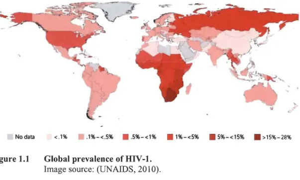
That year, the number of newly infected individuals reached 2.3 million people and approximately 2 million deaths resulting from AIDS-related diseases were reported (UNAIDS, 2013). Thanks to pharmacological treatments, education and interventions of various organizations, these numbers reached stable levels, however there is still no perspective to eradicate the disease. The most dramatic situation is in sub-Saharan Africa, where in some countries more than 25% of population is infected (Fig. 1.1).
No data <.1% .1%-<.5% • . 5%-<1% . 1%-<5% . 5%-<15% . >15%-28%
Figure 1.1 Global prevalence of HIV-1. Image source: (UNAIDS, 2010).
Following IDV-1 contraction a patient may experience an acute phase of infection with flu-like symptoms which gradually transfonns into an asymptomatic chronic infection. The latter usually lasts several years during which T lymphocytes bearing clusters of differentiation 4 receptor (CD4+ T -cells) are slowly eradicated (Organization., 2007). The CD4+ T -cell count in healthy individuals ranges from 500 cells/J!l to 1,500 cells/J!1. AIDS is defmed by low CD4+ T ..:cell count (less than 200 cells/J!l) and/or occurrence of opportunistic diseases, such as pneumonia caused by Pneumocystis carinii (Gottlieb, 2006) or Kaposi's sarcoma (Friedman-Kien, 1981; Rymes et al, 1981). People with AIDS usually die as a result of opportunistic diseases.
1.1.2 Treatment and prevention HIV-l infection
To this date, neither a cure nor a vaccine effective against RIV -1 have been developed. There are multiple obstacles in developing safe, effective and durable vaccines, including the fast mutation rate of the virus and high glycosylation of its spike proteins, which unables immune system detection of viral epitopes. Oruy one trial, RV144, consisting of two vaccines that individually shown no efficacy, resulted in a moderate, ~30%, protection (Rerks-Ngann et al, 2009). This study suggests that developing an effective vaccine could be feasible in the future.
The frrst drug effective against HIV-l azidothymidine (AZT) was introduced in 1987. (Yarchoan & Broder, 1987). This drug is now classified as nucleoside/nucleotide reverse transcriptase inhibitor (NRTI). NRTIs are nucleoside analogues. that resemble naturally occurring nucleosides and are incorporated into nascent DNA by viral
reverse transcriptase but not human polymerases. AZT, which was initially used as
monotherapy, quickly resulted in the emergence of drug-resistant virus es and subsequent
failure of the treatment. Between 1991 and 1995, four additional NRTIs were developed.
A breakthrough came in 1995 when a new protease inhibitor (PI, saquinavir) was
introduced, which led to the CUITent approach of combining multiple antiretroviral drugs targeting various steps of the viral replication cycle, known as highly active
antiretroviral therapy (HAART) (Flepp et al, 2001). In 1996 nevirapine, a drug
belonging to a new class of pharmaceuticals, was introduced, along with two additional PIs. Nevirapine is a non-nucleoside reverse transcriptase inhibitors (NNRTI) and acts directly on the reverse transcriptase. In 2003, the frrst fusion inhibitor was introduced (enfuvirtide), and in 2007 inhibitors of entry (maraviroc) and integrase (raIte gravir) were approved (palmisano & Vella, 20 Il), increasing therapy options for infected patients. Currently, there are over twenty pharmaceuticals available. Despite increased life
expectancy and overall health improvement of patients, HAAR T has serious drawbacks,
e.g. it has to be taken for the whole of the patient's life and administered daily; viral
targets might acquire drug resistance; fmally, side effects such as anemia,
hepatotoxicity, hyperglycemia, hyperlipidemia and lipodystrophy are common (Flepp et
al, 2001).
A very important aspect of fighting HIV -1 epidemics IS education about
its transmissibility and the prevention of new infections. In general, three
approved protocols employing antiretroviral pharmaceuticals to prevent HIV-l infection/transmission are commonly used. Treatment as prevention (TasP) is an approach, in which infected patients receive antiretroviral drugs in order to lower the chances of infecting others, and can be as effective as in 96% of cases; this approach is commonly used in HIV-discordant couples and to prevent mother-to-child transmission (De Cock et al, 2000). Also, healthy individuals can decrease the odds of becoming
infected by taking prophylactic antiretroviral drugs. This approach is known as pre-exposure prophylaxis (prEP) and is commonly practiced in HIV -discordant couples and sex workers; it has been shown to be effective in 70% of cases (Anglemyer et al, 2011; Liu & Chibwesha, 2013; Vaid et al, 2013). In the case of a healthy individual that has been exposed to HIV -1 a post-exposure prophylaxis (PEP) can be employed to reduce the chances of becoming infected. This is approach is commonly used in health workers after accidentaI exposure to the virus, for example a needlestick injury (Cardo et al, 1997; Smith et al, 2005). Interestingly, male circurncision has been found to decrease HIV -1 transmission by at least 60% in males that have been circumcised (Auvert et al, 2005). Additionally, it is estimated that male circumcision might prevent male-to-female transmission by 46% (Hallett et al, 20 Il).
In conclusion, while the past years have seen a tremendous development in antiretroviral therapy, mitigating adverse effects and simplifying treatments, finding either a cure or a vaccine has proven elusive.
1.1.3 Perspectives for a cure
The ultimate aim of HIV -1 research is a complete eradication of the virus from the infected organism, which is known as a 'sterilizing cure'. Up to this date, only one case ofthis kind of cure has been described, known as the 'Berlin patient'. Timothy Brown as a therapy for acute myeloid leukemia underwent myeloablative treatment and then received a bone marrow transplant from a donor whose cells were resistant to HIV-1 infection; this intervention resulted in a complete elimination of the virus (Hutter et al, 2009; Hutter & Thiel, 2011). The donor had a homozygotic deletion in the HIV-1co-receptor C-C chemokine receptor 5 (CCR5) gene (Alkhatib et al, 1996; Deng et al, 1996; Dragic et al, 1996); lack of which prevents infection with this virus (Liu et al, 1996). Until this date this remains the only case of a 'sterilizing cure'. The other famous cases turned out to be prematurely optimistic. For example, the famous 'Mississippi' infant was an infant reported to be cured as a result of an aggressive antiretroviral
treatment prior to, it was thought, the virus being able to establish its reservoir (Cohen, 2013). Unfortunately, in July 2014 the virus re-emerged in the baby's blood.
While the sterilizing cure is still beyond our reach a more feasible goal may be obtained, known as a 'functional cure'. The functional cure is defmed as spontaneous drug-free control of the viral replication. For example, the members of the ANRS VISCONTI study in which up to ~ 15% of early treated individuals wereshown to display spontaneous control of viremia (Saez-Cirion et al, 2013). The reasons for this viral control are still not fully understood, but it could be that early treatment, while limiting the viralload, enables the immune system to produce a specific response against the virus.
1.1.4 The HIV-l genome and its structure
Taxonomically HIV -1 belongs to the viral Group VI, family Retroviridae, subfamily Lentivirus. The genome of retroviruses exists in the form of a single-stranded RNA (ssRNA) which upon infection is reverse transcribed into a double-stranded complementary DNA (cDNA) by the viral enzyme reverse transcriptase. This DNA is later inserted into the host' s genome by the viral enzyme integrase. The hallmark of lentiviruses is their ability to actively import their cDNA into the nucleus through the nuclear pore complex (NPC), which allows for infection of non-dividing cells.
A
B Structural plO C Proteins SU TM PRNe
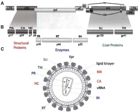
Rf p66 pU Enzymes Vpr Coat Proteins lIpld bilayer MA CA vRNA INFigure 1.2 Organization of HIV -1 genome and the structure of the virus.
(A) A schematic representation of the organization of HIV -1 genome. LTR - long terminai repeat. (B) A schematic representation of cleavage
products of HIV-1 polyproteins. Gag is cleaved to MA (matrix, p17),
CA (capsid, p24), NC (nucleocapsid, p7) and p6; Pol to PR (protease), RT (reverse transcriptase) and IN (integrase); Env to SU (surface protein, gp120) and TM (transmembrane protein, gp41). (C) A schematic representation of HIV -1 virion. vRNA -viral RNA.
Image source: (Krogan, 2011).
The HIV-1 genome consists of two positive-sense ssRNAs, ~10 kb in length
(Ratner et al, 1985; Wain-Hobson et al, 1985) that contain nine genes (Fig. 1.2A). Three encode the polyproteins Gag, Pol and Env, while the others encode regulatory and accessory proteins, sorne of which are the result of post-transcriptional splicing. The regulatory proteins regulator of expression of virion proteins (Rev) , trans-activator of transcription (Tat) and negative factor (Nef) are expressed during the viral replication. The accessory proteins viral infectivity factor (Vif), viral protein R (Vpr) and viral protein unique (Vpu) are usually packed into the virion and play roles during the early
steps of infection. Interestingly. the related HIV -2 does not encode Vpu, but in its place encodes viral prote in X (Vpx) (Coffin et al, 1997).
HIV-1 is an enveloped virus whose size is estimated to be approximately 120 nm (Kuznetsov et al, 2003). The structural proteins matrix (MA, p17), capsid (CA, p24),
nucleocapsid (NC, p7) and p6 are the result of cleavages of the Gag precursor (P55) (Fig. 1.2). The CA protein forms a conical-shaped core that encases the viral RNA (vRNA) associated with Ne. The CA core also contains three viral enzymes, products of the Pol precursor: reverse transcriptase (RT, p66), integrase (IN, p32) and protease (PR, plO). The virions also enclose accessory proteins Vif, Vpr, Vpu (or Vpx). MA forms an inner shell below the lipid envelope originating from a producer cell. The envelope, shielding the CA core, mediates viral entry. This lipid membrane is spanned with transmembrane (TM, gp41) viral prote in, one of the products of the Env precursor. Associated with gp41 is the surface protein (SU, gp120), which is responsible for receptor recognition and its binding (Coffin et al, 1997).
1.1.5 HIV-l replication cycle
The replication cycle of lentiviruses can be clearly divided into two phases, early and late, whereby the first consists of the steps leading to the integration of the viral genome into the host's genome. The late phase consists of all subsequent events.
1.1.5.1 Early phase
The primary targets for HIV -1 are cells of the immune system bearing the CD4 receptor, mainly macrophages, dendritic cells and helper T-cells (Dalgleish et al, 1984;
Klatzmann et al, 1984a; Klatzmann et al, 1984b). Initially, the gp120 (SU) triplet,
protruding from the viral envelope, attaches to the CD4 receptor and one of the two co-receptors; either CCR5 (Alkhatib et al, 1996; Deng et al, 1996; Dragic et al, 1996) or
C-X-C chemokine receptor type 4 (CXCR4) (Deng et al, 1996; Feng et al, 1996)
penetration of the cellular membrane by gp4l (TM). This event brings the two membranes together which causes their fusion (Melikyan, 2014). Next, the HIV-l CA core is released into the cytoplasm and this is followed by two processes: reverse transcription and core disassembly (uncoating), which are likely inter-dependent (Rulme et al, 2011; Roa et al, 2012). Interestingly, RT has no proofreading properties and its
fidelity is considerably low (1.4 x 10⁻⁵ errors per base pair (Abram et al, 2010)), thus the
reverse transcription process is extremely error-prone. This results in an enormous RIV -1 mutation rate which allows the virus to quickly adapt to a changing environment, such as the presence of an antiretroviral drug. Transcribed double-stranded cDNA together with IN and other remaining viral proteins form the pre-integration complex (PIC) which associates with the NPC. The distinctive feature of lentiviruses is their
ability to actively pass through the NPC and infect non-dividing cells (Lewis &
Emerman, 1994; ~oe et al, 1993). This process is not fully understood, but it is likely
supported by the viral protein Vpr (Kogan & Rappaport, 20 Il). Once in the nucleus, the
viral cDNA is inserted into the host's genome, with a preference towards actively transcribed regions (Schroder et al, 2002).
Host CelJ
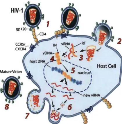
Figure 1.3
mv
-1 repli cation cycle.(1) Recognition of the cellular receptors. (2) Fusion of the viral and cellular membranes. (3) Core uncoating and reverse transcription. (4) Formation of the pre-integration complex (PIC). (5) Import and integration of vDNA (cDNA) into the host genome. (6) Synthesis of vRNAs and viral proteins. (7) Viral budding from the cellular membrane. (8) Maturation of released virions. .
Image source: (NIH, 2012).
1.1.5.2 Late phase
Once integrated, the viral genetic material is called a provirus and is actively transcribed by cellular RNA polymerase II. The transcription is mediated by a single promoter in the 5' LTR of the provirus and requires cellular transcription factors, of which the most crucial is NF-KB (Nabel & Baltimore, 1987). Notably, long-living cells carrying proviruses can be deactivated, during which the virus can remain transcriptionally dormant for many years while retaining the ability to be reactivated with full pathogenic capacity upon the cell's activation (Chun et al, 1998). This state is called latency and is the reason why HIV -1 infection remains incurable.
Post integration, the frrst transcripts are spliced and regulatory proteins Tat, Rev and Nef are expressed (Greene & Peterlin, 2002; Jordan et al, 2003). Tat and Rev proteins shuttle back to the nucleus. Tat activates the production of fulliength transcripts and in trans stimulates the expression of all HIV -1 genes (Kao et al, 1987). Rev binds to newly transcribed unspliced vRNA and allows it to leave the nucleus (Pollard & Malim, 1998). In the cytoplasm, unspliced vRNA either serves as a template for the production of the polyproteins Gag, Pol and Env, or is to be incorporated in newly produced virions. Nef is a virulence factor that significantly increases viral titers and is crucial for the development of AIDS (Kimpton & Emerman, 1992). Nefhas many functions, ofwhich the best described is its down-regulation of CD4 receptors (Aiken et al, 1994). When Env precursor (gp 160) is synthesized it remains attached to endoplasmic reticulum and then transported to Golgi where it is cleaved by cellular enzymes furin or other, related subtilisin-like proteases to gp120 and gp41(Decroly et al, 1994; Decroly et al, 1996). gp41 is embedded in the membrane and anchors gp 120, which protrudes into the Golgi lumen. Then the vesicles are transported to the cellular membrane at which in case of T -cells the viral assembly takes place (Finzi et al, 2007). Altematively, in the case of macrophages, newly synthesized virions bud inside so-called viral containing compartments (VCCs) (Nguyen et al, 2003; Pelchen-Matthews et al, 2003), which then frequently connect with the plasma membrane to release the virions into extracellular environment (Tan & Sattentau, 2013). gp41 associates with Gag and Gag-Pol precursors (Wyma et al, 2000), while the NC region of Gag interacts with vRNA (Sandefur et al, 1998). Gag has been reported to interact with the KIF4 kinesin motor (Martinez et al, 2008; Tang et al, 1999), Staufen 1 (Milev et al, 2010) and with components of intracellular vesicle trafficking pathways (Batonick et al, 2005; Camus et al, 2007; Dong et al, 2005). Viral budding is mediated by cellular endosomal sorting complexes required for transport (ESCRT) machinery, whose members are recruited by the p6 region of Gag (Martin-Serrano et al, 2003). Once virions detach, PR is activated and undergoes auto-cleavage releasing the other viral enzymes IN and RT, and then cleaves Gag precursor into MA, CA, NC and p6 (Freed, 1998; Fu & Rein, 1993). Subsequently, these proteins assemble into the CA core, which encapsulates NC together with vRNA. This process is called maturation and is necessary for HIV-l infectivity. Mature virions
also enclose accessory proteins and cellular molecules, such as tRNA Lys3 that serves as a
primer for reverse transcription (Ott, 2008; Sundquist & Krausslich, 2012).
1.1.6 The capsid core and its uncoating
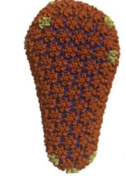
The mature HIV-l CA core is a ~60 nm x 120 nm (Briggs et al, 2003)
cone-shaped, protein lattice and is composed of 1,500 CA monomers arranged in
~250 hexamers and 12 pentamers (Briggs et al, 2004; Pomillos et al, 2009) through
interactions involving their N-terminal domains (NTD) (Pomillos et al, 2009), while CA
C-terminal domains (CTD) are important for homodimerization and connecting the rings
into a lattice (GambIe et al, 1997) (Fig. 1.4).
Figure 1.4 Model of HIV -1 capsid core.
Fullerene cone model composed of 1,056 CA subunits. NTD driven CA
hexamers are coloured in orange, pentamers in yellow, while CTD driven
homodimers in blue. NTD - N-terminal domain, CTD - C-terminal
domain.
Image source: (Pomillos et al, 20 Il).
For many years it was thought that the HIV-l CA core disassembled immediately
after its entry to the cytoplasm, which would then be followed by reverse transcription.
This reasoning stemmed from the difficulties of isolating particulate cores from infected cells, as this structure is very fragile (Bukrinsky et al, 1993; Famet & Haseltine, 1991;
Karageorgos et al, 1993). However, more recently we have gained critical insight into the importance of the HIV -1 CA core and its proper disassembly, referred to as uncoating.
1.1.6.1 Evidence supporting regulated HIV-1 uncoating
The fIfst line of evidence suggesting that uncoating is an important step in HIV-l replication was provided by mutagenesis. CA mutants resulting in either hypostable or hyperstable cores are not infectious (Forshey et al, 2002; Tipper & Sodroski, 2013).
The retroviral restriction factors TRIM5a and TRIMCyp inhibit viral infection (Sayah et al, 2004; Stremlau et al, 2004) through binding to incoming CA cores and destabilizing them (Bérubé et al, 2007; Stremlau et al, 2006). TRIM5 proteins will be discussed in the next section. Nevertheless, their action provides functional evidence of the signiticance of the 'intact' HIV-l capsid core during the tirst few hours post infection (p.i.). Additionally, the HIV-l small-molecule inhibitor PF-3450074 (PF74) is known to inhibit HIV -1 infectivity specitically by destabilizing the CA core (Shi et al, 2011).
Interesting evidence on the importance of uncoating was provided by linking uncoating with reverse transcription, since an inhibition of the latter led to a slower CA core disassembly (Hulme et al, 2011; Roa et al, 2012; Yang et al, 2013). The initiation of reverse transcription likely depends on an initial destabilization or increased
permeability of the CA core after entry into the cytoplasm (Hulme et al, 20 Il; Zhang et
al, 2000; Zhang et al, 1998). However, it has been suggested that the completion of reverse transcription takes place inside the CA core (Arhel et al, 2007; Schaller et al, 20 Il) which at that time would likely be at the NPC. It is most likely that uncoating takes place in several well-orchestrated steps instead of being a rapid single-step process; one report suggested it might happen in at least two phases (Xu et al, 2013).
Additionally, a recent study reported that cellular factors are recruited to the incoming CA core thus shielding actively transcribing viral genetic material from detection by pattern-recognition receptors (PRRs) (Rasaiyaah et al, 2013). The same study showed that HIV -1 viruses bearing mutations in the CA protein, which unable CA interaction with certain cellular partners, fail to replicate in macrophages due to activation of innate immune response.
Finally, CA mutagenesis also suggested that this protein has a role in nuclear entry (Dismuke & Aileen, 2006) and in the ability to transduce non-dividing cells (Yamashita & Emerman, 2004; Yamashita et al, 2007). It was also suggested that CA is linked with specificity to integration sites of proviral DNA (Schaller et al, 2011). Altogether, these reports demonstrate that the full uncoating does not happen immediately after the release of the CA core into the cytoplasm.
1.1.6.2 HIV-l uncoating
The knowledge regarding HIV -1 uncoating is very limited. This process is thought to be initiated soon after the exposure to the cellular environment, within the frrst hours from infection (Hulme et al, 2011). Large amounts of CA cores can still be observed in infected cells 6 hours post infection and longer (Li et al, 2009; Pawlica et al, 2014; Stremlau et al, 2006). Where uncoating takes place is still under debate: sorne reports proposed that it occurs in the cytoplasm (Hulme et al, 20 Il; McDonald et al, 2002), while others suggested that uncoating takes place at the NPC (Arhel et al, 2007; Rasaiyaah et al, 2013; Schaller et al, 2011). The latter is supported by microscopic observations in which HIV-1 CA cores reach the proximity of the nucleus 1-2 h p.i. (Arhel, 2010; Arhel et al, 2007; McDonald et al, 2002). Additionally, CA was also observed at the NPC and was proposed to be responsible for the docking of the virus at the nuclear membrane (Arhel et al, 2007; Di Nunzio et al, 2012). Interestingly, the NPC members Nup358 and Nup153 were shown to directly bind to CA protein, as demonstrated by immunoprecipitation (Matreyek et al, 2013; Schaller et al, 2011). Additionally, both CA-binding domains of Nup153 and Nup538 when fused to the
effector domain of the rhesus TRIM5a restricted IllV -1, providing an intracellular readout for the direct interaction during retroviral infection. One study reported that a small fraction of CA proteins might be transported inside the nucleus (Zhou et al, 2011).
1.1.6.3 Cellular proteins regulating uncoating
Following its entry, the IllV-1 CA core interacts with various cellular factors (Ambrose & Aiken, 2014). So far a handfu1 of cellular partners have been shown to directly bind CA core after its entry to the cytoplasm: Cyclophilin A (CypA) (Fricke et al, 2013; Li et al, 2009; Shah et al, 2013), cleavage and polyadenylation specific factor 6 (CPSF6) (De laco et al, 2013; Shah et al, 2013), peptidyl-prolyl isomerase Pin1(Misumi et al, 2010) and PDZ domain-containing prote in 8 (PDZD8) (Guth & Sodroski, 2014). It was proposed that CypA and CPFS6 are recruited to the CA core to conceal the viral genetic material from detection by innate immunity sensors (Lahaye et al, 2013; Rasaiyaah et al, 2013).
Another prote in playing a defmite yet unclear role in IllV -1 replication is transportin TNP03 (Ambrose & Aiken, 2014), and its importance for IllV-1 was genetically mapped to CA prote in (Krishnan et al, 2010). It has been suggested that the function of TNP03 is indirect and that it acts positively on RIV -1 replication by sequestering CPSF6 (De laco et al, 2013; Shah et al, 2013). At the later early stage of RIV-1 infection the CA core associates with nucleoporins Nup358 and Nup153 (Schaller et al, 2011; Shah et al, 2013), which mediate nuclear import of viral PIC. These two NPC members also directly bind to CA proteins (Matreyek et al, 2013; Schaller et al, 20 Il).
Interestingly, two recent studies have validated the existence of cellular proteins responsible for modulation of CA core stability, as evidenced by incubation with cellular lysates which increased the stability of CA cores (Fricke et al, 2013; Guth & Sodroski, 2014). These also support the hypothesis that following entry the CA core is initially shielded from the cellular environment and uncoats later in a well-orchestrated manner.
However, the knowledge about the uncoating process and its cellular partners remains very poor and additional studies are needed for its better understanding.
Sorne steps of the viral replication, such as intracellular transport, rely on the host's cytoskeleton. The eukaryotic cytoskeleton is a good target to be intercepted by viruses, as it is essential for cellular life and is involved in many cellular processes.
1.2 The cytoskeleton
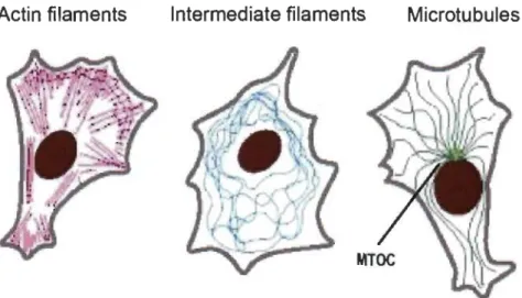
The eukaryotic cytoskeleton is an abundant, complex and dynamic network of various filaments and regulatory proteins associated with them. They are divided into three groups: actin filaments (also known as microfilaments), intermediate filaments and microtubules. These filaments have distinct roles, structure and organization (Fig. 1.5).
Actin filaments Intermediate filaments Microtubules
Figure 1.5 Schematic representation of cytoskeletal filaments. MTOC - microtubule-organizing centre.
Image source: (Alberts, 2002).
Actin filaments are the smallest, 5-9 nm in diameter, and are dispersed throughout the cytosol with the greatest concentration below the plasma membrane, known as the actin cortex. Microfilaments play major functions in cytokinesis, maintaining cellular shape and cell migration; they are the main components of lamellipodia and filopodia.
Intermediate filaments belong to a large heterogeneous family of rod-like
filaments ~ 10 nm in diameter. Most of them, such as vimentins, cytokeratins, desmins
neurofilaments, are found in the cytosol, while others, such as lamins, are in the nucleus. They are stress-resistant, conferring great mechanical strength, and are responsible for
maintaining the cell's integrity, organelle positioning and contributing to cellular
adhesion e.g. as a component of desmosomes (Alberts, 2002; Lodish, 2004).
1.2.1 The microtubule network
Measuring 25 nm in diameter, microtubules are the longest, most rigid and thickest components of the cytoskeleton. These filaments are important in many cellular processes. They are responsible for organelle positioning, intracellular transport, cell division and migration. Additionally, they serve as an internaI scaffolding of cilia and flagella.
The basic components of microtubules are heterodimers of (l- and P-tubulin which
are tightly associated with each other by a noncovalent bond. Tubulin heterodimers
assemble head-to-tail, forming polar protofilaments. Usually 13 protofilaments interact
laterally to form a hollow tube known as a microtubule. These fibres assemble towards
their plus-end, usually orientated towards the cell periphery, while the minus-end
is usually focused in microtubule-organizing centres (MTOC) (Fig. 1.5). The MTOC, which is often localized in the perinuclear region (except during the cell division),
consists of a pair of centrioles, built of nine microtubule triplets surrounded by a number
of proteins, including y-tubulin. Microtubule growth is initiated on the y-tubulin complex (Alberts, 2002; Lodish, 2004).
Microtubules are highly dynamic structures constantly switching between being
assembled and disassembled. This property is called 'dynamic instability' (Mitchison &
Kirschner, 1984). It allows the plus-end of microtubules to explore the cellular
environment in the process called 'search and capture' and adapt rapidly to changing conditions, e.g. during cell division (Kirschner & Mitchison, 1986). Once in the right
place, the filaments are bound by a specialized farni1y of proteins and undergo post-translational modifications modulating their stability (Alberts, 2002; Kueh & Mitchison, 2009; Lodish, 2004).
L2.l.l Regulation ofmicrotubule dynamics
Dynarnic instability provides the basis for rnicrotubule plasticity, which is highly regulated by cellular factors such as rnicrotubu1e-associated proteins (MAPs), molecular motors and others proteins, such as kinases (Mandelkow & Mandelkow, 1995). MAPs specifically bind to rnicrotubules and either stabilize (e.g. Tau, MAP2 or MAP4) or destabilize them (e.g. katanin, stathrnin). An important group ofMAPs are the plus-end tracking proteins (+TIPs) that usually prevent depolymeization of rnicrotubules. Additionally, +TIPs serve in connecting rnicrotubules to actin filaments and organelles (Gouveia & Akhmanova, 2010). The most well-known members of this group are end-binding (EB) proteins that promote growth of rnicrotubules (Komarova et al, 2009). Other important regulators of rnicrotubule dynarnics are kinesin motors, especially the kinesin-8 farnily, members of which are known to destabilize rnicrotubules (Su et al, 2012). Interestingly, the cortex-associated dynein motor complex was also proposed to play a role in binding the plus-end of rnicrotubules and stabilizing them (Hendricks et al, 2012).
Additional mechanisms regulating rnicrotubule dynarnics are post-translational modifications, usually carried out on polymerized rnicrotubules. It was proposed that these modifications generate a specific 'code' readable by MAPs and molecular motors that allows carrying divergent functions on rnicrotubules (Verhey & Gaertig, 2007). For example, acetylation increases binding of molecular motors (Friedman et al, 2010) while detyrosination decreases binding affmity of destabilizing MAPs and promotes formation of a stable sub-population of rnicrotubules (de Forges et al, 2012). Additionally, tubulin subunits have different isoforms, which contribute to the extreme diversity of rnicrotubules (Luduena, 1998). In conclusion, microtubule dynarnics is highly regulated by many cellular factors.
1.2.1.2 Treafments targeting microtubules
Drugs targeting microtubules can be divided into two groups: those preventing microtubule assembly and others preventing their disassembly. Sorne of the drugs preventing assembly bind to aJ~-tubulin heterodimers, such as colchicine (Ravelli et al, 2004) and nocodazole (Xu et al, 2002), while others bind between the dimers, e.g. vinblastine (Gigant et al, 2005). The second group of drugs (such as paclitaxel, originally named taxol (Schiff & Horwitz, 1981)), binds to and stabilizes tubulin polymers, also resulting in a loss of their dynamics (Derry et al, 1995). An alternative way to study micro tubule dynamics is to target various MAPs that regulate it: for instance a depletion of EB 1 results in a dramatic loss of microtubule dynamics (Rogers et al, 2002).
1.2.2 Molecular motors and their role in cellular transport
The cytoplasm is a crowded and viscous environment where only small molecules and proteins can freely diffuse. Movement of larger components, 20 nm of size and more, requires active transport (Ellis, 2001; Luby-Phelps, 2000). This transport, usually ATP-driven, is mediated by three classes of molecular motors: actin-based myosins and microtubule-based kinesins and dyneins. The myosin motor superfamily consists of 22 groups of proteins (Foth et al, 2006). Myosins are involved in various cellular processes, such as cytokinesis, actin-dependent transport, membrane trafficking and signal transduction. Interestingly, different types of myosin motors are responsible for movements in opposite directions; most of the myosins move towards the plus-end of microfilaments, while the members of group VI move towards the minus-end (Buss &
Kendrick-Jones, 2008).
There are 45 known members of the kinesin motors superfamily, grouped into 14 families. Kinesin motors are responsible for microtubule-dependent anterograde transport, towards the plus-end of microtubules (Hirokawa et al, 2009; Verhey &
minus-end (Ambrose et al, 2005). In addition to the transport, kinesin motors play roles in cell division, exocytosis and organelle positioning.
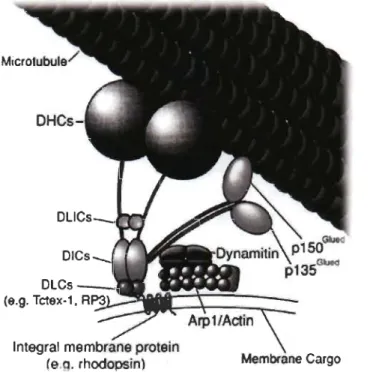
Dynein motors are the least diverse; there are only two members that are responsible for retro grade transport towards the minus-end of microtubules: the cytoplasmic dynein motor complex (known as dynein 1) and the intraflagellar transport dynein motor (known as dynein lB or dynein 2). In most cells, àll retrograde transport is carried out by dynein 1, thereafter referred to as the dynein motor complex. There are, additionally, 13 axonemal dyneins that are a part ofaxoneme and are responsible for cilia beating and are not involved in molecular transport. The dynein motor complex has multiple functions in the cell: transporting cargos towards the minus-end of microtubules; positioning organelles, such as Golgi, nucleus and lysosomes, and, during cell division, forming the mitotic spindle, attaching kinetochores and pulling astral microtubules (Gennerich & Yale, 2009; Roberts et al, 2013; Yale, 2003).
DLlCs_._.~ ..
Integral membrane protein
(e.q. rhodopsin
Figure 1.6 The dynein motor complex.
Membrane Cargo
DHC - dynein heavy chain, DLIC - dynein light-intermediate chains, DLC - dynein light chain, RP3 - dynein light chain Tctex-type 3, Arp 1 - actin-related protein 1.
1.2.2.1 The dynein motor comp/ex
The dynein motor complex, belongs to the AAA+ superfamily (ATPases associated with diverse activities) (Neuwald et al, 1999). This motor is a large protein complex consisting of two heavy chains (DHC, for dynein heavy chain) and several pairs of lighter chains (Fig. 1.6). DHC is a large protein (~520 kDa) and consists of a head - made of six AAA+ domains arranged into a ring -, a stalk with a microtubule-binding site, and a tail. The tail of DHC associates with five pairs of homodimers of intermediate, light-intermediate and three light chains. There are three classes of light chains: LC8 and Roadblock (also known as LC7) and T-complex testis-specific protein 1 (Tctex-l) (Pfister et al, 2005). 'Lighter' subunits of the dynein motor complex are responsible for determining cargo specificity and thus the function of the whole complex (Pfister et al, 2005). Additionally, ~he dynein motor complex interacts with regulatory and adaptor prote in complexes, including the dynactin complex, lissencephaly (LIS 1), nuclear distribution protein E (NUDE) and others. The dynactin complex is coniposed of eleven different subunits, such as pl50Glued, p50 (also known as dynamitin) and actin-related protein 1 (Arp 1), and seems to be essential for most of the functions performed by the dynein motor complex (Fig. 1.6). The dynactin complex enables inter aUa an association of dynein motor complexes with cargos via Aprl and pl50Glued• Additionally, this complex seems to be required for the processivity of the dynein motor complex (McKenney et al, 2014). Regulators, such as LISI and NUDE, mediate targeting the dynein motor complex to the plus-end of microtubules and thus are essential for mitotic spindle formation and in kinetochore activity, as well as in intracellular transport (Kardon & Yale, 2009).
The ATP-driven movement is carried out by DHCs that most likely associate and disassociate with microtubules in an altemating manner, which could be said to resemble 'walking'. The binding of ATP to the head of dynein DHC results in the disassociation of the stalk from microtubules (lmamula et al, 2007). Then DHC undergoes ATP-driven remodelling and the 'free' stalk can reach in search for a new binding site on microtubules. Initially, the binding is weak, but gets very strong as a result of the phosphate released through ATP hydrolysis (Roberts et al, 2013).