HAL Id: tel-03008821
https://tel.archives-ouvertes.fr/tel-03008821
Submitted on 17 Nov 2020
HAL is a multi-disciplinary open access archive for the deposit and dissemination of sci-entific research documents, whether they are pub-lished or not. The documents may come from teaching and research institutions in France or abroad, or from public or private research centers.
L’archive ouverte pluridisciplinaire HAL, est destinée au dépôt et à la diffusion de documents scientifiques de niveau recherche, publiés ou non, émanant des établissements d’enseignement et de recherche français ou étrangers, des laboratoires publics ou privés.
Joanna Zell
To cite this version:
Joanna Zell. Glycolipid - and Lectin-Dependant Endocytosis studies by a chemical biology Approch.. Other. Université Sorbonne Paris Cité, 2018. English. �NNT : 2018USPCC239�. �tel-03008821�
de l’Université Sorbonne Paris Cité
Préparée à l’Université Paris Diderot
Ecole doctorale Frontières du Vivant, CRI, Paris
UMR3666/U1143 Chemical Biology of Membranes and Therapeutic Delivery, Institut Curie
Glycolipid- and Lectin-Dependant Endocytosis
Studies by a Chemical Biology Approach
Joanna ZELL
Thèse de doctorat de chemobiologie
Dirigée par Ludger Johannes et Frédéric Schmidt
Présentée et soutenue publiquement à Paris, le 16 novembre 2018
Président du jury : Prof. Didier Dubreuil, Université de NantesRapporteur : Prof. Christoph Thiele, Universität Bonn, Allemagne Rapporteur : Dr. Boris Vauzeilles, CNRS ICSN/Université Paris Sud
Examinatrice : Dr. Lydia Danglot, Centre de Psychiatrie et Neurosciences/INSERM Directeur de thèse : Dr. Ludger Johannes, Institut Curie/INSERM
Co-directeur de thèse : Dr. Frédéric Schmidt, Institut Curie/CNRS Membre invité : Dr. Jean-Claude Florent, Institut Curie/CNRS
2
Title: Glycolipid- and Lectin-Dependant Endocytosis Studies by a Chemical Biology Approach
Abstract: Current biological techniques do not permit the functional reintroduction of long-chain glycosphingolipids (GSLs) into cells. We aim to develop molecular tools permitting cellular reconstitution of functional GSLs in a controlled manner, combining metabolic labelling of sphingolipids with copper-free click chemistry. Molecular tools are validated with the bacterial Shiga toxin, which gains entry to cells independently of the canonical intracellular clathrin machinery on binding to its GSL cell receptor, globotriaosylceramide (Gb3-Cer), through a recently described mechanism of endocytosis, termed GL-Lect (glycolipid-lectin). According to the GL-Lect hypothesis, cellular or pathogenic lectins drive the construction of endocytic pits by reorganising membrane lipids so as to favour the formation of narrow membrane curvature. The GSL reconstitution technique that is described here is applicable to the discovery of GSL binding partners in the study of GL-Lect-mediated endocytosis.
Keywords: Glycosphingolipid, metabolic labelling, copper-free click chemistry
Titre : Une approche de chemobiologie pour étudier l’endocytose dépendante des glycosphingolipides et des lectines
Résumé : Les techniques biologiques actuelles ne permettent pas la réintroduction fonctionnelle des glycosphingolipides (GSLs) à longue chaine dans les cellules. Nous proposons de développer des outils moléculaires permettant la reconstitution des GSLs fonctionnels de manière contrôlée, en combinant le marquage métabolique et la chimie click sans cuivre. Les outils moléculaires sont validés par la toxine de Shiga, qui pénètre dans la cellule hôte indépendamment de la machinerie clathrine intracellulaire, en se liant à son récepteur cellulaire GSL, le globotriaosylcéramide (Gb3-Cer), par un mécanisme d’endocytose récemment décrit, appelé GL-Lect (glycolipide-lectine). Selon l’hypothèse GL-Lect, les lectines cellulaires ou pathogéniques induisent la constitution de puits endocytiques en réorganisant les lipides membranaires de façon à favoriser la formation de courbures membranaires locales. La technique de reconstitution des GSLs qui est décrite ici s’applique à la découverte des partenaires d’interaction des GSL dans l’étude de l’endocytose GL-Lect.
3
Live is if you were to die tomorrow, learn as if you were to live forever
4
Acknowledgements
Je suis reconnaissante d’avoir fait partie de l’unité UMR3666, qui est multidisciplinaire, multiculturelle, étonnamment accueillante et ouverte à tous. À mes deux superviseurs, Ludger et Frédéric, qui ont eu tant de patience à travers les échecs et les désaccords, et les réussites toujours
trop courtes ; je vous remercie de m’avoir donné cette opportunité de découvertes, défis et fascinations qui m’ont énormément motivé tous les jours. Je remercie également Jean-Claude,
superviseur-ange gardien.
À mes camarades de labo, Steve, Stefan, Vesela, Haifei, Mélanie, Stéphanie, Alena, Carlos, Marco, Fabien, Antoine, Seb, Anne, Anne, Thomas, Tati, Raph, Yannick, Christine, Sylvie, Michelle, Alison,
merci d’avoir travaillé avec moi et partagé ces expériences délicieuses, passionnantes et inoubliables.
À Siau, qui m’a formé en bio avec Ludger et Christian, qui m’a soutenu, m’a écouté et m’a fait sourire tous les jours.
À Raphaël G, qui m’a donné la confiance dont j’avais besoin pendant ces longues périodes de syndrome de l’imposteur.
To Julio, who has driven this project with me, feeding all the creativity, imagination and dedication, and with whom I can always share a great moment in and out of the lab.
I thank all my friends from CRI, who managed to laugh in the face of PhD stress with me, whilst nourishing overpriced pints, whenever we managed to get out of the lab.
I thank the BIOPOL Innovative Training Network (European H2020 funding) that funded the project and my international training stations, and grouped together 16 PhD students, making us spend
quality-science-time together across Europe. We had never felt so different, yet so alike. After countless hours complaining on the phone, I retain some gems from my mum, who said:
“You don’t learn unless you’re suffering;” and my dad, who said
“You’re doing a PhD because once you’re a Dr., you never have to say if you’re a bloody Miss or Mrs., because you’ll be a Dr.!”
I would be nowhere without you.
Carolyn, I know one day your wisdom will come crashing down on me. I love you and M&M to the end and back.
5
Table of contents
Abbreviations………..………. 5
1 INTRODUCTION ... 9
1.1 Endocytosis mechanisms ... 9
1.2 Glycolipid- and Lectin-dependent endocytosis ... 11
1.3 GSL function studies ... 15
1.4 GSL function and biosynthesis ... 22
1.5 Biomedical relevance ... 27
1.6 Bioorthogonal conjugations and click chemistry ... 29
1.7 Metabolic labelling of glycans and lipids ... 36
1.8 Project aims... 39
2 RESULTS ... 42
2.1 Synthesis of biomolecule analogues ... 42
2.1.1 Reconstitutable globotriose phospholipid ... 42
2.1.2 Clickable sphingolipid and globotriose ... 46
2.1.3 Cyclooctyne-functionalised glycans ... 51
2.2 Biological validation of analogues... 58
2.2.1 Model membrane validation ... 58
2.2.2 Cellular metabolism and localisation of AzSph ... 61
2.2.3 Glycan click ... 68
2.2.4 Changing the lipid precursor to change localisation ... 75
3 DISCUSSION ... 79
3.1 Phospholipid strategy ... 79
3.2 Clickable sphingolipid synthetic strategy ... 82
3.3 Model membrane testing ... 89
3.4 Sphingolipid metabolism ... 90
3.5 In cellulo click ... 96
3.6 Optimisation of lipid localisation ... 100
4 PERSPECTIVES ... 103 5 CONCLUSIONS ... 106 6 EXPERIMENTAL ... 107 6.1 Biological validation ... 107 6.2 Chemical synthesis ... 114 7 BIBLIOGRAPHY ... 146
6
Abbreviations
A.U. Arbitrary units
Ac Acetyl AzCer 1-Azidoceramide AzPhy 1-Azidophytosphingosine AzSph 1-Azidosphingosine BCN Bicyclononyne Boc Tert-butyloxycarbonyl
BSA Bovine serum albumine
Bz Benzoyl
CAN Ceric ammonium nitrate
Cer Ceramide
CerS Ceramide synthase
CES Carboxylesterase
CHO Chinese hamster ovary
Chol Cholesterol
CIE Clathrin-independent endocytosis
COP 2-Chloro[1,3,2]dioxaphospholene-2-oxide CuAAC Copper(I)-catalysed azide-alkyne cycloaddition DBU 1,8-Diazabicyclo(5.4.0)undec-7-ene DCC N,N’-Dicyclohexylcarbodiimide DIBO Dibenzocyclooctyne DIFO Difluorocyclooctyne DIPEA Diisopropylethylamine DMAP 4-Dimethylaminopyridine DMF Dimethylformamide
DMSO Dimethyl sulfoxide
DOPC 1,2-Dioleoyl-sn-glycero-3-phosphocholine DOPE 1,2-Dioleoyl-sn-glycero-3-phosphoethanolamine
7 EC50 Half maximal effective concentration
EDC 1-Ethyl-3-(3-dimethymainopropyl)carbodiimide
EG Ethylene glycol
ER Endoplasmic reticulum
ESI Electrospray ionisation EtOAc Ethyl acetate
EWG Electron-withdrawing group FSC Fluorescence scanning microscopy FSL Function-spacer-lipid Gb3 Globotriose Gb3-Cer Globotriaosylceramide GCS Glucosylceramide synthase GL Glycolipid GlcCer Glucosylceramide Gly Glycine
GPI-AP Glycosylphosphatidylinositol-anchored protein GSL Glycosphingolipid
GUV Giant unilamellar vesicle HeLa Henrietta Lacks (cell line) HOBt Hydroxybenzotriazole
HPTLC High performance thin layer chromatography HRMS High resolution mass spectrometry
IF Immunofluorescence
IR Infrared
LacCer Lactosylceramide
Lo/Ld Liquid ordered/disordered LUV Large unilamellar vesicle MFCO Monofluorocyclooctyne MS Mass spectrometry Ms Mesylate NIS N-Iodosuccinimide PBS Phosphate-buffered saline PC Phosphatidylcholine
8
PE Phosphatidylethanolamine
PL Phospholipid
PM Plasma membrane
PS Phosphatidylserine
ROS Reactive oxygen species
rt Room temperature
S1P Sphingosine-1-phosphate
SIMS Secondary ion mass spectrometry
SM Sphingomyelin
SPAAC Strain-promoted azide-alkyne cycloaddition TAMRA Tetramethyl-rhodamine
TBAF Tetra-n-butylammonium fluoride TBDPSCl Tert-butyldiphenylsilyl chloride TFA Trifluoroacetic acid
TGN Trans-Golgi network
THF Tetrahydrofuran
TLC Thin layer chromatography
TMSOTf Trimethylsilyl trifluoromethanesulfonate TMSSCN Trimethylsilylisothiocyanate
UPLC-MS Ultra performation liquid chromatography - tandem mass spectrometry
w/o With or without
9
1 INTRODUCTION
1.1
Endocytosis mechanisms
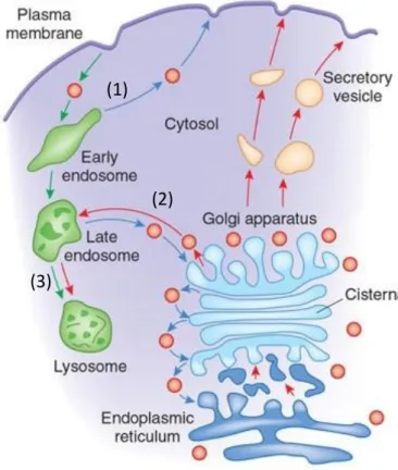
Endocytosis encompasses the cellular uptake of diverse cargo, including macromolecules, membrane components, nutrients and fluids, by their encapsulation in membrane vesicles which bud inwards to be pinched off from the plasma membrane (PM). From the PM, cargoes are trafficked through endosomes, where they can be (1) recycled back to the PM, (2) sorted via the retrograde route to the Golgi apparatus and in some cases to the endoplasmic reticulum (ER), or (3) targeted to the late endosomes and lysosomes for degradation (Figure 1.1.1a). The best characterised mechanism of endocytosis is mediated by clathrin, which coats the PM on binding to its adaptor proteins, forming a concave lattice structure (Figure 1.1.1b)1 to bend and engulf portions of the membrane containing endocytic
cargoes. These clathrin-coated vesicles (70-150 nm)2 are eventually scissionned from the PM
with assistance from intracellular protein machinery including a GTPase, dynamin (Figure 1.1.2), recruited by curvature-inducing BAR domain proteins, which themselves bind to and constrict negatively charged, curved membranes.
Figure 1.1.1: (a) Intracellular vesicular trafficking pathways3 (b) Electron
microscopy image of clathrin-coated pits in hypertonic fibroblasts, scale bar 100 nm1
(1)
(2)
10
Clathrin-independent endocytosis (CIE) pathways include macropinocytosis and phagocytosis (Figure 1.1.2), in which large portions of the membrane (>1 μm) are engulfed by membrane structures supported by actin filaments. In a different category of CIE, vesicles of closer size to clathrin-coated vesicles are formed by other cytosolic proteins. Caveolae are membrane invaginations formed from caveolin proteins and cavins (cavin1-4). They function as mechano-sensing domains which flatten out to buffer membrane tension. Caveolae can bend the membrane to form caveolar carriers (50-80 nm) that undergo endocytosis with the aid of dynamin, ATPase EHD2 and BAR domain protein pacsin2. Which cargoes are transported specifically by caveolae remains unclear,4–6 and their endocytic uptake is highly
dependent on cholesterol and sphingolipid content of the lipid bilayer.7 Flotillin can localise
in caveolae and itself outlines another clathrin- and caveolae-independent process.4,5 Unlike
clathrin, which is located in the cytosol and binds via membrane-bound adaptors, caveolins and flotillins are integral transmembrane proteins implicated in lipid rearrangement.4
Glycosylphosphatidylinositol-anchored proteins (GPI-ACs) have a high propensity to traffic through clathrin-, caveolin- and dynamin-independent endocytosis, giving the name to the clathrin-independent carrier (CLIC)/GPI-AC-enriched early endosomal compartment (GEEC) pathway.8 The protein machinery driving endocytic uptake of these high capacity
(100-200 nm)9 carriers is complex, implicating BAR domain proteins and Rho family GTPases.10 CIE
carriers lack regular protein coats, thus are harder to image by electron microscopy compared with clathrin-coated cargos. Several other CIE mechanisms have since been characterised, including those in which lipids are redistributed to bend membranes.8,11–13 The importance of
membrane lipid content is currently studied by indirect, secondary techniques (discussed in section 1.3-4 and 1.7), since these molecular mechanisms take place below the resolution limit of light microscopy (200 nm).
CIE mechanisms can be classified by the mechanical actors in vesicle formation, such as GTPases (e.g. dynamin, RhoA and CDC42), BAR domain proteins (e.g. endophilin, pacsin2) and actin, or by their cargoes, such as GPI-APs, CD44, major histocompatibility class I (MHCI) molecules and interleukin 2 receptor.9 Adding to this complexity in characterisation, these
categories present significant interchangeability and compensatory roles in both cargo and membrane-bending machinery. Experimentally removing, manipulating or probing one factor can often be compensated by another.14,15
11
Figure 1.1.2: Mechanisms of endocytosis. Dynamin-dependent pathways (+) are typically associated with small-scale circular carriers such as clathrin- or caveolin-mediated pits.5
1.2
Glycolipid- and Lectin-dependent endocytosis
Lectins are proteins that bind to glycan (sugar, carbohydrate, saccharide) moieties of glycoproteins or glycolipids (GL). The GL-Lect hypothesis proposes that certain mammalian or pathological Lectins, once bound to the PM through GLs and/or glycoproteins, utilise GlycoLipids to induce nanodomain formation in the lipid bilayer, membrane curvature, and thereby, the biogenesis of endocytic pits.16
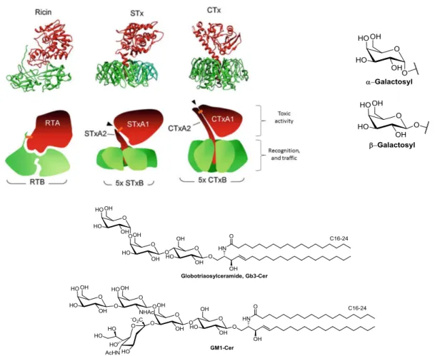
Clathrin-independent endocytosis (CIE) of the bacterial galactose-binding lectin, ricin, was the first CIE mechanism described.17,18 Bacterial ricin, Shiga and cholera toxins bind via
their respective B-subunits (RTB, STxB, CTxB) to their mammalian glycosyl receptors: galactosyl lipids and proteins, Gb3-Cer, or GM1-Cer, respectively (Figure 1.2.1). Once bound to the host cell and endocytosed to early sorting endosomes, the toxins bypass degradation in late endosomes and lysosomes. They are instead transported via the retrograde route (Figure 1.2.2), through the trans-Golgi network (TGN) and the Golgi stack to the endoplasmic reticulum (ER).19 In the ER, the disulphide bond linking A- and B-subunits (orange, Figure 1.2.1)
is cleaved to induce toxicity.12,20 In the case of ricin and Shiga toxin, the A-subunit, a catalytic
RNA N-glycosidase, translocates to the cytosol to induce toxicity by protein synthesis inhibition.
12
Figure 1.2.1: Crystal structures of ricin, Shiga and cholera toxins19 with their respective receptors: galactose
(presented on GLs and glycoproteins), globotriaosylceramide (Gb3-Cer), or GM1-ceramide.
13
The homopentameric B-subunits of AB5 Shiga and cholera toxin, STxB, CTxB, as well as
the pentameric VP-1 capsid protein of Simian polyoma virus SV40, have flat, doughnut-like structures (Figure 1.2.3a), where they dock onto the membrane via their glycosphingolipid (GSL) receptors. The lectins’ polyvalent binding is only of small individual affinity (dissociation constant Kd ~ 10-3 M for STxB) for each GSL, but combine to a large avidity (Kd ~ 10-9 M).21 STxB
contains three globotriose (Gb3) binding pockets per monomer, thus 15 total Gb3 binding sites. CTxB and SV40 protein have one GM1 binding pocket per monomer, totalling five GM1 binding sites.22
Of note, these three lectins have no sequence similarity, and yet the glycan receptors are all positioned around the edge of the protein in a seemingly overlapping geometry (Figure 1.2.3a), indicating a convergent evolution of structure. The glycan binding sites are of a geometry in which the membrane must bend up around the lectin to fit the glycan part of the GSL receptor molecules into their binding sites, thereby generating negative, inward-oriented curvature. This was recently demonstrated in silico for STxB-Gb313-15 in all-atom molecular
dynamics studies (Figure 1.2.3b,d).23 Effects on the membrane underneath the docked STxB,
such as suppression of lipid fluctuations,24 lead to membrane-mediated clustering to induce
wider membrane invagination (Figure 1.2.3c). These early endocytic pits are then recognised by cytosolic machinery, dynamin, endophilin and/or actin,15 for pit elongation and scission,
14
(a) (b)
(c)
(d)
Figure 1.2.3: (a) Crystal structure overlay of STxB, (green Protein Data Bank (PDB) 1BOS), CTxB (red; PDB 1FGB), VP1 protein (blue, PDB 3BWR) bound to GSL receptors (corresponding colour)8 (b, d) Local curvature
of STxB binding to 13-15 Gb3-Cer in phospholipid bilayer23 (c) STxB binding, inducing local curvature,
clustering and membrane invagination8
This membrane bending phenomenon is an intrinsic property of GL-Lect complexes and can be reproduced in simple model membrane systems, liposomes. One classical experimental technique applies hydrating with an aqueous solution, a lipid mixture of phospholipid with a small molar percentage of GSL, to obtain spherical giant unilamellar vesicles (GUVs) of homogenous size similar to cells (~10 μM).25,26 Membrane bending
properties of GL-Lect drivers have been widely investigated using GUVs: Gb3 with STxB (Figure 1.2.4a)27 or bacterial lectin LecA;28 GM1 with SV4029 or cholera toxin;30 and a GSL mixture with
GSL-15
dependent endocytosis after binding to glycoproteins in cells, spontaneously forms dynamic membrane invaginations when bound to GUVs containing 5% GSL.
Figure 1.2.4: Giant unilamellar vesicles (GUVs) showing membrane bending induced by lectins and GSLs. (a) DOPC/cholesterol/porcine Gb3-Cer/BodipyFL-C5-HPC (green) [64:30:5:1] incubated with STxB-Cy3 (red); (b) PC/cholesterol/sphingomyelin/PS/GSL-mix/Nickel-NTA-DOGS/lissamine-rhodamine-PE (0.1mol%) [56.9:20:12:3:5:3:0.1] incubated with His-tagged galectin Gal3-His31
1.3
GSL function studies
Unpicking the molecular mechanism of membrane curvature formation by the STxB-Gb3 complex can give novel insight on GL-Lect endocytosis. To investigate the relative contribution of driving forces in the clustering of STxB toxins, the system was recently studied
in silico and experimentally.24 There are no attractive interactions between STxB proteins, so
how do they cluster? A commonly cited membrane-mediated force, line tension (Gibbs free energy required to maintain domain interfaces) might contribute to clustering due to height or compositional mismatch within the lipid membrane.32,33 Gb3-Cer and other GSLs often
have longer lipid chains than bulk phospholipids, meaning the domain containing STxB-Gb3x≤15 (x indicates numbers of bound Gb3 molecules) should be held higher on the
membrane.34,35 When tested experimentally by varying lipid tail lengths of Gb3-Cer in GUVs,
no significant difference in clustering kinetics was observed,24 demonstrating that height
mismatch line tension is not strictly required for clustering. One could also implicate line tension caused by compositional mismatch of the lipids, due to thermodynamically favourable lipid-demixing effects. Pezeshkian et al. suggested that Gb3-Cer domains are not required for clustering to occur, since GUVs formed from 5 or 30 mol% Gb3-Cer showed no
16
significant difference in binding or invagination; 30 mol% being the Gb3 concentration that one
expects to find under STxB molecules.24 Solevyeva et al. showed that STxB binding induced
membrane lipid domain organisation in GUVs,36 but this reasons for the reverse causality;
that STxB clustering induces compositional mismatch.
To elucidate the mechanism, synthetic Gb3-spacer-Cer analogues (Figure 1.3.1a) were synthesised in the L. Johannes laboratory and tested in GUV experiments. The longer ethylene glycol (EG) spacers between the Gb3 head and the ceramide tail increasingly inhibited STxB clustering and invaginations in GUVs (n=3 or 7, Figure 1.3.1a, b). These experiments gave birth to the fluctuation force hypothesis, proposing that a suppression of fluctuation of the membrane is required for toxins to cluster (Figure 1.3.1c). This effect is commonly alluded to being like drifting boats on the ocean, or cheerios on milk, which aggregate together. This hypothesis was studied by course-grained molecular dynamics simulations, showing that two macromolecules, with dimensions corresponding to the membrane-bound STxB-Gb3x≤15
complex, would randomly drift on a phospholipid bilayer. In the simulation, only the macromolecules tightly bound to the bilayer (Figure 1.3.1d) would be attracted to each other from a distance of several nanometres, whereas loosely bound macromolecules (Figure 1.3.1e) would experience no attraction force.24 Hence, the tight binding of the toxin to the
membrane via Gb3 receptors is sufficient to induce clustering. These results will in turn guide this project, in the constraints on structure of Gb3 analogues proposed and synthesised, which should retain natural lectin binding and membrane bending actively.
17 (a) (b) (c) (d) (e)
Figure 1.3.1: (a) Structures of Gb3-ethyleneglycol (EG)n-Cer analogues, n=1,3,7 (b) quantification of GUV
experiments showing that long, flexible spacer region inhibits tabulation (c) Suppression of membrane fluctuation induces clustering of macromolecules bound to lipid bilayer. (d,e) Course-grain simulations of pentameric toxin-like nanoparticles bound either tightly (d, no spacer) or loosely (e, flexible spacer). 24
Despite the fact that low or high Gb3-Cer concentrations used in GUVs have little effect on STxB clustering, one must still consider that glycolipid organisation may have an effect on lectin binding and endocytosis.33 The lipid raft hypothesis describes the propensity
of cholesterol and sphingolipids, mostly sphingomyelin (SM) and GSLs, to phase separate into denser, more ordered regions in a lipid membrane.37,38 The theory gained interest because of
the dependence of certain biological processes on non-specific cholesterol depletion, or the easy isolation of detergent-resistant membrane domains by treating biological samples with cold detergent, justifying the binominal classification of proteins and processes as “raft” or “non-raft.” An enrichment of “raftophile” lipids, cholesterol and sphingolipids, in CIE cargos has been demonstrated in several non-direct methods.39–41 This phenomenon of lipid
18
extraction techniques, but the biological relevance is difficult to evaluate for natural, non-fluorescent lipids in cellulo or in vivo.
Lipid organisation can be driven by a multitude of factors such as actin cytoskeleton organisation,42 ectodomain association, and membrane pits, ruffles and contact sites.43
Specific lipid binding pockets in proteins and charge interactions also induce lipid organisation. 20-30% of the PM area is covered by proteins, and lipid solvation rings around proteins could account for the majority of the lipid area remaining.44 Eggeling et al. recently
used super resolution fluorescence scanning microscopy (FSC) to show that fluorescent phospholipids or cholesterol presented no significant lateral heterogeneity, whilst fluorescent SM showed transient trapping in ~80 nm domains, but hardly moved once trapped. This observation does not correspond to lipid raft criteria, but infers specific lipid interactions with membrane components.45,46
Another recent imaging study by Kraft et al. used secondary ion mass spectrometry (SIMS) with isotopically-labelled sphingolipids and cholesterol reconstituted into cells.47,48
After fixing and imaging cells, they show 15N-sphingolipids concentrated into microscale
domains (Figure 1.3.2b), and 18O-cholesterol evenly dispersed in the PM (Figure 1.3.2c),
contesting the existence of nanoscale lipid rafts. These findings correlate with recent super-resolution fluorescence microscopy performed with fluorescent analogues.49,50
19
Figure 1.3.2: Scanning electron microscopy (a) and high-resolution SIMS images (b,c) of a representative clone 15 fibroblast cell. (b) 15N-sphingolipids are enriched in
micron scale domains on the PM, (c) whereas 18
O-cholesterol showed no enrichment.
The high spatiotemporal resolution of fluorescence microscopy has promoted the development of fluorescent GSLs, obtained by some complex chemical syntheses.51–55 Despite
progress in smaller fluorophores and faster imaging techniques, the study of these molecular tools in GUVs and cells has highlighted the precariousness in studying lipid properties with modified lipids. Lipids, including fluorophore-tagged lipids, can separate into ordered (Lo, e.g.
saturated lipids) or disordered domains (Ld, e.g. unsaturated lipids) easily visible in GUVs by
confocal fluorescence microscopy.56,57 Patalag et al. recently showed that the synthetic
fluorescent Gb3-Cer analogues 1 and 2 (Figure 1.3.3) separated into the lipid disordered (Ld)
domains. The partitioning of natural Gb3-Cer cannot be verified or compared, but it is commonly conceived that saturated long-chain GSLs separate into Lo domains, based on the
thermodynamic theory of lipid de-mixing and lipid rafts. One can directly compare Gb3-Cer analogues and wild type when bound with fluorescent STxB (red, Figure 1.3.3b,d,e). STxB
20
localised to the Ld domains (green) in GUVs with 1 and 2 separately, whereas with natural
Gb3-Cer, STxB localised to Lo domains.58
Wild type
Figure 1.3.3: GUVs with 5mol% Gb3 analogue 1 (a,b) or 259 (c,d) or wild type Gb3-Cer (Figure 1.2.1)58 (e)
(DOPC/SM/Chol/Gb3-Cer [40:35:20:5] with <1% fluorescent phospholipid (green, Ld), incubated with
STxB-Cy3 (red).
Specific study of Gb3-Cer and other long-chain GSLs in cell culture is practically unattainable since they cannot be added directly to cells in culture in a controlled manner.60
Chinnapen et al., when investigating GM1 and cholera toxin traffic in live cells, reincorporated GM1 species bearing C18 chain length or shorter, despite specifying that the natural profile contains mostly C24 and C16 acyl chains.30 Pedrosa et al. showed that short C8 acyl chain
GlcCer could be taken up with a yield of 20% in cancer cells, and 5% in normal cells, but do not discuss longer chains.61 Fusogenic liposomes have been used to improve uptake,62,63
employing neutral and positively charged phospholipids to improve fusion of lipid vesicles with the PM.
Our laboratory and others have never shown natural retrograde transport of STxB by supplementing Gb3-Cer to cells not expressing Gb3-Cer natively. Attempted reconstitution of commercial Gb3-Cer with long ceramide acyl chains (majority C22, C24 chains)64 into
GSL-negative (GSL-) GM95 cells showed negligible STxB cell-surface binding (Figure 1.3.4a) and
endocytosis. Gb3-Cer with shorter C18 acyl chain could allow STxB binding to cells, at 4°C, before endocytosis (Figure 1. 3.4c), but its traffic is misdirected away from the Golgi (Figure 1. 3.4d). An example by Haicheur et al. shows the canonical retrograde transport of STxB to the Golgi in Gb3-expressing D1 murine dendritic cells (Figure 3. 3.4e),65 which is reproducible
in most Gb3-expressing (Gb3+) cell lines.40 Even when two strains of bacterial STx, verotoxin
1 and 2, bind to distinct PM domains, they coalesce into retrograde traffic to the Golgi.66
21
(a) (b)
(c) (d)
(e)
Figure 1.3.4: GM95 cells incubated with Gb3 commercial mix (Matreya) with bovine serum albumin (BSA) (20 μM) (a) or without (b), then incubated with STxB-Alexa488 to observe surface binding (4°C, 30 min). Short chain Gb3 C18:1 shows binding at 4°C (c) and uptake but misdirected traffic at 37°C (d). (e) When internalised on natural Gb3, STxB is transported to the Golgi and colocalises with CTR433, a Golgi marker.
22
1.4
GSL function and biosynthesis
The biosynthesis of glycosphingolipids (GSLs) is well documented. However, GSL function and their interactions is still mystifying scientists, apt to the etymology of the word “sphingolipids” – named by J. L. W. Thudichum in 1884 after the enigmatic sphinx.34 Over 400
unique oligosaccharide head groups make up the enigmatic “glycolipidome” in mammals.67
GSLs constitute 2-10% of total lipids in membranes,68 where sphingomyelin (SM) is estimated
at 15-20% of the plasma membranes (PM).69 GSLs are found at higher concentrations at the
apical side of epithelial cells, and in certain cell types including oligodendrocytes and neurons. Their fingerprint expression levels vary throughout development and pathogenesis, amongst cell and tissue type.
Functions of groups of GSLs have been studied by genetic suppressions. Mice deficient in all GSLs, obtained with a glucosylceramide synthase (GCS) gene knockout, do not survive beyond gastrulation,70 similar to lactosylceramide synthase knockout mice.71 Genetic
suppression of the gala- and ganglio-series GSLs in mice shows phenotypes in nervous system or neuronal functions respectively (Figure 1.4.1).72,73 GSLs have classically been used as
differentiation markers, especially the stage-specific embryonic antigens (SSEA1-4).74,75 GSLs
are implicated in cell migration, adhesion and signal transduction,76 as well as
clathrin-independent endocytosis.13,31 Many GSL-protein interactions are implicated in signal
transduction,77 such as the activation of epidermal growth factor receptor (EGFR).78 GLSs
undergo individual and polyvalent interactions with proteins, as proposed in the lipid raft hypothesis, where they play a direct role in membrane organisation and activation of raft-localised proteins.79 Such polyvalent interactions means that GSLs have low individual affinity
for protein interaction partners, making GSL-protein interactions difficult to identify by standard immunopurification techniques. High resolution mass spectrometry (HRMS) techniques are improving rapidly in GSL analysis, particularly shot-gun lipidomics.80,81 In spite
of this, a topological and temporal interaction map of GSL function and interactions is still far from reach.
23 Figure 1.4.1: Stepwise synthesis of GSLs from ceramide (Cer). Knockout of pathways shows phenotypes in early development, myelin (nervous system) or neuronal function. Modified from D’Angelo et al.82
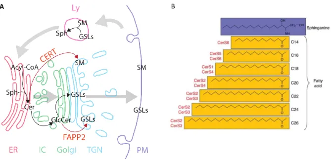
Ceramide, the central building block for sphingolipids (Figure 1.4.2), is synthesised in the de novo pathway at the cytosolic leaflet of the endoplasmic reticulum (ER) (Figure 1.4.3a). First, L-serine and palmitoyl-CoA are condensed by serine palmitoyl transferase (SPT) to form 3-ketodihydrosphinganine, which is reduced by 3-ketosphinganine reductase (3-KSR) to sphinganine. Sphinganine is N-acylated to dihydroceramide by the ceramide synthases (CerS1-6) to produce dihydroceramide. Each CesS has a specificity for CoA-fatty acid precursor(s) and catalyse formation of the corresponding N-acyl C14-26 products (Figure 1.4.3b).83 Dihydroceramides (C16-24) are dehydrogenated by dihydroceramide desaturase (DES) to ceramide (C16-24). In the ER, ceramide can be galactosylated to galactosylceramide by galactosylceramide synthase (GalCS), going on to form sulfatide GSLs in the Golgi.69 This
represents a small but pathologically important family of GSLs.67,84
Ceramide is transported from the ER to the Golgi to form the two major families of sphingolipids, SM and GSLs. Sphingomyelin synthase (SMS) transfers the phosphocholine head group from the cell’s most abundant glycerophospholipid, phosphatidylcholine (PC),69
to ceramide, forming SM and diacylglycerol (DAG), in the trans-Golgi and PM.85,86
Glucosylceramide (GlcCer), the precursor of most GSLs, is synthesised in the cytosolic leaflet of the Golgi by glucosylceramide synthase (GCS), a.k.a. UDP-glucose ceramide glucosyltransferase (UGCG). The stable UGCG gene knockout cell line, GM95, is a useful tool
24
in the study of GSL function.87 In lesser part, ceramide is phosphorylated to
ceramide-1-phosphate (C1P) by ceramide kinase (CK).88,89
The only irreversible degradation route leaving the sphingolipid network is the catabolic pathway. Ceramide is reversibly hydrolysed to sphingosine by ceramidases (CDase), which are organelle specific, pathway specific and substrate specific.85 Sphingosine is
phosphorylated to sphingosine-1-phosphate (S1P) by sphingosine kinase (SK), while the reverse reaction is catalysed by S1P phosphatase (S1PP). S1P can then be irreversibly degraded to ethanolamine-1-phosphate and C16 fatty acid by S1P lyase in the ER.
Ceramide is (re)formed in the salvage pathway, by reacylation of sphingosine, predominantly by the CerS5 enzyme.83,90 This pool cycles back to form 50-90% of total
sphingolipid content.91 Ceramide is also reformed by the degradation of GSLs by
glycosylceramidase (GCS) or galactosylceramidase (Gal-CDase) in lysosomes, the degradation of SM by sphingomyelinase (SMase), and of C1P by ceramide-1-phosphate phosphatase (C1PPase).
25
B
Figure 1.4.3: (a) GSL synthesis and transport through cellular compartments.72 (b) Ceramide synthase (CerS)
specificities.83
The stepwise biosynthesis of complex GSLs (Figure 1.4.4a) is fed by two distinct pools of lactosylceramide (LacCer), both formed by LacCer synthase from GlcCer, differing in localisation and glycosylation fate. The pool of GlcCer transported through the Golgi cisternae by vesicular transport forms LacCer to be sialylated to GM3 and ensuing gangliosides (sialosides, sialic acid-containing GSLs) (Figure 1.4.4b). Alternatively, GlcCer transported by the FAPP2 transfer protein to the trans-Golgi network (TGN) feeds the pool of LacCer which is galactosylated by Gb3 synthase to form Gb3 and the globo- and asialo-series.82 The biosynthesis of GSLs is mediated by glycosylation enzymes in the Golgi, which can act on multiple GSL substrates. However, the glycosylations are not random. Twelve glycans are used in the GSL “alphabet,” with sulfatation adding further complexity. However, only 400 different head groups have been characterised. D’Angelo et al. commented that, of the 144 linkages possible from 12 glycans, only 29 unique covalent bonds are observed.72 SphinGOMAP, an open source website compiled by the GSL research community (www.sphingomap.org), contains over 500 sphingolipid and GSL species arranged according to their biosynthesis, displaying nomenclature and stereochemistry.
26
(a)
Scheme 1.4.4: (a) Initial roots of GSL families in branched, stepwise biosynthesis.34 (b) Two distinct pools feed
the biosynthesis of complex GSLs.82
GSLs are localised between the Golgi, where they are synthesised, and the PM, where they are recognised by antibodies and lectins.92 Localising GSLs in cellular compartments is
challenging since functionalisation with a fluorophore can change considerably their localisation and traffic. Antibodies can be used to recognise GSLs as antigens. However, antibodies cannot cross the PM without disrupting the localisation of the GSL in question.93,94
27
1.5
Biomedical relevance
In this project, we develop tools for the investigation of GSLs in GL-Lect endocytosis. GSLs are of medical interest for the investigation of GSL lysosomal storage diseases, caused by mutations in sphingolipid degradation genes. Major examples include Gaucher disease, (GlcCer accumulation), Fabry (Gb3-Cer), Tay–Sachs and Sandhoff (GM2) diseases.86,96 These
can be treated with substrate reduction therapy, such as the glucosyl-ceramide synthase inhibitor N-butyldeoxyno-jirimycin,84 and enzyme replacement therapy at high cost.
Moreover, better comprehension of the molecular mechanism of toxins, which hijack mammalian GSLs for cellular uptake via GL-Lect endocytosis, will aid the development of treatment. Shiga toxin, the major virulence factor of Shigella dysenteriae, and Shiga-like toxins, also known as verotoxins, secreted by enterohemorrhagic strains of Escherichia coli bacteria, have a highly conserved structure.97 All bind specifically to Gb3-Cer, which is
expressed on kidneys and brain tissues,98 and some strains bind to Gb4-Cer with lower
affinity. Infection induces diarrhoea, and eventually haemolytic uremic syndrome (HUS) and acute renal failure, with high infant fatality rate across the world, and complex cases can have neurological symptons.20 Cholera toxin, secreted by Vibrio cholerae, causes cholera, a lethal
disease with high fatalities in the developing world, and outbreaks of disease in conflict zones is regularly reported in standard news media around the world.99 Currently no curative
treatment is known for disease caused by the Shiga and cholera toxin-producing bacteria.20
Gb3 is expressed in several cell types of interest to cancer research: cancer cells and cancer stem cells themselves,100,101 and antigen-presenting dendritic cells, which stimulate
the immune system. The interest in GSLs spans the development of cancer chemotherapeutics, in which it is paramount that effective drug targets are cell-specific to reduce non-specific toxicity. GSLs as cell biomarkers is under-explored territory in the state of chemotherapeutic research, and yet their expression levels are known to change drastically with pathogenesis.101,102 Furthermore, study of the GL-Lect mechanism will inform the
development of lectin- or STxB-conjugated drug-vectors; the rational for which aims at following the non-degradative retrograde pathway, from which STxB benefits, to target and invade host cells (Figure 1.5.1).98 This route is complementary to the standard pathway
28
clathrin-mediated endocytosis and sorted to lysosomes to be degraded. STxB-drug conjugates have shown promise in targeting Gb3-overexpressing cancer cells.104–110
Endogenous lectins and their GL-Lect interactions with GSLs are coming to light.2,16 For
example, galectin-3, which is deregulated in human cancers, is endocytosed with CD44 and β1-integrin in a clathrin-independent mechanism dependent on GSLs and N-glycosylation.31
Gb3-Cer is the cell receptor for the Shiga toxin, but no endogenous functions are currently known. Although GSLs are not classically “druggable” compounds due to their undesirable pharmacokinetic properties, research into their function and therapeutic role requires methods to manipulate their expression in cells. Hence a robust and versatile reconstitution method and related syntheses of these products are of great interest.
Figure 1.5.1: Cellular incorporation and traffic of antibody-drug conjugates (ADCs) and STx and its conjugates
In this project, we endeavour to reconstitute GSLs by conjugating in situ the two parts of a GSL (glycan and lipid) with a spacer group of the smallest possible size. We employ specific bioorthogonal chemistry, known as click chemistry, to achieve this in cell culture.
29
1.6
Bioorthogonal conjugations and click chemistry
Chemical biology uses chemical techniques to effect or probe biological systems at the molecular level. This includes the development of fluorophores, small-molecule protein inhibitors/activators, non-natural amino acids and any other synthetically modified biomolecule. Genetic tagging of proteins, for example, with green fluorescent protein (GFP), has significantly promoted research in molecular biology. However, smaller chemical modifications are needed for smaller metabolites, lipids, carbohydrates and post-translational modifications. Although chemical synthesis may never attain the complexity of nature’s highly regulated protein factory, progress is constantly being made in establishing better, subtler and more specific manipulation and higher-resolution study of the cellular system.
Current techniques for the study of lipids and GSLs are limited. Stable isotope labelling should not disrupt the biochemical interactions of the molecule under study, but incorporation is often low yielding, requiring technically complicated protocols for low sensitivity and high costs, and topological imaging techniques able to localise stable isotopes are still developmental (secondary ion mass spectrometry, SIMS, section 1.3). Radioisotope labelling can be localised but is innately hazardous. Mass spectrometry (MS) coupled to liquid chromatography (LC-MS) has traditionally been used for exact identification for the brute structure of the lipidome, all of the lipids present in a biological sample. Liquid chromatography separation of analyte species allows higher signal to noise ratio, but is not quantifiable. Now, shotgun lipidomics is coming to dominate the field, and is applied in this project. This rapidly developing technology permits identification and quantification of the lipidome using high resolution mass spectrometry (HRMS) to access exact masses at a parts-per-million (ppm, 10-6) mass unit accuracy, coupled with high-throughput data handling.
Highly adapted lipid extraction protocols are employed with direct infusion, without chromatographic separation. Direct infusion allows exact quantification since a constant flow of lipid sample of known concentration is analysed, with the use of appropriate internal standards.111,112
To investiage biomolecule localisation and function, small chemical reporter groups, such as fluorophores for fluorescence imaging, or affinity tags like biotin for purification, can be incorporated in vitro, before their incorporation and study in cells or organisms. For in vitro
30
tagging, selective or orthogonal chemical conjugations are required for specific tagging in a non-perturbing site on the biomolecule. For small biomolecules such as lipids and carbohydrates, where even the smallest fluorophores can perturb lipid function,57 the
reporter group can be added in cellulo by means of a minute, latently reactive tag or labelling group. After cellular incorporation and biological function, the tag can be specifically conjugated to a reporter group (e.g. fluorophore or biotin) for visualisation or isolation (Figure 1.6.1).113 Such chemical conjugation (or scission) reactions must occur specifically in a
complex biological environment, and are termed bioorthogonal. The bioorthogonal reactant partners must be specifically, mutually reactive whilst being inert to their environment, and as small as possible. Since applications are often in live cell culture, the reaction must occur at a similar timescale to standard cellular processes.114
Figure 1.6.1: Tagging a biomolecule and analysing its cellular localisation or metabolism products using a reporter molecule, often fluorophore or biotin affinity tag.
Early specific, orthogonal chemical reactions were used to chemically modify proteins
in vitro. Reactions include disulfide exchange or maleimides for cysteine residues, and
N-hydroxysuccinimides and iso(thio)cyanates for lysine residues (Figure 1.6.2a).115 However, for
complete regiospecificity, these reactions require that only one active thiol or amine is present in the conjugation environment. Ketones and aldehydes can be condensed with aminooxy and hydrazide forming oxime or hydrazine conjugations respectively (Figure 1.6.2b). These were among the first specific chemical conjugations applied to the surface of whole, live cells. Bertozzi et al. described the metabolic labelling of cell surface glycans using a ketone-labelled analogue of N-acetylmannosamine (ManNAc), N-levulinoylmannosamine (ManLev, Figure 1.6.3), which is metabolised into sialic acid-containing glycolipids and glycoproteins and presented at the cell surface, where they selectively conjugated with a hydrazide-biotin reporter.116–118 These aminooxy and hydrazide conjugations require high
31
concentrations of reactants, and fall prey to high cross-reactivity with natural ketones and aldehydes.
(a)
(b)
Figure 1.6.2: (Bio)orthogonal chemical conjugations. (a) Disulfide exchange and maleimide conjugations for cysteine residues and N-hydroxysuccinimides and iso(thio)cyanate and iso(thio)cyanates for lysine residues. (b) Oxime/hydrazine with aldehyde/ketones.
The Staudinger ligation uses azide- and arylphosphine-functionalised partners and has been applied to bioorthogonal live cell labelling119–121 and prodrug release,122 since bonds are
formed and broken in the reaction mechanism (Figure 1.6.3a). Azides are ideally small, non-perturbing and stable in cells and organisms.123 For example, an azide group is present in the
well-known anti-AIDS drug, 3’-azidothymidine (AZT).124 The bioorthogonal Staudinger ligation
derives from the classic reduction of azides with phosphines,125 using a functionalised
aromatic phosphine which traps the aza-ylide intermediate, forming a stable conjugation. The traceless Staudinger reaction (Figure 1.6.3b) leaves a perfectly small amide bond after the conjugation and intramolecular elimination of the arylphosphine. These variants are less applied in live cell studies due to poor kinetics, selectivity and solubility.126
Saxon and Bertozzi showed live cell metabolic labelling with an azide-labelled analogue of ManNAc, Ac4ManNAz (Figure 1.6.3c).119,127 The hydrophobic, peracetylated
glycan is used to increase cell permeability, and is deacetylated by non-specific cytosolic esterases to ManNAz in the cell.128 ManNAz then follows the endogenous anabolic sialic acid
pathway to SiaNAz, and is incorporated into sialic acid-containing proteins and glycolipids.129
The azide-functionalised sialosides were then conjugated specifically with an arylphosphine-biotin for quantification. Unfortunately, Staudinger ligations suffer from impractically slow kinetics (k2≈3×10⁻³ M-1s-1), require high concentrations (>250 µM triarylphosphine),127 and
32
(a)
(b)
(c)
Figure 1.6.3: Bioorthogonal chemical conjugations (a) Staudinger and (b) traceless Staudinger ligations. (c) ManNAc, Natural sialic acid precursor. Synthetic analogues ManLev and Ac4ManNAc, used for metabolic
labelling of sialic acid-containing glycoproteins and glycolipids.117,130
The term click chemistry was coined to describe reactions which are fast, specific, high yielding, occur in mild (aqueous, aerobic, biological) conditions, and produce inert or no byproducts.131 Reactions coming close to meeting these criteria are now so popular they are
used by chemists and non-chemists, in common, commercial laboratory kits. The heat-driven 1,3-cycloaddition between alkyne and azide was described by Huisgen in the the 1960s.132
The copper(I)-catalysed azide-alkyne 1,3-dipolar cycloaddition (CuAAC) variation (Figure 1.6.4a), first described in 2002,133,134 is still an extremely useful orthogonal conjugation in
organic synthesis,132,135 allowing complex molecules to be coupled specifically, in the
presence of diverse functional groups, without need for protecting groups. CuAAC reactions came to dominate click chemistry for its extremely fast reaction times and specificity (10-200 M-1s-1 in the presence of 20–500 mM Cu(I)). Sadly, the copper, solubilised in a ligand
complex, is abundantly taken up into cells and causes rapid toxicity by the formation of reactive oxygen species (ROS).136 This is augmented by ascorbate, the standard reductant
used to form Cu(I) from Cu(II), which also promotes ROS formation.136,137 Copper is also
reported to chelate to amino acids, impeding protein structure and function.
To combat this, ligands have been developed promising to reduce toxicity whilst retaining copper catalysis and solubility (Figure 1.6.2b).138 These have been successfully
applied in cell surface labelling.139 For example, Cairo et al. showed that they could induce cell
33
clicked with azide-coated beads and L-histidine ligand.140 Besanceney-Webley et al.
developed BTTAA (Figure 1.6.4b), which they showed chelates copper to catalyse the cell surface click reaction with azide-labelled sialic acids (SiaNAz) in 3 minutes. This is fast enough that copper could be used without significant cell death in Jurkat and HEK cells. They cite the use of 30 µM Cu(I), [ligand]/[Cu]=6:1, where 30 µM Cu(I) and no ligand shows no significant cell death, and yet 50 µM killed 90% of cells in the same time.141
(a)
(b)
Figure 1.6.4: (a) Copper-catalysed azide-alkyne cycloaddition (CuAAC). (b) Cu-chelating ligands for CuAAC; TBTA: R, R'=CH2Ph; THPTA: R, R'=(CH2)3OH; BTTAA: R=tBu; R'=CH2CO2H.
Cyclooctynes can react with azides without copper catalysis (Figure 1.6.5a). They have higher reactivity due to their strained cyclic structure, in a reaction termed the strain-promoted azide-alkyne cycloaddition (SPAAC).113 In 1961, Witting and Krebs reported that a
stable cyclooctyne reacted “like an explosion” with phenylazide, but it was not until 2004 that Agard, Prescher and Bertozzi synthesised OCT (Figure 1.6.5b) and showed its specific reaction on cells labelled with SiaNAz, without copper catalysis.142
The standard 1,3-dipolar SPAAC occurs between the LUMOπ , alkyne-HOMOπ,azide (Figure
1.6.5c).143 The electronic properties of the alkyne have been fine-tuned by adding
electron-withdrawing groups (EWGs, Figure 1.6.5c) to lower LUMO energy of the electron-rich alkyne.144,145 Difluorocyclooctyne (DIFO, Figure 1.6.5b) bares two small, biologically inert
fluorine atoms, with an inductive electron-withdrawing effect. Monofluorocyclooctyne (MFCO, Figure 1.6.5b) is less reactive – more stable – with one inductive EWG, fluorine, and a carbonyl or amide, slightly poorer, mesomeric EWGs. Dibenzocyclooctyne (DIBO) bares planar aromatic groups which increase reactivity by increasing ring strain.143 Unfortunately,
π-conjugated cyclooctynes can undergo side reactions with endogenous nucleophiles as Michael acceptors (electrophile prone to 1,4-addition),114,142 as well as reducing
DIBO-conjugates aqueous solubility. Bicyclononyne (BCN), alternatively, has greater reactivity due to ring strain caused by a fused cyclopropane ring. Many reported studies display impressive application of specific labelling in cells and in vivo with cyclooctynes (Introduction 1.7).146
34
When choosing a click reagent, one must consider that increasing reactivity is often detrimental to specificity and stability.
(a)
(b)
(c) Figure 1.6.5: (a) SPAAC click
reactions. (b) Derivatised cyclooctynes, rate constants for benzyl azides, relative to parent compound OCT.147,148 (c) SPAAC
forming triazole. Orbital diagram of bonding interactions between cyclooctyne LUMO and azide HOMO, activated by adding electron-withdrawing groups (EWG) to the cyclooctyne.144
Chemical evolution of click reagents has led to studies on nitrones, nitrile oxides and sydnones, which react quicker than azides with cyclooctynes (Figure 1.6.6 a-c). For example, strain-promoted alkyne-nitrone cycloaddition (SPANC) reactions demonstrate rate constants up to 60 M-1s-1.147 However, their syntheses are not commonly mastered and their use as tags
in biomolecules can prove more cumbersome than the classic azide. Nitrones were stable enough to efficiently label large proteins in live cells without degradation.149 However,
35
nitrone-labelled glycans fared less well, as the nitrone modification was not accepted into the biosynthetic pathway.150 Inverse electron demand Diels-Alder (IEDDA, Figure 1.6.6d, e)
conjugations come out ahead as the quickest bioorthogonal reactions, but leave a remarkably large scar in the structures of modified biomolecules.
(d) Figure 1.6.6: (a-c) 1,3 dipoles which react with cyclooctynes via SPAAC. Rate constants with BCN: (a) k<60 M-1s-1 (b)
k=0.054 M-1s-1. (c) k=1.6 M-1s-1
.147,151
Tetrazines (d) react specifically with strained alkenes (dienophile, e) via the inverse-electron demand Diels-Alder Reactions (IEDDA).147
36
1.7
Metabolic labelling of glycans and lipids
Click chemistry’s application in metabolic labelling has had great influence on several
advanced biological techniques. Genetic code expansion, using synthetically evolved derivatives of tRNA synthetases, now allows site-specific genetic encoding of clickable amino acids.152 With click chemistry, radiolabels can be added to proteins regioselectively under mild
conditions, for techniques such as positron emission tomography (PET) and radiotherapy, and which show potential in animal models.153 This project has been inspired and guided by recent
innovations in metabolic labelling to study lipid and glycan interactions, briefly outlined in this chapter.
Azide-modified glycans such as Ac4ManNAz (Figure 1.6.3),119 an analogue of the sialic
acid precursor, ManNAc, are popular substrates for labelling the cell surface, and have been applied to many high-tech labelling techniques. Laughlin et al. demonstrated the use of Ac4GalNAz for spatiotemporal labelling of zebrafish embryo morphology (Figure 1.7.1),
displaying early (red) and late (green) gestational post-translational glycosylation, using a fluorophore functionalised with difluorocyclooctyne (DIFO, Figure 1.6.5b). Wang et al. showed that Ac3ManNAz could be coupled with a tumour-selective self-immolative linker in
the anomeric (C1) position to specifically label tumour tissue in mice.154
Figure 1.7.1: Zebrafish embryo labeled with Ac4GalNAc.
DIFO-647 (red) click 60 hours post-fertilisation (hpf), then DIFO-488
click 62 hpf after an additional 1h
pulse of Ac4GalNAc labelling.
Scale bars 100 μm (A), 10 μm (B).
Click chemistry has been used to probe lipid localisation and metabolism in a myriad
of substrates. Jao et al. showed that incorporating clickable propargylcholine (Figure 1.7.2a),155 and later the azide analogue (Figure 1.7.2b)156 at high concentrations was
non-toxic, and could give dose-dependent labelling up to 44% phosphatidylcholine (PC), transferring to sphingomyelin labelling (SM, <15%) at later time points, with a similar profile to natural choline-containing lipids (Figure 1.7.2c). Labelling fixed cells with an azide-fluorophore and copper catalysis labelled all cellular membranes (Figure 1.7.2c).
37
Figure 1.7.2: Clickable choline analogues, (a) propargylcholine155 and (b) 1-azidoethylcholine,156 are
metabolised into phosphatidylcholine (PC), ether phospholipids (ePC) and sphingomyelin (SM). (c) Propargylcholine treatment (100 μM) shows similar profile of choline-containing lipids by ESI-MS/MS analysis. (d) Alkyne-lipids undergo CuAAC click reaction with alkyne-fluorophore (fluorescence microscopy image).
Lipids with terminal ω-alkyne or ω-azide groups (Figure 1.7.3a,b) have been widely used to study fatty acylated-protein localisation.157 Hang et al. first investigated the
metabolism of clickable fatty acids in proteins by Western blotting,158 whereas Thiele et al.
developed a lipid extraction technique which would permit the analysis of clickable lipid metabolism by thin layer chromatography (TLC).159,160 Clickable
ω-alkyne-oleoyl-lysophosphatidic acid, ω-alkynesphinganine and palmitoyl-lyso-propargyl-phosphatidyl-choline (Figure 1.7.3c-e) displayed canonical metabolism in cells.161 Enzyme affinity (K
m)
measurements of such substrates showed no significant difference in binding affinities with native biosynthesis enzymes compared with natural lipids.161 Localisation of clickable lipids,
clicked to a fluorophore with copper-catalysed or copper-free162,163 protocols, seem to reliably
demonstrate, with negative controls, that fluorescent labelling is specific to the lipid analogue.164 Most often the clicked lipid is lodged non-specifically in plasma membrane and
intracellular membranes, as is conceptualised for natural lipids. One less subtle modification in a ceramide analogue with trans-cyclooctene in the lipid tail (Figure 1.7.3f) provides the lipid strong Golgi specificity.165
38
Figure 1.7.3: Clickable lipids. (a,b) Fatty acids,158,159 (c) alkyne-oleoyl-lysophosphatidic acid (d) ω-alkynesphinganine
(e) palmitoyl-lyso-propargyl-phosphatidyl-choline.161 (f) Golgi-specific transcyclooctene ceramide.165
Clickable glucosylceramide (GlcCer) with short ω-alkyne C8 acyl chain gave
intracellular membrane staining on alkyne-fluorophore click reaction (1, Figure 1.7.4a). This localisation is in agreement with Pedrosa et al., who used a ω-azide C8 acyl chain on GlcCer.61
Inversely, an azide in the glycan head group or C4 acyl chain (2, Figure 1.7.4b) gave PM labelling. Long C14 GlcCer (3) and lactosylceramide (LacCer) analogues with an azide in the 6’’-OH position and short chain C4 or C8 acyl chain (47, 48, Figure 1.7.4) stained the PM, and required fusogenic liposomes to be incorporated.62
Figure 1.7.4: Clickable glycolipids localise in intracellular cellular membranes (a, analogue 1) and PM (b, analogue
2). Short chain LacCer analogues 47 and 48 localised to the PM. Analogues 3 and 49 were incorporated using
fusogenic liposomes, localising in the PM.
Discernibly from these examples and others, visualising modified lipids can indicate properties of that lipid with a large margin of error. So what can we learn from them about the natural system? Isolating lipid-protein interactions is of great interest, but non-covalent affinities are too weak to be used with standard immunoprecipitation techniques (Introduction 1.3-1.4). Photoactivable crosslinkers have been incorporated into lipids to study their interactions.166 This strategy, often known as affinity-based probes, is commonly used
39
common groups are benzophenone, diazirine and aryl azide (Figure 1.7.5a-c). These small chemical groups are activated by a flash of UV light, forming a reactive, radical species which bonds covalently to any molecule (often heteroatoms) close to the tagged molecule of interest. Their standard usage involves cellular incorporation of the photoactivatable small molecule, UV activation, cell lysis, and visualisation or isolation of the lipid-protein partners by clicking to a fluorophore or solid support respectively (Figure 1.7.5e). The use of photoactivatable crosslinking groups has led to the identification of interaction partners of plant glycoglycerolipids,168 phospholipids,169 sphingosine,170 cholesterol.171 A
photoactivatable, clickable GlcCer analogue was recently described, but no new interaction partners have yet been reported.172
Figure 1.7.5: Photocrosslinkable groups, benzophenone (a), 3-phenyl-3-(trifluoromethyl)diazirine (b), and aryl azide (c) with their reactive radical forms.167 (d) Confirmed photoactivatable sphingosine probe; protocol
involves live cell incubation, UV light activation, extraction and click with a fluorophore for visualisation, or biotin-streptavidin beads for purification and mass spectrometry characterisation.170
1.8
Project aims
Biosynthesis of GSLs (Figure 1.4.4) is stepwise and likely under equilibrium, and one gene does not code for one GSL but several. This makes genetic manipulation of a single GSL impossible without offsetting the expression of other GSLs. Current methods to supplement cells with pure long chain GSL species, a process here termed cellular reconstitution, are extremely inefficient (Introduction 1.3).60–62 The study of GSL function is thus challenging and
40
to reconstitute the biological and mechanochemical properties of GSLs. Tools must be reconstituted in an easy protocol, whilst retaining biological function.
We aim to develop a biorthogonal, clickable kit of GSL precursors from proof-of-concept studies on GSL globotriaosylceramide (Gb3-Cer) and the Shiga toxin (STxB). To reconstitute a functional Gb3-Cer analogue, we attempt to commandeer the native GSL biosynthesis and traffic; supplementing cells with a clickable 1-azidosphingosine (AzSph, Figure 1.8.1), which is natively metabolised to 1-azidoceramide (AzCer) and presented at the plasma membrane (PM), to which we click the functionalised oligosaccharide head group of choice by a highly selective, copper-free bioorthogonal click reaction.
Figure 1.8.1: GSL click reconstitution. Sphingolipid precursor, 1-azidosphingosine (AzSph) is natively metabolised to 1-azidoceramide (AzCer), ideally presented at the PM to be clicked with the prepared
clickable oligosaccharide (Gb3-MFCO) to form the final Gb3 analogue. (Lipid membrane not to scale)
The maintaining biological activity, by using the smallest least perturbing chemical groups possible is constantly balanced with having a fast and specific click reaction (Figure 1.8.2a). We base our design on previous studies showing that clustering of STxB was greatly inhibited with long, flexible linkers, but a small linker in the central region (n=1, 10Å) permitted native function (Figure 1.8.2b, Introduction 1.3).173 We design our final click
analogue with dimensions of this size or smaller. Previously a GM1-phospholipid analogue retained its native function in binding and trafficking with the cholera toxin and simian virus 40.29 We consider the use of a Gb3-phospholipid, proposed to be soluble enough to permit
41
If these neo-glycolipids are functional and reconstituted with a feasible yield, this strategy could potentially tolerate further chemical modifications, allowing further probing of native GSL interactions. One long-term goal is to reconstitute photoactivatable, traceable GSLs, for example containing a diazirine group (Figure 1.8.2c), to identify GSL interaction partners (cf. Perspectives).
(a)
(b)
(c)
Figure 1.8.2: (a) Balance between reactivity and specificity of click partners, with the biological functionality of final clicked product. (b) Gb3-EGn-Cer analogues able to bind Shiga toxin in liposome models, whilst only
Gb3-EG1-Cer is able to induce membrane bending.24 (c) Potential photoactivatable and reconstituable Gb3.
Specificity
42
2 RESULTS
2.1 Synthesis of biomolecule analogues
2.1.1
Reconstitutable globotriose phospholipid
We aim to synthesise a Gb3 with retained native function that can be reintroduced into cells in culture. Gb3-phospholipid analogues, or function-spacer-lipid (FSL) constructs, have shown to retain similar biological activity as the natural Gb3-Cer with known binding lectins: Shiga toxin (verotoxin1) and HIV-1.29,174,175 Crucially, phospholipids (PLs) are more
efficiently incorporated into cells in culture than GSLs (Introduction 1.3).60,62 Hence, we
proposed that a Gb3-PL would be a valuable tool. The spacer between glycan functional head and lipid tail of the FSL must be biostable, meaning it must not be recognised and digested by cellular clearance machinery. We require the spacer between the Gb3 head group and phospholipid tail to be short to retain membrane bending properties (Introduction 1.3).24 This
contributes to the originality of this endeavour, since only long Gb3-phospholipids have been synthesised in the literature.29,174,175
Synthetic routes to install isothiocyanate and azide functions at the anomeric position of glycans are well documented.176 Isothiocyanates conjugate specifically with amines to form
a thiourea (Scheme 2.1.1.1). The classic bioorthogonal copper-catalysed azide-alkyne cycloaddition (CuAAC, Introduction 1.6) forms a triazole. Both thiourea and triazole are stable in cells.
Our project is applied to the Gb3 triose, synthesised in a four-step synthesis (Section 2.1.3). We began testing syntheses on a lactose model for matters of cost and scale. Lactose (Scheme 2.1.1.2) was peracetylated with acetic anhydride in pyridine, to which the isothiourea (SCN) group was installed with the use of trimethylsilyl thiocyanic acid (TMSSCN) and SnCl4 as a Lewis acid catalyst to form peracetate lactosyl isothiocyanate 1.24,177 This was
conjugated to 1,2-dioleoyl-sn-glycero-3-phosphoethanolamine (DOPE, 15) under basic conditions to form thiourea conjugate 2. Despite an orthogonal deprotection of four acetyl groups being described,178 this hydrazine-mediated deprotection was unfeasible in our hands
without simultaneous breakdown of phospholipid ester groups, and conjugate 3 was not fully characterised. Alternatively, deprotection of isothiocyanate 1 was tested prior to conjugation
43
with DOPE, but isothiocyanate was unstable to methoxide conditions. It proved equally impossible to install the isothiocyanate group without hydroxyl protecting groups.
Scheme 2.1.1.1: Biostable chemical conjugations
Scheme 2.1.1.2: Attempted synthesis of lactose-phospholipid analogue with isothiocyanate linkage.
We wanted to avoid the use of protecting groups on the glycan part, which are difficult to remove once the phosphodiester tail is installed. A highly orthogonal CuAAC click was thus envisaged. The synthesis is here proposed with 1-azidoglobotriose, 4 (Scheme 2.1.1.3) since the high yielding click reaction would not require optimisation with a lactose model. Azide-functionalised glycan 4 could click with alkyne-Azide-functionalised phospholipid 5 to form a Gb3-PL 6.


![Figure 1.3.3: GUVs with 5 mol % Gb3 analogue 1 (a,b) or 2 59 (c,d) or wild type Gb3-Cer (Figure 1.2.1) 58 (e) (DOPC/SM/Chol/Gb3-Cer [40:35:20:5] with <1% fluorescent phospholipid (green, L d ), incubated with STxB-Cy3 (red)](https://thumb-eu.123doks.com/thumbv2/123doknet/14718618.750717/21.892.120.783.171.395/figure-guvs-analogue-figure-dopc-fluorescent-phospholipid-incubated.webp)