RESEARCH OUTPUTS / RÉSULTATS DE RECHERCHE
Author(s) - Auteur(s) :
Publication date - Date de publication :
Permanent link - Permalien :
Rights / License - Licence de droit d’auteur :
Bibliothèque Universitaire Moretus Plantin
Institutional Repository - Research Portal
Dépôt Institutionnel - Portail de la Recherche
researchportal.unamur.be
University of Namur
Management of Non-Vitamin K Antagonist Oral Anticoagulants in the Perioperative
Setting
Dincq, Anne-Sophie; Lessire, Sarah; Douxfils, Jonathan; Dogné, Jean-Michel; Gourdin,
Maximilien; Mullier, François
Published in:
BioMed research international
DOI:
10.1155/2014/385014 Publication date:
2014
Document Version
Early version, also known as pre-print
Link to publication
Citation for pulished version (HARVARD):
Dincq, A-S, Lessire, S, Douxfils, J, Dogné, J-M, Gourdin, M & Mullier, F 2014, 'Management of Non-Vitamin K Antagonist Oral Anticoagulants in the Perioperative Setting', BioMed research international, vol. 2014, 385014. https://doi.org/10.1155/2014/385014
General rights
Copyright and moral rights for the publications made accessible in the public portal are retained by the authors and/or other copyright owners and it is a condition of accessing publications that users recognise and abide by the legal requirements associated with these rights. • Users may download and print one copy of any publication from the public portal for the purpose of private study or research. • You may not further distribute the material or use it for any profit-making activity or commercial gain
• You may freely distribute the URL identifying the publication in the public portal ?
Take down policy
If you believe that this document breaches copyright please contact us providing details, and we will remove access to the work immediately and investigate your claim.
Review Article
Management of Non-Vitamin K Antagonist Oral
Anticoagulants in the Perioperative Setting
Anne-Sophie Dincq,
1Sarah Lessire,
1,2Jonathan Douxfils,
2Jean-Michel Dogné,
2Maximilien Gourdin,
1and François Mullier
31Department of Anesthesiology, CHU Dinant Godinne UcL Namur, Namur Thrombosis and Hemostasis Center (NTHC), Namur Research Institute of Life Sciences (NARILIS), 5530 Yvoir, Belgium
2Department of Pharmacy, University of Namur, Namur Thrombosis and Hemostasis Center (NTHC), Namur Research Institute of Life Sciences (NARILIS), 5000 Namur, Belgium
3Haematology Laboratory, CHU Dinant Godinne UcL Namur, Namur Thrombosis and Hemostasis Center (NTHC), Namur Research Institute of Life Sciences (NARILIS), 5530 Yvoir, Belgium
Correspondence should be addressed to Anne-Sophie Dincq; anne-sophie.dincq@uclouvain.be Received 30 May 2014; Accepted 5 August 2014; Published 3 September 2014
Academic Editor: Helen Mani
Copyright © 2014 Anne-Sophie Dincq et al. This is an open access article distributed under the Creative Commons Attribution License, which permits unrestricted use, distribution, and reproduction in any medium, provided the original work is properly cited.
The field of oral anticoagulation has evolved with the arrival of non-vitamin K antagonist oral anticoagulants (NOACs) including an anti-IIa agent (dabigatran etexilate) and anti-Xa agents (rivaroxaban and apixaban). The main specificities of these drugs are predictable pharmacokinetics and pharmacodynamics but special attention should be paid in the elderly, in case of renal dysfunction and in case of emergency. In addition, their perioperative management is challenging, especially with the absence of specific antidotes. Effectively, periods of interruption before surgery or invasive procedures depend on half-life and keeping a permanent balance between bleeding and thromboembolic risks. In addition, few data regarding the link between plasma concentrations and their effects are provided. Routine laboratory tests are altered by NOACs and quantitative measurements are not widely performed. This paper provides a review on the management of NOACs in the perioperative setting, including the estimation of the bleeding and thrombotic risk, the periods of interruption, the indication of heparin bridging, the usefulness of laboratory tests before surgery or invasive procedure, and the time of resuming. Most data are based on expert’s opinions.
1. Introduction
Three non-vitamin K antagonist oral anticoagulants (NOACs) [1] are already widely used in the clinical setting: rivaroxaban and apixaban, two direct factor Xa (FXa) inhi-bitors, and dabigatran etexilate (DE)—the prodrug of dabiga-tran, a direct thrombin inhibitor. Both of these drugs will progressively tend to replace vitamin K antagonists (VKAs) in most of their indications. NOACs indications vary among countries. They are licensed for long-term prevention of thromboembolic events in nonvalvular atrial fibrillation (NVAF), for thromboprophylaxis of venous thromboembol-ism (VTE) including deep venous thromboembolthromboembol-ism (DVT) and pulmonary embolism (PE) after hip and knee arthro-plasty, and for the treatment and secondary prophylaxis of
VTE. Rivaroxaban is also approved in Europe for secondary prevention of atherothrombotic events after acute coronary syndrome (ACS) with elevated cardiac biomarkers [2–7].
Advantages of NOACs include rapid onset and offset of action and relatively predictable anticoagulation effects [8]. In most patients, routine laboratory monitoring of the anticoagulant effect is not required but the assessment of the estimated renal clearance is necessary [9]. In some cases (e.g., emergencies, bleeding, overdose, and trauma), the anticoag-ulation status and the alteration of standard laboratory data must be known [10, 11]. An increasing number of patients on long-term treatment with NOACs are encountered in the perioperative setting and it is essential for physicians to be aware of the pharmacological properties of these drugs. The management of those patients requires an involvement of
Volume 2014, Article ID 385014, 16 pages http://dx.doi.org/10.1155/2014/385014
all participating teams (general practitioners, surgeons, anes-thesiologists, and other healthcare professionals involved in invasive procedures). Their cessation is indisputable in most elective procedure, but the risk between thrombosis and bleeding should be balanced [12]. In some settings, the therapeutic window is bridged by low molecular weight hep-arin (LMWH) or unfractionated hephep-arin (UFH) to prevent thromboembolic risk [13,14]. No specific antidote is currently available in case of bleeding so clinicians have to deal with rescue treatments [15]. The optimal time for NOAC’s resump-tion depends mainly on the postoperative risk of bleeding [16].
This paper aims at providing a review on the management of NOACs in the perioperative setting in accordance with the current literature. This includes the estimation of the bleeding and thrombotic risk of each patient, the period of NOAC’s interruption before an invasive procedure, the conditions for heparin bridging during this interruption, the usefulness of common and specific laboratory tests to assess the remaining anticoagulant effect preoperatively, and the time of NOAC’s resumption prerequisites for the perioperative management of NOACs. The literature search was performed in PubMed using the following keywords: perioperative, anticoagulant, dabigatran, rivaroxaban, and apixaban. Overall inclusion of papers was limited to studies published until May 30, 2014.
2. Indications and Posology of NOACs
Three molecules are currently available in the clinical setting: dabigatran etexilate (Pradaxa, Boehringer-Ingelheim Pharma GmBH, Ingelheim am Rhein, Germany): 75 mg, 110 mg, and 220 mg capsules, rivaroxaban (Xarelto, Johnson and John-son/Bayer HealthCare AG, Leverkusen, Germany): 2.5 mg, 10 mg, 15 mg, and 20 mg tablets, and apixaban (Eliquis, Bristol Myers Squibb/Pfizer, Bristol Myers Squibb House, Uxbridge, United Kingdom): 2.5 mg and 5 mg tablets.
Table 1summarizes the approved indications by the Food and Drug Administration and the European Commission, the posology, and the dose adaptation of the different NOACs. Table 2 summarizes the main studies leading to the approved indications of NOACs [17–27].
3. Clinician Oriented Overview of
Pharmacokinetic Properties of NOACs
3.1. Dabigatran Etexilate (Pradaxa, Boehringer-Ingelheim Pharma GmBH, Ingelheim am Rhein, Germany): 75 mg, 110 mg, and 220 mg Capsules. Dabigatran etexilate is the
prodrug of dabigatran, a selective and reversible oral direct thrombin inhibitor. The plasma peak after ingestion is at 1.5– 3.0 hours and the plasma trough level is 11–14 hours after ingestion in healthy volunteers [28]. Bioavailability varies from 3 to 7% depending on the pH encountered in the microenvironment of the gastrointestinal tract. Dabigatran is 35% bound to plasma proteins, allowing theoretically its elim-ination by hemodialysis. Eighty percent of the drug is directly eliminated in the urine, explaining that, in the setting of renal insufficiency, the anticoagulant effect accumulates. Its
elimination half-life rises to 18–24 hours in patients with significantly impaired renal function compared to healthy elderly subjects [29]. Creatinine clearance (CrCl) estimation based on the Cockroft and Gault formula [30,31] is recom-mended in elderly patients to calculate doses and avoid over-medication [9]. Twenty percent is conjugated as glucuronides by hepatic metabolism. Dabigatran etexilate is contraindi-cated in case of severe renal impairment (CrCl< 30 mL/min) in Europe while a lower dose is proposed in North America for CrCl between 30 and 15 mL/min [32]. Hepatic impairment or liver disease with expected impact on survival is also a contraindication [33,34].
3.2. Rivaroxaban. Rivaroxaban is a selective and reversible
oral direct FXa inhibitor. The plasma peak after ingestion is at 2–4 hours and the half-life elimination is 5–9 hours in healthy volunteers and 11–13 hours in the elderly [28]. Bioavailability is between 80 and 100% for 10 mg and around 66% for 15 or 20 mg under fasting conditions. Rivaroxaban is bound to plasma proteins in more than 90%, making hemodialysis ineffective to eliminate this drug. About one-third (36%) of the dose is eliminated by renal pathway as unchanged active drug, and the approximately remaining two-thirds of the dose is subject to metabolic degradation. Metabolites are elimi-nated equally via renal pathway and via hepatobiliary route in the feces [35]. Clearance is mildly influenced by renal func-tion [36]. Rivaroxaban is not recommended in severe renal impairment (ClCr< 15 mL/min) [32].
3.3. Apixaban. Apixaban is a selective and reversible oral
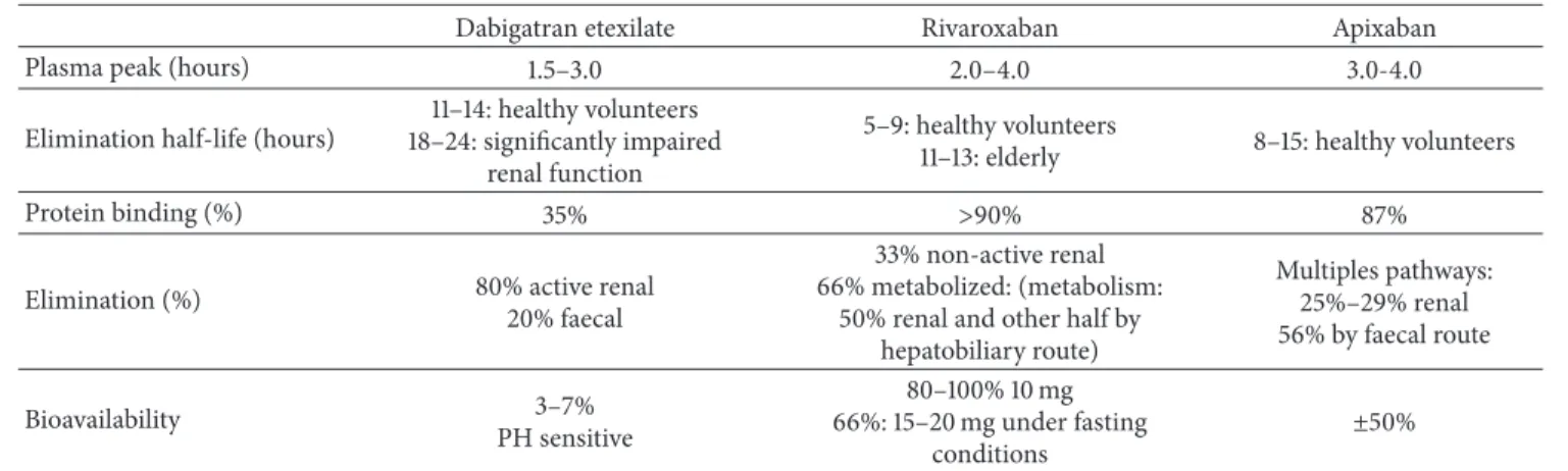
direct FXa inhibitor. The plasma peak after ingestion is at 2-3 hours and the half-life elimination is 8–15 hours in healthy volunteers [28]. Apixaban is 87% bound to plasma proteins. Bioavailability is around 50%. Apixaban is eliminated via multiple pathways: predominantly via the fecal route (56%) and 25–29% via renal excretion [37]. Apixaban is not recom-mended in severe renal impairment (ClCr< 15 mL/min) [32]. Table 3proposes an overview of the main pharmacoki-netic properties of direct oral anticoagulants.
4. Drugs Interactions
Two mechanisms are mainly involved in NOACs’ metabolism and elimination: the efflux operated by P-glycoprotein (P-gp) and the CYP450 isoform CYP3A4. Dabigatran etexilate is metabolized in dabigatran in the plasma and liver via CYP450-independent mechanisms [38], but DE acts as a substrate for P-gp. Therefore, strong inhibitors or inducers of P-gp may alter the absorption of DE [39]. Rivaroxaban and apixaban are metabolized by CYP3A4/5 and are both substrates for P-gp. Thus, drugs that strongly inhibit or induce CYP3A4 or P-gp or both influence the pharmacokinetic (PK) profile of these NOACs [39,40].
5. Perioperative Management of NOACs
As illustrated in the RE-LY (randomized evaluation of long-term anticoagulation therapy) study [41], 25% of the patients
T a ble 1: Summa ry o f ap p ro ve d indica tio n s, p os o log y and dos e ada p ta ti o n o f th e diff er en t N O A Cs. D ab iga tr an et exi la te (P radaxa) Ri va ro xa b an (Xa re lt o) A p ixa b an (E liq u is) VT E P ro p h yl ax is (i) 220 m g/ d ay (2 ca psules o f 11 0 m g O D) or (ii) 15 0 m g/ da y (2 ca psules o f 75 m g O D ) if CrC l3 0 –5 0 mL/min, if > 75 ye ars ,i f ve ra p am il, amio da ro ne an d q uinidine THR: 28 –3 5 d ay s TKR: 10 da ys 10 mg/ d ay (1 ta b let o f 10 m g O D ) THR: 5 w eeks TKR: 2 w eeks 5m g/ d ay (1 ta b let o f 2.5 m g B ID) THR: 32 –3 8 d ay s TKR: 10 da ys No n -v al vu la r at ri al fi b rilla tio n (i) 30 0 m g/ d ay (1 ca psule o f 15 0 m g BID) (ii) 220 m g/ d ay (EU) (1 ca psule 11 0 m g BID) (a) if > 80 y o r vera p am il 15 0 m g/ da y (US) (1 ca psule o f 75 m g B ID) (b) if C rC lb etw een 15–3 0 mL/min (c) if d ro neda ro ne/k et o co n azo le (US) (i) 20 m g/ da y (1 ta b let o f 20 m g O D ) (ii) 15 m g/ d ay (1 ta b let o f 15 m g O D ) if CrC lb etw een 15– 49 mL/min (i) 10 m g/ da y (1 ta b let o f 5 m g B ID) (ii) 5 m g/ da y (1 ta b let o f 2.5 m g B ID) if at le ast 2 o f th e fo llo win g co ndi tio n s: ≥ 80 ye ar s, ≤ 6 0 kg o r se ru m cr ea tinine ≥ 1.5 m g/ dL; o r if CrC l15–29 mL/min VT E tr ea tm en t an d se co nda ry p ro p h yl axis (i )1 50 m g B IDa ft er5 – 10d ay sp ar en te ra l an ti coa gula tio n (i i)1c ap su le 75 m g B ID if CrC l< 30 mL/min (US) (iii) A d o p ted indica tio n C HMP ∘ (a p ril 201 4 ) (EU) (i) T re at men t p h as e: 30 m g/ d ay (1 ta b let o f 15 m g BID) fo r 21 d ay s (ii) Se co nda ry p re ve n tio n: 20 m g/ d ay (1 ta b let o f 20 m g O D ) 15 m g/ d ay (1 ta b let o f 15 m g O D ) if CrC lb etw een 15– 49 mL/min an d the ri sk o f b leedin g o u tw eig h s th e risk o f rec ur re n t D V T o r PE M Pre ve n ti on of at h erot h romb ot ic ev en ts afte r A C S w it h el ev ate d ca rd iac b io ma rk er s M 5m g/ d ay (1 ta b let o f 2.5 m g B ID) in ass o cia tio n w it h A SA (7 5–10 0 m g) alo n e o r A SA + cl o p idogr el (7 5 m g) M M : O ff-la b el; BID: twice d ai ly ; C rCl: cr ea tinine cle ara nce; V T E: ve no us th ro m b o em b o lism; O D : o nce d ai ly ; P E: p u lmo n ar y em b o lism; THR: to ta l hi p rep la cemen t TKR: to ta l k nee rep lacemen t; ∘Co mmi tt ee fo r M edicinal P ro d u ct s fo r H u ma n U se (CHMP), A SA: acety ls alic yl ic acid .
T a ble 2: Summa ry o f the ma in st udies le adin g to ap p ro ve d indica tio n s o f N O A C s. C linical co n text N O A C O th er an ti coa gula n t C o n cl usio n (N O A C s ver sus o th er dr ugs) VT E 0 proph yl ax is aft er o rt h o p edic sur ger y RE-M O D E L D ab iga tra n et exila te 15 0 o r 220 m g O D Eno xa p ar in 4 0 m g O D SC 5 Sa m e effi ca cy an d safe ty p rofi le afte r T K R 1 RE-N O V A T E II D ab iga tra n et exila te 220 m g O D Eno xa p ar in 4 0 m g O D SC Sa me p ro fi le in te rm o f sa fe ty an d b leedin g aft er T HR 2 REC O RD Ri va ro xa b an 10 m g O D E no xa p ar in 4 0 m g O D SC M o re eff ec ti ve ,wi th o u t incr easin g m aj o r b leedin g aft er T HR/TKR A D V A NC E II A pi xa b an 2. 5 mg B ID E n o xa p ar in 4 0 m g O D SC M ore effe ct iv e w it h o ut in cr ea se d b le ed in g aft er T K R AD V A N C E III A p ixa b an 2.5 m g B ID Eno xa p ar in 4 0 m g O D SC L o w er ra te o f V TE wi th o u t incr eas ed b leedin g aft er T HR N o n-v al vula r at rial fi b rilla tio n RE-L Y 3 D ab iga tr an et exi la te 110 m g o r 15 0 m g BID A d ju st ed dos e wa rf ar in (INR 2-3) E ffi ca cy su p er ior for th e pre ve n ti on of st o ke w it h a si m il ar ra te o f ma jo r b leedin g RO C K ET -A F 4 Ri va ro xa ba n d ai ly dos e 20 m g A d ju st ed dos e wa rf ar in N o n-inf er io ri ty ,n o significa n t diff er ence in te rm o f b leedin g ARIS T O TLE A p ixa ba n 5 m g BID A d just ed d os e w ar fa ri n (INR 2-3) Su p er io r in p re ven ti n g st ro ke o r syst emic em b o lism, less b leedin g and lo w er m o rt ali ty VT E T re at m en t RE-C O V ER II D ab iga tr an et exi la te 15 0 m g BID aft er 5–11 da ys o f LMWH 6 or U F H 7 Wa rf ar in Simila r eff ec t o n V T E rec ur re nce ,lo w er risk o f b leedin g fo r th e tr ea tm en t o f acu te V T E EINS TEIN Ri va ro xa ba n 15 m g B ID fo r 3 w ee k s fo ll o w in gb y2 0 m gO D Eno xa p ar in SC fo llo w in g by vi ta min K an ta gon ist Si m p le ,s in gl e d ru g ap p ro ac h. Im p ro ve b enefi t-t o -r isk o f an ti coa gula tio n A cut e coron ar y sy n d rom e A T L A S A CS Ri va ro xa b an 2.5 BID P laceb o Red u ced the co m p osi te end p o in t o f d ea th fr o m ca rdio vas cu la r ca us es, m yo ca rdial infa rc tio n o r st ro ke .N o incr ea se o f fa ta lb leedin g. 0V T E :ve n o u s th romb o emb ol is m , 1TKR: to ta lk nee rep lacemen t, 2THR: to ta lhi p rep lacemen t, 3R E -L Y :R an d om iz ed E va lu at ion of L o ng -T er m A n ti co ag u la ti on th er ap y, 4R O CKET -A F :Ri va ro xa ba n O nce D ai ly Oral , D ir ect fa ct o r X a In h ib iti o n C o m p ar ed wi th V ita m in K an ta go n is m fo r P re ve n tio n o fS tr o ke and Em b o lism in A tr ial Fib rilla tio n , 5 SC: Su b cu ta n eo u sl y, 6 LMWH: lo w mo lec u la r w eig h t hepa ri n, 7 UFH: unf rac tio n at ed hep ar in.
Table 3: Overview of main pharmacokinetic properties of NOACs.
Dabigatran etexilate Rivaroxaban Apixaban
Plasma peak (hours) 1.5–3.0 2.0–4.0 3.0-4.0
Elimination half-life (hours)
11–14: healthy volunteers 18–24: significantly impaired
renal function
5–9: healthy volunteers
11–13: elderly 8–15: healthy volunteers
Protein binding (%) 35% >90% 87%
Elimination (%) 80% active renal
20% faecal
33% non-active renal 66% metabolized: (metabolism:
50% renal and other half by hepatobiliary route) Multiples pathways: 25%–29% renal 56% by faecal route Bioavailability 3–7% PH sensitive 80–100% 10 mg 66%: 15–20 mg under fasting conditions ±50% Table 4: CHA2DS2-VASc Score.
Acronym CHA2DS2-Vasc Score Points
C Congestive heart failure 1
H Hypertension 1
A2 Age≥ 75 years 2
D Diabetes mellitus 1
S2 Stroke, transient attack, or thromboembolism 2
V Vascular disease (prior myocardial infarction, peripheral arterial disease, aortic plaque) 1
A Age 65–74 years 1
Sc Sex category: female 1
on anticoagulant therapy required a transitory cessation during the two years of follow-up while, in the ROCKET-AF (rivaroxaban once daily oral, direct factor Xa inhibition com-pared with vitamin K antagonism for prevention of stroke and embolism in atrial fibrillation) study [42], 33% of the patients experienced a temporary interruption of anticoagu-lant therapy. Forty percent of these interruptions were before surgical or invasive procedure.
In the perioperative context, the balance between throm-bosis (in case of anticoagulation interruption) and bleeding (in case of anticoagulation continuation) should be assessed for each patient [43].
5.1. The Thromboembolic Risk
5.1.1. For the Arterial Side. In developed countries, atrial
fib-rillation (AF) affects about 1.5 to 2% of the population. This arrhythmia is associated with a 5-fold increased risk of stroke (AF is associated with 15–20% of all strokes [44]), a 3-fold increased incidence in congestive heart failure, and a higher mortality [45]. Scores that assess thrombotic risk in the peri-operative setting are not well established, whereas in certain chronic conditions risks like AF, stratification scores help in decision making when the risk of thrombosis and bleeding must be weighted [46,47]. The most widely used score is the CHADS2score (congestive heart failure, hypertension, age> 75 years, diabetes, and prior stroke/transient ischemic attack). It is validated for predicting AF-related thromboembolic risk events and helps for the optimal selection of VKAs and
NOACs therapies [46,47]. Since 2010, the CHA2DS2-VASc score (including cardiovascular disease, atherosclerotic dis-ease, and female sex as additional risk factors) (Table 4) improves the predictive value for thromboembolism. Only a small proportion of patients belong to the low risk and inter-mediate risk categories [46, 48]. Patients with CHA2DS2 -VASc score≥ 2 are considered at high risk of thrombosis [46]. The ARISTOTLE (apixaban for reduction in stroke and other thromboembolic events in atrial fibrillation) trial shows a superiority of apixaban over warfarin in terms of stroke or systemic embolism prevention [20,49] whatever the assess-ment of stroke risks by CHADS2and CHA2DS2-VASc [50]. Actually, the assessment of periprocedural thrombotic risk is extrapolated from the risk outside the periprocedural period [12].
5.1.2. For the Venous Side. Patients with venous
thromboem-bolism are at high risks of recurrent thrombosis, thrombus propagation, and embolization until 3 months after diagnosis and initiation of anticoagulation therapy [51]. The risk of recurrence is conditioned by the underlying cause. It decre-ases if the etiology is provoked (e.g., trauma, fractures, and pregnancy), but if the underlying cause is idiopathic, the risk of recurrence is higher [52].
5.2. The Bleeding Risk. The bleeding risk is multifactorial and
its assessment needs to consider patient-specific and proce-dure-specific variables [53].
Table 5: HAS-BLED score.
HAS-Bled Score Risk Factor Points
H Hypertension (uncontrolled, systolic blood pressure>160 mmHg) 1
A Abnormal renal function or liver function 1 or 2 (each 1)
S Stroke 1
B Bleeding history or predisposition to bleeding (e.g., bleeding diathesis, anemia) 1
L Labile INR 1
E Elderly (age> 65 years) 1
D Drug (antiplatelet, nonstero¨ıdal anti-inflammatory drugs) or alcohol abuse 1 or 2 (each 1)
5.2.1. Patient-Specific Variables
For the Arterial Side. HAS-BLED score is used to assess
1-year risk of major bleeding in patients with AF under VKAs [54] (Table 5). Other scores such as ATRIA [55] and HEMORR2HAGES [56] scores are also used [57]. All these scores offer a modest predictive performance of the estima-tion of the bleeding risk in NOACs treated patients with AF. However, HAS-BLED and HEMORR2HAGES scores are superior in terms of clinically relevant bleeding for patients on NOACs [58]. Nevertheless, a second analysis of the ARIS-TOTLE trial shows less bleeding under apixaban compared with warfarin, independently of the HAS-BLED score [50].
For the Venous Side. The RIETE score (Registro
Informati-zado de Enfermedad Tromboemb´olica) assesses the risk of fatal bleeding in VTE patients and seems better in predicting gastrointestinal than intracranial fatal bleeding [59].
5.2.2. Procedure-Specific Variables. There is little data
avail-able to identify the risk of blood loss related to the invasive procedure or surgery [60], except for cardiac surgery [61]. The estimation of bleeding risk for surgery/invasive procedure remains controversial for certain procedures and has low level of evidence [13]. Furthermore, there is a high intercenter variability in red cell blood loss for the main procedure, mainly reflecting differences in surgical techniques. Similarly, classifications into different surgery bleeding risks according to the severity of trauma and the risk of periprocedural bleeding [12, 60, 62–65] are not always easy to use in daily practice. Therefore, it is recommended to develop an institutional guideline and a hospital-wide policy concerning perioperative anticoagulation management in different pro-cedures [63].
5.3. Interval between Last Dose and Invasive Procedure or Surgery. The interval between the last dose and the invasive
procedure or surgery depends on the bleeding risk of the procedure and the drug half-life (Table 3).
5.3.1. The Invasive Procedure or Surgery. Some procedures
defined as minimal procedures with little tissue trauma are at low risk for bleeding [14, 63, 66] and can be achieved without interruption of NOACs (e.g., superficial skin and oral mucosal surgery, wound revisions, nonextraction dental treatment, or cataract surgery). Gastrointestinal endoscopic
biopsy without cessation of DE seems to be safe according to current Japanese guidelines [67]. It is recommended to perform the procedure at the trough drug concentration (12 or 24 hours after the last intake) [13,63,68].
5.3.2. Drug Metabolism and Elimination. The percentage of
drug eliminated after 2, 3, 4, and 5 half-lives is 75%, 87.5%, 93.8%, and 96.9%, respectively [69]. Dabigatran etexilate, rivaroxaban, and apixaban have different drug metabolic and elimination pathways (Table 3). Creatinine clearance (CrCl) must be assessed by the Cockroft and Gault method. Estimation of renal function by Modification of Diet in Renal Disease Equation 4 (MDRD 4) leads to an overestimation of the renal function at lower levels. Thus, many elderly patients with AF would either become incorrectly eligible for NOACs or would receive a too high dose, which may explain the serious incidences of bleeding reported [9,30].
Some specific populations have an increased half-life of NOACs’ elimination and need therefore special attention. This concerns patients with renal or hepatic impairment, par-ticularly the elderly with fluctuating renal function, diuretic use, hypovolemia, liver chronic infections, liver cirrhosis, alcohol abuse, obstructed bile flow, hepatorenal syndrome, and associated coagulopathy. Assessing both renal and liver function must be done preoperatively for every patient on NOACs. The three agents can be used in mild hepatic impairment (Child-Pugh A), while DE and apixaban are allowed in moderate hepatic impairment (Child-Pugh B), but not in severe hepatic impairment (Child-Pugh C) [70].
It is also important to check preoperatively any off-label use or misuse of these NOACs, including the concomitant medications (verapamil, dronedarone, ketoconazole, amio-darone, tacrolimus, etc.), older ages, and extreme body weights. All of these groups should probably require a longer interval of NOACs’ arrest, and a measurement of the anti-coagulant residual activity should be considered before an invasive procedure.
Several interval schemes based on expert’s opinions are proposed taking into account NOACs’ pharmacokinet-ics, patient, and/or type of invasive procedure or surgery (Table 6).
The “Groupe d’Int´erˆet en H´emostase P´eriop´eratoire” (GIHP) proposes a unique scheme: 1 day of interruption in case of low risk bleeding surgery or procedure and 5 for other procedures, whatever the molecule, renal function, and concomitant medications [13]. The duration of stopping is
proposed empirically to ensure no residual anticoagulant effect in the absence of a validated antidote.
Except for minimal procedures which can be achieved without stopping NOACs, other experts or expert groups propose a window without any NOAC, according to the CrCl and/or the type of surgery. An interval of at least 48 hours (about 3 half-lives) should be maintained for a patient with a CrCl above 50 mL/min to perform surgery or invasive pro-cedure, whatever the molecule. The free interval is increased to at least 4 days for patients on DE with CrCl between 30 and 50 mL/min or for patients on rivaroxaban/apixaban with a CrCl between 15 and 30 mL/min (Table 6). Further large prospective studies are needed to confirm the safety of these perioperative procedures.
5.4. To Bridge or Not to Bridge during the Perioperative Interruption. For patients on VKA, the procedure is well
established [12,60]. Guidelines recommend to bridge patients at high risk for thromboembolism (mechanical heart valve, AF, or VTE) with LMWH. For patients at low risk for throm-boembolism, they suggest no bridging in case of stopping [60,71]. Patients classified as being at high risk have more than 10% annual risk for thromboembolism while this risk is reduced to less than 5% for those classified as being at low risk [60].
For VKAs, Siegal et al. [71] as well as Feng et al. [72] showed an increased risk of bleeding with similar thrombotic risk among patients who underwent periprocedural bridging therapy with heparin bridging. Recently, Omran et al. [73] have validated a HAS-BLED score≥ 3 as the most predictive variable for hemorrhage for patients who had heparin bridg-ing durbridg-ing a perioperative interruption of VKAs before an elective invasive procedure.
Expert’s opinions recommend no heparin bridging for NOACs [62,63, 74], except the GIHP [13]. The last group proposes to stop NOACs 5 days before a surgery with medium or high risk of bleeding, to ensure a complete elimination of the drug in all patients. And if the patient is at high throm-boembolic risk (e.g., AF with a history of stroke), they suggest bridging with LMWH or UFH.
Beyer-Westendorf et al. [14] had recently presented the first prospective data from a national registry that supported the concept of short-term interruption without heparin bridging.
They concluded that if a surgery or an invasive procedure requires a NOAC’s arrest, most patients can safely interrupt NOACs for a short period without heparin bridging. In case of heparin bridging therapy, the rate of cardiovascular events is not reduced. There is a significantly higher rate of bleeding complications due to heparin bridging or major procedures. However, patients at cardiovascular risk undergoing major procedures may benefit from heparin bridging because their outcome in terms of cardiovascular risk is increased and because, in this particular setting, heparin bridging is not an independent factor for bleeding risk.
Those data do not support a systematic bridging therapy but highlight its probable benefit in patients at cardiovascular risk undergoing high risk surgery.Table 7shows categories of
procedures defined by the severity of tissue trauma and the risk for periprocedural bleeding [14].
During the ROCKET-AF trial, patients who required temporary interruption of anticoagulant therapy for surgery and invasive procedure were bridged only in 6% of the cases, predominantly by LMWH. The rate of major bleeding was similar in bridged and nonbridged patients. The incidence of bleeding (major and nonmajor clinically relevant bleed-ing accordbleed-ing to International Society on Thrombosis and Hemostasis definitions [75]) was higher in case of bridging therapy, while stroke and systemic embolism were similar in both groups [42]. The study suffers several biases such as the use of bridging therapy in a nonrandomized way and the too low number of events.
Devices implants in most of Canadian centers are per-formed with NOAC interruption without LMWH bridging [76]. Again, this study demonstrates the necessity of an indi-vidual assessment of each patient, on a case-by-case basis [77].
Thus, there is no universal strategy for periprocedural management of NOACs, but a stepwise approach can be considered. Some prerequisites are essential to allow peripro-cedural decision (Table 8).
The decision about perioperative NOACs management should be written in the medical record.
5.5. A Particular Case: Anesthesia Procedures
5.5.1. Neuraxial Anesthesia. For patients without thrombotic
risk (assessed by CHA2D2-VASc score), Benzon et al. [78] recommend 5 half-life intervals between NOAC’s discontin-uation and a neuraxial puncture (with or without epidural catheter placement). For patients with high risk of stroke or VTE, this interval can be shortened to 2 or 3 half-lives, or it can stay at 5 half-lives if LMWH bridging is associated. Llau and Ferrandis [79] provided recommendations based on NOAC’s pharmacokinetics in the setting of thromboprophy-laxis. For spinal anesthesia, if the puncture is atraumatic, the first dose of DE can be administrated 1–4 hours after the end of surgery and after 6–8 hours for rivaroxaban. If the puncture is traumatic or hemorrhagic, the first dose of DE or rivaroxaban is delayed by 24 hours. For epidural anesthesia, DE cannot be administrated if a permanent catheter is inserted. For rivaroxaban, the first dose after epidural anes-thesia is administered 6–10 hours after the end of surgery. An interval of 18 hours is recommended before the removal of the epidural catheter (22–26 hours for elderly patients) and at least 4 hours after removal. The European Society of Anaes-thesiology (ESA) [80] recommends extreme caution when using neuraxial blockade in the presence of rivaroxaban/ apixaban. For dabigatran, the manufacturer advises against its use in the presence of neuraxial blockade. Because of a poten-tial risk of retroperitoneal hematoma in lumbar plexus and paravertebral blocks, ESA recommends the same guidelines for these two peripheral nerve blocks as for neuraxial block-ades [78,80].
T a ble 6 :S ch emes o f dis co n ti n u at io n o f N O A Cs. D ab iga tr an Ri va ro xa b an A p ixa b an Ba ro n et al .[ 12 ] CrCl ≥ 50 mL/min: 1 o r 2 da ys CrCl < 50 mL/min: 3–5 d ay s ≥ 1d ay if C rC ln o rm al CrC l6 0 –9 0 mL/min: 2 d ay s CrC l3 0 –5 9 mL/min: 3 d ay s CrC l15–29 mL/min: 4 d ay s CrCl > 6 0 mL/min: 1 o r 2 da ys CrC l5 0 –5 9 mL/min: 3 d ay s CrC l3 0 – 49 mL/min: 5 d ay s Li ew et al .[ 64 ] N o /minimal re sid u al eff ec t at sur ge ry (4-5 dr ug ha lf-liv es) CrCl > 50 mL/min: 3 d ay s CrC l3 0 –5 0 mL/min: 4 to 5 da ys M il d -m o d er at e eff ec t at su rg er y (2 -3 d ru g ha lf-liv es) CrCl > 50 mL/min: 2 d ay s CrC l3 0 –5 0 mL/min: 3 d ay s N o /minimal re sid u al eff ec t at sur ge ry (4-5 dr ug ha lf-liv es) CrCl > 50 mL/min: 3 d ay s CrC l3 0 –5 0 mL/min: 4 to 5 da ys M il d -m o d er at e eff ec t at su rg er y (2 -3 d ru g ha lf-liv es) CrCl > 50 mL/min: 2 d ay s CrC l3 0 –5 0 mL/min: 3 d ay s N o /minimal re sid u al eff ec t at sur ge ry (4-5 dr ug ha lf-liv es) CrCl > 50 mL/min: 3 d ay s CrC l3 0 –5 0mL/min: 4 to 5 da ys M il d -m o d er at e eff ec t at su rg er y (2 -3 d ru g ha lf-liv es) CrCl > 50 mL/min: 2 d ay s CrC l3 0 –5 0 mL/min: 3 d ay s K o zek-L an genec ke r [ 93 ] 2 d ay s-in te rv al ma y b e sufficien t fo r lo w -r isk in te rv en ti o n 4 d ay s at le ast fo r hig h risk in ter ve n tio n and/o r CrCl < 50 mL/min 1 d ay fo r lo w -b leedin g in ter ve n tio n s 2 d ay s in hig h b leedin g risk in ter ve n tio n s 1 d ay fo r lo w -b leedin g in ter ve n tio n s 2 d ay s in hig h b leedin g risk in ter ve n tio n s Spy rop ou lo s an d D ou ke ti s [ 62 ] CrCl > 50 mL/min: L o w risk b leedin g sur ge ry ∘: last dos e 2 d ay s b ef o re sur ge ry H ig h ri sk b leedin g sur ge ry ∗ : L ast dos e 3 d ay s b ef o re sur ge ry CrC l3 0 –5 0 mL/min: L o w risk b leedin g sur ge ry ∘ : L ast dos e 3 d ay s b ef o re sur ge ry H ig h ri sk b leedin g sur ge ry ∗ : L ast dos e 4-5 d ay s b ef o re sur ge ry CrCl > 30 mL/min: L o w risk b leedin g sur ge ry ∘: last dos e 2 d ay s b ef o re sur ge ry H ig h ri sk b leedin g sur ge ry ∗ : L ast dos e 3 d ay s b ef o re sur ge ry CrC l15–29 .9 mL/min: L o w risk b leedin g sur ge ry ∘ : L ast dos e 3 d ay s b ef o re sur ge ry H ig h ri sk b leedin g sur ge ry ∗ : L ast dos e 4 d ay s b ef o re sur ge ry CrCl > 50 mL/min: L o w risk b leedin g sur ge ry ∘: last dos e 2 d ay s b ef o re sur ge ry H ig h ri sk b leedin g sur ge ry ∗ : L ast dos e 3 d ay s b ef o re sur ge ry CrC l3 0 –5 0 mL/min: L o w risk b leedin g sur ge ry ∘ : L ast dos e 3 d ay s b ef o re sur ge ry H ig h ri sk b leedin g sur ge ry ∗ : L ast dos e 4 d ay s b ef o re sur ge ry Si ´ee ta l. [ 13 ] L o w ri sk p ro ce du re 1 :1d ay O th er p ro ce du re s th an low ri sk b le ed in g pro ce d u re s 2 : st o p 5 d ay s b ef o re sur ge ry /in vasi ve p ro ced u re s L o w ri sk p ro ce du re 1 :1d ay O th er p ro ce du re s th an low ri sk b le ed in g pro ce d u re s 2 : st o p 5 d ay s b ef o re sur ge ry /in vasi ve p ro ced u re s L o w ri sk p ro ce du re 1 :1d ay O th er p ro ce du re s th an low ri sk b le ed in g pro ce d u re s 2 : st o p 5 d ay s b ef o re sur ge ry /in vasi ve p ro ced u re s ∘Lo w-ri sk b leedin g sur ge ry :2 -da y ri sk o f m ajo r b leed 0%–2%; ∗H ig h -r isk b leedin g sur ge ry :2 %– 4%. 1 Lo w risk p ro ced u re :i n cas e o f b leedin g ,if it o cc u rs, w ill b e lo w ab unda nce ,no n-cr it ica lin it s lo ca tio n and/o r easil y co n tr o lled by sim p le m echa nic al h em o st as is ; 2 H ig h ri sk p ro ced ur e: th e p ro b ab ili ty o f significa n t b leedin g ca n no t b e ex cl u ded o r, an y sur ge ry th at is usua ll y h emo rrhagic o r fo r w hich th e risk o f b leedin g w o u ld b e unaccep ta b le .
Table 7: Categories of procedures according to the severity of tissue trauma and the risk for peri-procedural bleeding.
Minor procedures with little tissue trauma
Superficial skin and oral mucosal surgery, skin biopsies Wound revisions
Non-extraction dental treatment
Minor procedures with little tissue trauma, but relevant bleeding risk
Transluminal cardiac, arterial, and venous interventions Pacemaker-related surgery
Pleura and ascites punctures Cataract surgery
Arthroscopy, endoscopy, laparoscopy Organ biopsies
Dental extraction Hernia repair
Intramuscular and paravertebral injections
Major procedures with relevant tissue trauma and high bleeding risk
Open pelvic, abdominal and thoracic surgery Brain surgery
Major orthopedic and trauma surgery Vascular surgery
5.5.2. Other Peripheral Nerve Blocks. ESA does not routinely
apply the same guidelines as for neuraxial blockades [81], but the American Society of Regional Anesthesia (ASRA) does [78].
5.6. A Particular Case: Atrial Fibrillation Ablation.
Uninter-rupted warfarin therapy with therapeutic anticoagulation has been shown to be associated with lower risk of periprocedural thromboembolic events after AF ablation and is increasingly being accepted as the preferred anticoagulation strategy [82]. However, increasingly more patients are being treated with NOACs, thereby complicating the periprocedural anticoagu-lation management.
For dabigatran, there is no consensus regarding the man-agement of patients taking dabigatran who are referred for AF ablation. The results depend on the period of dabigatran interruption. Nearly uninterrupted anticoagulation (holding the morning dose), which theoretically has a better protection from periprocedural thromboembolism, was associated with both increased bleeding and thromboembolic events, espe-cially in the nonparoxysmal AF [83]. Other observational studies with a more interrupted approach showed equivalent bleeding and embolic events compared to therapeutic war-farin [84–87].
For rivaroxaban, uninterrupted rivaroxaban appears to be a feasible and safe alternative to uninterrupted warfarin therapy in patients undergoing AF ablation. Future larger and randomized trials are needed to confirm those preliminary data [83].
6. Management in an Emergency Situation
In case of a surgery or invasive procedure, the degree of emer-gency should be assessed in order to decrease the bleeding
Table 8: Essential prerequisites allowing perioperative decisions.
Molecule Type Dose Indication
Patient
Thromboembolic risk—bleeding risk
Renal function (CrCl—Cockroft and Gault formula) Hepatic function
Concomitant drugs
Approved NOAC’s indication Procedure
Type and technique Bleeding risk
Date of its achievement (Day 0)
Anesthesia General and/or regional (neuroaxial or peripheralnerve blocks)
risk: NOACs must be discontinued, timing of the last dose intake should be known, and if possible, invasive procedure should be delayed until the NOACs reach trough concentra-tion. Waiting and postponing semiurgent procedures may be a reasonable strategy to prevent bleeding. In case of bleeding emergencies under NOACs, several difficulties are encoun-tered: there is currently no antidote and no rapid quantitative measurement of the anticoagulant effect (see laboratory testing for NOACs in a perioperative setting), strategies to reverse the anticoagulant effect are poor, and, depending on the residual concentration of NOACs, administration of factors is rendered ineffective [15,63].
It is very important to establish a hospital-wide policy for bleeding management in patients taking NOACs, based on the available blood products and laboratory assays in each institution. The procedure must be easily accessible (e.g., intranet site, personal digital assistant (PDA)). Some work-groups propose algorithms based on NOAC’s plasma concen-tration, but their quantitative measurements are not currently routinely performed [88].
In case of bleeding that does not respond to supportive measures (surgical hemostasis, embolization, fluid replace-ment, etc.) in patients taking DE, ensure adequate diuresis and suggest hemodialysis. Dabigatran is 35% bound to plasma proteins, theoretically allowing its elimination by hemodialysis, but clinical experience is limited [89–91]. How-ever, hemodialysis might be more effective than unspecific reversal treatment with factor concentrates. A single-center study including patients with end-stage renal disease mea-sured DE elimination with hemodialysis. Four hours of hemodialysis with 200 mL/min and 400 mL/min targeted blood flow removed 48.8% and 59.3% of total dabigatran from the central compartment, respectively. There was a linear relation between anticoagulant activity of dabigatran and its plasma levels. Minor redistribution of dabigatran (<16%) after the end of the hemodialysis session occurred. These results support hemodialysis as a suitable approach to elim-inate dabigatran in emergency situations [92]. An extracor-poreal renal replacement therapy filter can also be easily added during emergent cardiac surgery [93]. However, a
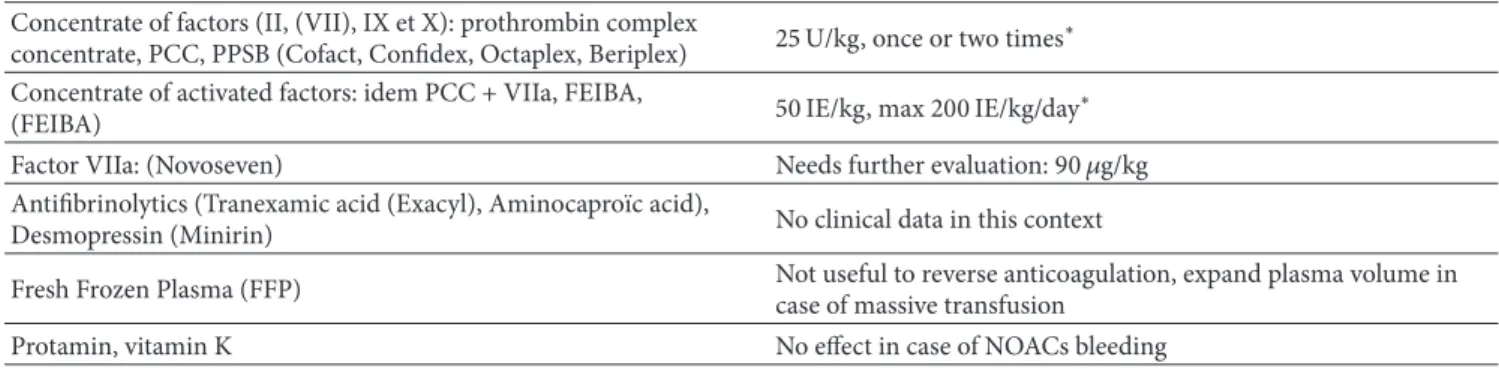
Table 9: Coagulation factor and pro-hemostatic agents. Concentrate of factors (II, (VII), IX et X): prothrombin complex
concentrate, PCC, PPSB (Cofact, Confidex, Octaplex, Beriplex) 25 U/kg, once or two times
∗
Concentrate of activated factors: idem PCC + VIIa, FEIBA,
(FEIBA) 50 IE/kg, max 200 IE/kg/day
∗
Factor VIIa: (Novoseven) Needs further evaluation: 90𝜇g/kg
Antifibrinolytics (Tranexamic acid (Exacyl), Aminocapro¨ıc acid),
Desmopressin (Minirin) No clinical data in this context
Fresh Frozen Plasma (FFP) Not useful to reverse anticoagulation, expand plasma volume incase of massive transfusion
Protamin, vitamin K No effect in case of NOACs bleeding
∗PCC or aPCC utilization is based of few experimental data, can be considered if immediate hemostatic support is essential in case of life-threating bleeding
(need for≥4 red cells transfusions and exogenous cathecolamins for hemodynamic stabilization).
special care to bleeding at the punctures sites is necessary and therefore ultrasound guided techniques are very useful. Hemoperfusion over a charcoal filter is under evaluation [89]. Oral activated charcoal may be effective only in case of a recent ingestion within 2 hours [89].
An analysis of major bleeding patients in the RE-LY study concludes that the overall resources required to manage bleeding were not greater than those after warfarin-related bleeding. For patients treated with DE, red cells transfusions were higher, plasma transfusions were lower, the stay in intensive care unit was shorter, and there was a lower trend in mortality compared with patients treated with warfarin. Based on these results, they concluded on a safety profile of DE [94].
There is currently no specific hemostatic agent for the reversal of NOACs in case of bleeding or in emergency situa-tions but different antidotes are under development. Andex-anet alfa (PRT064445) is a truncated form of enzymatically inactive FXa: it reverses dose-dependently the inhibitory activity and corrects ex vivo clotting times [95]. For dabi-gatran, a humanized selective and specific monoclonal anti-body fragment (idarucizumab) is under development [96]. Aripazine (PER977), another small synthetic molecule, reverses anticoagulant activity of all clinically used NOACs in rat bleeding models [97].
Hemostatic agents used for life-threatening bleeding are shown inTable 9.
7. Laboratory Testing for NOACs in
a Perioperative Setting
NOACs do not require routine coagulation monitoring. This point is considered as an advantage for the physicians and an improvement of quality of life for their patients. However, point measurement of NOACs may be required in several clinical situations including the perioperative setting [8,10, 11,78,98,99].
For VKAs, it is well established that, at time of surgery (INR on the day before surgery), an elevated INR (i.e.,≥2) will increase the risk of bleeding and a near normal or normal INR (i.e.,≤1.5) will not [60]. But, for NOACs, the residual drug level that can be considered as safe is not known, except
for dabigatran. The residual dabigatran concentration that is recommended before special intervention (such as surgery) is provided in the Committee for Medicinal Products for Human Use (CHMP) assessment report from the European Medicine Agency that states “(. . .) dabigatran concentration
under 48 ng/mL is equivalent to at least 75% of dabigatran’s elimination and should be recommended” [100]. A French group of experts called GIHP proposed the threshold of 30 ng/mL (for dabigatran and rivaroxaban) [88].
Details of these recommendations are presented in Table 10[88].
7.1. Which Laboratory Tests Should We Use in the Perioperative Setting and How to Interpret Them? Activated partial
throm-boplastin time (aPTT) and prothrombin time (PT) are global assays not reflecting plasma NOACs concentrations. They are not suitable for precise quantification of the anticoagulant effect. Thrombin time (TT) was demonstrated to be too sensitive towards dabigatran [89, 101]. However, this may guide the clinician in the perioperative setting since a nor-mal TT indicates no clinically relevant anticoagulant effect of dabigatran. The strong sensitivity of TT towards dabigatran leads to the development of a calibrated diluted thrombin time (dTT) with dabigatran standards to calculate the dabi-gatran plasma concentration. Several studies showed that the dTT or the ecarin chromogenic assay (ECA) highly correlates with dabigatran plasma concentrations measured by LC-MS/MS in patient’s plasma [102–104]. However, one limita-tion of dTT is that, in case of switching therapy (i.e., from heparins/heparinoids to dabigatran etexilate or from hirudin and derivatives to dabigatran etexilate), they will be slightly influenced by the presence of such inhibitors in the plasma. This implies the necessity of an accurate anamnesis of the drugs taken by the patient to avoid overestimation of drug concentrations in plasma. In addition, for the accurate determination of dabigatran plasma concentrations below 50 ng/mL, the more sensitive liquid chromatography-mass spectrometry/mass spectrometry (LC-MS/MS) method is still required [102,104].
For rivaroxaban, chromogenic anti-Xa assay has been proven to accurately estimate the plasma rivaroxaban con-centrations> 30 ng/mL [105]. Due to their good sensitivity
Table 10: Perioperative management of NOACs (dabigatran and rivaroxaban)—Proposal for recommendations from the GIHP (“Groupe d’Int´erˆet en H´emostase P´eriop´eratoire”).
Measured concentration Recommendations
<30 ng/mL Operate
30–200 ng/mL Wait up to 12 h and obtain new dosage or (if time is not compatible with emergency)Operate, if abnormal bleeding: antagonise the anticoagulant effect
200–400 ng/mL
Wait up to 12 h and obtain new dosage or (if time is not compatible with emergency) Maximise delay surgery
Discuss hemodialysis, especially if CrCl< 50 mL/min (with dabigatran only) Operate, if abnormal bleeding: antagonise
>400 ng/mL Overdose-Major haemorrhagic riskDiscuss haemodialysis before surgery (with dabigatran only)
Table 11: Influence of dabigatran, rivaroxaban and apixaban on coagulation tests used in the perioperative setting.
Dabigatran Rivaroxaban Apixaban
Prothrombin Time (PT) Time prolonged + (relative toreagent sensitivity) Time prolonged + to +++(relative to reagent sensitivity)
Time non-prolonged or prolonged + (relative to reagent sensitivity)
Activated Partial
Thromboplastin Time (aPTT)
Time prolonged + to +++ (relative to reagent sensitivity)
Time prolonged + (relative to reagent sensitivity)
Time prolonged + (relative to reagent sensitivity)
PT-based coagulation factors measurement
(II, VII, IX, X)
Slightly decreased (depending on the reagent)
Slightly decreased (depending on the reagent)
Slightly decreased (depending on the reagent)
APTT-based coagulation factors measurement (VIII, IX, XI)
Slightly decreased (depending on the reagent)
Slightly decreased (depending on the reagent)
Slightly decreased (depending on the reagent)
Fibrinogen No influence or slightly decrease
(depending on the reagent) No influence No influence
Thrombin Time Time prolonged +++++ No influence No influence
Anti-Xa based antithrombin
measurement No influence Increased: 10% per 100 ng/mL Increased: 10% per 100 ng/mL Anti-IIa based antithrombin
measurement Increased: 5–10% per 100 ng/mL No influence No influence
towards the inhibition of FXa by apixaban, chromogenic anti-Xa assays calibrated with specific apixaban calibrators should be performed to estimate plasma drug concentration [106–108]. However, taking into account the lower sensitivity of chromogenic assays compared to LC-MS/MS and the variability of coagulation analysers that may further increase the imprecision at the lowest concentrations, detection and quantitation of lower levels (<30 ng/mL for rivaroxaban and <15 ng/mL for apixaban) still require LC-MS/MS analyses [108,109].
Even if these specific coagulation tests to assess NOACs are promising, they suffer from difficulties to be implemented easily in the clinical setting. Moreover, assessment of lower levels of NOACs encountered in the perioperative setting is challenging due to the limit of quantitation of these tests but improvements in the low range are under development by several companies.
In addition, to correctly interpret the results of laboratory assay, some information needs to be collected: drug, indi-cation, and the timing of the last dose administration (due to the short half-life of NOACs) [110]. The interpretation of the results requires education of the front staff in many
specialties [98]. Finally, it has been clearly shown that NOACs influence the results of different coagulation assays used in the perioperative setting leading to misinterpretations [41] (Table 11).
7.2. A Particular Case: Atrial Fibrillation Ablation. The ideal
management of oral anticoagulation during catheter ablation for AF 112–114 is still controversial with a wide range of proce-dures available. During AF ablation, it is now recommended to achieve and maintain an ACT of 300 to 400 seconds in order to reduce the risk of systemic thromboembolism [111]. However, the ACT is affected by a lot of preanalytical [112] and analytical variables [113,114]. Finally, target ACT should be redetermined for the periprocedural use of NOACs for AF ablation [111–117].
8. Resumption of the NOACs after
Invasive Procedure or Surgery
Once again, in the postoperative period, the bleeding risk must be weighted with the thromboembolic risk; however the
risk for major bleeding exceeds the risk for thromboembolic complications after surgery [118]. Regarding the thromboem-bolic risk, Kaatz et al. [119] concluded that patients with chronic AF had twice as much risks of postoperative stroke, especially in neurosurgery and vascular surgery. The bleeding risk in the postoperative period is mainly due to patient’s characteristics (bridging therapy, mitral mechanic heart valve, active cancer, prior bleeding history, and reinitiation of heparin therapy within 24 hours after surgery), even if a premature heparin resumption is an avoidable risk factor [120]. But first of all surgical bleeding risk must be under control [121]. For NOACs, given the fact that full antico-agulation occurs between 2 and 4 hours (Table 3) and that no antidote is available, resumption of DE, rivaroxaban, and apixaban should be performed at least 48 hours after the high risk procedures [12]. This delay can expose patients at risk for thromboembolism. Twenty-four hours are recommended before resuming oral anticoagulation after a procedure at low risk of bleeding [60, 62]. Other schemes exist in order to minimize bleeding risks: consider a stepwise resumption of NOACs [16,77] or administer prophylactic doses of LMWH early after the surgery, and restart full doses of NOACs only after 3 or 4 days [122]. Heparin bridging can be useful if the patient cannot tolerate oral medication (e.g., in ileus or postoperative nausea and vomiting) [66]. In the postopera-tive setting, it is essential to reassess patient’s renal function, especially for elderly who are subject to dehydration and for patients taking DE. For LMWH, doses must be adapted in case of extreme body weight (body mass index above 30 kg/m2) and poor renal function. If CrCl is less than 30 mL/min, the use of unfractionated heparin is more indi-cated [12,123].
9. Conclusions
In the field of oral anticoagulation, clinicians will be more frequently confronted about their management of NOACs in the pre- and postoperative setting. Prerequisites are essential for NOAC’s pharmacokinetics, indications, drug interactions, and alterations of standard laboratory assays. Actual data are based mainly on expert’s opinions, except one national registry study. In some situation, interruption periods of NOACs are necessary and should be based on their respective half-life, on the bleeding risk of procedures, and on the thromboembolic risk of patients. Some scores such as CHA2DS2-VASc and HAS-BLED should help clinicians in their decisions. Given their shorter half-life, no heparin bridging during the interruption interval seems necessary, except for patients at high cardiovascular risk undergoing major surgery.
Further clinical trials over perioperative management of patients under NOACs are still required. Emergency surg-eries, invasive procedures, or bleeding patients remain a real challenge given the absence of antidote. Possibilities of rever-sal are poor and based on few experimental data and case reports. Furthermore, rapid quantitative measurements of the anticoagulant effect are not available in most institutions. Awaiting future clinical trial data, it seems to be important to
establish a hospital-wide policy for bleeding management in patients taking NOACs, in accordance to the available blood products and laboratory assays of each institution.
Conflict of Interests
The authors have no conflict of interests to disclose.
Authors’ Contribution
Anne-Sophie Dincq and Sarah Lessire contributed equally to this work.
References
[1] S. Husted, R. de Caterina, F. Andreotti et al., “Non-vitamin K antagonist oral anticoagulants (NOACs): no longer new or novel,” Thrombosis and Haemostasis, vol. 111, no. 5, pp. 781–782, 2014.
[2] European Medicine Agency, Xarelto: Summary of Product Characteristics, 2013,http://www.ema.europa.eu/docs/en GB/ document library/EPAR - Product Information/human/000944/ WC500057108.pdf.
[3] European Medicine Agency, “Pradaxa: Summary of Product Characteristics,” 2013,http://www.ema.europa.eu/docs/en GB/ document library/EPAR - Product Information/human/000829/ WC500041059.pdf.
[4] European Medicine Agency, Eliquis: Summary of Product Characteristics, 2014,http://www.ema.europa.eu/docs/en GB/ document library/EPAR - Product Information/human/002148/ WC500107728.pdf.
[5] Food and Drug Administration, “Pradaxa: Full Prescribing Information,” 2013,http://www.accessdata.fda.gov/drugsatfda docs/label/2013/022512s017lbl.pdf.
[6] Food and Drug Administration, Xarelto: Full Prescribing Information, 2014,http://www.accessdata.fda.gov/drugsatfda docs/label/2013/022406s004lbl.pdf.
[7] Food and Drug Administration—Eliquis: Full Prescribing Information, 2014,http://www.accessdata.fda.gov/drugsatfda docs/label/2013/022512s017lbl.pdf.
[8] J. H. Levy, D. Faraoni, J. L. Spring, J. D. Douketis, and C. M. Samama, “Managing new oral anticoagulants in the periopera-tive and intensive care unit setting,” Anesthesiology, vol. 118, no. 6, pp. 1466–1474, 2013.
[9] A. Helld´en, I. Odar-Cederl¨of, G. Nilsson et al., “Renal func-tion estimafunc-tions and dose recommendafunc-tions for dabigatran, gabapentin and valaciclovir: a data simulation study focused on the elderly,” BMJ Open, vol. 3, no. 4, Article ID e002686, 2013. [10] M. M. Samama, J. Amiral, C. Guinet, L. L. Flem, and J.
Segha-tchian, “Monitoring plasma levels of factor Xa inhibitors: how, why and when?” Expert Review of Hematology, vol. 6, no. 2, pp. 155–164, 2013.
[11] H. Ten Cate, “Monitoring new oral anticoagulants, managing thrombosis, or both?” Thrombosis and Haemostasis, vol. 107, no. 5, pp. 803–805, 2012.
[12] T. H. Baron, P. S. Kamath, and R. D. McBane, “Management of antithrombotic therapy in patients undergoing invasive proce-dures,” The New England Journal of Medicine, vol. 368, no. 22, pp. 2113–2124, 2013.
[13] P. Si´e, C. M. Samama, A. Godier et al., “Surgery and invasive procedures in patients on long-term treatment with direct oral anticoagulants: thrombin or factor-Xa inhibitors. Recommen-dations of the Working Group on perioperative haemostasis and the French Study Group on thrombosis and haemostasis,”
Archives of Cardiovascular Diseases, vol. 104, no. 12, pp. 669–676,
2011.
[14] J. Beyer-Westendorf, V. Gelbricht, K. Forster et al., “Peri-inter-ventional management of novel oral anticoagulants in daily care: results from the prospective Dresden NOAC registry,”
European Heart Journal, 2014.
[15] D. M. Siegal and M. A. Crowther, “Acute management of bleed-ing in patients on novel oral anticoagulants,” European Heart
Journal, vol. 34, no. 7, pp. 489–500, 2013.
[16] S. Schulman and M. A. Crowther, “How I treat with anticoag-ulants in 2012: new and old anticoaganticoag-ulants, and when and how to switch,” Blood, vol. 119, no. 13, pp. 3016–3023, 2012.
[17] J. L. Mega, E. Braunwald, S. D. Wiviott et al., “Rivaroxaban in patients with a recent acute coronary syndrome,” The New
England Journal of Medicine, vol. 366, no. 1, pp. 9–19, 2012.
[18] R. Bauersachs, S. D. Berkowitz, B. Brenner et al., “Oral rivaroxa-ban for symptomatic venous thromboembolism,” New England
Journal of Medicine, vol. 363, pp. 2499–2510, 2010.
[19] S. Schulman, A. K. Kakkar, S. Z. Goldhaber et al., “Treatment of acute venous thromboembolism with dabigatran or warfarin and pooled analysis,” Circulation, vol. 129, no. 7, pp. 764–772, 2014.
[20] C. B. Granger, J. H. Alexander, J. J. McMurray et al., “Apixaban versus warfarin in patients with atrial fibrillation,” The New
England Journal of Medicine, vol. 365, pp. 981–992, 2011.
[21] M. R. Patel, K. W. Mahaffey, J. Garg et al., “Rivaroxaban versus warfarin in nonvalvular atrial fibrillation,” The New England
Journal of Medicine, vol. 365, no. 10, pp. 883–891, 2011.
[22] S. J. Connolly, M. D. Ezekowitz, S. Yusuf et al., “Dabigatran versus warfarin in patients with atrial fibrillation,” The New
England Journal of Medicine, vol. 361, no. 27, pp. 1139–1151, 2009.
[23] M. R. Lassen, A. Gallus, G. E. Raskob, G. Pineo, D. Chen, and L. M. Ramirez, “Apixaban versus enoxaparin for thrombopro-phylaxis after hip replacement,” The New England Journal of
Medicine, vol. 363, no. 26, pp. 2487–2498, 2010.
[24] M. R. Lassen, G. E. Raskob, A. Gallus, G. Pineo, D. Chen, and P. Hornick, “Apixaban versus enoxaparin for thromboprophylaxis after knee replacement (ADVANCE-2): a randomised double-blind trial,” The Lancet, vol. 375, no. 9717, pp. 807–815, 2010. [25] B. I. Eriksson, O. E. Dahl, N. Rosencher et al., “Oral dabigatran
etexilate vs. subcutaneous enoxaparin for the prevention of venous thromboembolism after total knee replacement: the RE-MODEL randomized trial,” Journal of Thrombosis and
Haemostasis, vol. 5, no. 11, pp. 2178–2185, 2007.
[26] B. I. Eriksson, O. E. Dahl, M. H. Huo et al., “Oral dabigatran versus enoxaparin for thromboprophylaxis after primary total hip arthroplasty (RE-NOVATE II): a randomised, double-blind, non-inferiority trial,” Thrombosis and Haemostasis, vol. 105, no. 4, pp. 721–729, 2011.
[27] B. I. Eriksson, A. K. Kakkar, A. G. Turpie et al., “Oral rivarox-aban for the prevention of symptomatic venous thromboem-bolism after elective hip and knee replacement,” The Journal of
Bone and Joint Surgery, vol. 91, pp. 636–644, 2009.
[28] B. I. Eriksson, D. J. Quinlan, and J. I. Weitz, “Comparative pharmacodynamics and pharmacokinetics of oral direct throm-bin and factor Xa inhibitors in development,” Clinical
Pharma-cokinetics, vol. 48, no. 1, pp. 1–22, 2009.
[29] J. Stangier and A. Clemens, “Pharmacology, pharmacokinet-ics, and pharmacodynamics of dabigatran etexilate, an oral direct thrombin inhibitor,” Clinical and Applied Thrombosis/
Hemostasis, vol. 15, supplement 1, pp. 9S–16S, 2009.
[30] P. K. Maccallum, R. Mathur, S. A. Hull et al., “Patient safety and estimation of renal function in patients prescribed new oral anticoagulants for stroke prevention in atrial fibrillation: a cross-sectional study,” BMJ Open, vol. 3, Article ID e003343, 2013.
[31] D. W. Cockcroft and M. H. Gault, “Prediction of creatinine clearance from serum creatinine,” Nephron, vol. 16, no. 1, pp. 31– 41, 1976.
[32] R. Ferrandis, J. Castillo, J. de Andr´es et al., “The perioperative management of new direct oral anticoagulants: a question without answers,” Thrombosis and Haemostasis, vol. 110, no. 3, pp. 515–522, 2013.
[33] J. H. Levy, N. S. Key, and M. S. Azran, “Novel oral anticoagulants : implications in the perioperative setting,” Anesthesiology, vol. 113, no. 3, pp. 726–745, 2010.
[34] J. Stangier, K. Rathgen, H. St¨ahle, D. Gansser, and W. Roth, “The pharmacokinetics, pharmacodynamics and tolerability of dabigatran etexilate, a new oral direct thrombin inhibitor, in healthy male subjects,” British Journal of Clinical Pharmacology, vol. 64, no. 3, pp. 292–303, 2007.
[35] W. Mueck, J. Stampfuss, D. Kubitza, and M. Becka, “Clinical pharmacokinetic and pharmacodynamic profile of rivaroxa-ban,” Clinical Pharmacokinetics, vol. 53, no. 1, pp. 1–16, 2014. [36] J. D. Douketis, “Pharmacologic properties of the new oral
anticoagulants: a clinician-oriented review with a focus on perioperative management,” Current Pharmaceutical Design, vol. 16, no. 31, pp. 3436–3441, 2010.
[37] N. Raghavan, C. E. Frost, Z. Yu et al., “Apixaban metabolism and pharmacokinetics after oral administration to humans,” Drug
Metabolism and Disposition, vol. 37, no. 1, pp. 74–81, 2009.
[38] J. Stangier and A. Clemens, “Pharmacology, pharmacokinet-ics, and pharmacodynamics of dabigatran etexilate, an oral direct thrombin inhibitor,” Clinical and Applied
Thrombo-sis/Hemostasis, vol. 15, supplement 1, pp. 9S–16S, 2009.
[39] S. Lessire, A.-S. Dincq, J. Douxfils et al., “Preventive strategies against bleeding due to nonvitamin K antagonist oral antico-agulants,” BioMed Research International, vol. 2014, Article ID 616405, 14 pages, 2014.
[40] V. Pengo, L. Crippa, A. Falanga et al., “Questions and answers on the use of dabigatran and perpectives on the use of other new oral anticoagulants in patients with atrial fibrillation; a consen-sus document of the Italian federation of thrombosis centers (FCSA),” Thrombosis and Haemostasis, vol. 106, no. 5, pp. 868– 876, 2011.
[41] J. S. Healey, J. Eikelboom, J. Douketis et al., “Periprocedural bleeding and thromboembolic events with dabigatran com-pared with warfarin: results from the randomized evaluation of long-term anticoagulation therapy (RE-LY) randomized trial,”
Circulation, vol. 126, no. 3, pp. 343–348, 2012.
[42] M. W. Sherwood, J. D. Douketis, M. R. Patel et al., “Outcomes of temporary interruption of rivaroxaban compared with warfarin in patients with nonvalvular atrial fibrillation: results from the rivaroxaban once daily, oral, direct factor Xa inhibition com-pared with vitamin K antagonism for prevention of stroke and embolism trial in atrial fibrillation (ROCKET AF),” Circulation, vol. 129, pp. 1850–1859, 2014.
[43] A. C. Spyropoulos, R. M. Bauersachs, H. Omran, and M. Cohen, “Periprocedural bridging therapy in patients receiving chronic
oral anticoagulation therapy,” Current Medical Research and
Opinion, vol. 22, no. 6, pp. 1109–1122, 2006.
[44] P. A. Wolf, R. D. Abbott, and W. B. Kannel, “Atrial fibrillation as an independent risk factor for stroke: the Framingham study,”
Stroke, vol. 22, no. 8, pp. 983–988, 1991.
[45] A. J. Camm, G. Y. Lip, R. de Caterina et al., “2012 focused update of the ESC guidelines for the management of atrial fibrillation: an update of the 2010 ESC guidelines for the management of atrial fibrillation—developed with the special contribution of the European Heart Rhythm Association,” Europace, vol. 14, no. 10, pp. 1385–1413, 2012.
[46] G. Y. H. Lip, R. Nieuwlaat, R. Pisters, D. A. Lane, and H. J. G. M. Crijns, “Refining clinical risk stratification for predicting stroke and thromboembolism in atrial fibrillation using a novel risk factor-based approach: the Euro Heart Survey on atrial fibrillation,” Chest, vol. 137, no. 2, pp. 263–272, 2010.
[47] G. Y. H. Lip, F. Andreotti, L. Fauchier et al., “Bleeding risk assessment and management in atrial fibrillation patients: executive summary of a position document from the european heart rhythm association [EHRA], endorsed by the european society of cardiology [ESC] working group on thrombosis,”
Thrombosis and Haemostasis, vol. 106, no. 6, pp. 997–1011, 2011.
[48] I. G. E. Zarraga and J. Kron, “Oral anticoagulation in elderly adults with atrial fibrillation: integrating new options with old concepts,” Journal of the American Geriatrics Society, vol. 61, no. 1, pp. 143–150, 2013.
[49] R. D. Lopes, J. H. Alexander, S. M. Al-Khatib et al., “Apixaban for reduction in stroke and other ThromboemboLic events in atrial fibrillation (ARISTOTLE) trial: design and rationale,” The
American Heart Journal, vol. 159, no. 3, pp. 331–339, 2010.
[50] R. D. Lopes, S. M. Al-Khatib, L. Wallentin et al., “Efficacy and safety of apixaban compared with warfarin according to patient risk of stroke and of bleeding in atrial fibrillation: a secondary analysis of a randomised controlled trial,” The Lancet, vol. 380, no. 9855, pp. 1749–1758, 2012.
[51] C. Kearon, E. A. Akl, A. J. Comerota et al., “Antithrombotic ther-apy for VTE disease: antithrombotic therther-apy and prevention of thrombosis, 9th ed: American College of Chest Physicians evidence-based clinical practice guidelines,” Chest, vol. 141, no. 2, pp. e419S–e494S, 2012.
[52] P. G. de Jong, M. Coppens, and S. Middeldorp, “Duration of anticoagulant therapy for venous thromboembolism: balancing benefits and harms on the long term,” British Journal of
Haematology, vol. 158, no. 4, pp. 433–441, 2012.
[53] H. Gombotz and H. Knotzer, “Preoperative identification of patients with increased risk for perioperative bleeding,” Current
Opinion in Anaesthesiology, vol. 26, no. 1, pp. 82–90, 2013.
[54] R. Pisters, D. A. Lane, R. Nieuwlaat, C. B. De Vos, H. J. G. M. Crijns, and G. Y. H. Lip, “A novel user-friendly score (HAS-BLED) to assess 1-year risk of major bleeding in patients with atrial fibrillation: the euro heart survey,” Chest, vol. 138, no. 5, pp. 1093–1100, 2010.
[55] M. C. Fang, A. S. Go, Y. Chang et al., “A new risk scheme to predict warfarin-associated hemorrhage: the ATRIA (Antico-agulation and Risk Factors in Atrial Fibrillation) Study,” Journal
of the American College of Cardiology, vol. 58, no. 4, pp. 395–401,
2011.
[56] B. F. Gage, Y. Yan, P. E. Milligan et al., “Clinical classification schemes for predicting hemorrhage: results from the National Registry of Atrial Fibrillation (NRAF),” The American Heart
Journal, vol. 151, no. 3, pp. 713–719, 2006.
[57] S. Apostolakis, D. A. Lane, Y. Guo, H. Buller, and G. Y. H. Lip, “Performance of the HEMORR 2HAGES, ATRIA, and HAS-BLED bleeding risk-prediction scores in patients with atrial fibrillation undergoing anticoagulation: the AMADEUS (Eval-uating the use of SR34006 compared to warfarin or aceno-coumarol in patients with atrial fibrillation) study,” Journal of
the American College of Cardiology, vol. 60, no. 9, pp. 861–867,
2012.
[58] S. Apostolakis, D. A. Lane, Y. Guo, H. Buller, and G. Y. H. Lip, “Performance of the HEMORR2HAGES, ATRIA, and HAS-BLED bleeding risk-prediction scores in nonwarfarin antico-agulated atrial fibrillation patients,” Journal of the American
College of Cardiology, vol. 61, no. 3, pp. 386–387, 2013.
[59] J. A. Nieto, R. Solano, N. Trapero Iglesias et al., “Validation of a score for predicting fatal bleeding in patients receiving antico-agulation for venous thromboembolism,” Thrombosis Research, vol. 132, pp. 175–179, 2013.
[60] J. D. Douketis, A. C. Spyropoulos, F. A. Spencer et al., “Periop-erative management of antithrombotic therapy: antithrombotic Therapy and Prevention of Thrombosis, 9th ed: American College of Chest Physicians Evidence-Based Clinical Practice Guidelines,” Chest, vol. 141, pp. e326S–e350S, 2012.
[61] A. Vuylsteke, C. Pagel, C. Gerrard et al., “The Papworth Bleeding Risk Score: a stratification scheme for identifying cardiac surgery patients at risk of excessive early postoperative bleed-ing,” European Journal of Cardio-thoracic Surgery, vol. 39, no. 6, pp. 924–930, 2011.
[62] A. C. Spyropoulos and J. D. Douketis, “How I treat anticoag-ulated patients undergoing an elective procedure or surgery,”
Blood, vol. 120, no. 15, pp. 2954–2962, 2012.
[63] H. Heidbuchel, P. Verhamme, M. Alings et al., “European Heart Rhythm Association Practical Guide on the use of new oral anticoagulants in patients with non-valvular atrial fibrillation. Europace: European pacing, arrhythmias, and cardiac electro-physiology,” Europace, vol. 15, pp. 625–651, 2013.
[64] A. Liew and J. Douketis, “Perioperative management of patients who are receiving a novel oral anticoagulant,” Internal and
Emergency Medicine, vol. 8, pp. 477–484, 2013.
[65] J. D. Douketis, “Perioperative anticoagulation management in patients who are receiving oral anticoagulant therapy: a practi-cal guide for clinicians,” Thrombosis Research, vol. 108, no. 1, pp. 3–13, 2002.
[66] J. D. Douketis, P. B. Berger, A. S. Dunn et al., “The perioperative management of antithrombotic therapy: American College of Chest Physicians evidence-based clinical practice guidelines (8th edition),” Chest, vol. 133, no. 6, pp. 299S–339S, 2008. [67] M. Fujita, A. Shiotani, T. Murao et al., “Safety of gastrointestinal
endoscopic biopsy in patients taking antithrombotics,” Digestive
Endoscopy, 2014.
[68] J. I. Weitz, D. J. Quinlan, and J. W. Eikelboom, “Periprocedural management and approach to bleeding in patients taking dabigatran,” Circulation, vol. 126, no. 20, pp. 2428–2432, 2012. [69] D. J. Greenblatt, “Elimination half-life of drugs: value and
limi-tations,” Annual Review of Medicine, vol. 36, pp. 421–427, 1985. [70] J. Graff and S. Harder, “Anticoagulant therapy with the oral
direct factor Xa inhibitors rivaroxaban, apixaban and edoxaban and the thrombin inhibitor dabigatran etexilate in patients with hepatic impairment,” Clinical Pharmacokinetics, vol. 52, no. 4, pp. 243–254, 2013.
[71] D. Siegal, J. Yudin, S. Kaatz, J. D. Douketis, W. Lim, and A. C. Spyropoulos, “Periprocedural heparin bridging in patients