HAL Id: tel-02897826
https://tel.archives-ouvertes.fr/tel-02897826
Submitted on 13 Jul 2020HAL is a multi-disciplinary open access archive for the deposit and dissemination of sci-entific research documents, whether they are pub-lished or not. The documents may come from teaching and research institutions in France or abroad, or from public or private research centers.
L’archive ouverte pluridisciplinaire HAL, est destinée au dépôt et à la diffusion de documents scientifiques de niveau recherche, publiés ou non, émanant des établissements d’enseignement et de recherche français ou étrangers, des laboratoires publics ou privés.
Zhimin Song
To cite this version:
Zhimin Song. The Role of Phosphoinositides in the Regulation of NADPH Oxidase Activation in Neutrophils. Molecular biology. Université Paris Saclay (COmUE), 2017. English. �NNT : 2017SACLS152�. �tel-02897826�
NNT : 2017SACLS152
T
HÈSE DE DOCTORAT
DE
L’U
NIVERSITÉ
P
ARIS
-S
ACLAY
PRÉPARÉE A
L’U
NIVERSITÉ
P
ARIS
-S
UD
É
COLED
OCTORALE N°
568
S
IGNALISATION ET RÉSEAUX INTÉGRATIFS EN BIOLOGIES
PÉCIALITÉ DE DOCTORAT:
ASPECTS MOLÉCULAIRES ET CELLULAIRES DE LA BIOLOGIEPar
M.
ZHIMIN
SONG
THE
ROLE
OF
PHOSPHOINOSITIDES
IN
THE
REGULATION
OF
NADPH
OXIDASE
ACTIVATION
IN
NEUTROPHILS
Thèse présentée et soutenue à Orsay, le 12 Juillet 2017 : Composition du Jury :
Mme CUIF-LORDEZ Marie-hélène, Professeure, Université Paris-Sud, Présidente du Jury M. ZAHRAOUI Ahmed, Directeur de Recherche, Institut Cochin, Rapporteur
Mme BRÉCHARD Sabrina, Chargé de Recherche, Université du Luxembourg, Rapporteuse Mme JACKSON Catherine, Directrice de Recherche, Institut Jacques Monod, Examinatrice Mme BACIOU Laura, Directrice de Recherche, Université Paris-Sud, Examinatrice
I would like to express my deepest gratitude to my advisor Dr. DUPRÉ Sophie for the continuous support of my Ph.D. study and related research. Importantly, I thank her for her excellent guidance, immense knowledge, caring, patience, and providing me with an excellent atmosphere for doing research. Her guidance helped me in all the time of research and writing of this thesis.
I would like to thank the members of my committee, Dr. ZAHRAOUI Ahmed, Dr. BRÉCHARD Sabrina, Dr. CUIF-LORDEZ Marie-Hélène, Dr. JACKSON Catherine, and Dr. BACIOU Laura for their advice and helpful comments on my research work.
I greatly appreciate Dr. NÜßE Oliver for his continuous support, sharing of ideas and knowledge, and guidance for the experiments and writing. I would like to thank other members in the phagocyte lab for their kind help and support: HUDIK Elodie, BOUCHAB Leïla and JOLY Jérémy. I had a wonderful time working with all the members in the Phagocyte Lab.
I wish to express my appreciation to all the colleagues in the Inserm U1174 for their knowledge and friendship, in particular Dr. NG-BONAVENTURE Kim, GROSSE Brigitte, LAPIERRE Claudine, Dr. BOUCHERIE Sylviane, PRIGENT Sylvie, DOIGNON Isabelle, GARCIN Isabelle, COLLADO-HILLY Mauricette, FAYOL Olivier, and Dr. COMBETTES Laurent. I would like to thank all the members in LCP for their kindness and assistance, especially Dr. ERARD Marie, Dr. LEDERER Florence, Dr. BIZOUARN Tania and Dr. BRUN Emilie.
I would like to thank the members in I2BC (Platform Imagif) who have always been instrumental and supportive, in particular LE BARS Romain, BESSE Laëtitia, and BOURGE Mickael.
I would like to thank all the members in the group of Dr. EL BENNA Jamel, in particular DANG Pham My-Chan, for their kind assistance for blood experiment.
I would like to express my appreciation to all of my friends for the friendship and support: SONG Yuxiang, CAO Jing, SAFYA Hanna, ZIEGLER Cornelia, MELLOUK Amine, VILLEMAIN Laure, LE GUILCHER Camille, and all the friends from the “La Pacaterie” group.
I am so grateful to CSC (Chinese Scholarship Council) and Université Paris-Sud for making it possible for me to study in France.
Finally, I am continually thankful for the love and support of my parents (SONG Baolin and CAO Fenghua), my brother (SONG Zhiguo), my sister (SONG Qingzhen), and my girlfriend (YANG Xiuna).
TABLE OF CONTENTS
TABLE OF CONTENTS ... 1
LIST OF TABLES ... 5
LIST OF FIGURES ... 6
ABBREVIATIONS ... 9
CHAPTER ONE: INTRODUCTION ... 13
1.1 NEUTROPHIL BIOLOGY ... 13
1.1.1 The immune system ... 13
1.1.2 Neutrophil activation ... 15 1.1.2.1 Neutrophil production ... 15 1.1.2.2 Neutrophil recruitment ... 17 1.1.2.2.1 Rolling ... 17 1.1.2.2.2 Firm adhesion ... 19 1.1.2.2.3 Tissue neutrophils ... 19 1.1.2.3 Neutrophil receptors ... 20 1.1.2.3.1 G-protein-coupled receptors ... 22 1.1.2.3.2 Fc-receptors (FcRs) ... 24
1.1.2.3.3 Neutrophil adhesion receptors ... 26
1.1.2.3.4 Cytokine receptors ... 26
1.1.2.3.5 Innate immune receptors ... 27
1.1.3 Anti-microbial function of neutrophils ... 28
1.1.3.1 Granules ... 28 1.1.3.2 Degranulation ... 30 1.1.3.3 Antimicrobial Proteins ... 30 1.1.3.4 Phagocytosis ... 31 1.1.3.4.1 Non-opsonic phagocytosis ... 31 1.1.3.4.2 Opsonic-phagocytosis ... 31
1.1.3.5 Neutrophil extracellular traps ... 32
1.1.4 Neutrophil and extracellular matrix ... 34
2
1.1.4.2 Fibrinogen ... 36
1.1.4.3 Integrins ... 38
1.1.4.3.1 Integrin structures ... 39
1.1.4.3.2 Neutrophil integrins ... 41
1.2 REACTIVE OXYGEN SPECIES ... 44
1.2.1 NADPH oxidase ... 44
1.2.1.1 gp91phox ... 44
1.2.1.2 p22phox ... 45
1.2.1.3 The GTPase Rac ... 49
1.2.1.4 p40phox ... 51
1.2.1.5 p47phox ... 51
1.2.1.6 p67phox ... 55
1.2.1.7 NADPH oxidase assembly ... 55
1.2.2 ROS and immunity ... 58
1.2.2.1 Reactive oxygen species ... 58
1.2.2.1 Chronic granulomatous disease ... 60
1.2.2.2 Microbial antioxidant defense ... 60
1.2.2.3 Overproduction of ROS by NADPH oxidase ... 61
1.2.3 ROS and signaling ... 61
1.2.3.1 ROS signalling ... 61
1.2.3.2 Signalling and regulation of NADPH oxidase activation ... 63
1.2.4 ROS and adhesion ... 63
1.2.4.1 ROS and ECM ... 63
1.2.4.1.1 ECM regulates ROS production ... 63
1.2.4.1.2 ROS influence ECM production and deposition ... 64
1.2.4.2 ROS and integrins ... 64
1.3 PHOSPHOINOSITIDES ... 66 1.3.1 Phosphoinositide kinases ... 69 1.3.1.1 Class I PI3Ks ... 72 1.3.1.1.1 PI3Kα ... 72 1.3.1.1.2 PI3Kβ ... 73 1.3.1.1.3 PI3Kδ ... 75 1.3.1.1.4 PI3Kγ ... 75 1.3.1.2 Class II PI3K ... 77
3
1.3.1.3 Class III PI3K ... 77
1.3.1.3.1 Vps34 complex ... 79 1.3.1.3.2 Rubicon ... 79 1.3.2 Phosphoinositide phosphatases ... 82 1.3.2.1 5-phosphatases ... 82 1.3.2.2 4-phosphatases ... 82 1.3.2.3 3-phosphatases ... 83 1.3.2.3.1 PTEN ... 83 1.3.2.3.2 Myotubularins ... 84 1.3.3 Phosphoinositides signalling ... 86
1.3.3.1 Phosphoinositides and NADPH oxidase ... 86
1.3.3.1.1 PX domain of p40phox ... 86
1.3.3.1.2 PX domain of p47phox ... 89
1.3.3.2 Phosphoinositides during phagocytosis ... 91
1.3.3.3 Phosphoinositides during adhesion ... 93
1.4 RESEARCH GOALS ... 94
CHAPTER TWO: PHOSPHOINOSITOL 3-PHOSPHATE ACTS AS A TIMER FOR REACTIVE OXYGEN SPECIES PRODUCTION IN THE PHAGOSOME ... 95
ABSTRACT ... 97
INTRODUCTION ... 98
MATERIALS AND METHODS ... 101
RESULTS ... 109
DISCUSSION ... 135
REFERENCES ... 140
CHAPTER THREE: CLASS I PI3K IS REQUIRED FOR SUSTAINING BETA2-INTEGRIN DEPENDENT ROS PRODUCTION AT NEUTROPHIL PLASMA MEMBRANE ... 146
ABSTRACT ... 146
INTRODUCTION ... 147
MATERIALS AND METHODS ... 149
RESULTS ... 154
4
REFERENCES ... 180
CHAPTER FOUR: GENERAL DISCUSSION AND PERSPECTIVES ... 187
4.1 ROS PRODUCTION IN THE PHAGOSOME ... 187
4.1.1 Not all phagosomes produce ROS ... 187
4.1.2 Phagosome, endosome and generation of PI3P ... 188
4.1.3 Downregulation of the PI3P and the NADPH oxidase ... 189
4.2 ROS PRODUCTION IN ADHERENT NEUTROPHILS ... 189
4.2.1 Neutrophils and biomaterials ... 189
4.2.2 What is the function of the integrin mediated ROS? ... 190
4.3 MODULATION OF THE PHOSPHOINOSITIDES TO REGULATE NADPH OXIDASE ... 192
4.3.1 Pharmacological tools ... 192
4.3.2 Molecular tools ... 195
4.4 REGULATION OF NADPH OXIDASE AT PHAGOSOMAL VERSUS PLASMA MEMBRANE ... 196
5
LIST OF TABLES
TABLE 1. NEUTROPHIL RECEPTORS ... 21
TABLE 2. GRANULE PROTEINS. ... 29
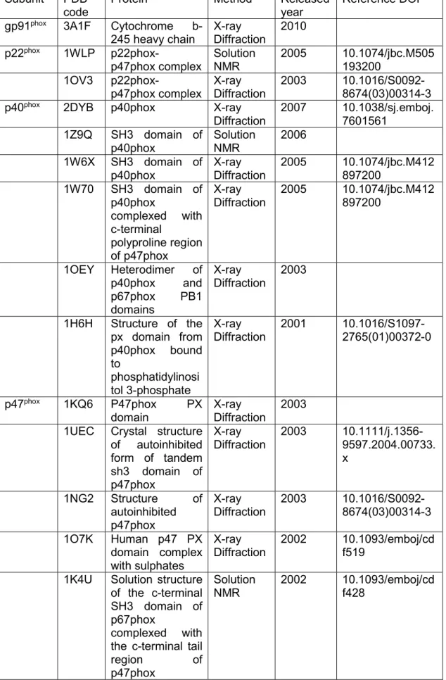
TABLE 3. PDB ENTRIES FOR STRUCTURE OF THE NADPH OXIDASE SUBUNITS. ... 47
TABLE 4. PPIS AMOUNT AND DISTRIBUTION IN CELLS. ... 68
TABLE 5. NOMENCLATURE OF THE PHOSPHOINOSITIDE KINASES. ... 70
TABLE 6. PHOSPHOINOSITIDES BINDING DOMAINS. ... 87
6
LIST OF FIGURES
CHAPTER ONE
FIGURE 1. INTEGRATED HUMAN IMMUNE SYSTEM. ... 14
FIGURE 2. DIFFERENCIATION OF THE HEMATOPOIETIC LINEAGES. ... 16
FIGURE 3. NEUTROPHILS RECRUITMENT TO THE SITES OF INFLAMMATION. ... 18
FIGURE 4. GPCR SIGNALING IN NEUTROPHILS. ... 23
FIGURE 5. NEUTROPHIL FC-RECEPTORS. ... 25
FIGURE 6. BACTERIA CAUGHT IN NETS. ... 33
FIGURE 7. SCHEMATIC REPRESENTATION OF THE NETOSIS PATHWAY. ... 34
FIGURE 8. TWO MAIN TYPES OF ECM. ... 35
FIGURE 9. SCHEMATIC OF THE DOMAIN STRUCTURE OF FIBRINOGEN. ... 37
FIGURE 10. PULL OUT HYPOTHESIS. ... 37
FIGURE 11. REPRESENTATION OF THE INTEGRIN FAMILY. ... 38
FIGURE 12. INTEGRIN STRUCTURE. ... 40
FIGURE 13. INSIDE-OUT AND OUTSIDE-IN SIGNALLING OF Β2-INTEGRINS. ... 43
FIGURE 14. MODEL OF THE CYTOCHROME B558 ... 46
FIGURE 15. MODE OF ACTIVATION OF THE RAC GTPASES. ... 50
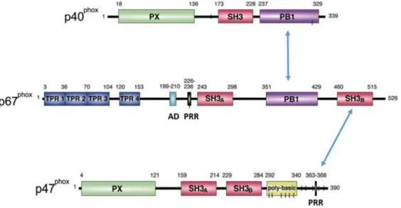
FIGURE 16. DOMAIN STRUCTURE OF THE CYTOSOLIC SUBUNITS P40PHOX, P47PHOX AND P67PHOX... 52
FIGURE 17. CONFORMATIONAL AND DOMAINS INTERACTIONS CHANGES AFTER P47PHOX ACTIVATION. ... 54
FIGURE 18. MODEL OF NADPH OXIDASE ASSEMBLY DURING PHAGOCYTOSIS. ... 57
FIGURE 19. ROS PRODUCTION DURING PHAGOCYTOSIS. ... 59
FIGURE 20. STRUCTURE AND METABOLISM OF PHOSPHOINOSITIDES. ... 67
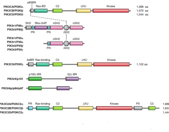
FIGURE 21. DOMAIN STRUCTURE OF CLASS I AND CLASS II PI3KS. ... 74
FIGURE 22. CLASS I PI3KS SIGNALLING. ... 76
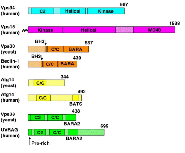
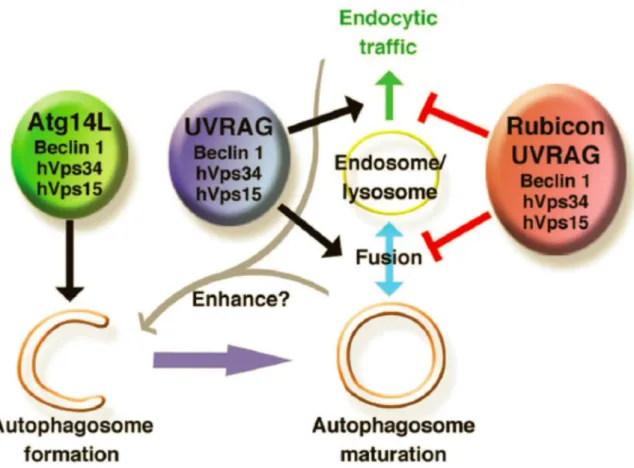
FIGURE 23. PROTEIN DOMAINS OF THE CORE COMPONENTS OF VPS34 COMPLEX I AND II.78 FIGURE 24. THE DIFFERENT DOMAINS OF THE PROTEIN: RUBICON... 80
FIGURE 25. MODEL OF THE ROLE OF THE THREE BECLIN 1-VPS34 COMPLEXES. ... 81
7
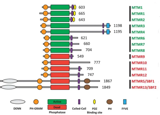
FIGURE 27. THE PROTEIN DOMAINS OF MYOTUBULARINS. ... 85
FIGURE 28. PI3P BOUND TO P40PHOX PX DOMAIN. ... 88
FIGURE 29. THE STRUCTURE OF P47PHOX PX DOMAIN... 89
FIGURE 30. BINDING POCKET OF P47PHOX PX DOMAIN. ... 90
FIGURE 31. PHOSPHOINOSITIDES DURING PHAGOCYTOSIS. ... 92
CHAPTER TWO FIGURE 1. VPS34-IN1 INHIBITS HVPS34 BUT NOT CLASS I PI3K. ... 112
FIGURE 2. PI(3)P INHIBITORS DECREASE ROS PRODUCTION. ... 113
FIGURE 3. EFFECTS OF WORTMANNIN ON PHAGOSOMAL ACCUMULATION OF MCHERRY-PXP40PHOX, P67PHOX-CITRINE AND CITRINE-P40PHOX. ... 116
FIGURE 4. KNOCKDOWN OF P40PHOX OR P67PHOX DECREASES THE ROS PRODUCTION AT THE PHAGOSOME. ... 120
FIGURE 5. GENERATION OF A STABLE PLB-CITRINE-P40PHOX CELL LINE. ... 120
FIGURE 6. ACCUMULATION OF CITRINE-P40PHOX AND P67PHOX-CITRINE AT THE PHAGOSOME. ... 121
FIGURE 7. RECRUITMENT OF MTM1 AND RUBICON DURING PHAGOCYTOSIS OF OPSONIZED ZYMOSAN. ... 124
FIGURE 8. KNOCKDOWN OF MTM1 AND RUBICON IN PLB CELLS. ... 125
FIGURE 9. KNOCKDOWN OF MTM1 AND/OR RUBICON INCREASE THE PI3P LEVEL AT THE PHAGOSOME. ... 127
FIGURE 10. KNOCKDOWN OF MTM1 OR/AND RUBICON INCREASES ROS PRODUCTION AND EXTENDS THE TIME OF PRESENCE OF P67PHOX AT THE PHAGOSOME. ... 130
FIGURE 11. MTM1 OVEREXPRESSION AT THE PHAGOSOMAL MEMBRANE PREVENTS YFP-P40PHOXPX RECRUITMENT. ... 133
FIGURE 12. MTM1 OVEREXPRESSION AT THE PHAGOSOMAL MEMBRANE PREVENTS ROS PRODUCTION AND THE ACCUMULATION OF P67PHOX ... 134
CHAPTER THREE FIGURE 1. DPI INHIBITS THE ROS PRODUCTION BY DIFFERENTIATED PLB CELLS IN RESPONSE TO FIBRINOGEN. ... 155
FIGURE 2. FMLF DOES NOT CHANGE THE FIBRINOGEN INDUCED ROS PRODUCTION BY DIFFERENTIATED PLB CELLS. ... 156 FIGURE 3. LY294002 INHIBITS THE FIBRINOGEN INDUCED ROS PRODUCTION IN
8
DIFFERENTIATED PLB CELLS. ... 157 FIGURE 4. LY294002 INHIBITS THE FIBRINOGEN INDUCED ROS PRODUCTION IN HUMAN NEUTROPHILS... 158 FIGURE 5. FMLF ENHANCES THE FIBRINOGEN DEPENDENT ROS PRODUCTION. ... 158 FIGURE 6. DISTRIBUTION OF THE CYTOSOLIC NOX SUBUNITS IN DIFFERENTIATED PLB CELLS ADHERENT TO FIBRINOGEN. ... 160 FIGURE 7. DISTRIBUTION OF THE CYTOSOLIC NOX SUBUNITS IN HUMAN NEUTROPHILS PLATED ON FIBRINOGEN AND STIMULATED WITH FMLF. ... 161 FIGURE 8. DPI TRIGGERS THE RELEASE OF P47PHOX-GFP FROM THE PLASMA MEMBRANE IN DIFFERENTIATED PLB P47PHOX-GFP CELLS PLATED ON FIBRINOGEN... 163 FIGURE 9. LY294002 RELEASES P47PHOX-GFP FROM THE PLASMA MEMBRANE OF
DIFFERENTIATED PLB P47PHOX-GFP CELLS PLATED ON FIBRINOGEN... 164 FIGURE 10. LY294002 RELEASES P67PHOX-CITRINE FROM THE PLASMA MEMBRANE OF DIFFERENTIATED PLB P67PHOX-CITRINE CELLS PLATED ON FIBRINOGEN. ... 165 FIGURE 11. LY294002 RELEASES P40PHOX-CITRINE FROM THE PLASMA MEMBRANE OF DIFFERENTIATED PLB P40PHOX-CITRINE CELLS PLATED ON FIBRINOGEN. ... 166 FIGURE 12. LY294002 ADDITION INDUCES A DECREASE IN P47PHOX AND P67PHOX PLASMA MEMBRANE FLUORESCENCE OF DIFFERENTIATED PLB CELLS. ... 167 FIGURE 13. RAC1G12V CAN NOT PREVENT THE DROP OF ROS PRODUCTION AND THE RELEASE OF P47PHOX AT THE PLASMA MEMBRANE INDUCED BY LY294002 IN NEUTROPHIL-LIKE CELLS PLATED ON FIBRINOGEN. ... 169 FIGURE 14. GENERATION OF THE STABLE PLB P47PHOX(R43Q)-GFP CELL LINE AND THE PLB P47PHOX-IRFP CELL LINE. ... 172 FIGURE 15. DIFFERENTIATED PLB P47PHOX(R43Q)-GFP CELLS DOES NOT PRODUCE ROS IN RESPONSE TO FIBRINOGEN. ... 174 CHAPTER FOUR
FIGURE 32. MODEL OF NADPH OXIDASE ASSEMBLY AND PI3P INVOLVEMENT DURING PHAGOCYTOSIS ... 198 FIGURE 33. PATHWAYS IN SUSTAINING THE INTEGRIN-MEDIATED ROS PRODUCTION ... 200
9
ABBREVIATIONS
AD Activation domain ADMIDAS site adjacent to MIDAS AIR Auto-inhibitory region AML Acute myeloid leukemia
ANCA Anti-neutrophil cytoplasmic antibody bTD b tail domain
CDG Chronic granulomatous disease CG Cathepsin G
CIB Calcium-and integrin-binding protein CLP Common lymphoid progenitor
CLR C-type lectin receptor
CMP Common myeloid progenitor CR3 Complement receptor 3 DAG Diacyl glycerol
DAMP Damage associated molecular pattern DAP12 DNAX activation protein of 12 kDa DC Dendritic cell
ECM Extracellular matrix EGF Epidermal growth factor ER Endoplasmic reticulum ESL-1 E-selectin ligand 1 FAK Focal adhesion-kinase FcR Fc receptor
FPR Formyl peptide receptor GAP GTPase activating protein
G-CSF Granulocyte colony stimulating factor GDI Guanine nucleotide dissociation inhibitor GEF Guanine nucleotide exchange factor
GM-CSF Granulocyte macrophage colony-stimulating factor GMP granulocyte-macrophage progenitor
GPCR G-protein-coupled receptor HSC Hematopoietic stem cell
10
ICAM Intercellular adhesion molecule
ICAP-1 Cytoplasmic domain associated protein-1 ILK Integrin-linked kinase
ITAM Immunoreceptor tyrosine-based activation motif ITIM Immunoreceptor tyrosine-based inhibitory motif JAK Janus kinase
JNK c-Jun N-terminal kinase LAD Leukocyte adhesion deficiency
LAF-1 Lymphocyte function-associated antigen 1 LPS Lipopolysaccharide
LTB4 Leukotriene B4 Mac-1 Macrophage-1 antigen MBL Mannan-binding lectin
MEP Megakaryocyte-erythroid progenitor MIDAS Metal-ion-dependent adhesive site MPO Myeloperoxidase
MTM Myotubularin
NCF Neutrophil cytosol factor NE Neutrophil elastase
NETs Neutrophil extracellular traps NF-κB nuclear factor κB
NGAL Neutrophil gelatinase-associated lipocalin
NLR Nucleotide-binding oligomerization domain-like receptor PA Phosphatidic acid
PAMP Pathogen associated molecular pattern PB1 Phox and Bem1
PBR Polybasic region
PDGF Platelet derived growth factor PI3P Phosphatidylinositol 3-phosphate PI(3,4)P2 Phosphatidylinositol (3,4)-biphosphate
PI(4,5)P2 Phosphatidylinositol (4,5)-bisphosphate
PI(3,4,5)P3 Phosphatidylinositol (3,4,5)-trisphosphate
11
PI4K Phosphoinositide 4-kinase
PI4P Phosphatidylinositol 4-phosphate PITP PtdIns transfer protein
PKC Protein kinase C PLCγ Phospholipase Cγ PR3 Proteinase 3
PRR Pathogen recognition receptor PRR Proline-rich region PS Phosphatidylserine PSGL-1 P-selectin ligand 1 PSI Plexin/semaphorin/intergrin PSM Phenol-soluble modulin PTB Phosphotyrosine-binding
PTEN Phosphatase and tensin homolog PTP Protein tyrosine phosphatase PX Phox homology
Rack1 Receptor for activated C kinase 1 RIAM Rap-1-mediated adaptor molecule RLR RIG-I-like receptor
ROCK Rho-associated protein kinase ROS Reactive oxygen species
Rubicon (RUN domain protein as Beclin-1 interacting and cysteine-rich containing SH3 Src homology3
SLE Systemic lupus erythematosus
SNARE Soluble N-ethylmaleimide sensitive factor or N-ethylmaleimide sensitive fusion protein t-SNARE target-membrane-specific SNARE
v-SNARE vesicle-membrane-specific SNARE SOCS Suppressor of cytokine signaling SOD Superoxide dismutase
STAT Signal transducers and activators of transcription Syk spleen tyrosine kinase
TLR Toll-like receptor TNF-α tumor necrosis factor-α
12
TPR Tetratricopeptide repeat
VAMP2 Vesicle- associated membrane protein-2 VCAM-1 Vascular cell adhesion protein 1
13
CHAPTER ONE: Introduction
1.1 Neutrophil biology
1.1.1 The immune system
The human anti-microbial defense system includes 3 different levels: (1) anatomic and physiologic barriers; (2) innate immunity; and (3) adaptive immunity (Figure 1). Failure in any of these systems will greatly increase susceptibility to infection (Turvey and Broide, 2010). The innate immune system first appeared 750 million years ago and has been notably conserved throughout the evolutionary tree of life. The innate immune system is the first line of defense against infection or self-damaged tissue injury (Hato and Dagher, 2014). Both innate and adaptive immune responses depend upon the activities of leukocytes (white blood cells) (Murphy, 2012). Neutrophils take up for 50% to 70% of all circulating leukocytes in humans (Mayadas et al., 2014). Neutrophils are highly motile and respond to a wide range of stimuli, especially pathogen- and damage-associated molecular patterns (PAMPs and DAMPs) (Jones et al., 2016). Researchers first believed that neutrophils were playing a passive role functioning only as pathogen killers. This view is not more prevailing since recent data suggest that activated neutrophils are able to also perform most of macrophages functions. Neutrophils can communicate with macrophages, dendritic cells (DCs) and cells of the adaptive immune response through direct cell-cell contacts or soluble mediators (Mayadas et al., 2014; Wright et al., 2010).
14
Figure 1. Integrated human immune system.
The human microbial defense system consists of 3 levels: (1) Anatomic and physiologic barriers. These barriers include intact skin, vigorous mucociliary clearance mechanisms, low stomach pH, and bacteriolytic lysozyme in tears, saliva, and other secretions. (2) Innate immunity. Hematopoietic cells including macrophages, dendritic cells, mast cells, neutrophils, eosinophils, natural killer (NK) cells, and NK T cells are involved in innate immunity. The skin and the epithelial cells lining the respiratory, gastrointestinal, and genitourinary tracts are also involved in innate immunity. Innate immunity augments the protection offered by anatomic and physiologic barriers and plays a central role in activating the subsequent adaptive immune response. (3) Adaptive immunity. T and B lymphocytes are the main self-defensive weapons of the adaptive immune system. Some elements are difficult to categorize, e.g. NK T cells and dendritic cells are classified as being on the cusp of innate and adaptive immunity rather than being firmly in one side (Turvey and Broide, 2010).
15
1.1.2 Neutrophil activation
1.1.2.1 Neutrophil production
Neutrophils come from hematopoietic stem cells (HSCs) (Figure 2). HSCs reside in bone marrow and produce all blood cells. HSCs localize to niches provided by osteoblasts, where there is little blood flow and low oxygen tension. The more mature and actively dividing stem cells localize close to the abluminal side of the sinusoids, which is a special vascular structure of the bone marrow (Borregaard, 2010; Winkler et al., 2010). Neutrophils are generated at a rate of 1 to 2x10¹¹ per day in a normal adult human. Granulocyte colony stimulating factor
(G-CSF) and granulocyte macrophage colony-stimulating factor (GM-CSF) are essential to trigger neutrophil production during infections. However, they are not absolutely required. Indeed mice lacking one or both of these cytokines still have approximately 20% of the normal level of fully mature neutrophils (Borregaard, 2010; Hibbs et al., 2007; Mayadas et al., 2014). Mature neutrophils leave the bone marrow and enter the circulation.
16
Figure 2. Differentiation of the hematopoietic lineages.
The long-term hematopoietic stem cell (LT-HSC) gives rise to short-term hematopoietic stem cells (ST-HSCs) that then give rise to the multipotent common myeloid progenitor (CMP) and common lymphoid progenitor (CLP). The CMP are the precursor of the megakaryocyte-erythroid progenitors (MEPs) and granulocyte-macrophage progenitors (GMPs). The maturation of lineage-committed erythroid progenitors is shown on the left side. MEPs give rise to erythrocytes, megakaryocytes and platelets. GMP give rise to eosinophils, basophils, neutrophils and monocytes/macrophages.CLP are the precursors of the B cells, T cells and Natural Killer (NK) cells. Adapted from (Sankaran and Weiss, 2015).
17
1.1.2.2 Neutrophil recruitment
Mature neutrophils will reach the sites of tissue inflammation or infection through postcapillary venules where the vessel wall is rather thin and the neutrophils can make contact with the endothelium. Neutrophil recruitment consists of the following steps: (1) capture, i. e. an initial attachment of the neutrophil to the endothelium, (2) rolling of the neutrophil along the endothelium, (3) firm arrest of the neutrophil with accompanying cell spreading, (4) crawling of the neutrophil along the endothelium, (5) transmigration of the neutrophil into the tissue (Figure 3) (Mayadas et al., 2014).
1.1.2.2.1 Rolling
TNF (tumor necrosis factor)-α, IL-1β, or IL-17 which are generated by immune cells during inflammation or infection result in stimulation of endothelial cells. Such stimulation leads to expression of P-selectin, E-selectin and members of the integrin superfamily on their luminal surface. Neutrophils constitutively express P-selectin glycoprotein ligand 1 (PSG1) and L-selectin, which bind other selectins on endothelial cells. PSGL-1 binding to P-selectin and E-selectin constitutes the initial contact between neutrophils and activated endothelial cells. Neutrophils also express E-selectin ligand 1 (ESL-1) and CD44, which bind both to E-selectin on endothelial cells. ESL-1 binding to E-selectin mediates the slower rolling, while CD44 binding to E-selectin contributes to a redistribution of PSGL-1 and L-selectin to form clusters. Ligands binding to PSGL-1 and CD44 activate Src family kinase (Hcr, Fgr, and Lyn), which activate DAP12 (DNAX activation protein of 12 kDa) and FcRγ (γ chain of immunoglobulin Fc receptors). DAP12 and FcRγ recruit and active spleen tyrosine kinase (Syk). Activated Syk mediates further activation of phospholipase Cγ (PLCγ), phosphoinositide 3-kinase (PI3K) and p38 mitogen-activated protein kinase (Borregaard, 2010; Hidalgo et al., 2007).
18
Figure 3. Neutrophils recruitment to the sites of inflammation.
Neutrophils attachment and rolling on the endothelium is mediated by the transient interaction of selectins/ligands. Selectins mediate the rolling of neutrophils along chemoattractant gradients, then integrins mediate the firm adhesion. Subsequently, the neutrophils transmigrate through the endothelium and arrive at the site of inflammation. Neutrophils kill pathogens through degranulation, phagocytosis and production of reactive oxygen species (Mayadas et al., 2014).
19
1.1.2.2.2 Firm adhesion
Firm adhesion is mediated by the β2 integrins LAF-1 (αLβ2) and αMβ2 (Mac-1) on neutrophils, with their ligands on endothelial cells: intercellular adhesion molecule-1 (ICAM-1) and ICAM-2. Before activation, integrins are in a bent conformation unable of binding the ligand. Then, upon inside-out activation (described below) due to selectin interaction, the conformation changes: the head of the integrin unfolds giving rise first to the “extended” conformation with a closed ligand-binding head that is of intermediate affinity for ligand binding, then to the “open extended” conformation with higher affinity for the ligand (Evans et al., 2009). As mentioned in the “Rolling” part, ligands binding between neutrophils and endothelial cells results in the activation of PLCγ in neutrophils. Activated PLCγ catalyses the hydrolysis of phosphatidylinositol (4,5) bisphosphate to produce diacyl glycerol (DAG) and inositol-3-phosphate (IP3), which actives protein kinase C (PKC) and induces a rise of intracellular Ca2+. This rise in Ca2+ activates the guanine exchange factor CalDAG-GEF1
which activates small GTPase Rap-1. Activated Rap-1 binds Rap-1-mediated adaptor molecule (RIAM), which recruits talins to bind to β chains of integrins and twist the two integrin (α and β) chains apart inducing a conformational change in the ectodomain and ligand binding. Talins tether the integrins to the actin skeleton. The activated integrin induces then an outside-in signalling (see below part) and cytoskeletal rearrangements in neutrophils (Borregaard, 2010).
1.1.2.2.3 Tissue neutrophils
Neutrophils migrate through the endothelial cell barrier in two ways: (1) paracellular way in which neutrophils squeeze between endothelial cells, (2) transcellular way in which neutrophils penetrate the individual endothelial cell. Both ways need the neutrophil integrins LFA-1 and αMβ2 and their ligands ICAM-1 and ICAM-2 (Borregaard, 2010; Mayadas et al., 2014). After their migration into tissues, neutrophils are more active as phagocytic cells than blood neutrophils (Sorensen et al., 2001). The migration of neutrophils in tissues distant from the site of injury is not dependent on activated β2 integrins (Nauseef and Borregaard, 2014). Neutrophils migrate to the site of injury in two waves of recruitment after leaving circulation. Firstly, neutrophils adjacent to the site of injury migrate toward the infection locus, then a second ‘swarm’ of neutrophils are recruited more than 200 µm from the site of tissue injury. Neutrophils in the reaction centre produce leukotriene B4 (LTB4), which drives the long-distance migration (Nauseef and Borregaard, 2014).
20
1.1.2.3 Neutrophil receptors
Neutrophil activation is a key step in the inflammatory response. Pre-activation of neutrophils upon exposure to stimuli such as lipopolysaccharide (LPS), TNF, chemokines, growth factors (which is also called neutrophil priming) or upon adhesion induces an enhanced response to a second stimulus such as fMLF (Mayadas et al., 2014). Neutrophils express numerous surface receptors involved in the recognition of microbial infection. Some of the receptors recognize microbial structures, others are related to the activation of adaptive immune response, while others recognize the inflammatory environment (Futosi et al., 2013b). Neutrophils express several class of receptors on the surface, including G-protein-coupled receptors (GPCRs), Fc-receptors, adhesion receptors such as selectins/selectin ligands and integrins, various cytokine receptors and innate immune receptors such as Toll-like receptors and C-type lectins. Neutrophil receptors are summarized in Table 1 and selected ones are described in details below.
21
Table 1. Neutrophil receptors
The most important neutrophil receptors (Futosi et al., 2013a).
G-protein-coupled receptors Fc-receptors Adhesion receptors Cytokine receptors Innate immune receptors Formyl-peptide receptors • FPR1 (FPR) • FPR2 (FPRL1) • FPR3 (FPRL2) Classical chemoattractant receptors • BLT1 (LTB4-rec.) • BLT2 (LTB4-rec.) • PAFR • C5aR Chemokine receptors • CXCR1 (human) • CXCR2 • CCR1 • CCR2 Fcγ-receptors • FcγRI • FcγRIIA (human) • FcγRIIB (inhibitory) • FcγRIII (mouse) • FcγRIIIB (human) • FcγRIV (mouse) Fcα-receptors • FcαRI (human) Fcε-receptors • FcεRI • FcεRII Selectins and selectin ligands • L-selectin • PSGL-1 Integrins • LFA-1 (αLβ2) • Mac-1 (αMβ2) • VLA-4 (α4β1) Type I cytokine receptors • IL-4R • IL-6R • IL-12R • IL-15R • G-CSFR • GM-CSFR Type II cytokine receptors • IFNAR (IFNα/β-rec) • IFNGR • IL-10R IL-1R family • IL-1RI • IL1RII (decoy) • IL-18R TNFR family • TNFR1 (p55) • TNFR2 (p75) • Fas • LTβR • RANK • TRAIL-R2 • TRAIL-R3 Toll-like receptors • TLR1 • TLR2 • TLR4 • TLR5 • TLR6 • TLR7 (?) • TLR8 • TLR9 C-type lectins • Dectin-1 • Mincle • MDL-1 • Mcl • CLEC-2 NOD-like receptors • NOD2 • NLRP3 RIG-like receptors • RIG-I • MDA5
22
1.1.2.3.1 G-protein-coupled receptors
Neutrophil express a large number of GPCRs including formyl peptide receptors (FPRs), classical chemoattractant receptors and chemokine receptors. These GPCRs strongly activate the chemotactic migration of neutrophils. Activation of the GPCRs triggers the dissociation of the Gα subunit from the Gβγ dimer and subsequently activate various signal pathways. One is the calcium signalling pathway. Gβγ dimer active phospholipase Cβ (PLCβ) leading to the generation of IP3 and DAG. IP3 triggers the release of intracellular Ca2+ and DAG activates
some isoforms of the PKC. The other one is the phosphatidylinositol (PtdIns) 3-kinase (PI3-kinase or PI3K) pathway. Gβγ dimer stimulates PI3Kγ isoforme, which leads to the generation of PIP3. PIP3 further activate Akt and ERK. Src tyrosine kinase (Hck, Fgr and Lyn) may be involved in neutrophil GPCR signalling. The mechanism of Src-family kinase activation by neutrophil GPCRs is poorly understand, it could be mediated by the direct interaction of Src-family kinase with β-arrestins, G-protein subunits or the GPCRs themselves (Figure 4.) (Futosi et al., 2013b).
Formyl-peptide receptors
Human neutrophils have three FPRs: FPR1 (FPR), FPR2 (lipoxin A4 (LXA4) receptor (ALX) or FPR-like receptor (FPRL)-1) and FPR3 (FPRL-2). These receptors bind numerous agonistic ligands including N-formyl and non-formyl peptides of different composition. FPR1 and FPR2 respond to very low concentrations of bacterial formyl peptides and phenol-soluble modulins (PSMs) (Kretschmer et al., 2010; Ye et al., 2009). N-formyl peptides, encoded only by bacterial and mitochondrial genes, are the only ligand class common to all three human receptors. Formyl peptides such as fMLF, fMIFL, fMIVIL or fMIGWI that result from the degradation of larger formylated proteins, have been found in culture supernatants of several bacterial species such as Escherichia coli, Staphylococcus aureus, L. monocytogenes and Helicobacter pylori. Certain formylated mitochondrial peptides have also been found to stimulate neutrophils. Thus FPR may have a role in infection associated inflammation as well as tissue injury (Bloes et al., 2015; Ye et al., 2009).
23
Figure 4. GPCR signaling in neutrophils.
GPCRs in neutrophils primarily dissociate Gα and Gβγ heterodimer, Gβγ activates PLCβ2/3 and PI3Kγ pathway. The activation of Src-family kinases is still poorly understand (Futosi et al., 2013a).
24
1.1.2.3.2 Fc-receptors (FcRs)
Neutrophil Fc-receptors participate in recognition of IgG-opsonized and immune complex-opsonized pathogens. One important role of FcRs is to trigger phagocytosis. Human neutrophils express FcγRIIA and FcγRIIIB which are low-affinity Fcγ receptors. FcγRIIA and FcγRIIIB are required in activation of human neutrophils by immune complex (Futosi et al., 2013b; Jakus et al., 2008). FcγRIIA is a single-chain transmembrane receptor with a cytoplasmic tail which bears an immunoreceptor tyrosine-based activation motif (ITAM). ITAMs are short consensus sequences of YxxL/Ix(6-12)YxxL/I (x refers to any amino acid). Crosslinking of the FcγRIIAs results in dual tyrosine phosphorylation of the ITAM sequence, which then activates the Syk tyrosine kinase. Syk tyrosine kinase initiates further downstream signalling (Mocsai et al., 2010). FcγRIIIB is an entirely extracellular molecule anchored to the plasma membrane by a GPI moiety (Figure 5). Neutrophils also express high-affinity FcγRI molecule, which is expressed in neutrophils activated by cytokines such as IFN-γ or G-CSF, but not in resting neutrophils. Neutrophils from patients with streptococcal pharyngitis, which is an infection of the back of the throat including the tonsils caused by group A streptococcus, express increased numbers of FcγRI, thus high-affinity FcγRI molecule is of great diagnostic value (Futosi et al., 2013b; Hoffmann, 2009).
25
Figure 5. Neutrophil Fc-receptors.
Cytoplasmic ITAM of Fcγ-receptors bind the Syk tyrosine kinase which activates further signaling. The human FcγRIIIB receptor is linked to the membrane by a GPI anchor (Futosi et al., 2013a).
26
1.1.2.3.3 Neutrophil adhesion receptors
Neutrophil adhesion receptors include selectins/selectin ligands and integrins. Selectins are single-chain transmembrane glycoproteins which mediate transient interactions between neutrophils and the vessel wall. P-selectin is expressed on platelets and endothelial cells, L-selectin is expressed on leukocytes. The expression of P-L-selectin increases in an inflammatory environment on endothelial cells and E-selectin is expressed on endothelial cells only in inflammatory condition (Futosi et al., 2013b; Sperandio et al., 2009). More informations about selectins/selectin ligands has been given in the “Rolling” paragraph. The structure and function of integrins are discussed in the “Firm Adhesion” paragraph and the “Integrin” paragraph.
1.1.2.3.4 Cytokine receptors
Neutrophil cytokine receptors include conventional cytokine receptors, IL-1-receptor/Toll-like receptor family members, and TNF-receptor family members. Neutrophil cytokine receptors participate in intercellular communication. Conventional cytokine receptors are represented by type I (which have a conserved extracellular WSXWS motif) and type II (which do not contain the WSXWS motif) cytokine receptors. Neutrophil type I cytokine receptors include IL-4, IL-6, IL-12, IL-15, G-CSF and GM-CSF receptors. Neutrophil type II cytokine receptors include IFNα, IFNβ, IFNγ and IL-10 receptors. G-CSF and GM-CSF are involved in neutrophil differentiation, survival and activation. IL-4, IL-6, and IL-15 participate in neutrophil activation. IL-10 inhibits neutrophil chemokine and cytokine production. IFNα, IFNβ, and IFNγ delay neutrophil apoptosis, while IFNγ can upregulate the gene expression of NADPH oxidase subunit, gp91phox, and enhance ROS production. Type I
and Type II cytokine receptors activate the Janus kinase/signal transducers and activators of transcription (JAK/STAT) pathway, but also Src-family kinases, the PI3K-Akt pathway, the ERK and p38 MAP kinases, and inhibitory SOCS (suppressor of cytokine signalling) molecules (Futosi et al., 2013b). Neutrophils express IL-1 and IL-18 receptors, which are members of the IL-1-receptor/Toll-like receptor family. IL-1 mainly prolongs the survival of neutrophils, IL-18 can trigger the release of chemokine and cytokine, enhance the ROS production and delay the apoptosis of neutrophils. Neutrophils also express the TNF receptor– related Fas, TRAIL receptors (TRAIL-R2 and TRAIL-R2), RANK, and LTβ receptor. TNF-α is a major cytokine which triggers neutrophils activation and primes neutrophils for the response to additional stimuli (Futosi et al., 2013b).
27
1.1.2.3.5 Innate immune receptors
Neutrophil innate immune receptors (pattern recognition receptors (PRRs)) include Toll-like receptors, C-type lectins, nucleotide-binding oligomerization domain-like receptors (NOD-like receptors, NLRs), and RIG-I-(NOD-like receptors (RLRs), which recognize PAMPs (Table 1). Neutrophils express all known TLRs except TLR3 (Hayashi et al., 2003), and there is still a debate about TLR7 (Berger et al., 2012). Various microbial structures can be recognized by neutrophil TLRs, such as bacterial lipopolysaccharide (TLR4) and peptidoglycans (TLR2) (Futosi et al., 2013b). Signalling through TLRs leads to the production of interleukin-8 (IL-8), triggers the shedding of L-selectin on their surface, primes for fMLF-mediated superoxide production, increases the rate of phagocytosis, and decreases IL-8-induced chemotaxis (Hayashi et al., 2003). Neutrophil C-type lectins include Dectin-1 (CLEC7A), Mincle (CLEC4E), MDL-1 (CLEC5A), Mcl (CLEC4D) and CLEC2 (Futosi et al., 2013b). Dectin-1 is an important phagocytic receptor for fungi and triggers ROS production in response to fungal exposure. Dectin-1 contains a C-type lectin domain, a stalk region, and a cytoplasmic tail with an ITAM motif. Dectin-1 binds exclusively to β-1,3-glucan. β-1,3-glucans are not expressed on mammalian cells, therefore they are classic pathogen-associated molecular patterns (PAMP) recognized by the innate immune system. Like other receptors (such as FcRs) with an ITAM motif, dectin-1 signalling relies on activation of Src and Syk family kinases (Huysamen and Brown, 2008; Plato et al., 2013). Dectin-1 is able to collaborate with many MyD88-coupled TLRs including TLR2, TLR4, TLR5, TLR7 and TLR9. It has been shown that collaboration between Dectin-1 and TLR2 is important in phagocytosis and MAPK activity in the control of Candida infections (Plato et al., 2013). Dectin-1 can also activate αMβ2 integrin via Vav proteins, this combined signalling enhances phagocytosis and ROS production of neutrophils in response to fungi (Li et al., 2011).
28
1.1.3 Anti-microbial function of neutrophils
Neutrophils can kill pathogens by multiple ways, including phagocytosis and formation of neutrophil extracellular traps (NETs). These process are both associated with neutrophil granules, which contains a series of anti-microbial proteins and proteolytic enzymes. In this part, granules, degranulation, antimicrobial proteins, phagocytosis and neutrophil extracellular traps are described.
1.1.3.1 Granules
There are three major granules in neutrophils azurophilic granules (primary/peroxidase-positive granules), secondary granules (specific granules) and tertiary granules (gelatinase granules). Azurophil granules are peroxidase-positive granules. The azurophil granules proteins contains the myeloperoxidase (MPO), the serine proteases: proteinase 3 (PR3), neutrophil elastase (NE), cathepsin G (CG), defensins etc (Amulic et al., 2012; Cowland and Borregaard, 2016). Both secondary granules and gelatinase granules are peroxidase-negative granules. The secondary granules contain a wide range of antimicrobial compounds including lactoferrin, neutrophil gelatinase-associated lipocalin (NGAL), hCAP-18 and lysozyme (Faurschou and Borregaard, 2003). Gelatinase granules are smaller than the secondary granules but are more easily exocytosed than specific granules. Except these granules, neutrophils also have secretory vesicles as a reservoir of membrane-associated receptors which is needed in the inflammatory response. The membrane of secretory vesicles are rich in the αMβ2 integrin, the complement receptor 1 (CR1), the FPRs, the LPS/lipoteichoic acid-receptor CD14, the FcγIII acid-receptor CD16 and the metalloprotease leukolysin (Faurschou and Borregaard, 2003). CR1 is the best validated maker for secretory vesicles and is absent from the plasma of unstimulated neutrophils and granules (Cowland and Borregaard, 2016). Major granules proteins are summarized in Table 2.
29
Table 2. Granule proteins.
Protein Azurophil Granules Specific Granules Gelatinase Granules Secretory Vesicles
Membrane CD63, CD68 + - - - CD10 - - - + CD11b/CD18 - ++ + + CD15 - - - + CD16 - - - + CD35 - - - + CD66 - + - - CD67 - + - - CD177 - ++(*) +(*) - NOX2 - ++ + + MMP-25 - - + ++ NRAMP2 - - - + fMLF-R - - + - SCAMP - + ++ +++ VAMP2 - - + ++ Syntaxin-4 - - - - Matrix Myeloperoxidase + - - - BPI +() - - - Defensins +() - - - Elastase + - - - Azurocidin + - - - Cathepsin G + - - - Proteinase 3 + - - - NSP4 + - - - Cathepsin C + - - - AI-at + + - ++ Lysozyme + ++ + - Arginase I - - + - β2-microglobulin - + - - Collagenase - + - - Gelatinase - +() + - Haptoglobin - + - + hCAP-18 - + - - Lactoferrin - + - - NGAL - + - - Ficolin I - - + - Pentraxin 3 - + - - SLPI - + - - OLFM4 - +(*) - - Plasma proteins - - - +
If a protein is present in present in more than one granule subset, the distribution is graded by the number of +. +() indicates that the protein is present in only a subset of the
particular type of granule. (*) indicates that the protein is present in granules of only a subset of neutrophils (Cowland and Borregaard, 2016).
30
1.1.3.2 Degranulation
After activation of the neutrophils by soluble or particular stimuli, granules are mobilized and fuse with the plasma membrane or the phagosome (Borregaard and Cowland, 1997). Secretory vesicles are the easiest released, followed by gelatinase granules, secondary granules, and azurophil granules (Faurschou and Borregaard, 2003). The amount of actin associated with the granules decreases from the secretory vesicles to the gelatinase granules, to the secondary granules, and to the azurophil granules (Jog et al., 2007), which indicates that the presence of an actin-binding protein on the granule membrane can possibly determine the degranulation (Cowland and Borregaard, 2016). It is reported that Rac-dependent actin remodelling can control the degranulation of azurophil granules (Mitchell et al., 2008). SNARE (soluble NSF (N-ethylmaleimide sensitive factor or N-ethylmaleimide sensitive fusion proteins) attachment protein receptor) family is highly conserved and ubiquitous in vesicle budding and fusion. The t-SNARE (target-membrane-specific SNARE proteins) and syntaxin-4 are present in the plasma membrane. The v-SNARE (vesicle-membrane-specific SNARE proteins) and VAMP2 (vesicle- associated membrane protein-2) are present predominately in the membrane of secretory vesicles, followed by gelatinase granules secondary granules and none in azurophil granules (Cowland and Borregaard, 2016). SNARE plays a critical role in the process of vesicle fusion, however the relationship between cytoskeleton and SNAREs in the regulation of neutrophil degranulation is not clear, which need further investigation.
1.1.3.3 Antimicrobial Proteins
Neutrophil antimicrobial proteins are against a wide range of microbes, such as Gram positive and Gram negative bacteria, virus and fungi. There are three main types of antimicrobial proteins: cationic peptides and proteins which bind to microbial membrane, proteolytic enzymes, and proteins that deprive microorganisms of necessary nutrients (Amulic et al., 2012). Neutrophils contain cationic antimicrobial peptides such as defensins and cathelicidins, and also cationic antimicrobial proteins such as BPI and histones. Neutrophils have a broad range of proteolytic enzymes that destruct the microbessuch as lysozyme, the serine proteases (PR3, CG and NE) and azurocidin. Lysozyme destroys the bacterial wall independently of its enzymatic activity (Markart et al., 2004; Nash et al., 2006).Although azurocidin lacks protease activity, it still kills microbes (Amulic et al., 2012; Campanelli et al., 1990).
31
Neutrophils also contain proteins that chelate essential metals from microbes and possibly affect the growth of microbes, such as lactoferrin which inhibits the proliferation of bacteria by sequestering iron (Levay and Viljoen, 1995) and calprotectin that inhibits S. aureus growth through chelation of nutrient Mn2+ and Zn2+ (Corbin et al., 2008).
1.1.3.4 Phagocytosis
Phagocytosis is essential for tissue homeostasis and innate immune response. It is defined as the regulated uptake of particles, larger than 0.5 µm in diameter, into cytosolic membrane-bound vacuoles named phagosomes (Levin et al., 2016). Phagocytosis is mediated primarily by white blood cells, such as macrophages, neutrophils and dendritic cells (DCs). Most mechanisms of phagocytosis have been described in macrophages. Much less is known about neutrophils due to their short lives and the difficulties to make genetic modified neutrophils by either transfection or microinjection. Neutrophils can internalize both non-opsonized and opsonized particles.
1.1.3.4.1 Non-opsonic phagocytosis
Non-opsonic phagocytosis is initiated by direct recognition of phagocytic target. Pathogens express essential molecules for survival and pathogenicity which are known as PAMPs. PAMPs can be recognized by host sensors known as pathogen recognition receptors (PRRs). For example, the fungi cell wall composed of β-glucans, mannans and chitins which is recognized by immune cells as PAMPs. Neutrophils express PRRs for binding PAMPs, such as C-type lectin receptors (CLRs) and Toll-like receptors (TLRs). Human neutrophils express CLRs including Dectin-1, Dectin-2 and mincle which can recognize β-glucans and α-mannose (Gazendam et al., 2016). Zymosan is a cell wall particle of Saccharomyces cerevisiae yeast and is made up primarily of α-mannans and β-glucans (Di Carlo and Fiore, 1958). Dectin-1 has been reported to mediate the non-opsonic phagocytosis of yeast and zymosan in macrophage (Brown and Gordon, 2001) and also later in human neutrophils (Kennedy et al., 2007).
1.1.3.4.2 Opsonic-phagocytosis
A variety of distinct molecular and morphological processes occurs in phagocytosis, which also leads to the diversity of phagocytic mechanisms. Researchers use IgG or complement
32
opsonized beads to study the phagocytosis mechanism. Internalization of IgG opsonized particles requires phagocyte membrane extension, Syk tyrosine kinase and produce pro-inflammatory mediators. But internalization of Complement opsonized particles internalisation involve particle “sinking” into the cell and does not produce inflammatory mediators. Usually both process are involved in the phagocytosis of pathogens (Underhill and Ozinsky, 2002).
Fcγ-receptor mediated phagocytosis
IgG opsonized particles can be recognized by FcRs on neutrophils though binding to the Fc region of IgG. FcRs can be divided into two classes. One kind of receptors that contain ITAMs in their intracellular domains which recruit kinases and activate phosphorylation cascades, such as Fcγ RI (high affinity receptor), Fcγ RIIA and Fcγ RIIIA (low affinity receptor).
The other kind of receptors that contain ITIM (immunoreceptor tyrosine-based inhibitory motif) motifs which recruit phosphatases that inhibit signalling, such as Fcγ RIIB (Freeman and Grinstein, 2014; Underhill and Ozinsky, 2002).
Complement-receptor mediated phagocytosis
Microbes can be opsonized by complement proteins in the serum. The complement proteins are activated through antibody-dependent or antibody-independent mechanisms. Complement-opsonized particles can be recognized and internalized by specific complement receptors. Neutrophils express phagocytic complement receptors: complement receptor 1 (CR1), complement receptor 3 (CR3, αMβ2 integrin, CD11b/CD18, or Mac-1) and complement receptor 4 (CR4, αXβ2 integrin, CD11c/CD18, or gp150/95). CR1 can bind complement component c1q, c3b, c4b and mannan-binding lectin (MBL), but CR1 alone cannot mediate internalization of a particle without additional signals. αMβ2 integrin and CR4 can bind ic3b-opsonized particles (Underhill and Ozinsky, 2002). αMβ2 can mediate both non-opsonic and opsonic phagocytosis(Le Cabec et al., 2002). αMβ2 mediated phagocytosis requires additional signals such as chemokines, cytokines (e.g. TNF-α) and microbial products (e.g. LPS) (Aderem and Underhill, 1999; Underhill and Ozinsky, 2002).
1.1.3.5 Neutrophil extracellular traps
Neutrophil can kill pathogens extracellularly by releasing extracellular traps (NETs) (Brinkmann et al., 2004). NETs can be induced by bacteria, fungi, HIV parasites etc (Figure 6) (Brinkmann and Zychlinsky, 2012). NETosis is a different process from necrosis or
33
apoptosis. During NETosis, the nuclei swells and the chromatin decondenses. Then large strands of decondensed DNA is ejected into the extracellular space, carrying along with them some proteins (Figure 7) (Brinkmann et al., 2004; Brinkmann and Zychlinsky, 2007). 24 neutrophil proteins are present in NETs. Most of NETs proteins come from granules, few are from the nucleus and rare proteins are cytosolic. NETs proteins are primarily the cationic (DNA-binding) bactericidal proteins: histones, defensins, elastase, proteinase 3, heparin binding protein, cathepsin G, lactoferrin and MPO (Urban et al., 2009). The formation of NETs appears to require attachment of neutrophils to a substrate that stimulates the αMβ2 integrins (Neeli et al., 2009). Neutrophils make NETs poorly in suspension, probably preventing excessive formation of NETs in circulation and avoiding thrombus formation(Brinkmann and Zychlinsky, 2012). NETs formation depend on hydrogen peroxide generated by the NADPH oxidase and also MPO. CDG (chronic granulomatous disease) neutrophils and MPO-deficient neutrophils do not form NETs (Bianchi et al., 2009; Metzler et al., 2011). NET are a doubled-edged swords. NETs can also cause damage to the host. NETs expose self molecules extracellularly which lead to autoimmunity. NETs are involved in systemic lupus erythematosus (SLE), which is a chronic autoimmune disease affecting multiple tissues and organs. SLE produce autoantibodies, which are predominantly anti-neutrophil cytoplasmic antibodies (ANCA) (MPO and PR3 are the main targets) or directed against chromatin (Brinkmann and Zychlinsky, 2012; Villanueva et al., 2011). NETs have also been found in the airway fluids of cystic fibrosis patients. They may increase the viscosity of the sputum and decrease lung function (Marcos et al., 2010).
Figure 6. Bacteria caught in NETs.
Scanning electron microscopy of human neutrophils incubated with Salmonella, the bacteria are trapped in NETs. Bar, 1 µm. (Brinkmann and Zychlinsky, 2012)
34
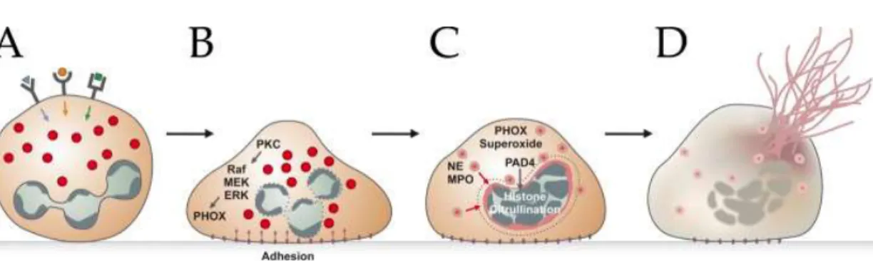
Figure 7. Schematic representation of the NETosis pathway.
After stimulation of receptors (A), neutrophils adhere to the substrate (B) and mobilize granule components, such as NE and MPO (C). Granules are shown as red circles. Histones are citrullinated and the intracellular membranes are disintegrated. In the end, the mixture of cytoplasm (granule proteins) and nucleoplasm (DNA and histones) are released into the extracellular space to form NETs (D). (Brinkmann and Zychlinsky, 2012).
1.1.4 Neutrophil and extracellular matrix
Pathogens can be recognized by a number of cells in the tissue, such as macrophages and mast cells. These cells release pro-inflammatory chemokines and cytokines, which activate the neutrophils and endothelial cells for an inflammatory response. Neutrophils firstly adhere to the vascular wall, then migrate through the endothelial cell bilayer, and subsequently through the extracellular matrix to reach the site of inflammation. During this process, neutrophils need to interact with multiple extracellular matrix proteins.
1.1.4.1 Extracellular matrix
All tissues and organs contain non cellular component, the extracellular matrix (ECM), which provides essential physical scaffolds and also regulate many cellular process such as growth, migration, differentiation, survival, homeostasis and morphogenesis (Theocharis et al., 2016). The two main ECMs are the interstitial connective tissue matrix, which surrounds cells and provides structural scaffolding for tissues, and the basement membrane, which separates the epithelium from the surrounding stroma (Figure 8) (Bonnans et al., 2014) . Cells can modulate the composition and structure of the ECM, in contrast, ECM can also regulate cellular process (Ayres-Sander and Gonzalez, 2012). These processes are complicated and
35
need to be tightly regulated to sustain tissue homeostasis, especially in response to injury. Dysregulated ECM remodeling is related with pathological conditions and can aggravate disease progression, such as in osteoarthritis, fibrosis and cancer (Bonnans et al., 2014). The mammalian core ECM, also known as core matrisome, is composed of around 300 proteins including 43 collagen subunits, three dozen or so proteoglycans, and around 200 glycoproteins (Hynes and Naba, 2012).
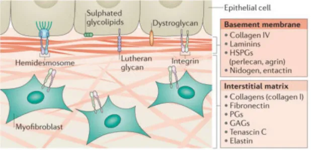
Figure 8. Two main types of ECM.
There are two main types of ECM. The interstitial connective tissue matrix and the basement membrane. The interstitial matrix surrounds cells and is mainly made up of collagen I and fibronectin. The basement membrane is more compact than the interstitial matrix and mainly consists of collagen IV, laminins, heparin sulfate proteoglycans (HSPGs) and proteins, such as nidogen and entactin. PG: Proteoglycan, GAG: Glycosaminoglycan. (Bonnans et al., 2014).
36
1.1.4.2 Fibrinogen
Fibrinogen together with Von Willebrand factor and vitronectin are belong to vascular ECM glycoproteins (Hynes and Naba, 2012). Fibrinogen normally present in human blood plasma at 1.5-3.5 g/L (Pulanic and Rudan, 2005) and is important for haemostasis, wound healing, inflammation and angiogenesis. Fibrinogen is soluble, but it will form a clot or insoluble gel when it is converted to fibrin by thrombin, which is activated by a cascade of enzymatic reactions induced by injury or a foreign surface (Weisel, 2005). Fibrinogen are comprised of two sets of three polypeptide chains named Aα, Bβ and γ, which form two outer D-domain and a central E-domain (Figure 9). There is a minor γ chain variant γ’ by alternative processing of the primary mRNA transcript, γ’ accounts about 8% of the total γ chain population (Mosesson et al., 2001). Fibrinogen has two integrin binding sites at Aα95-98(RGDF) and at Aα572-575(RGDS). A lot cellular interactions with fibrinogen and fibrin occur through binding to one or two of these binding sites (Henschen et al., 1983; Mosesson et al., 2001). The leukocyte αMβ2 integrin on stimulated monocytes or neutrophils has high affinity for binding fibrinogen. αMβ2 integrin binds to the fibrinogen D-domain at a site corresponding to γ-module sequences (Figure 10) : γ190-202(P1) and γ377-395(P2) (Ugarova et al., 1998). P1 is an integral part of the γ-module central domain, while P2 is inserted into this domain forming an antiparallel β-strand with P1. P2 is implicated as the major binding site for αMβ2 integrin. Soluble fibrinogen poorly binds to αMβ2 integrin, but fibrin or immobilized fibrinogen bind to αMβ2 with great affinity. Immobilization of fibrinogen can expose P2. (Lishko et al., 2002; Loike et al., 1992; Mosesson, 2005). Yakovlev et al. has hypothesized that P2 insert may be removed (pulled out) (Figure 10) from the γ-module. They hypothesized that P1 and P2 could be separated by this mechanism and that such separation could enhance the αMβ2-binding activity of the γ-module, thus modulating fibrinogen-leukocyte interaction during an inflammatory response (Yakovlev et al., 2000a; Yakovlev et al., 2001; Yakovlev et al., 2000b; Yakovlev et al., 2005).
37
Figure 9. Schematic of the domain structure of Fibrinogen.
The D and E regions are boxed. The γC, βC, and αEC domains are shown as shaded balls. The connector is the coiled-coil region composed of segments of the α-, β-, and γ-chains. (Lishko et al., 2004).
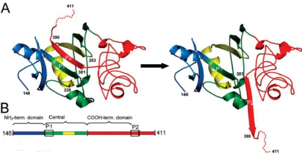
Figure 10. Pull out hypothesis.
A. Schematic ribbon diagram of the known γ-module crystal structure (residues γ148–411; left), and the hypothetical structure resulting from ‘pull out’ of the γ381–390 β-strand insert (P2; right). B. The NH2-terminal domain (residues γ148−191) is colored in blue, the central
domain (residues 192−286) is in green and yellow, and the COOH-terminal domain of the γ-module is in red. Adapted from (Yakovlev et al., 2005).
38
1.1.4.3 Integrins
Cell adhesion to the ECM is mediated by ECM receptors including integrins, discoidin domain receptors and syndecans (Frantz et al., 2010). Neutrophils encounter the tissue ECM and then they emigrate from the bloodstream to a site of inflammation. Cell-cell and cell-ECM adhesive events take place in extracellular matrix, integrins mediate the adhesion events and trigger signalling inside cells which leads to cell response. Integrins are expressed in metazoan, sponges and primitive bilateralia (Gahmberg et al., 2009; Hynes, 2002). Integrins are heterodimers consisting of two subunits, α and β subunits. In vertebrates, there are 18 α(α1-11, αD, αE, αL, αM, αV, αX and αIIb) and 8 β subunits (β1-8) forming 24 αβ heterodimers by noncovalent bonds(Hynes, 2002). The integrins are grouped into subgroups according to ligand-binding properties or their subunit composition (Figure 11). The α and β subunits show no homology, but different α subunits or β subunits have similarities among themselves (Barczyk et al., 2010).
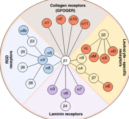
Figure 11. Representation of the integrin family.
In higher vertebrates, the integrin family has 24 members. They are classified into collagen receptors, RGD receptors, laminin receptors and leukocyte-specific receptors. (Barczyk et al., 2010).
39
1.1.4.3.1 Integrin structures
Both α and β subunits contain a large extracellular domain, a single-spanning transmembrane domain and a short cytoplasmic tail (except for β4) (Pan et al., 2016). The extracellular domains of α subunit contain the following domains: I-domain, β-propeller, Thigh, Calf-1 and Calf-2. Nine of the α subunit (α1, α2, α10, α11, αD, αE, αL, αM, αX) have an I-domain (around 200 amino acids) which is crucial for ligand binding. In the other α subunit, the β-Propeller domain constitute the ligand binding site. Integrin β subunits contain a I-like domain, which is crucial for ligand binding, a plexin/semaphorin/intergrin (PSI) domain, a hybrid domain, four epidermal growth factor (EGF) repeats and a membrane proximal b tail domain (bTD) (Figure 12) (Campbell and Humphries, 2011; Pan et al., 2016). There are around 1 mM Ca2+ and Mg2+ in blood. Ca2+ plays an important role in keeping integrins in
inactive state. For many integrins, millimolar Ca2+ often has an inhibitory effect on ligand
binding. For almost all integrins, removal of Ca2+ or addition of Mn2+ will remarkably
increase their binding affinity and adhesiveness (Zhang and Chen, 2014). The extracellular domains of α subunits contain a MIDAS (metal-ion-dependent adhesive site) which can bind Mg2+ and Ca2+. The β I- like domain contains an Mg2+ coordinating MIDAS and a site
adjacent to MIDAS (ADMIDAS) which binds an inhibitory Ca2+. When this ADMIDAS site
binds Mn2+, the conformation will change and lead to an active form of the integrin (Barczyk
et al., 2010). Integrin α and β transmembrane segments are associated in the inactive resting receptor, they dissociate upon talin binding during inside-out activation(Wegener and Campbell, 2008). β integrins share homology in the cytoplasmic tail with NPXY sequences that binds proteins with phosphotyrosine-binding (PTB) domains (Bouaouina et al., 2008). Integrin cytoplasmic domains are directly associated with several cytoskeletal proteins including α-actinin, talin, filamin, paxillin and tensin. Integrin cytoplasmic domains may also associated directly with several intracellular signaling proteins including cytohesion-1, focal adhesion-kinase (FAK) , integrin-linked kinase (ILK) ,β3-endonexin, cytoplasmic domain associated protein-1 (ICAP-1), receptor for activated C kinase 1 (Rack1) and calcium-and integrin-binding protein (CIB) (Pan et al., 2016).
40
Figure 12. Integrin structure.
( A) Domain structure of αxβ2 (Upper, αx; lower, β2); ( B) structure of αxβ2 using same color code as A; (C) Cartoon representation of bent and upright conformations (Campbell and Humphries, 2011).
41
1.1.4.3.2 Neutrophil integrins
Neutrophils express αLβ2 (LFA-1, CD11a/CD18), αMβ2, αXβ2, αDβ2 (CD11d/CD18) and low levels of α4β1, which are involved in slow rolling, adhesion, post-adhesion strengthening, neutrophil migration, ROS production, phagocytosis and polarization(Nathan et al., 1989; Zarbock and Ley, 2008). αMβ2 is the most abundant integrin on neutrophils, αDβ2 is basally expressed on the majority of circulating human neutrophils (Arnaout, 2016). αMβ2 integrin engagement is necessary for phagocytosis and ROS production (Mayadas and Cullere, 2005; Nathan et al., 1989). αMβ2 integrin is stored in neutrophil secretory, secondary and tertiary granules (Borregaard et al., 1994). αMβ2 integrin has more than 40 reported ligands, including ICAMs 1-4, vascular cell adhesion protein 1 (VCAM-1), bacterial and fungal glycoproteins, heparin, coagulation factor Xa, fibrin(ogen), fibronectin, proteinase 3 and complement C3bi (Arnaout, 2016; Ross, 2000; Zarbock and Ley, 2008).
Integrin are normally expressed in an inactive state on the cell surface, which allows the leukocytes or platelets to circulate in the blood with minimal aggregation or interaction with blood vessel walls (Arnaout, 2016). Integrin-ligand binding is regulated by the structural state of activation. When in a bent conformation the integrin is in a low-affinity state, which is modulated upon inside-out or outside-in activation. Outside-in activation involves changes in integrin conformation: Ligand binding to the external domains of integrin induce conformational changes and activates signaling patways. Outside-in signaling is important for adhesion strengthening, spreading and intraluminal crawling. During outside-in activation, Src-family kinases phosphorylate the transmembrane adapters DAP12 and FcRγ, which recruit and activate the Syk. Syk further activate different downstream molecules, such as Vav1, the regulatory subunit of Class I PI3K p85, and PLCγ2 (Mocsai et al., 2015). In most cases, the activation of integrin requires first an inside-out activation: signals originate from various cell surface receptors, such as chemokine receptors (Gahmberg et al., 2009). However integrins can be activated by immobilized fibrinogen alone without inside-out activation , resulting in ROS production, upregulation of TNF-α mRNA and induction of IL-1β (Ugarova and Yakubenko, 2001).
43
Figure 13. Inside-out and Outside-in signalling of β2-integrins.
(A) Inside-out signalling of β2-integrins induces the shit of the bent conformation (low ligand binding affinity) to the extended conformation (intermediate ligand binding affinity) and the induction of the open extended conformation (high ligand binding affinity). (B) Outside-in signalling of β2-integrins is initiated upon ligand binding by clusters of high affinity integrins. Src-family kinases are involved in both signalling ways. Src-family kinases phosphorylate the ITAM motif of transmembrane adapters DAP12 and FcRγ, which recruit and activate Syk. Syk mediates different downstream signals. In inside-out activation, talin finally mediate the integrin to form open-extended conformation. In outside-in activation, Sky can activate Vav1, PLCγ2 and the regulatory subunit of Class I PI3K p85 (Mocsai et al., 2015).
44
1.2 Reactive oxygen species
Neutrophil produce superoxide upon activation by particulate or soluble inflammatory stimuli such as fMLF, inomycin, and LPS. Superoxide is the precursor of other reactive oxygen species (ROS). ROS production by neutrophils is an important step in human innate immune system. NADPH oxidase is the major source of ROS.
1.2.1 NADPH oxidase
The NADPH oxidase consists of membrane-associated heterodimer, cytochrome b558
(gp91phox and p22phox), cytosolic subunits p40phox, p47phox and p67phox, and small GTPase Rac.
After activation, all the subunits assemble together to form the active enzyme at the plasma membrane or at the phagosomal membrane. In mammals, there are seven isoforms of the transmembrane subunit gp91phox or NOX2 of the NADPH oxidase: Nox1-5, Duox1 and
Duox2. Like gp91phox, all other NOX proteins have six transmembrane domains, motifs for
NADPH and FAD binding, and a conserved paired histidines for the binding to the haem groups (Nauseef, 2008).
1.2.1.1 gp91phox
gp91phox (β subunit of the cytochrome b
558) is encoded by the gene CYBB, which is located
on the X chromosome at position 21.1. The human gp91phox is synthesized as a 58 kDa core
protein that is subsequently glycosylated to form a 65 kDa protein gp65 in the endoplasmic reticulum (ER). Gp65 binds to the hem and forms the heterodimer with p22phox. The
heterodimer then traffics to Golgi, and gp91phox is further glycosylated to the mature 91 kDa
form (three N-linked glycosylation sites, Asn132, Asn149, Asn240). (Casbon et al., 2012; DeLeo,
2000; Groemping and Rittinger, 2005; Kleniewska et al., 2012; Yu, 1999). Gp91phox consists
of 570 amino acids. 300 amino acids in the N-terminal are predicted to form six trans-membrane α-helices, and the cytosolic domain in the C-terminal contains the FAD and NADPH binding sites. Gp91phox have conserved paired histidines for binding to the heme
groups (Figure 14). Electrons are transferred from NADPH to FAD and then to the heam groups to reduce O2 to O2.- . The 3D structure of only a fragment of gp91phox, the Cytochrome