HAL Id: tel-01159535
https://tel.archives-ouvertes.fr/tel-01159535
Submitted on 3 Jun 2015HAL is a multi-disciplinary open access archive for the deposit and dissemination of sci-entific research documents, whether they are pub-lished or not. The documents may come from teaching and research institutions in France or abroad, or from public or private research centers.
L’archive ouverte pluridisciplinaire HAL, est destinée au dépôt et à la diffusion de documents scientifiques de niveau recherche, publiés ou non, émanant des établissements d’enseignement et de recherche français ou étrangers, des laboratoires publics ou privés.
Serotonin & developmental axonal refinement : microglia
contribution ?
Marta Kolodziejczak
To cite this version:
Marta Kolodziejczak. Serotonin & developmental axonal refinement : microglia contribution ?. Tis-sues and Organs [q-bio.TO]. Université Pierre et Marie Curie - Paris VI, 2015. English. �NNT : 2015PA066073�. �tel-01159535�
I
PhD thesis
University of Pierre and Marie Curie Specialty
Physiology & Physiopathology
Presented by
Marta KOLODZIEJCZAK
To obtain
DOCTEUR de l’UNIVERSITÉ PIERRE ET MARIE CURIE
Title:
SEROTONIN & DEVELOPMENTAL AXONAL REFINEMENT:
MICROGLIA CONTRIBUTION?
Date of defense: 20 Mars 2015
Examining committee members:
Ann Lohof President
Etienne Audinat Referee
Alexandre Dayer Referee
Sonia Garel Examiner
Anne Roumier PhD advisor
III
Acknowledgements
First of all, I would like to express thanks to the jury members: the referees Etienne Audinat and Alexandre Dayer, the examiner Sonia Garel and the president Ann Lohof. It is a great pleasure and honor to be able to discuss my work with you.
I am also grateful to Luc Maroteaux for welcoming me into his team. Thank you for creating such a friendly and stimulating environment and for your door always being open. You showed me the bigger picture of my research subject which pushed my thinking further and kept me enthusiastic about my work.
I would like to express my heartfelt gratitude to my amazing supervisor Anne Roumier. Thank you that after these four years of work under your supervision I want to do science more than ever! Thank you for guiding me in a way that I could still make my own choices and that made me feel autonomous. Thank you that despite your very busy schedule you were always able to find time to discuss my work ensuring that I never felt left on my own. Thank you for showing me how to be a good scientist. Your motivation and joy of doing science is contagious and motivational. With you I learned how important it is to clearly define a scientific goal and persistently pursue it until getting some clear answers. You have amazing pedagogic skills and every student who gets the chance to work with you should feel truly lucky!
I am grateful to Catherine Béchade for her encouraging and constructive feedback while writing my thesis. Catherine, you are an amazing scientist. Your help with the bibliography was invaluable! I also really appreciate your straightforwardness.
It has been a great pleasure to do a collaboration with Nicolas Gervasi. Thank you for showing me the beauty of the live cells imaging. To see microglia moving around on a brain slice was one of the most spectacular experiments during my PhD!
I acknowledge the invaluable help of Patricia Gaspar and members of her team:
Alexanda Rebsam for help with the CTb experiments, Cedric Francius for the ISH and Ahlem Assali for her friendly advice as a more experienced PhD student. Thank you Aude, Sebastian, Maria, Mariano, Sophie & Teng Teng. It has been a great pleasure to have you all as lab neighbors.
IV
I would like to express thanks to Michel Mallat and Christine Metin for their proficient advice during my mid-thesis committee. I would like to additionally thank Christine for providing me with the tomato reporter mouse which allowed me to generate some interesting results.
I am indebted to: Mythili Savariradjane & Théano Eirinopoulou from the imaging platform. Mythili, thank you for the introduction to the amazing world of epifluorescence imaging (one of my favorite experimental tools). I really appreciated your always open door and the constant smile on your face. Théano, thank you for the extended lesson on confocal imaging and for the beautiful 3D reconstructions. I love your sense of humor. I would like to acknowledge the animal house stuff (especially Natacha) for their useful help despite difficult conditions.
A big thank you to the former PhD student in our lab. Thank you Dr. Arno for introducing me to the graduate student life. I have always admire your joy and passion for science. The lab is not the same without you and your Bob Marley songs!
I could not forget to mention the two amazing M2 students, Marine & Emily, with whom I quickly became friends. Marine, it was a great pleasure to have you around! Besides the protocols for DNA extractions you wrote; I also loved our little “pauses-cafés”.
Ma chère Emily, I feel extremely lucky to have found a friend like you. My work days became much more enjoyable when you came around! The little favors you did for me over the past years (putting brains into sucrose, checking on my mice or sending me pictures of my lab notes) are countless… (I’ve lost the track of how many cakes I owe you! :P). I will miss our nights out and the “climbing Thursdays”!
I would also like to thank other present and past members of the lab. Sophie, for her help with breeding animals. I have enjoyed our numerous discussions about scientific and non-scientific subjects. Stephane, for his help during the preparation of my PhD application. I am a big fan of the salad dressing you used to make at the lab “plateaux fromage” (never found out your secret…). Silvina, for the advice in animal care. It is amazing how one person can bring such an amount of positive energy to the whole office. Imane, for her very clear explanations and help with molecular biology. And also: Elodie, Damian & Takis.
V
It has been an amazing journey and I will keep many warm memories from the years we worked together!
Finally, but by no means least, I would like to thank my loved ones for their infinite support during all of these years.
My friends: Chris, Emily, Maja, Ina, Ola & Milka for all the amazing trips, common meals & inspiring discussions.
My family: Mum, Dad, Hubert, Grandma Renata, and all the others, thank you for always believing in me. Dear parents, thank you for letting me go my way even if at the time it sounded like a crazy idea. Your unconditional support gave me strength and courage to reach for the “impossible”. Mum, thank you for teaching me the courage to follow my dreams and not try to fit to some standards. Thanks to this precious knowledge I made sure I enjoyed every single moment of my PhD experience!
I could not forget to thank my amazing boyfriend. Frederik, you have supported me all through the years of my PhD. Thanks to you, this journey turned out to be an amazing experience. Thank you for introducing me Matlab and for your huge help with image analysis. I love our life and our numerous amazing trips! I could not imagine a better life companion. I love you.
VII
Table of Contents
Acknowledgements ... I Table of Contents ... VI Table of Figures ... XI List of acronyms ... XIV
Abstract ... 1
A - INDRODUCTION ... 3
A1 Microglia ... 5
A1.1 Microglia History ... 5
A1.2 Developmental Origin of Microglia ... 6
A1.2.1 Microglia Originate from Extra-Embryonic Yolk Sac ... 6
A1.2.2 Transition to the Committed Microglia Progenitors ... 8
A1.2.3 Microglia Brain Colonization ... 9
A1.3 Microglia in Adult Brain ... 11
A1.3.1 Origin of Microglia in Adult Brain ... 11
A1.3.2 Adult Microglial Phenotype ... 13
A1.3.3 Microglia in Non-Pathological Conditions... 14
A1.3.4 Microglia Activation ... 16
A1.3.5 Microglia Activation Markers ... 18
A1.3.6 Microglia vs. Macrophages ... 19
A1.4 Early Postnatal Microglia ... 21
A1.4.1 Developmental Microglial Phenotype ... 21
A1.4.2 Developmental Neuronal Survival ... 23
A1.4.3 Synaptogenesis, Synaptic Maturation & Plasticity during Development ... 24
A1.4.4 Neurogenesis & Plasticity in Adult Brain ... 25
VIII
A2.1 Anatomy ... 29
A2.2 From Synthesis to Degradation ... 31
A2.3 Serotonin Receptors ... 33
A2.3.1 5-HT2B Receptor: Expression ... 34
A2.3.2 5-HT2B Receptor: Pharmacology ... 35
A2.3.3 5-HT2B Receptor: Signaling Pathways... 36
A2.3.4 5-HT2B Receptor: Functions ... 37
A2.4 Serotonin & Immune System Interactions ... 38
A2.4.1 In the Periphery ... 38
A2.4.2 In the CNS ... 38
A3 Axonal Refinement ... 41
A3.1 Definition of Axonal Refinement ... 41
A3.2 Models of Axonal Refinement ... 42
A3.2.1 Barrel Formation in the Somatosensory Cortex ... 43
A3.2.2 Maturation of Synapses between Pyramidal Cells ... 44
A3.2.3 Segregation of Retinal Axons in Thalamus ... 46
A3.2.3.1 Model Description ... 46
A3.2.3.2 General Mechanisms ... 47
A3.3 Serotonin & Axonal Refinement ... 48
A3.3.1 Models to Study Serotonin Effects ... 48
A3.3.2 Importance of Timing of Serotonin Action ... 50
A3.4 Immune System & Axonal Refinement ... 51
A3.4.1 Mechanisms involving microglia ... 51
A3.4.1.1 Complement System ... 51
A3.4.1.2 Fractalkine Receptor (Cx3Cr1) ... 54
A3.4.2 Immune System Molecules not (yet) related to Microglia ... 55
IX
A4.1 Developmental Role of Serotonin in Psychiatric Disorders ... 57
A4.1.1 Serotonin level perturbations ... 57
A4.1.2 5-HT2B Perturbations ... 58
A4.2 Developmental Role of Immune System in Psychiatric Disorders ... 59
A4.2.1 Role of Adult Microglia in Psychiatric Disorders ... 61
B - RESULTS & DISCUSSION ... 63
B1 Article ... 65
B1.1 Axonal Refinement Deficits of Htr2B-/- Mice ... 103
B1.1.1 Heterogeneity Within wild-type Mice ... 103
B1.1.2 Axonal Refinement Deficits in Htr2B-/- Mice ... 104
B1.1.2.1 Retina Injury Consequences ... 105
B1.2 5-HT effect on microglia ... 106
B1.2.1 In situ Chemoattractant Effect of Serotonin on Microglial Processes ... 106
B1.2.2 Phagocytosis assay ... 107
B2 Potential Mechanisms & Cells Involved in Htr2B-/- Mice Deficits ... 111
B2.1 Cells Expressing 5-HT2B in dLGN ... 111
B2.2 Effect of the Invalidation of the 5-HT2B in Pet1Cre/0; Htr2Bfl/fl mice ... 112
B2.3 Putative Role of Retinal Ganglion Cells ... 114
B2.4 Effect of the Invalidation of Htr2B in microglia... 115
B3 Conditional Knockouts of 5-HT2B Receptor in Microglia ... 119
B3.1 Cre lox Recombination ... 119
B3.1.1 Inducible Cx3cr1CreERT2/+; Htr2Bfl/fl ... 120
B3.1.2 Constitutive Cx3cr1Cre/+; Htr2Bfl/fl ... 124
B4 Supplementary Data ... 127
B4.1 Invasion of Microglia into dLGN of wild-type Mice ... 127
B4.2 Overexpression of inflammatory markers ... 129
X
B5.1 Involving Microglia ... 132
B5.2 Alternative ... 133
B6 Importance of Results & Perspectives ... 135
XI
Table of Figures
Figure 1: Different types of glial cells ... 6
Figure 2: Schematic representation of developmental microglia origin. ... 8
Figure 3: Microglia colonization of the brain ... 9
Figure 4 Classification of developmental microglia morphology ... 10
Figure 5: Postnatal distribution of microglia in the somatosensory cortex. ... 11
Figure 6: Homogeneous microglia distribution in adult mouse brain ... 14
Figure 7: Classification of microglial morphology ... 14
Figure 8: Microglia control neuronal activity ... 15
Figure 9: Proportion of shared genes between microglia and macrophages ... 20
Figure 10: Developmental microglia express CD68 activation marker ... 22
Figure 11: Serotonergic system in the rodent brain. ... 30
Figure 12: Serotonin transporter (SERT) functioning ... 32
Figure 13: Serotonin metabolism in CNS ... 33
Figure 14: 5-HT2B signaling pathways. ... 37
Figure 15: Main steps of developmental axonal refinement ... 42
Figure 16: Barrel cortex. ... 43
Figure 17: Terminals of thalamocortical afferents in the rat barrelfield ... 44
Figure 18: Dendritic spine dynamics in pyramidal neurons of mouse somatosensory cortex. ... 45
XII
Figure 20: Deficit of RGC axons segregation in the dLGN of MaoA- deficient mouse. ... 49
Figure 21: “Barrelless” phenotype in MaoA- deficient mouse. ... 50
Figure 22: Complement-mediated synapse elimination during development. ... 53
Figure 23: Both, activity and the complement system regulate phagocytosis of synapses by microglia. ... 54
Figure 24: Microglial processes moved rapidly toward the source of serotonin ... 65
Figure 25: Retina injury-induced recruitment of microglia to the dLGN ... 106
Figure 26: Microglia phagocytic capacities. ... 109
Figure 28: Cells in the murine dLGN susceptible to express 5-HT2B receptor. ... 112
Figure 29: Synaptic refinement in Pet1Cre/0; Htr2Bfl/fl mice. ... 114
Figure 31: Synaptic refinement in the Cx3cr1CreERT2/+; Htr2Bfl/fl mice ... 116
Figure 32: Schematic representation of different invalidations of genes of interest in microglia. ... 119
Figure 33: Schematic of modified Cx3cr1 loci ... 120
Figure 34: Recombination of the Htr2Bfl/fl by the Cre recombinase. ... 121
Figure 35: TXF-induced recombination in Cx3cr1CreERT2/+; Htr2Bfl/fl P11 mouse ... 122
Figure 36: TXF-independent recombination in Cx3cr1CreERT2/+; Htr2Bfl/fl P6 mouse... 123
Figure 37: Recombination in the Constitutive Cx3cr1Cre/+; Htr2Bfl/fl P6 mouse. ... 125
Figure 38: Recombination efficacy and specificity in the conditional models of 5-HT2B deletion from microglial cells.. ... 126
Figure 37: One of the first microglia within the dLGN are aligned along blood vessels 128 Figure 40: Microglia invasion in barrel cortex (P5-P7-P9) ... 129
XIII
Figure 41: Schematic representation of the RGC axons segregation in the dLGN of the WT and Htr2B-/-. ... 131
Figure 42: Transient location of SERT in the dLGN ... 132 Figure 43: Putative consequences of 5-HT2B receptor absence. ... 134
XIV
List of acronyms
ATP: Adenosine triphosphate
BDNF: Brain derived neurotrophic factor cAMP: Cyclic adenosine monophosphate CNS: Central nervous system
CR3: Complement receptor 3 CSF1: colony stimulating factor 1
CSF1R: colony stimulating factor receptor1 CX3CL1, FKN: Fractalkine
DAG: Diacylglycerol
dLGN: Dorsal lateral geniculate nuclei DT: Diphtheria toxin
DTR: Diphtheria toxin receptor E: Embryonic day
FKN, CX3CL1: Fractalkine GFP: Green fluorescent protein
Iba1: Ionized calcium-binding adaptor molecule 1
IGF1: insulin-like factor 1
iNOS inducible nitric oxide synthase LPS: Bacterial endotoxin
lipopolysaccharides
MaoA: monoamine oxidase A
MHC class I: major histocompatibility complex class I
MHC II: Histocompatibility complex class II NGF: Nerve growth factor
NO: Nitric oxide P: Postnatal day
PCPA: (para-chlorophenylalanine), Blocker of 5-HT synthesis through inhibition of Tph2
PKC: Protein kinase C
Poly I:C: Synthetic analog of viral double-stranded RNA
RGC: Retinal ganglion cell ROS: Reactive oxygen species
Runx1: Runt-related transcription factor SERT: Serotonin transporter
TGFβ: Transforming grow factor beta TLR: Toll-like receptor
TPH: tryptophan hydroxylase
TTX: Tetradotoxin, a voltage-gated sodium channel blocker
TXF: 5-OH-Tamoxifen
1
Abstract
Maturation of functional neuronal circuits during development of the central nervous system relies on sophisticated mechanisms. First, axonal and dendritic growth and synapse elaboration should reach appropriate targets. Second, pruning and neuronal death are required to eliminate redundant or inappropriate neuronal connections. Serotonin, besides its functions as a neurotransmitter, actively participates in postnatal establishment and refinement of brain wiring in mammals. Another important role is played by the brain resident macrophages, microglia, in developmentally-regulated neuronal death as well as in synaptic maturation or elimination.
Interestingly, the unpublished laboratory data show a major expression of the serotonin 5-HT2B receptor by postnatal microglia, suggesting that serotonin could participate in
temporal and spatial synchronization of microglial functions.
During my thesis, I tested the hypothesis of cross-regulations between microglia and serotonin during postnatal brain development in a mouse model. First, I investigated whether some developmental steps known to be controlled by serotonin, could potentially result from microglia sensitivity to serotonin. Using an in vivo model of synaptic refinement during early brain development, the maturation of retinal projections to the thalamus, I observed that Htr2B-/- mice present anatomical alterations
of the projecting area of retinal axons into the thalamus.
Parallelly, I tested the effects of serotonin on microglial cells. Although neither chemotaxis nor phagocytosis assays have revealed any serotonin effect on microglia in vitro, a local delivery of serotonin attracted microglial processes on acute brain slices (two-photon microscopy). Indeed, we observed that microglial processes moved rapidly toward the source of serotonin in wild-type mice, but not in Htr2B-/- mice lacking the
5-HT2B receptor. Moreover, after comparing mRNA expression level in microglial primary
cultures, we have found that some activation markers are upregulated in microglia from Htr2B-/- supporting the hypothesis that serotonin modulates developmental microglia via
2
In the second part of my PhD, by using a number of conditional Htr2B-/- mice, I
investigated which cell type(s) could be responsible for the altered segregation of retinal axons in the thalamus of the total Htr2B-/- mice. The analysis of Pet1-Cre mice in which
the 5-HT2B receptor is absent specifically in the serotonergic raphe neurons have not
revealed any significant abnormalities. The investigation of the involvement of the microglial 5-HT2B in this process is still ongoing.
Overall, my results support the hypothesis that serotonin interacts with microglial cells and that these interactions could participate in brain maturation.
3
5
A1 Microglia
A1.1
Microglia History
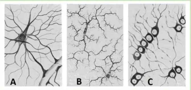
The word glia comes from Greek γλία which means glue. Glia have been described for the first time in 1856 by a German pathologist Rudolf Virchow. He referred to glia as “neuronal glue”, considering them just as a brain filling tissue that keeps the neurons together (Fields 2009). In the early 20th century, Pío del Río Hortega, a student of the
famous histologist Ramon Cajal, has reported the existence of three types of glia: astrocytes (Figure 1 A), oligodendrocytes (Figure 1 C), and microglia (Figure 1 B). However, research over the past decades led to the extension of the glial family to include the polydendrocytes (NG2 cells) (Nishiyama et al. 2009).
After a long time during which glial cells have been neglected, their golden time in science has arrived. Although, the glial research is still very young, and we are far from fully understanding glial functions, these cells have already been shown to be in charge of brain development, support and defense.
The proportion of glial to neuronal cells has been a matter of debate for a long time. It varies among species, and also depends upon the quantification method. For example, until recently, it was accepted that glial cells represent even 90% of the human brain, and that this large amount is due to its important size comparing to other species (Allen and Barres 2009). Today however, a novel isotropic fractionator method quantifying proportions of different brain cells simply by using brain cell nuclei suspension, allowed to estimate that the human brain consist 50% of glial cells, whereas mouse brain of 35% (Herculano-Houzel 2014).
Moreover, scientists argue that the glial to neuronal cells ratio is not necessary higher in larger brains but it is rather correlated with lower neuronal density (Herculano-Houzel 2014).
The microglial cells, which were already distinguished by Río Hortega in 1919 as the ‘third element’, are different to neurons and macroglia (astrocytes and
6
oligodendrocytes). As the name suggests, microglia have much smaller cell bodies than other members of the glial family (Figure 1). Although, these resident brain macrophages are best known for their role in the adult brain (discussed in the chapter: “Microglia in Adult Brain”) recent studies have shown st their involvement in developmental neuronal death, synaptic maturation and establishment of brain wiring.
Before discussing the functions of microglia in adult and developing brains, I will present the current knowledge about their origination from extra-embryonic yolk sac and the transcription factors required for their differentiation.
Figure 1: Different types of glial cells. A: astrocytes, B: microglia, C: oligodendrocytes. Drawings by Río Hortega, 1920. Source: (Fields 2009)
A1.2
Developmental Origin of Microglia
A1.2.1 Microglia Originate from Extra-Embryonic Yolk Sac
Macroglia (astrocytes, oligodendrocytes and N52 cells), same as neuronal cells, originate from the neuroectoderm. Microglia, however, alike other macrophages, differentiate from hematopoietic precursors originating from the mesodermal tissue. In mouse embryo, there are two distinct waves of hematopoiesis: primitive and definitive. Primitive hematopoiesis takes place in the extra-embryonic yolk sac before E8, whereas
7
definitive hematopoiesis takes place within the embryo around E10.5 (Ginhoux et al. 2010; Schulz et al. 2012; Kierdorf et al. 2013).
Only recently, generation of a transgenic mouse expressing a green fluorescent protein (GFP) under the control of CX3CR1 promoter (Cx3cr1GFP/+), a marker of committed
macrophage progenitors, enabled to show that microglia arrive for the first time to the brain through blood vessels around E9.5 (Ginhoux et al. 2010). Before achieving their definitive phenotype in adult animal, microglia are highly proliferative throughout their embryonic life (Ginhoux et al. 2010).
In order to investigate the potential contribution of primitive myeloid precursors to the microglial pool, the Cre recombinase has been expressed under a promoter of runt-related transcription factor (Runx1), which is expressed exclusively in the yolk sac, in mice harboring an YFP reporter construction (Ginhoux et al. 2010). TXF-induced activation of Cre recombinase at E7.5 resulted in YFP expression in 30% of yolk sac macrophages, 30% of microglia and close to 0% of peripheral macrophages. However, when the Cre recombinase has been activated later during embryogenesis there was no YFP positive cells in the brain. Instead, 40% of leukocytes expressed YFP (Ginhoux et al. 2010; reviewed in Prinz et al. 2014). Moreover, this yolk sac cell population of uncommitted stem cells expressing a stem cell growth factor receptor (c-kit), identified in mouse at E8 by flow cytometry experiments, has been reported to represent the earliest potential microglia progenitors (Kierdorf et al. 2013).
Shortly after the formation of blood circulation (around E8.5) the microglial progenitors populate the brain (Kierdorf et al. 2013). These committed developmental microglia express also three other markers: a member of adhesion G protein-coupled receptors family, the F4/80, a subunit of the complement receptor, the integrin CD11b and Runx1 (Ginhoux et al. 2010; Kierdorf et al. 2013; reviewed in Derecki et al. 2014). In the adult brain, the F4/80 and CD11b are upregulated in case of an inflammation.
Another population of the yolk sac-derived progenitors gives rise to a number of tissue macrophages such as liver Kupffer cells, epidermal Langerhans cells or pleural macrophages (Schulz et al. 2012). Definitive hematopoiesis, on the other hand, gives rise to the remaining peripheral macrophages such as circulation monocytes and dendritic cells (Schulz et al. 2012).
8
A1.2.2 Transition to the Committed Microglia Progenitors
The transition from the uncommitted stem cells to the committed microglial progenitors is regulated by a number of proteins such as Runx1, Pu.1, Irf8 and CSF1R (Figure 2). The absence of one of these proteins results in disturbed microglial differentiation and brain colonization and can have a fatal outcome. For instance, Pu.1 deficient mouse, which lack macrophages, die shortly after birth. The deficiency of interferon regulatory factor (Irf8), which acts downstream of Pu.1, also leads to significant overall decrease in microglia density that persist into adulthood (Kierdorf et al. 2013). Similarly, in mice that lack the colony stimulating factor receptor 1 (CSF1R), microglial cells are missing and the animals die before adulthood. Additionally, the absence of the CSF1R also leads to a severe depletion of most populations of peripheral macrophages. Interestingly, in animals with a null mutation in the main CSF1R ligand, the colony stimulating factor 1 (CSF1 or M-CSF; expressed by the mononuclear phagocytic lineage), the population of the peripheral macrophages is greatly reduced, whereas the microglia are mostly normal. This could be explained by the existence of another CSF1R ligand in the brain. Indeed, an alternative CSF1R ligand, interleukin 34 (IL-34) (produced by neurons), has been proposed to modulate microglia differentiation (Pollard 2009). A more recent study, however, demonstrated that IL-34 is dispensable for developmental microglia and their yolk sac precursors and it is only required for their maintenance in the adult brain (Greter et al. 2012).
Unlike in the case of peripheral macrophages, the expression of transcription factor Myb is dispensable for microglial differentiation.
9
The results summarized above show that microglia differentiate early in embryogenesis from yolk-sac derived myeloid progenitors. Their source in the adult brain, however, is still controversial and I will discuss it in the chapter: “Microglia in Adult Brain”.
A1.2.3 Microglia Brain Colonization
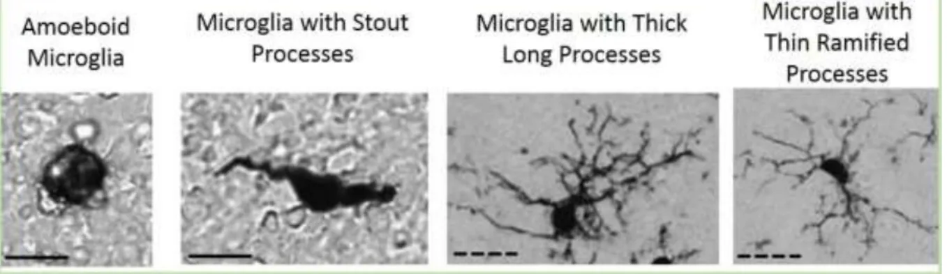
As introduced previously, microglial precursors are generated in the yolk sac and start invading the brain shortly after formation of blood circulation. The first microglia precursors arrive to brain at E9.5. These precursor cells have an amoeboid phenotype and accumulate at several sites (e.g. at plexus choroideus and corpus callosum) from where they invade the brain parenchyma. These sites have been first described as “fountains of microglia” by Kershman in 1939 (summarized in Figure 3). Once in brain parenchyma, microglia start to adapt a ramified morphology (Figure 4).
Figure 3: Microglia colonization of the brain. On the left: drawing of human embryonic brain and fountains of microglia (from book: “Glial physiology and pathophysiology” 2013, Alexei Verkhratsky, Arthur Butt). On the right: microglial localization in the embryonic mouse forebrain, showing sites of higher concentration of microglial cells (open arrowheads). Scale bar: 500µm. (Squarzoni et al. 2014).
10
Figure 4 Classification of developmental microglia morphology (Iba1 IHC) in the E80 macaque neocortex. Amoeboid microglia (D), ramified microglia (A) and two intermediary phenotypes (B, D).Scale bare: 20µm. (Cunningham et al. 2013).
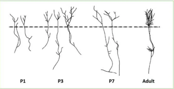
The microglia invasion is a complex process and it remains largely unknown. A very recent study provided some insights into microglia colonization of the mouse cortex. It occurs in three phases. First, from E10,5 to E14,5 a slow increase in microglia number is observed. Next, from E14,5 to E15,5 a rapid phase characterized by a massive increase in microglia takes place. And the last slow phase occurs from E15,5 to E17,5 (Swinnen et al. 2013). At E14.5 amoeboid and more ramified microglia accumulate focally, whereas they are absent in other specific regions such as cortical plate (Arnoux et al. 2013; Squarzoni et al. 2014) (Figure 5). In a timely manner, a progressive invasion of microglia can be observed. However, before achieving their homogenous distribution observed in adult brain, early postnatal microglia are distributed heterogeneously. Until P5, higher densities of microglia have been observed in the vicinity of meninges and ventricular surfaces, whereas there are almost no microglia in cortical layers (Arnoux et al. 2013) (Figure 5).
11
Figure 5: Postnatal distribution of microglia in the somatosensory cortex at P0, P4 and P7 of Cx3cr1GFP/+ mouse. At P0, microglia have an uneven distribution and are notably very rare in
the cortical plate. At P4, hotspots of microglia are still visible notably near hippocampal fissure. At P7, the distribution is mostly homogenous. Scale bar: 300µm. (Arnoux et al. 2013).
In mouse, the brain colonization by microglia lasts until the closure of the blood brain barrier at around P10 (Verkhratskiĭ and Butt 2013). The gradual increase in microglia density could be explained both by the microglial precursors invasion and by cell proliferation.
A1.3
Microglia in Adult Brain
In this part, I will introduce the role of microglia in an adult brain. First, after explaining their origin, I will present a classification of their phenotypes. Secondly, I will discuss the dynamism of the unchallenged ramified microglia. Next, I am going to review their different activation states and give a brief overview of markers of microglial activation. In the end, I will point out some of the similarities and the differences between microglia and peripheral macrophages.
A1.3.1 Origin of Microglia in Adult Brain
As I have introduced previously, microglia originate from extra-embryonic yolk sac during development. Surgically adjoined parabiotic mice were used in order to investigate the contribution of blood-circulating progenitors to the adult microglia pool. One of these mice had GFP positive hematopoietic cells (except blood cell precursors). The analysis of their brains five months after the surgery have not revealed any GFP
12
fluorescent microglia (Ajami et al. 2007). This suggests, that in physiological conditions microglial cells are independent from adult hematopoiesis.
Previous approaches to study the origin of microglia in adult brain consisted on a transplantation of GFP positive, mononuclear bone marrow cells into GFP negative mouse (recipient mouse). In order to enable the transplantation, the immune system of the recipient mouse had to be eliminated by irradiation. One of the first experiments of this sort was performed using donor bone marrow cells transduced with a GFP positive vector (murine stem cell virus). Four weeks after transplantation of these cells into an irradiated mouse, a population of ramified GFP fluorescent microglia has been identified in the brain parenchyma. This indicats that, at least after irradiation, adult microglia pool can be replenished from bone-marrow derived progenitors (Priller et al. 2001). A few years later, it has been proposed that the Ly-6hiCCR+ monocytic population contains
microglial precursors. However, the same study showed that engraftment of circulating microglia progenitors in the healthy brain is dependent on irradiation. The mice with selective-body irradiation (where the head was omitted) were shown to be protected from engraftment (Mildner et al. 2007).
Other experiments have been conducted to investigate whether a drastic increase in microglia number, which is characteristic of many CNS pathologies (injury, disease), is dependent on the peripheral precursors recruitment.
First, five weeks after irradiation/transplantation procedure, an injury (i.e. facial nerve axotomy-induced neurodegeneration) has been inflicted in order to induce a microgliosis in a non-GFP mouse. As a result, around 20% of microglia in the recipient mouse were GFP positive, suggesting that a part of microglial cells originates from blood-circulating microglia precursors (Ajami et al. 2007). Next, a similar injury was inflicted in the recipient mouse after two weeks of parabiosis. Surprisingly, in this model, the injury-induced microgliosis did not result in recruitment of microglia precursors from the circulation (Ajami et al. 2007). The following demonstrates that the GFP positive cells
13
observed in the brains of transplanted animals originate from immature hematopoietic precursors present in the injected transplantation material, but absent in the blood circulation under physiological conditions (Ajami et al. 2007). These results suggest that the recruitment of GFP positive precursors might be due to the irradiation/transplantation procedure and it does not occur in a physiological situation. Another approach to study their origin in adult brain consists on inducing microglia repopulation after their pharmacological elimination. Selective CSF1R inhibitor treatment leads to an efficient microglia depletion (most of microglia are eliminated after one week of treatment). Interestingly, microglia repopulation takes place already within one week after stopping drug administration. Further analysis have demonstrated that such a rapid microglia replenishment has been established by a pool of nestin-expressing CNS latent progenitors (Elmore et al. 2014).
In summary, these experiments demonstrate that, in both physiological and pathological conditions, adult microglia pool depends on the self-renewing nestin-expressing CNS latent progenitors, and that it is independent from the bone marrow-derived monocytes and adult hematopoiesis (Ajami 2007, Ginhoux 2010). Throughout life, microglia can use a pool of CNS nestin-expressing progenitors (Elmore 2014, reviewed in Hughes 2014).
A1.3.2 Adult Microglial Phenotype
In adult animals, microglia distribution is homogenous covering the entire brain parenchyma (Figure 6). As discussed before, these brain resident macrophages constitute around 10-30% of total mouse brain cells, depending on the method used for the estimation (Lawson et al. 1992; Herculano-Houzel 2014).
14
Figure 6: Homogeneous microglia distribution in adult mouse brain. A: Coronal section of
Cx3cr1GFP/+ P20 mouse. Green: microglia; Ncx: neocortex; Str: striatum (Squarzoni et al. 2014). B,
C: region corresponding to white square on A (thalamic dLGN); B: nuclei stain (Bis-Benzimide); C: microglia stain (Iba1); Scale bare: solid line: 500µm; dashed line (B, C): 200µm.
In physiological conditions, microglia have small soma and long, thin ramified processes (Figure 7). However, in an activated state, their soma might become larger and their processes shorter and thicker. Microglia can also adopt an amoeboid shape resembling peripheral macrophages (Kettenmann et al. 2011)(Figure 7).
Figure 7: Classification of microglial morphology found in adult brain in normal or pathological conditions. Scale bare: solid line: 10 µm, dashed line: 50 µm.
A1.3.3 Microglia in Non-Pathological Conditions
Since there was no direct evidence of movement or activity of ramified microglia in non-pathological conditions, they were long referred to as “resting”. However, their quick transformation in response to even subtle changes of the micro-environment is
15
contradictory to their quiescent state (Kreutzberg 1996; Nimmerjahn et al. 2005). Only a decade ago, studies using powerful in vivo imaging techniques, such as two-photon microscopy, have shown that the ramified microglia are indeed very dynamic. For instance, the time lapse recordings of the brain of Cx3cx1GFP/+ mouse have demonstrated
that, even though the microglial somata are immobile, their processes are very motile. Cortical ramified microglia have been shown to rapidly extend and retract their branches at an average velocity of 1.5 µm/minute (Nimmerjahn et al. 2005; Davalos et al. 2005). A few years later, another study has shown that the dynamic processes of ramified microglia frequently (once per hour) interact with spines of surrounding neurons, suggesting their ability to carefully monitor brain activity (Wake et al. 2009). Electron microscopy together with three-dimensional reconstructions allowed to visualize appositions of microglial processes with synapses in the visual cortex of a juvenile mouse during normal visual experience (Tremblay et al. 2010). An examination of the optic tectum (brain region responsible for vision reflex) of larval zebrafish has demonstrated that microglia preferentially contact neurons with an increased activity (i.e. in response to a visual stimulation), which in turn leads to a decrease of activity in the contacted neuron (Li et al. 2012). This is summarized in Figure 8.
Figure 8: Microglia control neuronal activity. Microglial processes (red) preferentially contact more active neurons (orange). This microglia-neuron interaction results in a decrease of activity (grey) of these previously active neurons. (Salter and Beggs 2014).
In summary, these studies clearly show that the ramified microglia are not “resting”. They monitor and control their microenvironment and they stay in a constant dialogue with the surrounding neurons and very probably other neighboring cells.
16
A1.3.4 Microglia Activation
Alike other macrophages, microglia can quickly respond to changes in their microenvironment. In response to brain injury or infection microglia undergo complex changes including their morphology. Activated microglia can adopt an amoeboid shape resembling peripheral macrophages (Kettenmann et al. 2011) (Figure 7). Their physiologic characteristics and gene expression can also shift to favor e.g. an increased motility and phagocytosis.
Microglial response to injury can be investigated in vivo on acute brain slices thanks to two-photon microscopy. Using this technique it has been shown that a laser disruption of blood-brain barrier at the level of a small capillary induces an immediate response of microglia in a radius of 90µm. They direct their processes toward the lesion site without translocating their soma. 10 to 15 minutes later, at the site of the lesion, microglial processes form phagocytic spherical engulfments. The number of responding microglia depended on the severity of the injury (Nimmerjahn et al. 2005). Pharmacologically induced hydrolysis of adenosine triphosphate (ATP), a purinergic neurotransmitter, significantly reduced microglial response to the laser injury. Moreover, the same effect has been achieved by administration of an inhibitor of one of the ATP receptors: the P2Y receptor (Davalos et al. 2005). Another study has demonstrated that P2Y12 receptor,
which is highly expressed by surveying microglia and downregulated upon microglial activation, is required for early microglial response to injury. Microglia from the P2Y12
-deficient mice showed dramatically reduced chemotaxis toward a focal laser ablation in vivo and toward ATP in vitro (Haynes et al. 2006). Moreover, the P2Y12 receptor has been
identified among recently described microglia specific proteins involved in sensing ligands and microbes in microglia environment (so-called microglial “sensome”). These proteins were identified thanks to a novel direct RNA sequencing (DRS) technology which enables robust quantitative analysis of small amounts of RNA without the need for cDNA synthesis or amplification (Hickman et al. 2013).
These examples show that microglia, in response to environmental stimuli, can rapidly switch from their “resting” state and undergo activation. They also show that their response to the laser injury is, at least partially, mediated by ATP.
17
Different environmental context (i.e. bacterial infection or neuronal death), characterized by different combinations of cytoactive factors, results in a polarized activation of microglia. In parallel to the classification proposed in peripheral macrophages (Mantovani et al. 2002), on the basis of Th1/Th2 lymphocytes classification, two distinct complimentary activated states of microglia could be distinguished: classical, M1-like activation, and alternative M2-like activation. This dichotomy has been proposed to describe also neurotoxic and neuroprotective microglia, respectively (Colton 2009). Although this classification is now debated because it is too simple to describe the multiple activation states of microglia. I will describe it briefly.
“Classic” activation generates M1-like microglia, which orchestrate the tissue defense mechanisms. In case of brain injury or infection microglia recognize a pathogen or damage-associated molecular patterns (PAMPs, DAMPs) via their broad range of pattern-recognition receptors (such as TLRs). Upon activation these receptors trigger a microglial signaling cascade resulting in activation of transcription factors and in differential gene expression. The differentiation of M1-like microglia can be triggered experimentally by inflammatory agents such as bacterial endotoxin lipopolysaccharides (LPS), synthetic analog of viral double-stranded RNA (Poly I:C) or IFNɣ (Roy et al. 2008; O'Connell et al. 2007). These microglia participate in the “killing phase” of the innate defense of the brain and they are characterized by enhanced antigen presentation and expression of pro-inflammatory and neurotoxic factors such as nitric oxide (NO), TNFα, IL-6, IL-1β (reviewed in Belarbi and Rosi 2013).
“Alternative” activation generates M2-like microglia, which may be required to terminate the inflammatory response to brain injury or infection. Their differentiation can be induced by anti-inflammatory cytokines IL-4 and IL-13. The neuroprotective function of M2-like microglia is mediated by a panel of anti-inflammatory factors such as Il-4, IL-10, transforming grow factor beta (TGFβ), and brain derived neurotrophic factor (BDNF; reviewed in Belarbi and Rosi 2013).
Both, the M1-like and M2-like microglia are equally important and maintaining a balance between these two states is essential for brain homeostasis. Moreover, a disturbance in this balance could be at the origin of numerous brain pathologies. For instance, M1-like
18
microglia, which normally constitute the first line of defense against pathogens, have been proposed to play a role in neurodegenerative disorders. A recent study has shown that ROS production mediated by microglial NADPH2 enzyme, through a positive feedback loop, activates microglial NFkB and results in persistent neurotoxic microglial phenotype (Qin et al. 2013). Another recent study has demonstrated that decreased microglial proliferation observed in mice lacking galectin-3, a microglia activation marker, results in increased size of an ischemic lesion (Lalancette-Hébert et al. 2012). These examples clearly show that mechanisms ensuring balance between neurotoxic and neuroprotective microglial states are required. One of such mechanisms involves the alternative arginine pathways. Arginine is a substrate of two distinct enzymes: nitric oxide synthase (iNOS) and Arginase-1. The production of neurotoxic NO is mediated by iNOS, which’s expression is upregulated in M1-like microglia. Arginase-1, on the other hand, is an enzyme implicated in cell proliferation and extracellular matrix reconstruction, upregulated in neuroprotective M2-like microglia. Depending on the context one of these two pathways is favored, which creates a balance between neurotoxic and neuroprotective microglial states (Morris et al. 1998; reviewed in Colton 2009).
The M1 and M2-like states constitute the extreme poles along a continuum of activation. There are many more intermediary, parallelly overlapping and complimentary functional microglial states, which share both, inflammatory and anti-inflammatory characteristics.
A recent investigation of microglia at different ages revealed that at any time microglia do not express exclusively M1 or M2 markers and that differential expression of those markers vary with age (Crain et al. 2013).
A1.3.5 Microglia Activation Markers
Switching of microglial functional state does not always require morphological changes. For instance, blood-brain barrier disruption which induces immediate microglia activation and attraction of their processes toward the injured site, does not trigger any major microglia morphological changes (Nimmerjahn et al. 2005). Thus, analysis of morphology alone is often unsufficient to identify functional state of microglia. Several
19
techniques (i.e. immunohistochemistry, FACS, qPCR) allow us to look at the panel of microglial markers, and to better differentiate distinct differentially activated microglial populations. Below, I will present a number of immunohistochemical markers which allow to identify activated microglial cells.
Iba1 (ionized calcium-binding adaptor molecule 1) expression is restricted to monocytic lineage cell line. Indeed, in the brain, microglia are the only Iba1 positive cells (Imai et al. 1996). An upregulation of this marker is induced by activation such as ischemic stroke (Ito et al. 2001) and status epilepticus (Avignone et al. 2008). Another microglial molecule induced upon activation is the beta integrin, CD11b, which together with CD18 forms a heterodimeric complement receptor (named CR3 or Mac1). The markers listed above are present in the unchallenged ramified microglia but their expression level is upregulated in response to an immune challenge. There are also molecules that are absent in basal state and which expression is induced upon activation. For instance, major histocompatibility complex class II (MHC II) is absent in microglia of healthy young rodents. Induction of its expression has been observed in both aged and infected mice. Microglia neither express the galectin-3 (Mac2) in normal physiological conditions. However, its expression has been shown to be required for microglia activation and proliferation in response to ischemic injury (Lalancette-Hébert et al. 2012). Finally, CD68 (or macrosialin) is a cytosolic marker of activated, notably phagocytic, microglia.
A1.3.6 Microglia vs. Macrophages
As I introduced previously, microglial cells are brain resident macrophages. However, despite numerous similarities between microglia and macrophages, there is also an increasing evidence highlighting their unique molecular and functional signature. On one hand, alike macrophages, microglia can rapidly respond to an immune stimuli: recognize chemotactic and purinergic cues to find the injury site, adapt neuroprotective or neurotoxic phenotype and release inflammatory cytokines, and are able to engulf pathogens via phagocytosis. On the other hand, there is an increasing evidence showing that macrophages and microglia constitute two distinct cell populations. Besides the differences in their developmental differentiation, which I pointed out in one of the previous chapters (“Developmental Origin of Microglia”), they can also be distinguished
20
by the panel of genes they express. An example illustrating the proportion of similarities and differences between microglia and macrophages is a recent study which compared the top 10% of the most abundant transcripts of microglia with these of peritoneal macrophages. This analysis revealed that 70% of these genes are in both of these cell types, suggesting a substantial number of similarities between microglia and macrophages. The expression levels of the remaining 30% of analyzed genes, however, were dramatically different (fold change ranging from 4.8 to 15.1) contributing to a distinct transcriptomic signature of microglia and macrophages (Hickman et al. 2013).
Figure 9: Proportion of shared genes between microglia and macrophages. Diagram representing proportion of similarities and differences of the top 10% of transcripts expressed by microglia and macrophages (Hickman et al. 2013).
Another recent study has identified 239 genes and 8 microRNAs which are uniquely or highly expressed by microglia comparing with other myeloid cells (including macrophages). Moreover, this unique microglial signature has been proposed to be dependent on TGFβ signaling (Butovsky et al. 2014).
Thus, microglia should be considered “cousins” of macrophages. Despite a shared pool of similarities microglia is a distinct cell type with its own molecular signature.
To summarize, the results presented in this chapter show that adult microglia are able to sense and rapidly respond to changes in the brain microenvironment such as injury, cell death/damage or presence of pathogens. Microglial response to such stimuli is mediated by a wide panel of receptors, including purinergic receptors. Parallelly to the
21
eventual morphological changes, the environmental stimuli might trigger a switch in microglial gene expression profile, increasing their motility or phagocytic capacities. Activated microglia act as double-edged sword: on one hand they can be neurotoxic and actively remove damaged neurons, on the other hand they can secrete neuroprotective and anti-inflammatory factors. A balance between different microglial phenotypes is essential for brain homeostasis.
Additionally to these adult brain functions, microglia might also actively participate in some steps of brain development which I will discuss in the following section.
A1.4
Early Postnatal Microglia
Before discussing microglial involvement in the developmental neuronal death, synaptogenesis and synapse plasticity, I will shorty introduce the phenotype of microglia in an early postnatal brain. Microglial involvement in synaptic pruning and axonal refinement, as well as the behavioral outcome of their alterations will not be discussed here. These subjects will be reviewed in a following chapter (“Developmental Role of Immune System in Psychiatric Disorders”).
A1.4.1 Developmental Microglial Phenotype
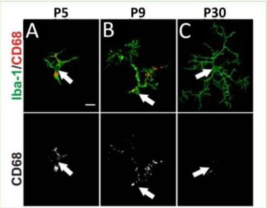
In mouse, microglia progenitors populating the brain during embryogenesis have an amoeboid morphology (Hanisch and Kettenmann 2007). Early postnatal microglia share similarities with both ramified and activated microglia. It has been reported that P5 and P9 microglia express CD68, a lysosomes marker specific to phagocytic activated microglia (Figure 10 A, B). CD68 is downregulated in adult P30 ramified microglia (Figure 10 C). On the other hand, P9 developmental microglia have processes characteristic of ramified microglia (Schafer et al. 2012)(Figure 10).
More generally, microglia in developing brain express differentially a number of chemokines-related genes (Kierdorf et al. 2013).
22
Figure 10: Developmental microglia express CD68 activation marker. Green: microglia stain (Iba1); lower panel: CD68 stain; upper panel: merge. Confocal images. Arrows indicate position of cell soma. Scale bar: 10 μm (Supplementary data Schafer et al. 2012).
Finally, functional electrophysiological studies in mouse cortex have revealed a population of early postnatal microglia which share some characteristics with adult activated microglia. Furthermore, the sharp increase of the number of these cells is observed at the time window of cortex colonization and it is delayed in CX3CR1-deficient mice, in which microglia arrival is retarded. Additionally to their distinguishable electrophysiological properties, these cells also express a particular panel of purinergic receptors. Whereas all of the developing microglia express the P2X purinergic receptors, the heterogeneous expression of P2Y receptors is enriched only in the rectifying microglia population (Arnoux et al. 2013).
These results suggest that developmental microglia are distinct from adult microglia population. Moreover, the existence of differences (such as differential receptor expression) between the developmental microglia supports their putative functions in distinct aspects of brain development.
23
A1.4.2 Developmental Neuronal Survival
Dynamic changes in cell and synapse numbers during development require rigorous regulation of cell survival. For instance, cell production should ensure but not exceed demand in the developing brain. Furthermore, regulation of cell survival is also essential during maturation of functional neuronal circuits which requires on one hand, axonal and dendritic growth and synapse elaboration with appropriate targets but on the other hand, pruning and neuronal death to eliminate redundant or inappropriate neuronal connections. In the following, I will discuss microglia involvement in this process which has been investigated during the past twenty years.
Microglial production of ROS has been shown to be required for most of the neuronal death in the developing mouse hippocampus. Indeed, CR3, which in the brain is expressed exclusively by microglia, has been demonstrated to act in a convergent pathway with an adaptor molecule, DAP12, to lead to microglial production of superoxide and to induce neuronal death (Wakselman et al. 2008). Moreover, elimination of microglia from organotypic cerebellum slices reduced apoptosis of Purkinje cells, indicating their role in inducing neuronal death in the developing mouse cerebellum (Marín-Teva et al. 2004).
Developmental neuronal death is also observed in the chick retina. Strikingly, a dramatic decrease of neuronal death has been reported in retinas that have been detached from the surrounding tissue before microglia infiltration. Since it was restored by the addition of microglial cells to the retina and since this rescue effect was blocked by nerve growth factor (NGF) antibodies, these results show that the microglia-derived NGF induces developmental neuronal death (Frade and Barde 1998).
Another recent study investigated the role of microglia during perinatal cortical development in macaques and rats. It demonstrated that, early in development, microglia accumulate in the proliferative zones of the cortex where they phagocytose neural precursors and therefore restrain production of cortical neurons (Cunningham et al. 2013) further supporting active microglial contribution to the regulation of developmental neuronal survival.
24
Moreover, both pharmacologically induced inhibition of microglial activity (with minocycline treatment) and a transient microglia ablation have led to an increased death of cortical neurons in early postnatal mouse brain (Ueno et al. 2013). The microglia ablation was achieved by diphtheria toxin (DT) treatment (at P3) of a transgenic mouse harboring human diphtheria toxin receptor (DTR) under the control of the microglia specific CD11b promoter. The effect of microglia ablation on cortical neurons has been reproduced by inhibition of insulin-like factor 1 (IGF1) signaling. Therefore, since IGF1 is expressed specifically by developmental microglia, these results endorse their survival-promoting effects (Ueno et al. 2013).
The results summarized above support a dual function of developmental microglia which could modulate both, programmed neuronal death and neuronal survival.
A1.4.3 Synaptogenesis, Synaptic Maturation & Plasticity during Development
Developmental microglia function is not limited to the control of neuronal death. Their involvement in synaptogenesis, synaptic maturation and plasticity has also been proposed.
For instance, DAP12 is a transmembrane signaling polypeptide, which was first described in the immune cells, and which expression in the hippocampus is restricted to the perinatal microglia. The absence of DAP12 has been shown to alter the composition of glutamatergic postsynaptic densities and to enhance long term potentiation, suggesting microglial role in synaptic maturation (Roumier et al. 2004).
CX3CR1-deficient animals, which present a delay of microglia arrival to the hippocampus, provide an interesting model to study microglial role in the synaptogenesis, synaptic function and plasticity. In the juvenile (P15) hippocampus of CX3CR1-deficient mice there is an increased number of weak excitatory synapses (Paolicelli et al. 2011). A following study has reported also a decreased functional connectivity between adult brain regions of these mutant mice (Zhan et al. 2014). Moreover, even though in the CX3CR1-deficient mice there was no overall decrease in microglia density in the layer IV of somatosensory cortex, microglia recruitment inside the barrel centers was transiently delayed.
25
During the maturation of neuronal circuits there is an increase of postsynaptic AMPA receptor proportion and a shift toward GluN2A subunit of NMDA postsynaptic receptors. Both of these changes are impaired in the CX3CR1-deficient animals (Hoshiko et al. 2012) supporting microglia involvement in synaptic maturation.
All the evidence summarized above strongly supports the role of microglia in the synaptogenesis, synaptic function and plasticity.
A1.4.4 Neurogenesis & Plasticity in Adult Brain
Additionally to microglial involvement in the developmental synaptic refinement, there is an increasing evidence supporting their role in the adult neurogenesis and synaptic plasticity, required for neuronal maintenance and learning.
Recently, it has been demonstrated that microglial morphology and the interactions of microglia with synapse-associated elements are modified by visual experience (i.e. light deprivation), supporting the notion if microglial role in modulation of the experience-dependent plasticity in the visual cortex of an adult mouse (Tremblay et al. 2010). Moreover, an in vivo time-lapse imaging of ramified microglia in the optic tectum of larval zebrafish have revealed that microglial processes are attracted toward highly active neurons and that neuronal ATP could act as a “find me” signal to guide them. Interestingly, these microglia-neuron interactions result in a reduction of activity of the contacted neurons, supporting the idea that microglia stay in constant dialog with the elements of the surrounding environment (Li et al. 2012).
Another study, showing microglial involvement in adult synaptic plasticity analyzed a model of adult brain microglia depletion. It was achieved by using a transgenic mouse which harbors DTR specifically in microglia. This mouse was obtained by crossing a Cx3cr1CreERT2/+ mouse, expressing Cre recombinase under the control of Cx3Cr1
promoter, with a Rosa26-stop-DTR mouse, expressing DTR in presence of Cre recombinase. In this animal, a 5-OH-Tamoxifen (TXF) administration at birth induced activation of Cre recombinase and therefore also the expression of the DTR in all CX3CR1 positive cells (including microglia and peripheral monocytes). 30 days later, the
26
recombination and thus DTR expression persisted in the long living microglia, whereas it was lost in most of the peripheral macrophages undergoing a rapid turnover. Thus, in these mice, DT injection leads to a specific depletion of microglia. Two-photon imaging has revealed a reduced formation and elimination of dendritic spines on the cortical motoneurons of these microglia-depleted mice. Further analysis of their brains has revealed an altered protein composition of excitatory synapses which has been associated with a decreased spontaneous glutamate release. In the same study, a similar phenotype was observed in mice in which BDNF has been removed specifically from microglia. These results suggest that the microglial involvement in adult synaptic plasticity is BDNF-dependent (Parkhurst et al. 2013). However, the behavioral effects of the microglia depletion in adult brain are still debated. A study reporting an impairment of memory and learning (Parkhurst et al. 2013) is in discord with another study which has not found any behavioral differences in mice where adult microglia were depleted (Elmore et al. 2014). Since in the first study microglia have been depleted during late postnatal period (P19) or young adult (P30) mice (Parkhurst et al. 2013), whereas in the second study only in an adult (P60) mice (Elmore et al. 2014), the observed difference could be possibly due to the age at which microglia were depleted.
An example of microglial role in adult brain plasticity has been also provided by investigation of CX3CR1-deficient mice. It revealed an impaired hippocampal LTP and defects in memory formation. Pharmacological inhibition of the IL-1b receptor corrected this phenotype, suggesting that an increased inflammation is implicated in the synaptic plasticity deficits observed when fractalkine/CX3CR1 signaling is disrupted (Rogers et al. 2011).
Microglia has been also implicated in neurogenesis regulation. The generation of new neurons is not restricted to the developmental period. Indeed, in the hippocampus neurogenesis occurs throughout adulthood. However, only a minority of the newborn neuroblasts in the subgranular zone of the dentate gyrus gives rise to neurons. The vast majority undergo apoptosis and is phagocytized by ramified, unchallenged microglia (Sierra et al. 2010) endorsing their role in adult neurogenesis.
The evidence summarized above demonstrates that microglia are not just a brain “garbage man”, responsible for engulfing neuronal corpses, but that they are also actively
27
modulating the developmental neuronal death, survival, and synapse formation and function. Moreover, these developmental mechanisms might also underlie the adult plasticity and neurogenesis. In the following chapters we will see that microglia are involved in axonal refinement (see chapter “Immune System & Axonal Refinement”), which could be important for behavior in adulthood (see chapter Developmental Role of Immune System in Psychiatric Disorders”).
29
A2 Serotonin
Besides microglia, also serotonin has been shown to play a role in the development of functional neuronal circuits. However, before further discussing this subject I will introduce the brain serotonergic system. First I will discuss its anatomy and serotonin biosynthesis. Then, I will introduce brain serotonin receptors and their signaling pathways. Since during my PhD I was investigating the developmental role of 5-HT2B
receptor, I will discuss it in more details. In the end, I will give examples of how serotonin could modulate the immune system.
5-hydroxytryptamine (5-HT), commonly known as serotonin, is a monoamine neurotransmitter that has been first identified in 1948 as a vasoconstrictor stored by blood platelets (Rapport et al. 1948). Five years later its presence has been reported also in the brain (Twarog and Page 1953). Serotonin participates in a wide variety of physiological effects including the regulation of intestinal movements, the sleep-wake cycle, pain perception, appetite, and the shaping of emotional and sexual behavior. The pleiotropic effects of serotonin are driven by more than 15 subtypes of its receptors. It has been proposed that serotonin exists since over 750 million years, which could explain such a functional diversity of this monoamine (Peroutka 1995).
A2.1
Anatomy
In the periphery, serotonin is synthesized in enterochromaffin cells of the intestine and it plays multiple physiological roles such as regulation of intestinal movements. Moreover, it is stored in platelets to induce vasoconstriction during clotting. Interestingly, recent studies report presence of serotonin also in immune cells. For instance, human mast cells have been shown to both synthesize and store serotonin, supporting its role in the modulation of the immune system (which I further discuss in the chapter “Serotonin & Immune System Interactions”).
Even though most of serotonin in the body is found in the periphery, as it is stored in platelets, this peripheral serotonin does not enter the nervous system under normal conditions. In the mammalian brain, serotonin is synthesized exclusively in serotonergic
30
neurons, originating from nuclei of the brainstem. The serotonergic neurons are organized into nine raphe nuclei: B1-B9 (Figure 11 A). Two distinct subdivisions can be distinguished: a rostral division containing nuclei B4-B9 and a caudal division with nuclei B1-B3.
In mouse, serotoninergic neurons are generated from E10 to E11. Already one day after, they are able to synthesize serotonin and they start to progressively extend their projections. Serotonergic fibers reach cortex by E14. In mouse brain, a mature pattern of serotoninergic axons is achieved by P21 (reviewed in Vitalis et al. 2013; Gaspar et al. 2003).
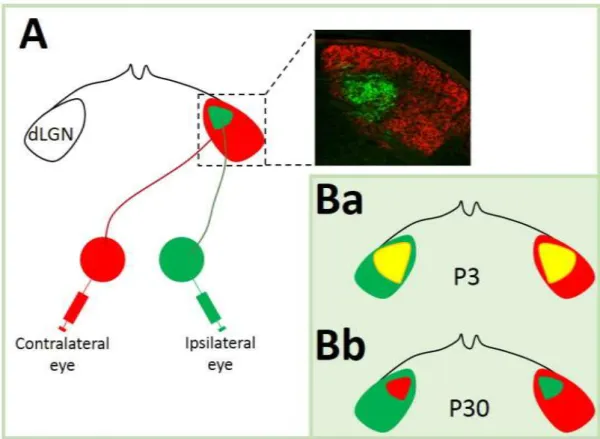
Early immunohistochemical analysis have revealed that serotonergic fibers originating from the rostral raphe nuclei innervate mostly forebrain targets, whereas the caudal B1-B3 nuclei mainly innervate the spinal cord (Törk 1990). Although, since neurons are often intermingled, the anatomical analysis of serotonin projections were ambiguous. In order to overcome this limitation a novel technique using conditional tracing has been proposed. It consists on injecting viral vectors which express GFP protein under the control of SERT to a small portion of raphe nuclei cells, providing an unambiguous way to determine their targets. Using this technique it has been possible to demonstrate that, for instance, serotoninergic neurons originating from B7 terminate, among others, in the thalamic dLGN (Muzerelle et al. 2014)(Figure 11 B).
Figure 11: Serotonergic system in the rodent brain. A: Schematic representation of raphe nuclei (B1-B9) organization within the brainstem, and of serotonergic fibers distribution (Lesch
31
2012). B: Projections of serotonergic neurons from B7 raphe nucleus to the thalamic lateral geniculate nuclei. Scale bare: 200µm. (Muzerelle et al. 2014).
Additionally to a classical synaptic transmission, where the neurotransmitter released by a presynaptic neuron into the synaptic cleft binds only to the receptors of the postsynaptic neuron, serotonin can also be released in a non-synaptic way from varicosities present along the serotoninergic raphe neurons. From there it can diffuse several microns away and activate remote receptor sites by a mechanism called volume transmission (Umbriaco et al. 1995; Bunin and Wightman 1998; Zoli et al. 1999; Daubert and Condron 2010).
A2.2
From Synthesis to Degradation
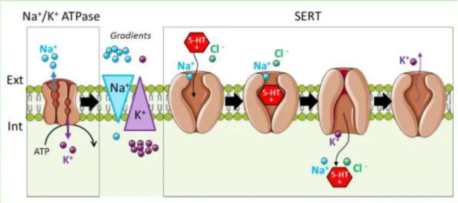
Serotonin is synthesized from tryptophan, an essential amino acid in animals (Figure 13). There are two enzymes necessary for the 5-HT production: the tryptophan hydroxylase (TPH) and tryptophan decarboxylase (AADC). In mammals, there are two TPH genes, which encode two distinct isoforms of this enzyme. The isoform “1” of TPH is expressed in the periphery (skin and intestine), while isoform “2” is brain specific and expressed in serotonergic neurons of the raphe (Walther et al. 2003; Millan et al. 2008). 5-HT is actively packed into synaptic vesicles by a vesicular monoamine transporter (VMAT2) that transport positively charged serotonin against a proton gradient (H+/5-HT) (Eiden et al. 2004).
The specific serotonin transporter, SERT, uptakes 5-HT from the extracellular environment. The energy to transport the serotonin is generated from the co-transport of sodium ions. Thus, the energy for 5-HT uptake is provided by the Na+/K+ ATPase that generates the Na+ gradient (Figure 12).