HAL Id: tel-02951816
https://tel.archives-ouvertes.fr/tel-02951816
Submitted on 29 Sep 2020HAL is a multi-disciplinary open access
archive for the deposit and dissemination of sci-entific research documents, whether they are pub-lished or not. The documents may come from teaching and research institutions in France or abroad, or from public or private research centers.
L’archive ouverte pluridisciplinaire HAL, est destinée au dépôt et à la diffusion de documents scientifiques de niveau recherche, publiés ou non, émanant des établissements d’enseignement et de recherche français ou étrangers, des laboratoires publics ou privés.
Contribution of actin cytoskeleton to C. elegans
embryonic elongation
Alicia Lardennois
To cite this version:
Alicia Lardennois. Contribution of actin cytoskeleton to C. elegans embryonic elongation. Develop-ment Biology. Sorbonne Université, 2019. English. �NNT : 2019SORUS236�. �tel-02951816�
SORBONNE
UNIVERSITÉ
Ecole
doctorale 515 Complexité du Vivant
Laboratoire
de Biologie du Développement UMR7622 IBPS
Contribution
of actin cytoskeleton to C. elegans embryonic elongation
Contribution
du cytosquelette d’actine
lors
de l’élongation embryonnaire de C. elegans
Thèse
présentée par :
Alicia
LARDENNOIS
Soutenue publiquement le 27 Septembre 2019Pour obtenir le grade de : Docteur de Sorbonne Université Discipline/Spécialité : Biologie du développement Thèse dirigée par : Dr. LABOUESSE Michel Directeur de Recherche, Sorbonne Université, IBPS Rapporteur INTERNE/ Présidente de Jury : Dr. PLASTINO Julie Directrice de Recherche, Sorbonne Université, Institut Curie Rapporteurs EXTERNES : Dr. MANGO Susan Professeur, Université de Bâle, Biozentrum Dr. HEISENBERG CarlPhilipp Professeur, Institute of Science and Technology Austria Membre invité : Dr. LLENSE Flora Maître de conférences, Sorbonne Université, IBPS
ACKNOWLEDGMENTS
It had been quite some journey for me. Science was not my first career choice, but life decided it otherwise. I learned to make the most of it and today I am very happy of the result.
I wish to thank, first and foremost, my thesis director, Michel, for handing me over such a wonderful and challenging project, and for trusting me to carry it out. Thank you for your patience, motivation, enthusiasm, and immense knowledge. Your guidance helped me in all the time of research and writing of this thesis. It has not been easy every day, but it was totally worth it. Thank you also for encouraging me when I needed it, for finding the right words. I consider it an honor to work with Flora, my supervisor in the lab, and I am extremely thankful for her support and guidance during these four intense years. Thank for encouraging me all the way, especially during the difficult moments. Thank you for your help with the experiments, your scientific advices, and very stimulating discussions (scientific or else).
I also would like to express my profound gratitude to Teresa. Without your scientific contribution, my thesis would not have been the same. Thank you for your support, various scientific discussions and also everyday talks.
Along with Michel, Flora and Teresa, I would also like to thank all present and past members of Labouesse and Robin teams: François for insightful scientific discussions (especially about cytoskeleton) but also for precious life hacks Saurabh for teaching some basics about C. elegans and how to design CRISPRs Shashi for being available each time I had questions about protocols or else (especially about CRISPR or VAB 10…) Loan for many shared laughs at the bench and in the office, and his kind help with injections Serena for great scientific discussions (as well as cats related and United Wizard discussions) Asma for her kindness and for being a Potterhead just like me Camille, Thanh, Xinyi, Vlad and Dilyana for nice conversations about science and life. I want to thank students who helped me with bench work: Antoine, Leïla and Camille. Thank you also to the Galy team members for their kind welcome whenever we go for injections. And most importantly, thank you to all for just being there, it was great sharing such a nice time with all of you during these last four years.
I cannot find words to express my gratitude to Emma, Manon and Dylan, who had been there for me all along particularly in the very difficult times. You bear with me anytime I discussed my PhD project, my paper, the difficulties I was facing and most of all the frustration that came with it. I really owe you a lot (and that bottle of Champagne I promised you!).
Special regards to Michel, Flora and Teresa for correcting the science of my thesis, as well as to Emma and my parents, for editing my thesis, tracking orthograph and layout mistakes of the documents during the last days. Finally, I deeply thank my family, and Nounoutte, my beloved cat. Thank you, Mom and Dad, for your unconditional support and love and always standing next to me whenever I need. It has been fifteen years that you trust me with the life choices I make, even the hardest ones. Your trust made me who I am today, I could not have done it without you. I will never be grateful enough.
REMERCIEMENTS
Cette expérience a été déterminante pour moi. La science n'était pas mon premier choix de carrière, mais la vie en a décidé autrement. J’en ai tiré le meilleur parti et aujourd'hui, je suis très heureuse du résultat.
Je tiens avant tout à remercier mon directeur de thèse, Michel, de m'avoir confié un projet aussi intéressant et stimulant et de m'avoir fait confiance pour le mener à bien. Merci pour ta patience, ta motivation, ton enthousiasme et tes immenses connaissances. Tes conseils m'ont aidé tout au long de cette thèse et de son écriture. Cela n’a pas été facile tous les jours, mais cela en valait la peine. Merci également de m'avoir encouragée lorsque j'en ai eu besoin et d'avoir trouvé les mots justes.
Ce fut un honneur de travailler avec Flora, ma superviseure dans l’équipe, et je suis extrêmement reconnaissante de ton soutien et de tes conseils au cours de ces quatre années intenses. Merci de m'avoir encouragée jusqu'au bout, surtout pendant les moments difficiles. Merci pour ton aide pour les expériences, tes conseils scientifiques et nos discussions très stimulantes (scientifiques ou autres). Je voudrais également exprimer ma profonde gratitude à Teresa. Sans ta contribution scientifique, ma thèse n'aurait pas été la même. Merci pour ton soutien, nos diverses discussions scientifiques et celles plus légères.
En plus de Michel, Flora et Teresa, je voudrais également remercier tous les anciens membres et membres actuels des équipes Labouesse et Robin: François pour nos échanges scientifiques approfondis (en particulier sur le cytosquelette) mais aussi pour tes précieux conseils Saurabh pour m’avoir appris quelques manipulations de base sur C. elegans et la conception des CRISPRs Shashi pour ta disponibilité à chaque fois que j’avais des questions sur les protocoles ou autres (en particulier sur les CRISPR ou VAB10…) Loan pour avoir supporté avec bonne humeur de partager la paillasse avec moi pendant deux ans et pour ton aide avec les injections Serena pour nos grandes conversations scientifiques (ainsi que celles sur les chats et United Wizard) Asma pour ta gentillesse et notre passion partagée pour Harry Potter Camille, Thanh, Xinyi, Vlad et Dilyana pour nos conversations sur la science et le reste en général. Je tiens à remercier les étudiants qui m'ont aidée sur les expériences : Antoine, Leïla et Camille. Merci également aux membres de l’équipe Galy pour leur accueil chaleureux chaque fois que nous injectons. Et surtout merci à tous d’avoir été présents, j’ai beaucoup apprécié votre compagnie au cours de ces quatre dernières années.
Je n’ai pas de mots pour exprimer ma gratitude envers Emma, Manon et Dylan, qui ont toujours été là pour moi, particulièrement dans les moments très difficiles. Vous m’avez supportée à chaque fois que je parlais de ma thèse, de mon papier, de mes difficultés et surtout de la frustration qui en découlait. Je vous dois beaucoup (nous sabrerons le champagne bientôt !).
Merci particulièrement à Michel, Flora et Teresa pour la correction de ma thèse, ainsi qu’Emma et mes parents pour en avoir perfectionné la mise en page au cours des derniers jours.
Enfin, je remercie profondément ma famille, et Nounoutte, mon chat adoré. Merci, papa et maman, pour votre soutien inconditionnel et votre amour et pour être à mes côtés à chaque fois que j'en ai eu besoin. Cela fait quinze ans que vous me faites confiance, quel que soient mes choix de vie, même les plus difficiles. Votre confiance a fait de moi ce que je suis aujourd'hui, je n'aurais pas pu le faire sans vous. Je ne serai jamais assez reconnaissante.
TABLE OF CONTENTS
ACKNOWLEDGEMENTS / REMERCIEMENTS ... I TABLE OF CONTENTS ... II ABBREVIATIONS ... III
Common abbreviations for mutant phenotypes in C. elegans Most commonly mentioned genes in this work
ABSTRACTS (in English and French) ... 1
ABSTRACT ... 3
RÉSUMÉ COURT ... 4
RÉSUMÉ LONG ... 5
1. Phénotype de rétraction et activité musculaire ... 7
2. Remodelage et dynamique du cytosquelette d’actine ... 7
2.a. Organisation de l'actine ... 7
2.b. Remodelage durant l’élongation ... 8
3. Modélisation de l’élongation... 9 Méthodes ... ... 10 Publications et Conférences ... 10 Communications orales... 10 Posters ... ... 10 INTRODUCTION ... 11 I. Epithelia in morphogenesis ... 15
1. General characteristics and function of the epithelia ... 17
2. The organization of an epithelial cell ... 18
2.1. Epithelial junctions ... 19
2.2. Extracellular matrix (ECM) ... 22
2.3. Cytoskeleton ... 22
2.3.1. Actin and myosin ... 23
2.3.2. Microtubules ... 28
2.3.3 Intermediate filaments ... 29
II. Epithelia remodeling during morphogenesis ... 31
1. Contractility and intrinsic forces ... 33
1.1. Apical constriction ... 33
1.2. Cell migration... 35
1.3. Convergent extension ... 38
2. Extrinsic forces – Mechanotransduction ... 41
2.1. Matrix control of stem cell fate ... 41
2.2. Role of cytoskeleton in mechanotransduction ... 43
2.3. Protein unfolding under force: in vitro example, spectrin ... 44
2.4. Protein unfolding under force: in vivo example, talin ... 46
2.5. The plasma membrane as a tension sensor ... 48
III. Introduction to our model: the nematode Caenorhabditis elegans ... 53
1. C. elegans general anatomy ... 54
2. C. elegans muscles ... 55
3. C. elegans epidermis ... 56
3.1. C. elegans Adherens Junctions (CeAJs) ... 57
3.2. C. elegans hemidesmosomes (CeHDs) ... 58
3.3. C. elegans extracellular matrix ... 61
4. Cytoskeleton ... 62
4.1. Actin ... 62
4.2. Actin remodeling proteins ... 64
4.2.1. Formin ... 64 4.2.2. Gelsolin ... 66 4.2.3. Vilin ... 67 4.3. Microtubules ... 68 4.4. Spectrin cytoskeleton ... 69 4.4.1. βGspectrin/UNC70 ... 71 4.4.2. βHspectrin/SMA1 ... 72 4.4.3. αspectrin/SPC1 ... 73
4.4.4 The spectrin heterotetramers ... 74
5. C. elegans life cycle and its embryonic development ... 76
5.1. Overview of the embryonic elongation ... 78
5.2. Early elongation ... 80
5.3. Late elongation ... 81
IV. Aim of this thesis ... 83
MATERIAL AND METHODS ... 85
Animal strains, conditions of maintenance ... 87
Yeast Two Hybrid Screening ... 87
RNAi screens ... 87
Fluorescent translational reporter constructs... 88
SPC1::GFP and PAK1::mKate ... 88
FHOD1 full length and alternative constructs ... 89
LifeAct::mMaple3 photoconvertible construct ... 89
ACT1::GFP overexpression ... 90
Pdpy7::GFP overexpression ... 90
CRISPR/Cas9 fluorescent knockin transgenic worm generation ... 91
ACT1 / SMA1 / SPC1 ... 91
UNC70 ... 92
Table recapitulating the different strategies used ... 93
Fluorescence imaging ... 94
TIRFSIM ... 94
Image analysis and quantification of actin filament contraction, continuity and orientation ... 95
Anisotropy of the orientation ... 96
Straightness ... 96
Bundle organization ... 97
Statistical Analysis ... 97
RESULTS ... 99
I. Introduction to the results ... 101
II. Previous work: molecular and functional screens identify SPC1 as a potential PAK1 partner . 102 1. SPC1 and PAK1 loss leads to a retraction of the embryo ... 103
2. This retraction phenotype depends on the muscle input ... 105
III. PAK, SPC1, SMA1 colocalize with actin near the epidermal cell membrane ... 106
1. PAK1 and SPC1 localize along the actin filaments ... 106
2. Superresolution shows precise colocalization of actin and spectrin cytoskeletons ... 108
3. Other tools have been developed to investigate actin dynamics ... 109
IV. Genetic interactions of pak1, sma1, unc70, vab10b affects elongation ... 112
V. The organization and remodeling of actin filaments is a key element of the retraction ... 115
1. Spinningdisk characterization of the actin disorganization in spc1 pak1 embryos ... 115
2. Enhanced visualization of actin filament abnormalities in spc1 pak1 defective embryos ... 119
3. The intensity of actin varies over time in spc1 pak1 defective embryos ... 122
VI. The embryo diameter decreases during elongation ... 123
VII. Muscle contractions bend actin filaments leading to the recruitment of severing proteins ... 125
1. Actin filaments are bend at very sharp angles upon muscle input ... 125
2. GSNL1 and VILN1 are involved in the remodeling of actin in our system ... 127
VIII. A KelvinVoigt model recapitulates the elongation of the embryos of various phenotypes .... 130
IX. Combined loss of FHOD1 and SPC1 leads to the same retraction phenotype ... 137
1. fhod1 spc1 retracts and their actin show the same abnormalities as spc1 pak1 ... 137
2. FHOD1 bundling activity is important for the remodeling ... 141
3. FHOD1 and PAK1 localization are affected the same way by the lack of SPC1 ... 142
DISCUSSION AND PERSPECTIVES ... 145
I. Identification of a novel morphogenetic ratchet ... 150
II. Modelization of our ratchet mechanism ... 152
III. Unraveling the internal organization of actin ... 154
IV. Interaction of actin and spectrin cytoskeleton ... 156
V. SPC1 as a major player in a mechanotransduction pathway ... 157
VI. Identification of other players that could also be involved ... 159
APPENDIX I. Caption for movies and tables ... 163
APPENDIX II. Tables ... 167
APPENDIX III. An actinbased viscoplastic lock ensures progressive body axis elongation ... 175 REFERENCES
ABBREVIATIONS
ABBREVIATIONS
A/P Anterior/Posterior ABD Actin Binding Domain ABP Actin Binding Protein ABS Actin Binding Site
AFM Atomic Force Microscopy AJ Adherens Junctions BMD Body Morphology Defect C. elegans Caenorhabditis elegans CeAJ C. elegans Adherens Junctions CeHD C. elegans Hemidesmosomes CH Calponin Homology (domain)
CRIB Cdc42/Rac Interactive Binding (domain)
CRISPR Clustered Regularly Interspaced Short Palindromic Repeats DIC Differential Interference Contrast
DLG1 (Drosophila) DiscsLarge (homologue)1 DNA Deoxyribonucleic Acid
D/V Dorsal/Ventral ECM Extracellular Matrix FA Focal Adhesions
FH Formin Homology (domain)
FRAP Fluorescence Recovery After Photobleaching FRET Förster Resonance Energy Transfer
GAS2 GrowthArrestSpecific protein 2 GBD GTPase Binding Domain GFP Green Fluorescence Protein GIT G proteincoupled receptor kinase InTeractor IF Intermediate Filament L1L4 Larval stages of C. elegans development MHC Myosin essential Heavy Chain MLC Myosin essential Light Chain MSC Mesenchymal Stem Cells MT Microtubule
MTOC Microtubule Organizing Center NGM Nematode Growth Medium NMYII Nonmuscle MyosinII PAK p21Activated Kinase PAM Protospacer Adjacent Motif PCR Polymerase Chain Reaction PH Plecktrin Homology (domain)
PIP2 Phosphatidylinositol4,5bisphosphate PIX PAK interacting exchange factor PR Plectin Repeat
RLC Regulatory Light Chain (of nonmuscle myosinII) RNA Ribonucleic Acid
RNAi RNA interference ROCK Rhoassociated Kinase ROI Region Of Interest sgRNA single guide RNA SH3 Src Homology (domain) SR Spectrin Repeat (domain)
TIRFSIM Total Internal Reflection Fluorescence Microscopy Structured Illumination Microscopy
VBS Vinculin Binding Site Y2H Yeast Two Hybrid ZP Zona Pellucida Common abbreviations for mutant phenotypes in C. elegans Dpy Dumpy Let Lethal
Pat Paralysed at twofold Sma Small size
Unc Uncoordinated movement Vab Variable abnormal Morphology
Most commonly mentioned genes in this work
fhod1 encodes for C. elegans formin.
mutants are viable and fertile although they show some body wall muscle defects.
gsnl1 encodes for C. elegans gelsolin. mutants are viable and fertile.
pak1 encodes for C. elegans p21activated kinase1.
mutants are viable and fertile, slight BMD (enlarged head) in L1 larvae.
spc1 encodes for C. elegans αspectrin.
mutants show embryonic lethality and fails to elongate beyond 2fold.
sma1 encodes for C. elegans βHspectrin.
mutants display a Sma phenotype with shortbody length.
unc70 encodes for C. elegans βGspectrin.
mutants are short and paralyzed; the defects develop gradually through the larval stages. However embryonic elongation is not affected.
unc112 encodes for a C. elegans dense body component, vertebrate kindlinhomologue mutants display a Pat phenotype: embryonic arrest due to muscle dysfunction and paralyzation at the 2fold stage.
vab10 encodes for C. elegans spectraplakin.
mutants have defects in embryonic elongation, body morphology, uterusvulva connection, and gonad arm migration, the animals have poor mating efficiency. generates isoforms related either to plectin (VAB10A) or to microtubule actin cross linking factor plakins (VAB10B) that have distinct functions and localizations.
viln1 encodes for C. elegans villin. mutants are viable and fertile.
ABSTRACTS
(in
English and in French)
ABSTRACT
Body axis elongation is a fundamental morphogenetic process, involving cell shape changes powered by mechanical forces through small incremental steps which need to be stabilized. During my PhD, I studied C. elegans embryonic elongation to define how the embryo, an elastic material, lengthens progressively upon muscle contractions. Previously, the lab found a kinase, PAK1, to be mediator of an epidermal mechanotransduction pathway downstream of muscles. Two screens in a pak1(Ø) background identified αspectrin SPC1 as an interactor of PAK1. spc1()pak1() embryos elongate up to 1.5fold and then retract to 1fold in a muscle dependent manner. I used super resolution microscopy to show that epidermis circumferential actin bundles are highly disorganized in these embryos; suggesting that actin rearrangement could be the lock counteracting elasticity. With a screen in spc1()pak1() background, I identified two severing proteins helping break actin filaments when muscle activity bends them at sharp angles. In addition, the actin bundling formin FHOD1, was also shown to induce retraction in fhod1();spc1() embryos. I overexpressed a C terminally truncated FHOD1(ΔFH2/DAD) that partially rescued the spc1()pak1() retraction suggesting that FHOD1 blocks further actin depolymerization at each cycle of contraction. To test it, we modeled the embryo as a KelvinVoigt material under actomyosin force from the epidermis and muscle tension. We predicted embryo lengthening using a viscoplastic component accounting for actin shortening. Altogether I characterized a cellular network conferring mechanical plasticity to stabilize cell shape during a morphogenetic process.
4
RÉSUMÉ COURT
Les processus morphogénétiques impliquent des changements de forme de cellules, via des forces mécaniques, qui doivent être stabilisés. Ma thèse vise à élucider comment l'embryon de C. elegans, matériau élastique, s'allonge progressivement sous l’effet des contractions musculaires. Un crible ARNi en fond pak1(Ø), kinase de l’épiderme impliquée dans une cascade de mécanotransduction en aval des muscles, a identifié SPC1/αspectrine, comme partenaire probable. Les embryons spc1()pak1() s'allongent jusqu'à 1.5fold, puis reviennent à leur taille initiale sous l’effet des muscles. Avec la microscopie à superrésolution, j’ai montré que leurs faisceaux d'actine épidermiques sont très désorganisés ; suggérant que le remodelage de l'actine pourrait contrer l'élasticité des cellules. Avec un crible en fond spc1()pak1(), j'ai identifié deux protéines de fragmentation aidant à rompre les filaments d'actine quand les muscles les courbent fortement. Par ailleurs, la formine de “pontage” FHOD1 induit aussi une rétraction dans des embryons fhod1( );spc1(). J'ai surexprimé une construction FHOD1(ΔFH2/DAD) tronquée en Cterminal qui a partiellement sauvé la rétraction spc1()pak1() suggérant que FHOD1 bloque la dépolymérisation de l'actine à chaque cycle de contraction. Pour tester cela, j’ai modélisé l'embryon en tant que matériau KelvinVoigt soumis à une force épidermique et à la tension musculaire et prédit son allongement en utilisant un composant viscoplastique symbolisant le raccourcissement de l'actine. J'ai donc caractérisé un réseau cellulaire conférant une plasticité mécanique et stabilisant la forme des cellules dans un processus morphogénétique.
5 RÉSUMÉ LONG En tant qu'organismes vivants, nous interagissons avec notre monde grâce à nos sens. Nous sommes continuellement exposés à des signaux mécaniques, et il en va de même pour nos cellules. Ces dernières ont la capacité de détecter leur environnement physique en traduisant des forces mécaniques et des déformations en signaux biochimiques. À leur tour, ces signaux peuvent ajuster les structures cellulaires et extracellulaires. Au cours des deux dernières décennies, de nombreux processus biologiques répondant à des stress mécaniques ont été mis en évidence tels que : le changement de pression de turgescence dans la réponse au toucher chez Mimosa pudica, la différenciation variable des cellules souches mésenchymateuses en fonction de la rigidité du milieu de croissance sousjacent, la cicatrisation des plaies ou encore la prolifération et la dissémination des cellules cancéreuses. Les différents mécanismes par lesquels le tissu cellulaire convertit le stimulus mécanique en une activité électrochimique ou biochimique sont appelés mécanotransduction. La mécanotransduction est à l’origine d'un certain nombre de sens et de processus physiologiques dans le corps, notamment la proprioception, le toucher, l'équilibre et l'audition. Au niveau cellulaire, elle est importante pour de nombreuses fonctions telles que l'adhésion, la migration, la prolifération, la différenciation et l'apoptose et elle est essentielle au développement d'organes. Une étape clé du développement de l’animal est la mise en place de l’axe du corps définissant sa forme finale lors de la morphogenèse. Ce processus détermine la forme et la structure des tissus, des organes et des organismes. Plus spécifiquement, la morphogenèse embryonnaire désigne des modifications de la forme et de la position des cellules dans l’embryon, en particulier de l’épithélium. L'importance de la morphogenèse épithéliale a été rapportée dans des processus de développement cruciaux tels que la gastrulation (étudiée de manière approfondie dans les ascidies, la drosophile, le poisson zèbre ou le xénope) ou dans la formation du tube neural. Cette étape dépend de manière critique des changements de forme de cellules, euxmêmes dépendants de l'interaction entre les forces intrinsèques et extrinsèques aux cellules. Comprendre comment les forces mécaniques influencent la morphogenèse au niveau cellulaire et moléculaire reste un défi majeur.
Pour étudier la mécanotransduction et son importance dans la morphogenèse à l'échelle d'un organisme, nous travaillons avec un modèle simple, le nématode C. elegans. Depuis son introduction, par Sydney Brenner, dans les laboratoires à la fin des années 1960, C. elegans est utilisé comme organisme modèle en biologie du développement, génétique et biologie moléculaire grâce à sa progéniture abondante, son développement rapide, son corps transparent et son génome complètement séquencé. Son épiderme est un épithélium à monocouche séparé des muscles par une mince membrane au niveau basal. Les muscles sont disposés en quatre rangées, deux attachées à l'épiderme dorsal et deux à l’épiderme ventral via les hémidesmosomes, rendant l’épiderme
6
sensible au stress mécanique résultant de leurs cycles de contraction. Au cours de son élongation embryonnaire, le nématode C. elegans voit sa longueur multipliée par un facteur 4 et sa circonférence réduite par un facteur 2,5. Il s’agit d’un processus rapide (trois heures environ) qui a lieu sans réarrangement ni division cellulaire, seulement permis par la déformation des cellules latérales et dorsoventrales de son épiderme. Cette élongation est initialement activée par la contractilité de l'actomyosine dans les cellules latérales, via la régulation de la Rho kinase. A partir du stade « 2fold », la tension fournie par les contractions musculaires permet le recrutement de la protéine adaptatrice GIT1 aux hémidesmosomes, permettant la seconde phase de l’élongation notamment via activation de la kinase PAK1. Lorsque l’activité musculaire est inhibée, les embryons sont paralysés à un stade « 2fold » et GIT1 n’est plus recrutée. Pourtant les mutants git1 et pak1 sont viables, suggérant qu'une autre voie agit en parallèle. Par ailleurs, chez C. elegans, la contribution du cytosquelette d’actine à la morphogenèse embryonnaire a été rapportée depuis la fin de la gastrulation. Au début, l'actine forme un maillage près du cortex épidermique, et au fur et à mesure elle se réorganise pour former des faisceaux circonférentiels et parallèles d'abord dans les cellules dorsoventrales, puis dans les cellules latérales. Une fois l'élongation embryonnaire terminée, les faisceaux se désassemblent. Dans ce contexte, l'objectif de mon projet de thèse a été de caractériser de manière plus détaillée l'apport du cytosquelette d'actine pendant l'élongation embryonnaire de C. elegans et la façon dont les câbles d'actine sont remodelés au cours de ce processus.
De précédents travaux de l’équipe ont montré que PAK1 fait partie d’une voie de mécanotransduction induisant le remodelage des hémidesmosomes, structures de couplage entre les muscles et l’épiderme susjacent. Deux cribles ont alors été réalisés en fond sensible pak1() pour rechercher de potentiels partenaires de PAK1 et l’αspectrine SPC1 s'est révélée être un fort candidat. Les spectrines ont été décrites pour la première fois dans les érythrocytes en 1968 par Marchesi et Steers. Elles sont un des composants majeurs du cytosquelette de la membrane cellulaire et sont responsables des propriétés mécaniques (stabilité, plasticité et déformabilité) de celleci.
À l'aide d'outils génétiques et d'approches d'imagerie in vivo, une ancienne doctorante de l’équipe, Gabriella Pàsti, a montré que l'αspectrine et PAK1 interagissent génétiquement. En effet, elle a constaté que chez les mutants spc1 () pak1 (), les embryons s’allongent jusqu’à « 1,5fold », puis se rétractent jusqu’à leur taille initiale (phénotype de rétraction). Fait intéressant, ce phénotype avait déjà été décrit suite à la dépolymérisation massive de l'actine, via l’usage d’une drogue, suggérant que le réarrangement de l'actine pourrait fonctionner comme un verrou lors de la
7
morphogenèse embryonnaire. Mon travail a donc consisté à caractériser plus en détail cette interaction entre le cytosquelette d'actine et l'αspectrine en répondant aux questions suivantes : − Comment fonctionne le mécanisme de verrou au niveau moléculaire ? − Comment les câbles d'actine sontils remodelés au cours du temps ? − Quels sont les rôles des différents acteurs identifiés ? − Peuton modéliser l'allongement progressif de l’embryon pour comprendre comment les forces contribuent au remodelage de l’actine ? 1. Phénotype de rétraction et activité musculaire
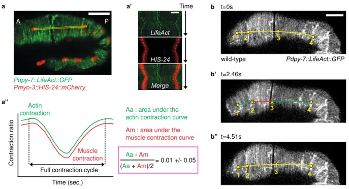
Sachant que les embryons spc1() pak1() amorcent leur rétraction lorsque les muscles comment à être actifs, j'ai essayé de déterminer la manière dont l'apport musculaire affecte la rétraction des embryons. Précédemment, Gabriella a montré que si l’activité musculaire était bloquée que chez des mutants spc1() pak1(), grâce l’utilisation d’un ARNi contre unc112 (kindlin homologue et composant essentiel des muscles), cela empêchait la rétraction des embryons. J'ai terminé cette expérience en ajoutant plusieurs contrôles supplémentaires en mesurant l’élongation des vers unc112(); pak1() et unc112(); spc1(). Ils ont montré une croissance plus lente très semblable aux vers unc112() seuls. Pour comprendre comment les contractions musculaires contribuent au phénotype de rétraction, j'ai quantifié la différence de comportement entre embryons de type sauvage et mutants en utilisant des points de repère sur l'actine épidermique. J'ai mesuré le temps de contraction / relaxation et l'ampleur de la contraction / relaxation. Les doubles mutants ont présenté des contractions plus rapides et plus profondes que les embryons de type sauvage et pak1(). Ces deux expériences ont montré que l'apport musculaire est crucial pour le phénotype de rétraction bien qu'il soit perturbé dans le double mutant spc1() pak1().
2. Remodelage et dynamique du cytosquelette d’actine 2.a. Organisation de l'actine
En plus de contracter plus rapidement, les embryons spc1() pak1() présentent également des défauts d'organisation de l’actine : les câbles d'actine ne sont plus complètement parallèles, ils se regroupent et semblent parfois discontinus. Pour mieux caractériser ces défauts observés avec un microscope « spinningdisk », j'ai effectué une analyse de texture développée dans l'équipe avec l'aide de notre ingénieure, Teresa Ferraro. Nous avons analysé des images d'embryons anesthésiés à différents stades («1,7fold», «2fold» et «3fold») pour obtenir une résolution plus élevée de l'actine. L'analyse du signal de fluorescence associé aux filaments d'actine dans l'épiderme dorso ventral a révélé davantage de discontinuité dans les embryons spc1() pak1() par rapport aux témoins ; de plus, leur degré d'anisotropie par rapport à l'axe circonférentiel était anormal. Avant
8
que les muscles commencent à se contracter, aucune différence significative n'est visible. Dès que l’activité musculaire est établie, l'organisation de l’actine devient significativement différente entre les contrôles et les spc1() pak1() mais également entre spc1(ARNi) et spc1() pak1(). Dans un second temps, j'ai utilisé la microscopie à super résolution pour affiner ces observations et éventuellement accéder à des évènements de remodelage en temps réel. L'analyse de segmentation du signal de fluorescence associé aux filaments d'actine a été confirmée et même améliorée puisque nous avons pu faire une différence entre la zone de l’épiderme située juste audessus des muscles et celle située à côté : les défauts d’organisation sont plus prononcés au niveau des muscles, là où la tension est plus forte. Par ailleurs, l'intensité du signal entre les faisceaux d'actine adjacents était également moins nette chez les embryons spc1() pak1() et ils étaient plus souvent courbés, indiquant que les faisceaux pourraient s'être partiellement défasciculés.
Ces phénotypes apparaissent une fois que les muscles entament leur activité suggérant que les contractions musculaires contribuent au remodelage de l'actine. Pour étudier plus en détail leur rôle dans ce processus, nous avons examiné les filaments d'actine spécifiquement lors de la contraction des muscles. De manière frappante, la microscopie « spinningdisk » a révélé que les contractions musculaires sont suffisamment fortes pour courber localement des faisceaux d'actine avec un angle moyen de 57 °, ce qui induirait une cassure du filament d'actine d’après des analyses réalisées in vitro. Les cycles répétés d'activité musculaire pourraient avoir pour effet d'induire localement la rupture du filament d'actine épidermique, suivie de leur stabilisation. Dans les doubles mutants spc 1() pak1(), des contractions musculaires plus courtes pourraient faire pencher la balance entre rupture et stabilisation conduisant ainsi à leur désorganisation. 2.b. Remodelage durant l’élongation Tous ensemble, les résultats décrits cidessus établissent un lien entre l'activité musculaire et désorganisation de l’actine chez les embryons spc1() pak1(). L’hypothèse la plus simple est que l’activité musculaire induit chez les embryons normaux une rupture et une stabilisation de l’actine bien contrôlée qui se détériore chez les embryons spc1() pak1(). En effet, une autre analyse sur ces images a montré que la circonférence de l'embryon diminuait de 20% au cours de l'allongement, impliquant que les filaments d'actine dans les cellules dorsoventrales doivent également se raccourcir. Pour comprendre au niveau moléculaire comment ce remodelage pourrait se produire, j'ai effectué un crible ARNi en fond sensible spc1() pak1() à la recherche d'un sauvetage du phénotype de rétraction. De manière remarquable, avec l’aide de Flora Llense, maître de conférences dans l’équipe, nous avons identifié des homologues de deux « protéines de fragmentation » de l'actine, la gelsoline et la villine suggérant que les contractions musculaires
9
induisent une possible rupture du filament d'actine et donc une stimulation directe ou indirecte de l'activité des protéines.
Pour aller plus loin dans cette caractérisation moléculaire, Gabriella a effectué un dernier crible ARNi en fond sensible spc1() pour trouver d’autres partenaires potentiels permettant de reproduire le phénotype de rétraction. Nous avons identifié une protéine de « câblage », la formine atypique FHOD1. Pour évaluer le lien entre PAK1, SPC1 et FHOD1 dans le phénotype de rétraction, Flora a cloné plusieurs constructions de FHOD1 basées sur la littérature existante. Il a en effet été démontré que FHOD1 est initialement inactive en raison d’une interaction autoinhibitrice entre son domaine autorégulateur en Cterminal et son domaine inhibiteur en Nterminal (DID). Des expériences in vitro ont montré qu’en supprimant le domaine DAD, l'autoinhibition est levée et conduit à une forme constitutivement active de la protéine. L'objectif était de savoir si je pouvais sauver le phénotype de rétraction d'embryons spc1() pak1() en surexprimant FHOD1. Effectivement j'ai observé que 2/3 des embryons exprimant la construction FH2/DAD ne se rétractent plus et qu'ils sont significativement plus longs que les doubles mutants spc1() pak1().
3. Modélisation de l’élongation
Pour rendre compte de l’élongation du ver à l’échelle mésoscopique, nous avons développé un modèle physique prédictif. Changer le statut d'une entité physique nécessite l'intervention d'une force (mécanique ou chimique) et l'embryon de C. elegans n'échappe pas à cette règle de la physique. Pendant la première phase d'élongation et jusqu'à ce que les muscles deviennent actifs, l'élongation est permise par une force active dans l'épiderme latéral et par une force passive exercée par les cellules épidermiques dorsales et ventrales suffisante pour allonger l'embryon jusqu'au stade « 2fold ». Cependant, cette force n'est pas suffisante pour expliquer l'allongement jusqu'au stade « 4fold », puisque les embryons avec des muscles non fonctionnels ne s'allongent pas audelà du stade « 2fold ». Par conséquent, les muscles fournissent une deuxième force motrice d’allongement. Considérant cela, nous avons décidé de modéliser l’embryon de C. elegans comme un matériau de KelvinVoigt soumis à deux forces actives principales : la force épidermique, qui est une force positive continue, et la force musculaire, qui est une force pulsatile puisque les muscles se contractent alternativement. En utilisant un ensemble d'équations mathématiques, nous avons pu prédire l'allongement de l'embryon en introduisant un composant viscoplastique dans le système, symbolisant le raccourcissement de l'actine. Grâce à cela, nous avons également pu modéliser la rétraction des doubles mutants spc1() pak1(). Comme la continuité des filaments d'actine est modifiée chez ces mutants, leur résistance au stress provenant des cellules latérales ne serait pas maintenue et la force épidermique diminuerait progressivement, ce qui réduirait la longueur du
10
système. L'absence combinée de SPC1 et de PAK1 coïncide avec l'incapacité d'étendre plastiquement la longueur du tissu.
Globalement, nos résultats identifient deux groupes de protéines impliquées dans la stabilisation de la forme des cellules dans un épiderme soumis à un stress répété : SPC1, PAK1 et FHOD1 qui stabilisent les câbles d’actine lors du remodelage, GSNL1 et VILN1 qui les sectionnent pour les aider à raccourcir. Ce réseau cellulaire confère une plasticité mécanique (en termes physiques, il implique une déformation irréversible sous contrainte) stabilisant la forme des cellules pendant la morphogenèse. Comprendre comment les cellules s'adaptent à un stimulus mécanique est essentiel et pourrait aider à élucider les dernières étapes de la morphogenèse chez C. elegans. Dans un contexte plus médical, la dérégulation de PAK1 et des spectrines a été décrite dans de nombreuses pathologies. PAK1 apparaît surexprimée dans de multiples formes de cancers humains tels que le cancer du pancréas, de la prostate, du sein ou encore les mélanomes. De nombreuses études ont montré que la capacité à détecter une entrée mécanique, sa transduction dans la cellule et sa réponse sont défectueuses dans les cellules cancéreuses. In fine, nos résultats pourraient aider à mieux comprendre la dérégulation de la réponse à un stress mécanique lors de maladies. Méthodes : Les méthodes suivantes ont été utilisées dans le cadre de ce travail. Analyse génétique, ARN interférent pour inhiber l’expression de gènes, Construction de plasmides, Édition du génome par CRISPRCas9, Microscopie superrésolutive, Microscopie à disque rotatif, Microscopie à lapse de temps, Microinjection, Analyse d’images, Modélisation mathématique
Publications et Conférences :
Alicia Lardennois*, Gabriella Pásti*, Teresa Ferraro, Flora Llense, Pierre Mahou, Julien Pontabry, David Rodriguez, Samantha Kim, Emmanuel Beaurepaire, Christelle Gally, Michel Labouesse. An actinbased viscoplastic lock ensures progressive body axis elongation. Nature. Communications orales : European Worm Meeting (EWM), Juin 2016, MDC, Berlin Séminaire de restitution PHC Maimonide, Novembre 2017, Jérusalem VerMidi XXI, Janvier 2018, CIML, Marseille European Worm Meeting (EWM), Juin 2018, Barcelone Building The Cell, Septembre 2018, Institut Pasteur, Paris – Prix du Meilleur Flask Talk International Worm Meeting (IWM), Juin 2019, UCLA, Los Angeles Posters : Retraite de l’école doctorale, ED 515, October 2016, Roscoff VerMidi XX, Janvier 2017, ENS Lyon Journée Boris Ephrussi, Avril 2017, Institut Pasteur, Paris International Worm Meeting (IWM), Juin 2017, UCLA, Los Angeles Building The Cell, Septembre 2018, Institut Pasteur, Paris Journée Boris Ephrussi, Avril 2019, ENS (Campus Jourdan), Paris
INTRODUCTION
INTRODUCTION
The range of animal forms is immense, both in terms of obvious external appearances and in terms of the internal anatomies of organs and tissues. Observing the vast number of different shapes, we can wonder how does this diversity arise? Morphogenesis means the generation of form, and usually in the context of developmental biology it means the generation of tissue organization and shape in animal and plant embryos. It is one of three fundamental aspects of developmental biology along with the spatiotemporal control of cell growth and cellular differentiation. The process controls the organized spatial distribution of cells during the embryonic development of an organism. Morphogenesis can also take place in a mature organism, in cell culture or inside tumor cell masses.
Some of the earliest ideas and mathematical descriptions on how physical processes and constraints affect biological growth, and hence natural patterns such as the spirals of phyllotaxis, were written by D'Arcy Wentworth Thompson (Thompson, 1917; Montell, 2008) and Alan Turing (Turing, 1952). Thompson explained animal body shapes as being created by varying rates of growth in different directions, for instance to create the spiral shell of a snail. Turing correctly predicted a mechanism of morphogenesis, the diffusion of two different chemical signals, one activating and one deactivating growth, to set up patterns of development, decades before the formation of such patterns was observed (Hiscock and Megason, 2015). The first major approach to investigating morphogenesis was to look at the intrinsic morphogenetic properties of cells. It showed that randomized aggregates of cells from a mix of amphibian embryonic tissues would not only sort themselves out into their cell types but also generate some structure (Townes and Holtfreter, 1955). They demonstrated that the cells themselves had morphogenetic properties that they could use and stimulated a great deal of work in the '60 '70s and '80s on the morphogenetic abilities of cells. A second approach was to analyze cell behavior in tissues that that will develop in culture where they can be experimentally manipulated. As chick and amphibian embryos are large and accessible, they have been the model species of choice for studying morphogenesis (e.g. neural crest and nerve migrations, corneal development, gastrulation, and epithelial morphogenesis) (Trelstad et al., 1967; Summerbell and Wolpert, 1972; Summerbell and Wolpert, 1973; Meier, 1981; Heasman et al., 1984; Weliky et al., 1991). The mouse Mus musculus, the zebrafish Brachidanio rerio, the fruitfly Drosophila melanogaster and the roundworm Caenorhabditis elegans have also supplied continuous and extensive insights into cellular and molecular aspects of morphogenesis (Knust and Müller, 1998; Chisholm and Hardin, 2005; Ellertsdóttir et al., 2010; RiveraPérez and Hadjantonakis, 2014).
Morphogenesis was a major area of research in the 70s and early '80s, but activity then declined as the focus of research in development moved to discovering and studying the genes involved in networks that regulate differentiation. However the discovery of molecules controlling tissue organization and the development of transgenic animals led to an enormous amount of work in the first decade of the 21st century that has explained much about the molecular basics of morphogenesis, but less is known about how these are integrated at the cellular level.
For decades, the morphogenesis of epithelial tissues has fascinated scientists at the interface of cell, developmental, and molecular biology. Epithelial morphogenesis includes the various processes by which epithelia contribute to organ formation and body shape. During embryonic development, definitive epithelial organs often arise from preexisting epithelial tissues. Indeed, epithelia form transient structures, such as the neural tube, somites, and the precardiac epithelium, that serve as progenitors for the development of more complex organs (Affolter et al., 2003; Mango, 2007; Bryant and Mostov, 2008). Different epithelia acquire diverse morphological forms appropriate for their specific functions, such as the kidney tubules or the complex branching structures found in the lung for example (Miura, 2015; Marciano, 2017). Recently, the characterization of some of the molecular mechanisms involved in epithelial morphogenesis has provided an abundance of new information on the role and regulation of the cytoskeleton, cellcell adhesion, and cellmatrix adhesion in these processes (Ding et al., 2004; Blankenship et al., 2006; Armenti and Nance, 2012; Bosveld et al., 2012, Chauhan et al, 2015).
Here I will first discuss the epithelia as layers of cells and their cellular characteristics. Then I will detail the dramatic change they undergo to drive morphogenetic processes, such as shape changes, increase in number, or rearrangements. Finally, I will talk about the mechanical forces, generated by actomyosin and transmitted by adhesive junctions, driving most of these changes.
I. Epithelia in morphogenesis
Maintaining the homeostasis of a multicellular organism requires compartmentalization of the internal environment of the external environment. In metazoans, the epithelial tissues fulfill this function. These tissues consist of epithelial cells that line the entire body and cavities of the body. In addition, these cells are histologically compact and rest on a basal lamina. Nevertheless, the epithelium denomination includes tissues with vastly different forms and functions within the same organism. Thus, the different epithelia can first be classified empirically according to the overall morphology of their cells (squamous morphology, cubic or cylindrical). Epithelia can also be distinguished by the number of layers of the tissue: layered epithelia consisting of several stacked cell layers (eg. skin epithelium), pseudostratified epithelia (eg. tracheal epithelium) and simple epithelia, consisting of a single layer of epithelial cells light (ex: epithelium of the jejunum) (Marieb, 1995; Guillot and Lecuit, 2013).
Biologically, epithelia support the structures of organs and protect the body from the outside pathogens. They separate the interior from the exterior environment as a barrier or control the substance exchange across the plasma membrane as a regulated barrier. They also take part in sensing the environment, repairing wounds, contributing to various steps of development, and playing a pivotal role in shaping organs. To fulfill these roles, the epithelial cells can present additional specific structures, such as microvilli (eg intestinal epithelium), vibratile eyelashes (eg bronchial epithelium) or stereocilia (eg inner ear cells). However, despite this morphological and functional diversity, epithelial cells have a set of molecular characteristics that define them. They have an apicobasal polarity that allows polarized secretion, communication, and uptake of material from extracellular sources. This epithelial polarity is central in the biology of epithelia (Munro et al., 2004; Houk et al., 2012; Von Stetina and Mango, 2015) (Fig. 1). Indeed, its implementation precedes and is required for the morphogenesis of epithelial tissues and the maintenance of their homeostasis.
Figure 1: Morphological and functional diversity of epithelia.
Epithelial cells can form tissues of very different shapes and functions within the same organism. Nevertheless, their apicobasal polarization is required for morphogenesis to take place. Adapted from Kévin Sollier’s thesis. In addition, they also have several cytoskeletal elements with distinct functions that allow both their shape maintenance and force generation. Finally, they have multiple sets of junctions and attachment structures that allow them to form layers with each other and to set up connection with neighboring tissues. These contacts do not solely serve structural and barrier functions: they allow intra and intertissular communication in two ways. One is the classical, chemical signaling. The other one involves physical forces generated in one cell or tissue, which are transmitted and turned into biochemical signals in another cell or tissue. This way of communication is termed as mechanotransduction that is a wellcharacterized process in sensory mechanisms. On the other hand, a relatively new and dynamically developing research field establishes its role in various developmental processes, as for instance epithelial morphogenesis. This crossing point of epithelial morphogenesis and mechanotransduction stands in the very center of our research limelight.
I.1. General characteristics and function of the epithelia
Epithelia, along with the connective, the muscle, and the nervous tissue, are one of the four basic animal tissue types. Epithelia are continuous sheets of cells (one or more layers thick) that cover the exterior surfaces of the body, line internal closed cavities and body tubes that communicate with the outside environment (the alimentary, respiratory and genitourinary tracts), make up the secretory portions of glands and their ducts, and are found in the sensory receptive regions of certain sensory organs (e.g. ear & nose). Epithelia cover and lining surfaces (e.g. skin), take part in absorption (e.g. the intestine), secretion (e.g. glands), can be sensory (e.g. neuroepithelium) or contractile (e.g. myoepithelial cells). They secrete a basement membrane which supplies a site of attachment for the epithelium, and acts as a selective filtration barrier. There is little intercellular material. Epithelia do not have their own blood supply (avascular), they rely on diffusion for exchange of oxygen and metabolites. Epithelia hold specialized cellcell junctions that bind adjacent cells to each other. They also have communicating junctions (gap junctions) that allow communication between adjacent cells. Cells in epithelia show a polarity along the axis between the external and internal environment, called apicalbasal polarity. When organized in epithelial sheets, they can also exhibit a planar polarity, defined as polarity in a plane other than the apicobasal axis. All the three germ layers, the endoderm, mesoderm, and ectoderm can give rise to epithelial tissues. The main functions of an epithelial tissue are:
to protect the tissues that lie beneath from radiation, desiccation, toxins, invasion by pathogens, and physical trauma
the regulation and exchange of chemicals between the underlying tissues and a body cavity the secretion of hormones into the circulatory system, as well as the secretion of sweat,
mucus, enzymes, and other products that are delivered by ducts (Quitin et al., 2016) to provide sensation (Takeichi, 2014)
Beyond that, the epithelium oversees the formation of the invertebrate exoskeleton as well (e.g. cuticle in worms and arthropods, mineralized shell in molluscs). The role of the exoskeleton is isolation and protection. But it also serves as an insertion for the muscles that enables the active movements and displacement of these animals (Wolpert et al., 1998). In vertebrates the epidermis can be keratinized and can form various structures like hair, nails and claws or protective structures (like spines, scales, or carapaces).
I.2. The organization of an epithelial cell
Epithelial cells have evolutionary conserved features at the level of their structural organization. First, they have an apicobasal polarization that enables polarized secretion, helps to better fulfill their barrier function, and allows polarized communication with their environment (Johnston and Ahringer, 2010). This polarization divides the epithelial cells into an apical side that is most commonly in contact with the environment and could have various surface structures (membrane protrusions, microvilli or sensory structures) and a basolateral side that is in contact with the internal environment, with the neighboring tissues and it also secretes the basal lamina (Fig. 2). Figure 2: Features of the polarized epithelial phenotype. A typical vertebrate epithelial cell is shown with components of the polarized vesicle sorting machinery and the apical junctional complex depicted. Note that invertebrate (for example in Drosophila) epithelial cells lack primary cilia and the junctional complex is organized differently with adherens junctions (AJs) located more apically than the sealing junction (named septate junction instead of tight junction). In C. elegans, AJs and sealing junctions are combined into a single structure.
Adapted from RodriguezBoulan and Macara, 2014.
Second, epithelial cells have multiple sets of cell junctions that are relatively conserved in form and to some extent also in function throughout the evolution. They set the barrier between the apical and the basolateral side of the cell, this way promoting polarity maintenance. Furthermore, they are the elements that hold together the epithelial cells. They are important in resisting and
Primary cilium Apical membrane Apical sorting endosome Apical recycling endosome Common recycling endosome Multi-vesicular body Trans-Golgi network Golgi complex Basal sorting endosome
Po la riz e d traf fi ck in g mac hi ne ry Microvilli Basolateral membrane Extracellular matrix Integrins Nucleus Basal body Microtubule Tight junction Adherens junction
transmitting forces within an epithelial sheet, which is pivotal during the different morphogenetic processes (Lecuit and Yap, 2015).
Epithelial cells also possess four main interconnected cytoskeletal systems. Microtubules (MTs), intermediate filaments (IFs), and actin are the commonly discussed “classical” cytoskeletal networks, shared by all animal cells. Although there is a fourth skeletal network, the spectrin skeleton. It has been initially characterized and extensively studied in red blood cells and its role was little understood in nonerythroid cells for long. Nevertheless, recent research efforts started to recognize the impact of nonerythroid spectrins that seem to be just as important as the first three cytoskeletal entities (Machnicka et al., 2012). I.2.1. Epithelial junctions
Epithelial cells are tightly connected to form a sheet. This tight connection is achieved through specialized junctions. They interconnect the membrane of adjoining cells, or between a cell and the extracellular matrix (ECM). They also build up the paracellular barrier. The primary function of cellcell junctions is to resist the external forces that pull cells apart. For instance, epithelial cells must remain tightly linked when they are stretched and pinched. These junctions are dynamic to accommodate the changes in cellular environment such as growth and remodeling.
In epithelia, junctions can be divided into six distinct types, tight junctions, adherens junctions (AJs), desmosomes, gap junctions, hemidesmosomes (HDs), and focal adhesions (FAs) (Fig. 3). At the apical pole, some types of junctions can even combine to form junction complexes.
The apicalmost junction in vertebrates is the tight junction or zonula occludens, responsible of the paracellular gating function (Hartsock and Nelson, 2008; Meng and Takeichi, 2009). The intercellular space where tight junction happens is very narrow and filled with this dense junction material. Therefore, the passage of substances between epithelial cells is impossible because the intercellular space is sealed (SullivanBrown and Goldstein, 2012). Its analogous structure in Drosophila is the septate junction (Hall and Ward, 2016), while in C. elegans the paracellular gating is established through different mechanisms and the zonula occludens ortholog protein functions differently (Labouesse, 2006).