HAL Id: hal-01230716
https://hal.archives-ouvertes.fr/hal-01230716
Submitted on 18 Nov 2015
HAL is a multi-disciplinary open access
archive for the deposit and dissemination of
sci-entific research documents, whether they are
pub-lished or not. The documents may come from
teaching and research institutions in France or
abroad, or from public or private research centers.
L’archive ouverte pluridisciplinaire HAL, est
destinée au dépôt et à la diffusion de documents
scientifiques de niveau recherche, publiés ou non,
émanant des établissements d’enseignement et de
recherche français ou étrangers, des laboratoires
publics ou privés.
disease patients from colonization by adherent-invasive
[i]E. Coli[/i]
Emilie Vazeille, Marie-Agnès Bringer, Aurelie Gardarin, Christophe Chambon,
Christoph Becker-Pauly, Sylvia L. F. Pender, Christine Jakob, Stefan Mueller,
Daniel Lottaz, Arlette Darfeuille-Michaud
To cite this version:
Emilie Vazeille, Marie-Agnès Bringer, Aurelie Gardarin, Christophe Chambon, Christoph
Becker-Pauly, et al.. Role of meprins to protect ileal mucosa of Crohn’s disease patients from colonization
by adherent-invasive [i]E. Coli[/i]. PLoS ONE, Public Library of Science, 2011, 6 (6),
�10.1371/jour-nal.pone.0021199�. �hal-01230716�
Role of Meprins to Protect Ileal Mucosa of Crohn’s
Disease Patients from Colonization by Adherent-Invasive
E. coli
Emilie Vazeille1,2, Marie-Agne`s Bringer1,2., Aure´lie Gardarin1,2., Christophe Chambon3, Christoph Becker-Pauly4, Sylvia L. F. Pender5, Christine Jakob6, Stefan Mu¨ller6, Daniel Lottaz7, Arlette Darfeuille-Michaud1,2*
1 Clermont Universite´, JE2526 Universite´ d’Auvergne, Clermont-Ferrand, France, 2 INRA USC 2018, Clermont-Ferrand, France, 3 INRA, Plate-Forme d’Exploration du Me´tabolisme, Saint-Gene`s-Champanelle, France,4 Institute of Zoology, Cell and Matrix Biology, Johannes Gutenberg University, Mainz, Germany, 5 Division of Infection, Inflammation, and Immunity, School of Medicine, University of Southampton, Southampton, United Kingdom,6 Division of Gastroenterology, Department of Clinical Research, University of Bern, Bern, Switzerland,7 Department of Rheumatology, Clinical Immunology and Allergology, Inselspital, University of Bern, Bern, Switzerland
Abstract
Ileal lesions in Crohn’s disease (CD) patients are colonized by pathogenic adherent-invasive Escherichia coli (AIEC) able to adhere to and invade intestinal epithelial cells (IEC), and to survive within macrophages. The interaction of AIEC with IEC depends on bacterial factors mainly type 1 pili, flagella, and outer membrane proteins. In humans, proteases can act as host defence mechanisms to counteract bacterial colonization. The protease meprin, composed of multimeric complexes of the two subunits alpha and beta, is abundantly expressed in IECs. Decreased levels of this protease correlate with the severity of the inflammation in patients with inflammatory bowel disease. The aim of the present study was to analyze the ability of meprin to modulate the interaction of AIEC with IECs. In patients with ileal CD we observed decreased levels of meprins, in particular that of meprin b. Dose-dependent inhibition of the abilities of AIEC strain LF82 to adhere to and invade intestinal epithelial T84 cells was observed when bacteria were pre-treated with both exogenous meprin a and meprin b. Dose-dependent proteolytic degradation of type 1 pili was observed in the presence of active meprins, but not with heat-inactivated meprins, and pretreatment of AIEC bacteria with meprins impaired their ability to bind mannosylated host receptors and led to decreased secretion of the pro-inflammatory cytokine IL-8 by infected T84 cells. Thus, decreased levels of protective meprins as observed in CD patients may contribute to increased AIEC colonization.
Citation: Vazeille E, Bringer M-A, Gardarin A, Chambon C, Becker-Pauly C, et al. (2011) Role of Meprins to Protect Ileal Mucosa of Crohn’s Disease Patients from Colonization by Adherent-Invasive E. coli. PLoS ONE 6(6): e21199. doi:10.1371/journal.pone.0021199
Editor: Hang Thi Thu Nguyen, Emory University, United States of America Received December 21, 2010; Accepted May 23, 2011; Published June 16, 2011
Copyright: ß 2011 Vazeille et al. This is an open-access article distributed under the terms of the Creative Commons Attribution License, which permits unrestricted use, distribution, and reproduction in any medium, provided the original author and source are credited.
Funding: The research leading to these results has received funding from the Ministe`re de la Recherche et de la Technologie (JE2526), by the INRA (USC 2018), by grants from the Association F. Aupetit (AFA) and from the European Community’s Seventh Framework Programme (FP7) under agreement No 200931 (project IBDase). IBDase Partners are D. Lottaz (University of Bern, Switzerland), S. Vermeire (Katholieke Universiteit Leuven, Leuven, Belgium), A. Darfeuille-Michaud (Universite´ d’Auvergne, Clermont-Ferrand, France), C. Becker-Pauly (Johannes Gutenberg Universita..t Mainz, Germany), M. Artieda (Progenika Biopharma, S.A., Spain), M. Rescigno (Istituto Europeo di Oncologia, Italy), G. C. Hansson (University of Gothenburg, Sweden), S. L.F. Pender (University of Southampton, United Kingdom), G. Monteleone (University Tor Vergata of Rome, Italy), K. Leufgen (SCIPROM Sa`rl, Switzerland). The funders had no role in study design, data collection and analysis, decision to publish, or preparation of the manuscript.
Competing Interests: The authors have declared that no competing interests exist. * E-mail: arlette.darfeuille-michaud@u-clermont1.fr
.These authors contributed eqaully to this work.
Introduction
Crohn’s disease (CD) and ulcerative colitis (UC) are the two major forms of idiopathic inflammatory bowel disease (IBD), with a combined prevalence of about 150–200 cases per 100,000 in Western countries. They are multifactorial diseases, occurring in individuals with genetic predisposition in whom an environmental or infectious trigger causes an abnormal immune response [1,2]. Several lines of evidence suggest that bacteria play a role in the onset and perpetuation of IBD [3]. Intestinal bacteria are essential for the development of intestinal inflammation. In patients with CD, post-surgical exposure to luminal contents of the terminal ileum is associated with increased inflammation, and diversion of the faecal stream is associated with improvement [4]. The presence of intramucosal Escherichia coli or mucosa-associated E.
coli with invasive properties in CD patients has been reported in independent studies performed in Europe and the United States [5,6,7,8,9,10]. The phenotypic characterization of CD-associated E. coli showed that they are highly adherent and invasive, and accordingly they were termed adherent-invasive E. coli (AIEC) [11]. They form a biofilm on the surface of the ileal mucosa owing to abnormal expression of the specific host receptor CEACAM6 that recognizes the type 1 pili variant expressed by CD-associated E. coli bacteria [12,13]. Flagella and outer membrane proteins (OMPs) act in concert with type 1 pili to promote AIEC bacteria adhesion to and invasion of intestinal epithelial cells and to induce intestinal inflammation [13,14,15,16].
The intestinal mucosal surface is endowed with high proteolytic activity involving numerous types of endo- and exoproteases, thereby providing a broad substrate specificity. MEP1A has been
identified as a genetic susceptibility factor for IBD [17,18]. It encodes meprin a, an astacin-like metalloprotease synthesized as zymogen, which is activated by tryptic proteolytic processing [19,20,21]. Meprin a is secreted into the intestinal lumen or it is retained at the brush border membrane in association with transmembrane meprin b [22]. A variety of substrates that include extracellular matrix proteins, growth factors, and cytokines [23,24,25,26,27,28] are cleaved by meprins whose biological function however is still poorly understood. The location and the proteolytic activity of meprins are evidence of functions at the interface of the host and the luminal environment. Meprin a knock-out mice were more susceptible to DSS-induced experi-mental colitis and underwent greater colon damage and inflammation than wild-type mice [17,29]. Meprins may act as a
mucosal defence mechanism that protects the intestinal epithelium against potential toxic peptides and also against enteric commensal and pathogenic bacteria by modulating the interaction between microbes and the host mucosa. The aims of the present study were to investigate meprin mRNA expression in ileal biopsies of CD patients since AIEC bacteria show a tropism for ileal colonization in CD patients and to analyse the role of meprins in the interaction of CD-associated E. coli and intestinal epithelial cells.
Results
Intestinal expression of meprins a and b
Meprin expression was analysed by quantitative RT-PCR. The level of meprin a mRNA expression was not significantly lower in
Figure 1. Intestinal meprin a and meprin b mRNA. A and B, Mep1A (A) and Mep1B (B) mRNA levels were determined by TaqMan quantitative real time PCR and are displayed as amounts relative to the intestinal epithelial marker villin-1. Healthy controls (hc) were compared with ulcerative colitis (UC) and Crohn’s disease (CD) patients. CD patient biopsies were separated into groups with normal appearance or with macroscopic inflammation, as determined by an experienced endoscopist. Statistics were performed using GraphPad Prism 5.0 Software, and P values were calculated with the non-parametric Mann-Whitney test. C and D, Mep1A (C) and Mep1B (D) mRNA levels in C57Bl/6J mouse ileum and colon uninfected or infected with AIEC LF82 bacteria. The effect of AIEC LF82 infection on meprin expression was determined in mouse ileum and colon by quantitative real time PCR. Data are displayed as meprin amounts relative to the housekeeping TATA box binding protein (TBP) gene. Statistics were performed using GraphPad Prism 5.0 Software, and P values were calculated with the one-way ANOVA test. Dot plots show individual samples with relative mRNA (cDNA) amounts on a linear scale. Horizontal bars represent the median.
the ileal biopsies of UC or CD patients than that of healthy controls (Fig. 1A). In contrast the level of meprin b mRNA expression was significantly reduced in ileal biopsies of CD patients, compared to that of healthy controls (Fig. 1B, P = 0.0067 Mann-Whitney U test). Interestingly, this was independent of macroscopic inflamma-tion, as uninvolved ileal CD biopsies alone also showed significantly less meprin b mRNA expression than did controls (Fig. 1B, P = 0.0387 after Bonferroni correction for multiple testing). Ileal biopsies from UC patients showed equally high meprin b mRNA expression as healthy controls. Of note, among non-IBD controls two patients had diverticulitis and presented expression levels for meprin a (1.6 and 2.4) and b (2.7 and 3.9) close to the median, and one patient had pouchitis but the pouchitis-derived biopsy showed a rather low level for meprin a (0.8) and almost average meprin b expression (2.2).
Meprin expression was also analyzed in an adherent-invasive E. coli (AIEC)-induced colitis model in C57BL/6J mice. We observed that both meprin a and b were highly expressed at the mRNA level in the ileum compared to the colon (Fig. 1C and D). No significant modified expression of both meprins was observed following AIEC reference strain LF82 infection.
Exposure of AIEC bacteria to meprins a and b decreased their ability to adhere to and to invade intestinal epithelial cells
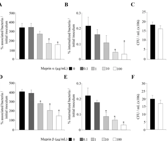
As endogenous meprins are synthesized in intestinal epithelial cells as inactive zymogen, which has to be activated, to investigate whether meprins can modulate the ability of AIEC bacteria to interact with intestinal epithelial cells, AIEC bacteria were pretreated with exogenous meprins a and b. The ability of the AIEC LF82 to adhere to and to invade T84 cells was assessed (Fig. 2). Quantitative adhesion assays showed that pretreatment of AIEC LF82 bacteria with increased concentrations (from 0.1 to 100mg/ml) of meprins a and b significantly (P,0.05) decreased in a dose dependent manner the ability of bacteria to adhere to differentiated T84 epithelial cells, compared to untreated bacteria (Fig. 2A and 2D). Meprin-treated LF82 bacteria were also significantly (P,0.05) impaired in their ability to invade differentiated T84 cells, compared to untreated bacteria (Fig. 2B and 2E). To ensure that the decreases in adhesion and invasion levels of AIEC LF82 to cells were not due to bactericidal activity of meprins, we checked that treatment with these proteases did not affect bacterial viability (Fig. 2C and 2F). In addition, we
Figure 2. Meprins impair AIEC LF82 ability to adhere to and to invade differentiated intestinal epithelial cells. AIEC LF82 bacteria were treated before infection with increased concentrations of exogenous meprins a and b (from 0.1 mg/ml to 100 mg/ml, see Materials and Methods section). Differentiated T84 cells were infected at a MOI of 10 with untreated or meprin pretreated bacteria. A and D, the number of cell-associated bacteria was determined after a 3 h infection period. B and E, the number of internalized bacteria was determined after a 3 h infection period followed by gentamicin treatment for 1 h. Results are expressed as percentage of cell-associated (adherent+ intracellular) (A and D), or intracellular (B and E) bacteria relative to initial inoculum. C and F, effect of meprin treatment on AIEC bacteria viability. Equal amounts of bacteria were exposed or not with a dose of 100 mg/ml of meprin a or b for 120 min. Thereafter, the number of viable bacteria was determined by plating on agar plate. Results are expressed as colony forming units (CFU) per ml (C and F). Data are mean 6 SEM for at least three independent experiments. Student’s t-test, * P,0.05.
doi:10.1371/journal.pone.0021199.g002
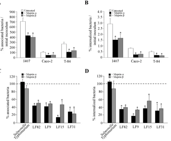
observed that meprin-treated AIEC LF82 bacteria showed significantly decreased adhesion to and invasion of undifferenti-ated intestinal epithelial cells T84, Caco-2 and Intestine-407 (Fig. 3A and 3B). We also analyzed whether the effect of meprin a and b on AIEC adhesion to and invasion of intestinal epithelial cells was not limited to the AIEC strain LF82 and can be extended to other AIEC strains and other enteric bacteria such as Salmonella enterica serovar Typhimurium. Pretreatment of S. Typhimurium strain LT2 with 10mg/ml of meprin a or b did not induce any significant decrease in either bacteria adhesion to or invasion of T84 cells (Fig. 3C and 3D). In contrast, for all the other AIEC strains tested (LF9, LF15 and LF31) a significant decreased ability to adhere to and invade undifferentiated T84 cells was observed when bacteria were treated with 10mg/ml of both meprins (Fig. 3C and D, P,0.05). Together, these results show that meprins can modulate the interaction between AIEC bacteria and intestinal epithelial cells. We further investigated which bacterial components involved in the abilites of AIEC bacteria to adhere to and invade intestinal epithelial cells were affected by meprin treatment.
Meprins a and b do not induce proteolytic cleavage of OMPs and flagellin
We have previously shown that outer membrane proteins (OMPs), by binding to the Gp96 receptor and flagella, are
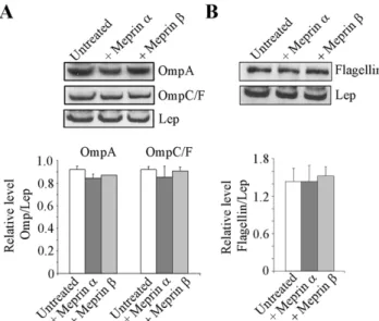
involved in the interaction of AIEC bacteria with intestinal epithelial cells [14,16,30,31]. Meprins a and b at 100mg/ml had no proteolytic effect on LF82 OMPs as shown by Western blot analysis using anti-OmpA, and OmpC/F antibodies and stan-dardization against the inner membrane protein Lep (Fig. 4A). Similarly, treatment of LF82 flagella with meprins a or b (100mg/ ml) did not induce any proteolytic degradation of flagella as observed by Western blot analysis of flagellin (Fig. 4B). Together these results show that at the highest dose of meprins, the decreased abilities of AIEC bacteria to adhere to and invade intestinal epithelial cells were not due to degradation of LF82 OMPs or flagella.
Meprins a and b induce proteolytic cleavage of AIEC LF82 type 1 pili
Experiments performed in vitro with cultured intestinal epithelial cells, ex vivo with human isolated enterocytes from CD patients, or in vivo using transgenic mice expressing the human CEACAM6 receptors showed that type 1 pili play a key role in the ability of AIEC bacteria to adhere to and invade intestinal epithelial cells [12,13,15]. We investigated whether the decrease in the abilities of AIEC bacteria to adhere to and invade epithelial intestinal cells observed after pretreatment of bacteria with meprin a or meprin b could be the result of proteolytic degradation of type 1 pili by these proteases. A dose-dependent proteolytic degradation of FimA, the
Figure 3. Effect of meprins on the ability of AIEC strains andSalmonella Typhimurium strain LT2 to adhere to and to invade intestinal epithelial cells. Bacteria were pretreated with exogenous meprin a or meprin b at 10 mg/ml. A and B, undifferentiated Intestine-407 (I407), Caco-2 and T84 cells infected with AIEC LF82 at a MOI of 10. The number of associated (A) and internalized (B) bacteria was determined. Results are expressed as the percentage of cell-associated (A) or intracellular bacteria (B) relative to initial inoculum. C and D, undifferentiated T84 cells were infected at a MOI of 10 with Salmonella Typhimurium and AIEC strains LF82, LF9, LF15 and LF31. The number of associated (C) and internalized (D) bacteria was determined. Results are expressed as the percentage of cell-associated (C) or intracellular bacteria (D) relative to untreated bacteria, defined as 100% (bar, C and D). Data are mean 6 SEM for at least three independent experiments. Student’s t-test, * P,0.05.
major subunit of type 1 pili, was observed after treatment of AIEC LF82 purified type 1 pili with meprins a and b (Fig. 5A). Treatment of LF82 purified type 1 pili with 100mg/ml of meprin a or meprin b induced a strong decrease in the FimA band observed on SDS-PAGE compared to untreated type 1 pili (89% for meprin a and 72% for meprin b). We also analyzed the proteolytic activity of meprins on type 1 pili present on the surface of whole bacteria. The amount of FimA subunit relative to that of the inner membrane protein Lep was determined by Western blot after treatment of LF82 bacteria with active or heat-inactivated meprin a or b. We observed 58% and 34% decreased amounts of FimA after treatment of the bacteria with active meprins a and b, respectively compared to those of untreated bacteria (Fig. 5B). In contrast, when bacteria were treated with heat-inactivated meprins the amount of FimA was unchanged. The degradation products of purified AIEC LF82 type 1 pili following meprin treatment were analyzed by mass spectrometry. Treatment of purified LF82 type 1 pili with 100mg/ml of active meprins a and b, compared to heat inactivated meprins, modified the mass spectrometry profile of type 1 pili (Fig. 5C and 5D). A decrease in the intensity of the base peak of spectrum corresponding to the protonated form ([M + H]+) of major analyte detected between 16259 and 16267 m/z was observed after treatment with meprins. This was no longer observed with heat inactivated meprins. The same observation was made with the doubly protonated form ([M + 2H]2+) of this analyte, detected between 8130 and 8134 m/z after treatment with active meprins. Together, these results indicate that type 1 pili are a major bacterial target of meprins and that the decreased adhesion and invasion of AIEC bacteria to intestinal epithelial cells after pretreatment of bacteria with exogenous meprins could be a direct consequence of the proteolytic activity of these proteases against type 1 pili.
Meprins affect mannose residue recognition mediated by AIEC LF82 type 1 pili and the ability of AIEC bacteria to induce IL-8 secretion
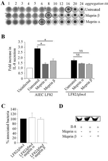
Adhesion of AIEC bacteria to cells that involved type 1 pili is mediated by recognition between the FimH adhesin, located at the tip of the pilus, and mannose residues on cellular receptors [32]. The ability of LF82 type 1 pili to bind D-mannose residues was tested using a yeast aggregation assay. Pretreatment of bacteria with meprin a and meprin b strongly reduced their ability to induce yeast aggregation (Fig. 6A). The impaired recognition of mannosylated receptor by AIEC bacteria could result from meprin-mediated proteolysis of type 1 pili.
Pathogenic E. coli interaction with host cells generally induce enhanced epithelial cell IL-8 secretion [33,34]. We quantified by ELISA the levels of IL-8 secreted by T84 cells infected with AIEC LF82 bacteria pretreated or not with 100mg/ml of meprin a or b (Fig. 6B). The amount of IL-8 secreted by T84 cells infected by meprin a- or meprin b-treated bacteria was significantly lower than that of cells infected with non-treated bacteria (24% and 28%, respectively, P,0.05) and was similar to the amount observed with T84 cells infected by a non-piliated LF82 mutant (LF82DfimA). To analyse whether bacterial components inducing IL-8 secretion other than type 1 pili susceptible to meprin degradation could exist, we performed assays with LF82DfimA bacteria treated with meprins (Fig. 6B). IL-8 secretion by T84 cells remained similar following infection with LF82DfimA bacteria treated or not with meprins. In addition, we did not also observe any modified adhesion to T84 cells of LF82DfimA mutant treated or not with meprins (Fig. 6C). Decreased IL-8 amounts as measured by ELISA were not due to proteolytic degradation of IL-8 by meprins (Fig. 6D). Thus, these results demonstrate that meprins affect AIEC interaction with IEC, since degradation of type 1 pili by meprins impaired recognition of D-mannose residues by LF82 bacteria and subsequently decreased the ability of AIEC LF82 to adhere to and invade IEC, resulting in decreased IL-8 secretion.
Discussion
According to the currently accepted hypothesis, both UC and CD result from a dysregulated response of the intestinal immune system to antigens of microbial origin or pathogenic bacteria in genetically predisposed individuals. MEP1A has been identified as a genetic susceptibility factor for IBD [17,18]. It encodes meprin a, a metalloprotease highly expressed in the intestine. Meprin a is secreted into the intestinal lumen or accumulates at the apical brush border membrane of polarized epithelial cells retained by meprin b. Thus any decrease in meprin a or b expression can lead to similar defects in the host. In the present study, we analyzed meprins a and b mRNA expression in ileal biopsies of CD patients since AIEC are more frequently found in intestinal tissue samples of CD patients, than in those of UC patients or healthy controls and because AIEC bacteria show a tropism for ileal colonization. Our real time PCR analysis with patient biopsies revealed that, irrespective of macroscopic inflammation, the ileal mucosa of CD patients had significantly weaker expression of meprin b than that of non-IBD control biopsies. As meprin b is required to retain meprin a, this may aggravate the deficit in meprin a/b secretion/ retention on the luminal side of the epithelial cell membrane. In addition, we analyzed whether AIEC infection could modulate meprin expression and observed no modified expression of either meprin following AIEC LF82 mouse infection, indicating that the exacerbation of colitis by infection did not interfere with meprin expression.
Figure 4. Proteolytic activity of meprins a and b on AIEC LF82 outer membrane proteins and flagellin. Total protein extracts from untreated or meprin-treated (100 mg/mL) whole bacteria were immunoblotted for OmpA and OmpC/F (A) or Flagellin (B). The inner membrane protein Lep was used as internal control. Amounts of proteins were quantified by using Image J software. Results are expressed as protein amount relative to Lep. Data are mean 6 SEM for at least three independent experiments. Student’s t-test, * P,0.05.
doi:10.1371/journal.pone.0021199.g004
We speculated that defects in meprin expression on the surface of the ileum could affect microbe-host interactions since proteases could create a proteolytic environment which either kills bacteria or degrades their virulence factors. This was well exemplified for neutrophil elastase, which induces considerable degradation of E. coli OmpA [35] or Pseudomonas aeruginosa flagellin [36], and for lactoferrin, which cleaves IgA1 protease protein and Hap adhesin of Haemophilus influenzae, [37]. We show that pretreatment of AIEC bacteria with exogenous meprins a and b impaired the ability of bacteria to adhere to and invade various differentiated or non differentiated intestinal epithelial cells, indicating that any decrease in meprin expression in the gut could result in increased AIEC colonization. This indicated that meprins can participate in modifications of the bacterial population associated with the gut mucosa. Interestingly, no decreased adhesion or invasion was observed with the enteroinvasive pathogen S. Typhimurium strain LT2 treated with meprins, probably because these bacteria use a type three secretion system to invade epithelial cells
We previously reported that AIEC colonization of the intestinal mucosa is dependent on binding of type 1 pili to the glycosylated CEACAM6 receptor, which is abnormally expressed by ileal epithelial cells in CD patients [12]. We also previously reported that flagella and outer membrane proteins (OMP) via outer membrane vesicles are involved in the ability of AIEC bacteria to adhere to and to invade cultured IEC in vitro [14,30,31]. We investigated whether meprins target these virulence factors and observed a proteolytic degradation of AIEC LF82 type 1 pili by meprins a and b, but not of flagella and OMPs. This effect was found with active meprins but no longer observed when bacteria were treated with heat-inactivated meprins. The impaired ability of meprin-treated bacteria to adhere to and invade various intestinal epithelial cells could result from proteolytic degradation of type 1 pili. Previous molecular dissections of virulence factor expression in the AIEC reference strain LF82 suggested that these bacteria are highly piliated under physiological conditions in the gastrointestinal tract [16]. Any proteolytic degradation of type 1 pili could modify the behaviour of the AIEC bacteria within the host, and any decrease in protease expression could therefore increase AIEC colonization of the ileal mucosa.
AIEC infection of IECs induces the secretion of high amounts of the pro-inflammatory cytokine IL-8, a potent chemoattractant for neutrophils [7]. We observed that when AIEC LF82 bacteria were treated by meprin a or b, IL-8 secretion by infected IECs was significantly decreased. This effect was probably due to the decrease in the ability of bacteria to adhere to and invade intestinal epithelial cells as a consequence of proteolytic degradation of type 1 pili. These results are in accordance with our previous study showing that the inflammation induced by AIEC infection of mice expressing the human CEACAM6 was not observed when the AIEC LF82 type 1 pili–negative mutant was used [13]. It can be speculated that down-regulation of meprin expression observed in CD patients leading to impaired meprin secretion/retention at the epithelial brush border might favor not only colonization of the
intestinal mucosa by AIEC bacteria but also AIEC-induced inflammation of the gut.
Materials and Methods Patients and controls
CD: n = 27, mean age 41 years (25-88), 12 male and 15 female patients. UC: n = 6, mean age 45 years (21–65), 2 male and 4 female patients. Controls: n = 18, mean age 55 years (38–74), 9 male and 9 female non-IBD individuals/patients. Biopsies were collected during routine (patients) or screening (controls) endo-scopic examination at the Gastroenterology unit, University Hospital Bern, Bern, Switzerland. Biopsies were chosen solely upon their location and macroscopic appearance, as judged by the experienced endoscopist. CDAI and Mayo scores, although determined routinely, were not used to stratify the cohort of patients used in the present study. Informed written consent to use biopsies for the present study was obtained from all patients, and all procedures were approved by the ethical committee of the local authorities of the Canton of Bern.
Mouse model and infection
Nine-week-old C57BL/6J (B6) mice (<19.5 g) were bred and reared at the central animal facility of the medical faculty of the University of Bern, Switzerland. They received DSS (MW = 36,000–50,000, MP Biomedicals, Illkirch, France) in drinking water for 7 days with 3.5% (w/v) DSS for the first 3 days and 2.5% (w/v) DSS for the last 4 days. The mice were orally challenged by 5 mg of streptomycin (Sigma) at day 3 and by 108 AIEC LF82 bacteria at day 4. They were sacrified at day 7 and the ileum and colon were collected. Animal experiments were approved by the local authorities of the canton of Bern and were also in accordance with the Committee for Research and Ethical Issues of the IASP.
Real-time mRNA quantification
Human ileal biopsies and mouse ileum and colon were placed in RNAlater (Qiagen, Hombrechtikon, Switzerland). Samples were kept for 24 h at 4uC and then at 220uC until RNA extraction. RNAlater-preserved biopsies were homogenized in RLT buffer (Qiagen) in a TissueLyzerTM (Qiagen) and RNA were isolated using the mirVanaTM (mi)RNA Isolation Kit (Ambion, Applied Biosystems, Rotkreuz, Switzerland). RNA quality was checked with Agilent Bioanalyzer Nano Chips (Agilent Technologies, Basel, Switzerland). Onemg of RNA was reverse transcribed using Promega’s ImProm-II Reverse Transcription System (Promega, Mannheim, Germany) with random primers. Meprins a and b mRNA were quantified from human samples using intron-spanning TaqMan gene expression assays (Applied Biosystems) and from mouse samples using Light Cycler 1.5 (Roche Diagnostics) and SYBR green Taq ReadyMix (Sigma, St. Louis, MO) with specific human and mouse oligonucleotides. Each sample was run in duplicate. Results were normalized to the
Figure 5. Proteolytic activity of meprins a and b on AIEC LF82 type 1 pili. A, proteolytic effect of various concentrations (from 1 mg/ml to 100 mg/ml) of meprins on purified AIEC LF82 type 1 pili. Proteins were loaded on 15% polyacrylamid gel for SDS-PAGE and stained with Coomassie brilliant blue. Results are expressed as FimA protein amount for meprin-treated type 1 pili relative to that of untreated type 1 pili. B, proteolytic effect of meprins on type 1 pili expressed at the surface of whole AIEC LF82 bacteria. AIEC bacteria were untreated or treated with active or heat-inactivated meprins at 100 mg/mL. Total proteins were immunoblotted with rabbit antiserum raised against purified type 1 pili and the inner membrane protein Lep, as an internal control. Results are expressed as FimA amount relative to Lep. Amounts of proteins were quantified by using Image J software. Data are mean 6 SEM for at least three independent experiments. Student’s t-test, * P,0.05. C and D, MALDI-TOF MS profile of purified AIEC LF82 type 1 pili treated with active or heat-inactivated meprin a or meprin b. Spectra were acquired in a mass spectrometer MALDI-TOF from LF82 type 1 pili treated with 100 mg/ml of meprin a (C) or with meprin b (D).
doi:10.1371/journal.pone.0021199.g005
human epithelial marker villin gene or mouse TATA box binding protein (TBP) housekeeping gene.
Bacterial strains
The four AIEC strains (AIEC reference strain LF82 and AIEC strains LF9, LF15 and LF31) were isolated from patients with
Crohn’s disease [6,11]. The LF82-DfimA isogenic mutant does not synthesize type 1 pili [38]. Salmonella Typhimurium strain LT2 was purchased from ATCC (ATCC 700720). Bacteria were grown routinely in Luria Bertani (LB) broth or on LB agar plates overnight at 37uC.
Cell culture
The intestinal epithelial cell lines T84 (ATCC, CCL-248), Intestine-407 (ATCC, CCL-6) and Caco-2 (ATCC, HTB-37) were maintained in an atmosphere containing 5% CO2at 37uC in the
culture medium recommended by ATCC. For infection assays, undifferentiated T84, Intestine-407, and Caco-2 cells were seeded in 24 well plates at a concentration of 4.105per cm2. To obtain differentiated T84 cells, cells were seeded onto Transwell filters at 8.105cells/filter (5mm pore size, 4.6 cm2; Costar, Corning Inc.) and were grown for 21 days in an atmosphere containing 5% CO2 at 37uC.
Adhesion and Invasion Assays
Before infection, bacteria were pretreated for 120 min with 0.1mg/ml to 100mg/ml of exogenous meprin a or b in PBS (See paragraph below). Meprins were then inactivated, and pretreated bacteria were washed and used for infection. Cells were infected at a multiplicity of infection (MOI) of 10 bacteria per cell. Adhesion and invasion assays were performed as previously described [6]. For adhesion assays, after 3 h of incubation period at 37uC, monolayers were washed five times in phosphate buffer saline (PBS). To determine the numbers of intracellular bacteria (Invasion assay), cell culture medium containing 100mg of gentamicin per ml was added for 1 h to kill extracellular bacteria. The epithelial cells were then lysed with 1% Triton X-100 (Sigma) in deionized water. Samples were diluted and plated onto LB agar plates to determine the number of colony-forming units (CFU).
Bacterial viability assay
Bacteria were pretreated with 100mg/ml meprin a or meprin b (expression and purification described previously [39,40]) at 37uC for 120 min. Meprins were then inactivated and pretreated bacteria were compared to untreated bacteria. Numbers of CFU were determined from samples diluted and plated onto LB agar plates.
Extraction of type 1 pili and total proteins
Type 1 pili were extracted as previously described [15,16]. Purified type 1 pili were subjected to HCl hydrolysis before SDS-PAGE analysis because fimbriae are resistant to SDS disaggrega-tion. After an overnight incubation at 37uC in LB broth, bacteria were centrifuged and resuspended in SDS-PAGE loading buffer (2% SDS, 50 mM Tris-HCl pH 6.8, 12.5% glycerol, 400 mM b-mercaptoethanol and 0.01% Bromophenol Blue).
Treatment of whole bacteria, purified bacterial surface components and recombinant human IL-8 with meprins
Whole bacteria were pretreated for 120 min with exogenous meprin a or b (from 0.1mg/ml to 100mg/ml) in PBS. PBS supplemented with 4 mM EDTA pH 7.4, was then added (20:1) in order to inactivate meprin activity. The same protocol was used for bacteria in the absence of meprin or in presence of 100
mg/ml of meprin a and b inactivated at 100uC for 15 min.
Bacteria were then washed twice in PBS by centrifugation and resuspended in PBS for infection or in SDS-PAGE loading buffer for Western-blotting.
Figure 6. Meprin treatment affect mannose residue recognition by AIEC and AIEC-induced IL-8 secretion by T84 cells. A, ability of type 1 pili to bind D-mannose residues as determined by a yeast aggregation test. AIEC LF82 bacteria were treated with 100 mg/ml of meprin a or b at 37uC for 120 min. A fixed amount of inactivated yeast cells (Saccharomyces cerevisiae) suspension and decreasing concentra-tions of treated and untreated bacteria were mixed, and the loss of the ability to form homogenous aggregation was used as the read-out for impaired type 1 pili-yeast interaction. B, Amount of IL-8 secreted by uninfected or AIEC LF82- or type 1 pili negative mutant LF82-DfimA-infected T84 cells, at 24 h post-infection. AIEC LF82 and LF82-DfimA bacteria were treated with 100 mg/ml of meprins. Il-8 secretion was determined by ELISA. Data are expressed as fold increase in the amount of secreted IL-8 6 SEM by T84 cells infected with untreated or treated bacteria relative to non infected cells. Student’s t-test, * P,0.05 for comparison between IL-8 secretion induced by untreated versus meprin-treated AIEC LF82 or LF82-DfimA bacteria. C, LF82-DfimA bacteria were pretreated with exogenous meprin a or meprin b at 100 mg/ml and undifferentiated T84 cells were infected at a MOI of 10. The number of associated bacteria was determined. Results are expressed as the percentage of cell-associated bacteria pretreated with exogenous meprins relative to untreated bacteria, defined as 100%.D, effect of meprins on recombinant human IL-8. Recombinant human IL-8 (110 ng/ml) was treated with meprin a or b (100 mg/ml), electroblotted and detected with mouse anti-human IL-8.
Purified type 1 pili were treated with increasing concentrations (1 to 100mg/ml) of meprin a and b, at 37uC for 120 min and subjected to SDS-PAGE and mass spectrum analysis. In addition, purified type 1 were also treated with 100mg/ml of meprin a and b inactivated at 100uC for 15 min.
Recombinant human IL-8 (110 ng/ml, R&D systems) was treated with 100mg/ml of meprin a or b at 37uC for 120 min
and was resuspended in SDS-PAGE loading buffer for Western-blotting.
Western blotting
Purified bacterial surface components or total proteins were subjected to SDS-PAGE on 12-17% gels. Protein concentrations were determined by Bradford assay and the gels were stained for protein with Coomassie brilliant blue. Western immunoblotting was performed according to the procedure of Towbin [41]. Proteins were electroblotted onto nitrocellulose membranes (Amersham International), and the membranes were immuno-blotted for type 1 pili (rabbit antiserum raised against purified type 1 pilus preparations, diluted 1:1,000), Lep (rabbit anti-Lep, diluted 1:1,000), OmpA (rabbit anti-OmpA, diluted 1:1,00), OmpC/F (rabbit anti-OmpC/F, diluted 1:1,000), flagellin (rabbit anti-H1, diluted 1:500) and IL-8 (mouse anti-human; diluted 1/250; R & D Systems). Immunoreactants were detected using horseradish peroxidase-conjugated anti-rabbit or anti-mouse immunoglobulin G antibody, ECL reagents (Amersham Biosciences) and autora-diography. Image J software was use to estimate protein quantity.
Yeast cell aggregation assay
Bacteria were washed and resuspended to an optical density of 0.1 at 620 nm in PBS and treated or not with 100mg/ml of meprin a and meprin b at 37uC for 120 min. Equal volumes of fixed commercial baker’s yeast cell (Saccharomyces cerevisiae) suspension (10 mg dry weight/ml) in PBS and decreasing concentrations of E. coli suspension were used, and aggregation was monitored visually.
Mass spectrum analysis
Mass spectra were acquired by a mass spectrometer-DE PRO (Applied Biosystems, Cortaboeuf) in positive ionization, linear
MODE. Briefly, 1ml of type 1 pili was mixed on the MALDI plate with 1mL of Sinapinic acid matrix, using the standard dried-drop method. Spectra were then generated in the mass-to-charge ratio (m/z) range of 4,000 m/z to 20,000 m/z. Each spectrum was calibrated by calibrant protein mixture (C110, LaserBiolabs, Antibes, France).
Enzyme-linked immunosorbent assay (ELISA)
The amount of Il-8 released in the culture supernatant was determined by ELISA (R&D systems). Cytokine concentrations were assessed according to the manufacturer’s instructions.
Statistical Analysis
Data generated from adhesion and invasion assays or ELISA were analysed by Student’s t-test. All experiments were performed at least three times. A P-value #0.05 was considered statistically significant. Data are expressed as the mean 6 SEM. Statistical analysis of data generated from RT-PCR with human biopsies and with mouse colon and ileum was performed using GraphPad Prism 5.0 Software. For data generated from RT-PCR with human biopsies, overall differences between groups were estimated with the Kruskall Wallis test and P values were calculated with planned posteriori tests using the non-parametric Mann-Whitney U test. P-values as indicated in figure 1B were further corrected for multiple testing by Bonferroni correction for final statements in results and discussion. For data generated from RT-PCR with mouse ileum and colon, overall differences between groups were estimated with one-way ANOVA.
Acknowledgments
We gratefully acknowledge Karen Krogfelt for providing the type 1 pili antiserum, Frank Seibold for collection biopsies and for providing us the corresponding clinical information.
Author Contributions
Conceived and designed the experiments: EV M-AB AG SM DL AD-M. Performed the experiments: EV AG M-AB SM CJ SLFP. Analyzed the data: EV M-AB AG SM DL AD-M. Contributed reagents/materials/ analysis tools: CC CB-P SLFP. Wrote the paper: EV M-AB AD-M.
References
1. Strober W, Fuss I, Mannon P (2007) The fundamental basis of inflammatory bowel disease. J Clin Invest 117: 514–521.
2. Xavier RJ, Podolsky DK (2007) Unravelling the pathogenesis of inflammatory bowel disease. Nature 448: 427–434.
3. Sartor RB (2008) Microbial influences in inflammatory bowel diseases. Gastroenterology 134: 577–594.
4. Rutgeerts P, Goboes K, Peeters M, Hiele M, Penninckx F, et al. (1991) Effect of faecal stream diversion on recurrence of Crohn’s disease in the neoterminal ileum. Lancet 338: 771–774.
5. Baumgart M, Dogan B, Rishniw M, Weitzman G, Bosworth B, et al. (2007) Culture independent analysis of ileal mucosa reveals a selective increase in invasive Escherichia coli of novel phylogeny relative to depletion of Clostridiales in Crohn’s disease involving the ileum. Isme J 1: 403–418.
6. Darfeuille-Michaud A, Boudeau J, Bulois P, Neut C, Glasser AL, et al. (2004) High prevalence of adherent-invasive Escherichia coli associated with ileal mucosa in Crohn’s disease. Gastroenterology 127: 412–421.
7. Eaves-Pyles T, Allen CA, Taormina J, Swidsinski A, Tutt CB, et al. (2007) Escherichia coli isolated from a Crohn’s disease patient adheres, invades, and induces inflammatory responses in polarized intestinal epithelial cells. Int J Med Microbiol.
8. Martin HM, Campbell BJ, Hart CA, Mpofu C, Nayar M, et al. (2004) Enhanced Escherichia coli adherence and invasion in Crohn’s disease and colon cancer. Gastroenterology 127: 80–93.
9. Martinez-Medina M, Aldeguer X, Siles M, Gonzalez-Huix F, Lopez-Oliu C, et al. (2009) Molecular diversity of Escherichia coli in the human gut: New ecological evidence supporting the role of adherent-invasive E. coli (AIEC) in Crohn’s disease. Inflamm Bowel Dis.
10. Sasaki M, Sitaraman SV, Babbin BA, Gerner-Smidt P, Ribot EM, et al. (2007) Invasive Escherichia coli are a feature of Crohn’s disease. Lab Invest 87: 1042–1054. 11. Boudeau J, Glasser AL, Masseret E, Joly B, Darfeuille-Michaud A (1999) Invasive ability of an Escherichia coli strain isolated from the ileal mucosa of a patient with Crohn’s disease. Infect Immun 67: 4499–4509.
12. Barnich N, Carvalho FA, Glasser AL, Darcha C, Jantscheff P, et al. (2007) CEACAM6 acts as a receptor for adherent-invasive E. coli, supporting ileal mucosa colonization in Crohn disease. J Clin Invest 117: 1566–1574. 13. Carvalho FA, Barnich N, Sivignon A, Darcha C, Chan CH, et al. (2009)
Crohn’s disease adherent-invasive Escherichia coli colonize and induce strong gut inflammation in transgenic mice expressing human CEACAM. J Exp Med 206: 2179–2189.
14. Barnich N, Boudeau J, Claret L, Darfeuille-Michaud A (2003) Regulatory and functional co-operation of flagella and type 1 pili in adhesive and invasive abilities of AIEC strain LF82 isolated from a patient with Crohn’s disease. Mol Microbiol 48: 781–794.
15. Boudeau J, Barnich N, Darfeuille-Michaud A (2001) Type 1 pili-mediated adherence of Escherichia coli strain LF82 isolated from Crohn’s disease is involved in bacterial invasion of intestinal epithelial cells. Mol Microbiol 39: 1272–1284. 16. Rolhion N, Darfeuille-Michaud A (2007) Adherent-invasive Escherichia coli in
inflammatory bowel disease. Inflamm Bowel Dis 13: 1277–1283.
17. Banerjee S, Oneda B, Yap LM, Jewell DP, Matters GL, et al. (2009) MEP1A allele for meprin A metalloprotease is a susceptibility gene for inflammatory bowel disease. Mucosal Immunol 2: 220–231.
18. Bond JS, Matters GL, Banerjee S, Dusheck RE (2005) Meprin metalloprotease expression and regulation in kidney, intestine, urinary tract infections and cancer. FEBS Lett 579: 3317–3322.
19. Grunberg J, Dumermuth E, Eldering JA, Sterchi EE (1993) Expression of the alpha subunit of PABA peptide hydrolase (EC 3.4.24.18) in MDCK cells. Synthesis and secretion of an enzymatically inactive homodimer. FEBS Lett 335: 376–379.
20. Johnson GD, Bond JS (1997) Activation mechanism of meprins, members of the astacin metalloendopeptidase family. J Biol Chem 272: 28126–28132. 21. Ohler A, Debela M, Wagner S, Magdolen V, Becker-Pauly C (2010) Analyzing
the protease web in skin: meprin metalloproteases are activated specifically by KLK4, 5 and 8 vice versa leading to processing of proKLK7 thereby triggering its activation. Biol Chem 391: 455–460.
22. Eldering JA, Grunberg J, Hahn D, Croes HJ, Fransen JA, et al. (1997) Polarised expression of human intestinal N-benzoyl-L-tyrosyl-p-aminobenzoic acid hydrolase (human meprin) alpha and beta subunits in Madin-Darby canine kidney cells. Eur J Biochem 247: 920–932.
23. Bertenshaw GP, Turk BE, Hubbard SJ, Matters GL, Bylander JE, et al. (2001) Marked differences between metalloproteases meprin A and B in substrate and peptide bond specificity. J Biol Chem 276: 13248–13255.
24. Herzog C, Haun RS, Kaushal V, Mayeux PR, Shah SV, et al. (2009) Meprin A and meprin alpha generate biologically functional IL-1beta from pro-IL-1beta. Biochem Biophys Res Commun 379: 904–908.
25. Herzog C, Kaushal GP, Haun RS (2005) Generation of biologically active interleukin-1beta by meprin B. Cytokine 31: 394–403.
26. Kruse MN, Becker C, Lottaz D, Kohler D, Yiallouros I, et al. (2004) Human meprin alpha and beta homo-oligomers: cleavage of basement membrane proteins and sensitivity to metalloprotease inhibitors. Biochem J 378: 383–389. 27. Schutte A, Hedrich J, Stocker W, Becker-Pauly C (2010) Let it flow: Morpholino knockdown in zebrafish embryos reveals a pro-angiogenic effect of the metalloprotease meprin alpha2. PLoS One 5: e8835.
28. Villa JP, Bertenshaw GP, Bylander JE, Bond JS (2003) Meprin proteolytic complexes at the cell surface and in extracellular spaces. Biochem Soc Symp. pp 53–63.
29. Banerjee S, Jin G, Bradley SG, Matters GL, Gailey RD, et al. (2011) Balance of meprin A and B in mice affects the progression of experimental inflammatory bowel disease. Am J Physiol Gastrointest Liver Physiol 300: G273–282. 30. Rolhion N, Barnich N, Bringer MA, Glasser AL, Ranc J, et al. (2010)
Abnormally expressed ER stress response chaperone Gp96 in CD favours adherent-invasive Escherichia coli invasion. Gut 59: 1355–1362.
31. Rolhion N, Barnich N, Claret L, Darfeuille-Michaud A (2005) Strong decrease in invasive ability and outer membrane vesicle release in Crohn’s disease-associated adherent-invasive Escherichia coli strain LF82 with the yfgL gene deleted. J Bacteriol 187: 2286–2296.
32. Ofek I, Hasty DL, Abraham SN, Sharon N (2000) Role of bacterial lectins in urinary tract infections. Molecular mechanisms for diversification of bacterial surface lectins. Adv Exp Med Biol 485: 183–192.
33. Betis F, Brest P, Hofman V, Guignot J, Bernet-Camard MF, et al. (2003) The Afa/Dr adhesins of diffusely adhering Escherichia coli stimulate interleukin-8 secretion, activate mitogen-activated protein kinases, and promote polymorpho-nuclear transepithelial migration in T84 polarized epithelial cells. Infect Immun 71: 1068–1074.
34. Steiner TS, Nataro JP, Poteet-Smith CE, Smith JA, Guerrant RL (2000) Enteroaggregative Escherichia coli expresses a novel flagellin that causes IL-8 release from intestinal epithelial cells. J Clin Invest 105: 1769–1777. 35. Belaaouaj A, Kim KS, Shapiro SD (2000) Degradation of outer membrane
protein A in Escherichia coli killing by neutrophil elastase. Science 289: 1185–1188.
36. Lopez-Boado YS, Wilson CL, Parks WC (2001) Regulation of matrilysin expression in airway epithelial cells by Pseudomonas aeruginosa flagellin. J Biol Chem 276: 41417–41423.
37. Hendrixson DR, Qiu J, Shewry SC, Fink DL, Petty S, et al. (2003) Human milk lactoferrin is a serine protease that cleaves Haemophilus surface proteins at arginine-rich sites. Mol Microbiol 47: 607–617.
38. Chassaing B, Rolhion N, de Vallee A, Salim SY, Prorok-Hamon M, et al. Crohn disease—associated adherent-invasive E. coli bacteria target mouse and human Peyer’s patches via long polar fimbriae. J Clin Invest 121: 966–975. 39. Becker C, Kruse MN, Slotty KA, Kohler D, Harris JR, et al. (2003) Differences
in the activation mechanism between the alpha and beta subunits of human meprin. Biol Chem 384: 825–831.
40. Becker-Pauly C, Howel M, Walker T, Vlad A, Aufenvenne K, et al. (2007) The alpha and beta subunits of the metalloprotease meprin are expressed in separate layers of human epidermis, revealing different functions in keratinocyte proliferation and differentiation. J Invest Dermatol 127: 1115–1125. 41. Towbin H, Staehelin T, Gordon J (1979) Electrophoretic transfer of proteins
from polyacrylamide gels to nitrocellulose sheets: procedure and some applications. Proc Natl Acad Sci U S A 76: 4350–4354.