PaPer TyPe
OncoImmunology 2:9, e26011; September 2013; © 2013 Landes Bioscience
revIew
Introduction
The classification of cancers deriving from the hematopoi-etic system has become increasingly complex with the advent of novel techniques of molecular and cellular biology that can be used to precisely characterize malignant cell clones.1 Nonetheless,
hematological neoplasms can be roughly classified into lympho-mas and leukemias. The former are lymphoid tumors initially confined to peripheral lymphoid organs and extranodal tissues, while the latter include both lymphoid and myeloid malignan-cies that originate in the bone marrow but generally invade the peripheral blood. All hematological cancers are therefore exposed very early during oncogenese and throughout tumor progression to effectors of the immune system. Thus, the immunological microenvironment should be taken into particular consideration to fully understand and treat hematological malignancies.
The term “cancer immunosurveillance” is generally employed to describe the process whereby the immune system eliminates newly formed malignant cells. After an initial debate on the physiological relevance of this progress, it is now widely accepted that the interaction between malignant cells and immune cells
is one of the most prominent parameters determining disease outcome in cancer patients. In line with this notion, Hanahan and Weinberg have recently added two novel features that high-light the complex interplay between developing tumors and the immune system to the six hallmarks of malignancy that they had originally proposed in 2000.2 These novel hallmarks are the
ity of neoplastic cells to avoid immune destruction, and the abil-ity of chronic inflammation to promote tumor progression.3 As
a result, pharmaceutical companies are now developing several anticancer drugs that operate via the immune system, both in its innate and adaptive components.
Natural killer (NK) cells are innate lymphocytes recently reclassified as members of the group 1 of innate lymphoid cells (ILC1).4 NK cells are defined by their capacity to kill target cells
upon recognition through a set of activating and inhibitory recep-tors. In the course of immune responses, NK cells are rapidly activated by monocytes5 and dendritic cells6 trans-presenting the
immunostimulatory cytokine interleukin-15 (IL-15). This rapid (6–12 h) process primes NK cells to kill their targets mainly through the polarized release of cytotoxic granules that contain the pore-forming factor perforin, granzymes, and several other proteins. NK cells also secrete interferon γ (IFNγ) and other cytokines upon stimulation, in particular when this is mediated by the combination of IL-12 and IL-18.
NK cells play an important role in the early defense against intracellular pathogens.7 Within lymphoid organs, they are
stra-tegically positioned in the proximity of sentinel macrophages that line the lymphatic sinus, where they can efficiently respond to cytokine signals emanated from pathogen-sensing phagocytes by secreting IFNγ.8 NK cells have been shown to kill not only
infected cells, but also malignant cells of various origin, in vitro and in vivo. This latter property underpinned their discovery in the 1970s and drew considerable interest from tumor immu-nologists. Subsequently, it was found that NK cells are capable to sense the absence of MHC class I molecules on the surface of target cells through inhibitory receptors of the killer cell immu-noglobulin-like receptor (KIR) family in humans and Ly49 in mice.9 Such an absence of MHC class I molecules, which is often
referred to as “missing-self,” characterize many cancers, in par-ticular of hematological origin, and is thought to originate from a step of T cell-dependent selection. NK cells are also equipped
*Correspondence to: Thierry Walzer; Email: Thierry.walzer@inserm.fr Submitted: 07/12/2013; Accepted: 08/02/2013
Citation: Viel S, Charrier E, Marçais A, Rouzaire P, Bienvenu J, Karlin L, Salles G, Walzer T. Monitoring NK cell activity in patients with hematologic malignancies. OncoImmunology 2013; 2:e26011; http://dx.doi.org/10.4161/onci.26011
Monitoring NK cell activity in patients
with hematological malignancies
Sébastien viel1,2,3,4,5,6, emily Charrier1,2,3,4,5,6, antoine Marçais1,2,3,4,5, Paul rouzaire,7 Jacques Bienvenu1,2,3,4,5,6, Lionel Karlin8,
Gilles Salles8, and Thierry walzer1,2,3,4,5,*
1CIrI, International Center for Infectiology research; Université de Lyon; Lyon, France; 2Inserm, U1111; Lyon, France; 3ecole Normale Supérieure de Lyon; Lyon, France; 4Université Lyon 1; Centre International de recherche en Infectiologie; Lyon, France; 5CNrS, UMr5308; Lyon, France; 6Laboratoire d’Immunologie; Hospices Civils de Lyon;
Centre Hospitalier Lyon Sud; Lyon, France; 7Laboratoire d’Immunologie; CHU Clermont-Ferrand; Clermont-Ferrand, France; 8Hospices Civils de Lyon; Service d’Hématologie;
Centre Hospitalier Lyon Sud; Lyon, France
Keywords: natural killer cells, multiple myeloma, flow cytometry, cytotoxicity, Lenalidomide
Natural killer (NK) cells are lymphocytes of the innate immune system that can recognize and kill various types of malignant cells. Monitoring the activity of peripheral NK cells in patients affected by hematological malignancies may provide prog-nostic information or unveil ongoing tumor-specific immune responses. Moreover, further insights into the biology of NK cells might also promote the development of novel strategies for stimulating their anticancer activity. Here, we review the main methods to monitor phenotypic and functional NK cell properties in cancer patients, focusing on individuals affected by multiple myeloma, a hematological malignancy currently treated with immunomodulatory drugs.
with a variety of activating receptors that altogether contribute to their ability to recognize and kill neoplastic cells. The proto-typical NK-cell activating receptor is killer cell lectin-like recep-tor subfamily K, member 1 (KLRK1, best known as NKG2D), which recognizes various proteins expressed on the surface of target cells in response to several forms of cellular stress, includ-ing DNA damage, infection and oncogenic stress.10 In humans,
many malignancies of hematopoietic or non-hematopoietic ori-gin (but not healthy tissues) also express natural killer cell cyto-toxicity receptor 3 ligand 1 (NCR3LG1, best known as B7-H6) on their surface, which can be recognized by the NK-cell activat-ing receptor natural cytotoxicity receptor 3 (NCR3, also known as NKp30).11
Taken together, these observations suggest that NK cells are an important component of the endogenous arsenal of antican-cer defenses, especially at early stages of oncogenesis and tumor progression. In this context, NK cells might indeed detect and kill transformed cells, in turn favoring the activation of tumor-associated antigen (TAA)-specific T and B lymphocytes. Later on, NK cells might also play an important effector functions, in particular in the presence of TAA-targeting antibodies. In fact, human NK cells are believed to be among the most prominent executor of antibody-dependent cellular cytotoxicity (ADCC), owing to a robust expression of Fc fragment of IgG, low affinity IIIa, receptor (FCGR3A, also known as CD16a).12 Nonetheless,
NK cells are often insufficient to mediated tumor regression, and a general decrease of NK-cell functions is frequently observed in cancer patients.13–15 Presumably, this originates from
tumor-derived mediators that negatively regulate NK-cell activity, such as transforming growth factor β (TGFβ), or to the intense chemo-therapeutic regimens that are often employed in cancer patients.
Here, we review the rationale and the methods for monitor-ing the function of circulatmonitor-ing NK cells in individuals affected by hematopoietic malignancies. In addition, we summarize the results of a series of studies on NK-cell function in multiple myeloma (MM) patients, to illustrate what principles can be learned from such a monitoring and what questions should be addressed in the near future.
Why Monitoring the Activity of NK Cells in Cancer Patients?
NK-cell activity as surrogate marker of general immunological functions
Jérôme Galon’s group has defined the immune contexture as the type, functional orientation, density and location of adap-tive immune cells within distinct regions of solid neoplasms.16
They then quantified all these parameters in patients with colon cancer to delineate an “immunoscore” that turned out to have a better prognostic value than the classical scoring system rely-ing on histological and anatomical features of neoplastic lesions.17
This study highlighted not only the critical importance of immunological functions in tumor rejection but also the value of monitoring the immunological status of individuals affected by cancer. Of note, the immunoscore in its present form is not particularly adapted to tumors of hematological origin, as these
grow within lymphoid organs that are naturally infiltrated with immune cells. Moreover, as T cells infiltrating solid tumors are generally believed to be TAA-specific, their level is supposed to reflect the intensity of antitumor immune responses. In the cir-culation, the situation is completely different, as TAA-specific T cells are diluted within a huge amount of T cells with unrelated specificities. Thus, the assessment of immune responses against hematological cancers must proceed by alternative techniques.
Circulating immune cells have been monitored for a long time for different purposes, as the peripheral blood is rapidly acces-sible without the need for invasive procedures. Often, the status of circulating immune cells is argued not to reflect that of their tumor-infiltrating counterparts. In the case of hematological malignancies, however, the blood de facto constitutes the tumor site or a site of intense cancer cell transit. Evaluating circulating immune cells is therefore highly relevant for many types of hema-tological tumors. But, what should be evaluated? How? Why? We believe that monitoring the activity of circulating NK cells is use-ful for the following reasons. First, this can be performed with simple assays (see below), as most NK cell subsets (in spite of their notable repertoire) can respond to malignant cells (be them nude or coated with antibodies) in a few hours. Second, it may provide indirect insights into the general proficiency of the immune sys-tem, as often the two parameters are correlated with each other. For example, in the course of chronic infection18 or during
meta-static tumor dissemination,19 the activity of NK and T cells often
decreases concomitantly, a phenomenon that may reflect a sig-nificant degree of interdependence between innate and adaptive components of the immune system. Third, it could give indica-tions on tumor immunogenicity. In fact, the activation of NK cells may represent a hint of ongoing antitumor immune response. By analogy to the immunoscore approach, it would therefore be interesting to consider the prognostic value of the activity of circu-lating NK cells in patients with hematological malignancies. This parameter has already been shown to influence disease outcome in patients affected by other types of cancer.20
Predicting or monitoring the effect of anticancer (immune) therapy
Monitoring the function of circulating NK cells is obviously essential when patients are treated with drugs that are supposed to directly target NK cells. IPH2101 is a human monoclonal antibody specific that blocks the interaction between common KIRs (including KIR2DL1, KIR2DL2, and KIR2DL3) and their ligands, thus potentiating the cytotoxic activity of NK cells against autologous cancer cells in vitro.21 At least theoretically,
IPH2101 might be beneficial for the treatment of various can-cers, provided that it really increases the cytotoxic activity of NK cells in patients. Phase I clinical trials involving IPH2101 have already been conducted in patients affected by refractory MM, a setting in which IPH2101 significantly increased the cytotoxic activity of NK against autologous myeloma cells ex vivo.22
The efficacy of several new anticancer agents relies (as a whole or in part) on the elicitation of anticancer immune responses. These therapeutic agents include an increasing large panel of tumor-targeting monoclonal antibodies. Among them, ritux-imab as well as second-generation anti-CD20 antibodies are
now commonly used for the treatment of B-cell malignancies, based on their ability to stimulate ADCC. However, a subset of patients does not respond well to anti-CD20 antibodies, some of them because of a polymorphism in the FCGR3A-coding gene that lowers the affinity of the receptor on NK cells for IgG1.23
Monitoring the activity of NK cells against autologous tar-get cells coated with anti-CD20 antibodies prior to use would hence be helpful to identify those patients that have the high-est chances to respond to therapy (an example of personalized medicine). A number of strategies are also under investigation to stimulate ADCC in patients receiving tumor-targeting therapeu-tic antibodies. In this context, monoclonal antibodies are often combined with immunostimulatory cytokines such as IL-2 and granulocyte macrophage colony-stimulating factor (GM-CSF) or immunomodulatory drugs (IMiDs). It will therefore be impor-tant to correlate the ability of peripheral NK cells to mediated ADCC with the therapeutic efficacy of these combinatorial regimens.24
Along similar lines, many conventional chemotherapeutic agents exert anticancer effects that depend on the immune sys-tem, especially when given at “metronomic,” non-toxic doses.25
Monitoring the activity of circulating NK cells in this setting may lead to potentially important discoveries. Vice versa, it will be important to understand which of the immunosuppressants that are currently employed in the clinics inhibit NK-cell func-tion.26 Indeed, as immunosuppressants are generally given to
inhibit adaptive autoimmune responses, they should ideally spare the innate immune system, leaving patients with an important barrier against infections.
Understanding the impact of cancer cells on immunological functions
Why can NK cells function normally in cancer patients or, more often, be significantly impaired in their activity? Obtaining precise insights into the cell-intrinsic and cell-extrinsic circuitries that regulate the function of NK cells in physiological and patho-logical settings may lead to the development of novel strategies to stimulate for therapeutic purposes. It is therefore essential to monitor the activity of NK cells with appropriate protocols prior to the initiation of in-depth analyses.
Techniques to Monitor the Activity of Circulating NK Cells Effector functions
For several decades, the activity of NK cells has been mea-sured using the 51Cr release assay. In this assay, target cells (most
often the erythroblastic leukemia K562 cells) were loaded with
51Cr and then co-cultured for 4 h with peripheral blood
mono-nuclear cells (PBMCs). Eventually, the release of 51Cr in culture
supernatants was quantified as a marker of target cell death. This simple assay has several drawbacks. First, its results are highly dependent on the ratio between effector and target cells. Thus, limited extents of target cell death may originate from a low per-centage of NK cells in the PBMC sample as well as from a poor cytotoxic activity of individual NK cells. Second, it does not pro-vide insights into the activity of individual NK cells relative to
other parameters, such as cytokine secretion. For these reasons, the activity of NK cells nowadays is preferentially measured by means of the so-called lysosomal-associated membrane protein 1 (LAMP1, best known as CD107a) assay.
The LAMP1 assay measures the release of secretory lysosomes (cytotoxic granules) by NK cells that enter in contact with tar-get cells, a phenomenon commonly referred to as degranulation. The membrane of cytotoxic granules contains proteins, such as LAMP1, that are transiently exposed on the surface of NK cells upon fusion of granules with the plasma membrane. Thus, the staining of NK cells maintained in non-permeabilizing condi-tions with fluorochrome-labeled LAMP1-specific antibodies coupled to flow cytometry identifies cells that have recently undergone degranulation. Importantly, degranulation has been shown to correlate with the cytotoxic activity of NK cells,27
and occurs rapidly after their contact with target cells (usually within an hour). The frequency of degranulating NK cells also turned out to be less dependent on the effector to target cell ratio than lysis of target cells (TW et al., unpublished observa-tions). Moreover, the measurement of degranulation by flow cytometry can be combined with the evaluation of cytokine/ chemokine expression levels through an intracellular staining with specific antibodies.28,29 Upon ex vivo stimulation, NK cells
mainly secrete IFNγ, tumor necrosis factor α (TNFα), chemo-kine (C-C motif) ligand 3 (CCL3), CCL4, and CCL5.30 A low
fraction of activated NK cells may also secrete GM-CSF, IL-10, and IL-13.31 Flow cytometry allows for the precise
identifica-tion of NK cells expressing one or more cytokine/chemokine(s). Usually, all cytokine-expressing cells also show signs of degranu-lation. Of note, a wide range of stimuli can be used to stimulate NK cells to degranulate and secrete cytokines/chemokines. For example, transformed cells including K562 cells, which do not express MHC class I molecules on their surface, and lymphoma B cells coated with anti-CD20 antibodies induce robust NK cell responses.
Expression of cell surface receptors and intracellular proteins
Multiparametric flow cytometry now allows for the measure-ment of up to 12 fluorescent signals in a single tube. Hence, even with limited blood samples, multiple phenotypic and functional parameters of circulating NK cells can be analyzed (Table 1). Mean fluorescence intensity is generally used to quantify the level of expression of each receptor. If both the cytofluorometer and the staining procedure are well calibrated, data from patients analyzed on different occasions can be compared with accuracy. This information can relate to:
NK-cell activation
Upon stimulation with cytokines such as IL-2 or IL-15 or interaction with target cells, NK cells express increased levels of CD25, CD69, CD25, NCR2 (also known as NKp44), and gran-zyme B. Recent reports have also demonstrated that in the course of various infectious diseases, NK cells upregulate expression the expression of KLRC2 killer cell lectin-like receptor subfamily C, member 2 (KLRC2, also known as NKG2C).32,33
NK-cell receptors and maturation
NK cells are equipped with a complex set of activating and inhibitory receptors that critically regulate their function in
response to putative target cells, including malignant cells. In addition, NK cells express various cytokine and chemokine receptors, which also profoundly influence their activation and effector functions. Tumors often alter the expression of these receptors on the surface of NK cells, a process that may favor the escape of malignant cells from immunosurveillance.34 Upon
development in the bone marrow, NK cells operate a maturation process during which they stop proliferating, acquire effector functions, and express a peculiar profile of receptors (including activatory and inhibitory, as well as cytokine/chemokine recep-tors) on their surface. Also the maturation of NK cells is often inhibited by neoplasms.35 The monitoring of this process by flow
cytometry has been recently refined on the basis of several mark-ers including selectin L (SELL, also known as CD62L)36 and
killer cell lectin-like receptor subfamily C, member 1 (KLRC1, also known as NKG2A).37
Repertoire of NK cells
Multiparametric flow cytometry and epitope-specific anti-bodies also allow for the study of the NK-cell repertoire. Using a panel of 5 antibodies against inhibitory receptors of the KIR and NKG2D family and a boolean gating strategy, Björkström et al. have recently demonstrated that 27 different NK-cell popula-tions can be identified that express different combinapopula-tions of the 5 receptors analyzed.38 How the repertoire of NK cells evolves in
the course of tumor progression is currently unknown.
Intracellular mediators
Intracellular staining procedures can be implemented to measure not only the expression level of cytokines and chemo-kines, but also that of proteins involved in the cytotoxic func-tions of NK cells, such as granzyme A, granzyme B, and perforin. Recently, several antibodies specific for transcription factors that regulate the activity of NK cells have also been generated. In particular, this is the case of T-box 21 (TBX21, best known as T-bet) and eomesodermin (EOMES), 2 transcription factors that are essential for the development and maturation of murine NK cells. The expression of these transcription factors by cytotoxic lymphocytes is altered in the course of chronic infection,39 and
could therefore be deregulated also in the context of oncogenesis and tumor progression.
Gene expression
Microarrays are valuable tools to measure global gene expres-sion profiles and identify molecular pathways that are altered by tumors. So far, only a few studies have investigated global changes of gene expression in NK cells isolated from cancer patients. One study reported no difference in the gene expression profile of NK cells obtained from healthy individuals and melanoma patients.40
Conversely, signs of activation were detected in NK cells infiltrat-ing non-small cell lung carcinoma lesions as compared with NK cells located in the healthy lung parenchima of the same patient.41
Clearly, this type of approach, which is not extensively used yet, will help deciphering the role of NK cells in cancer immunosur-veillance and will open avenues to manipulate the antineoplastic functions of these cells.
Monitoring NK Cells in Multiple Myeloma Patients Phenotype of NK cells in MM patients
MM is a malignant disease of plasma cells that, despite major therapeutic progresses, is incurable. Like many neoplasms, MM is associated with a marked deficiency in general immunological function, which presumably contributes to the escape of cancer cells from immunosurveillance.42 The role of NK cells in the
con-trol of MM has been intensively investigated. A seminal study by Carbone et al. demonstrate that NK cells could kill autolo-gous MM cells in a NKG2D- and NCR-dependent manner.43
Moreover, MM cell lines derived from early-stage patients were found to express low levels of MHC class I molecules and high levels of the NKG2D ligand MHC class I polypeptide-related sequence A (MICA), while cell lines obtained from late-stage patients displayed the reverse phenotype, suggesting the exis-tence of an NK cell-mediated pressure in the course of MM progression.43
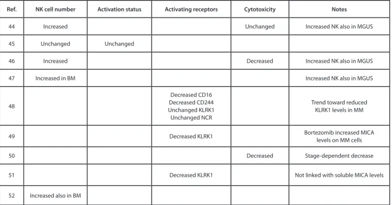
Table 2 summarizes the results of several studies44–52 that
monitored the number, phenotype and functional activity of cir-culating NK cells in patients affected by MM at different stages of the disease, as compared with NK cells from the blood of healthy individuals. These studies suggest that patients with active MM have an increased number of circulating NK cells.44,46,52 Of note,
such an increase is also detectable in patients affected by mono-clonal gammopathy of undetermined significance (MGUS), an asymptomatic pre-malignant disorder characterized by the pro-liferation of a plasma cell clone in the bone marrow that may pre-cede overt myeloma. The expansion of NK cells in MM patients is not associated with their activation, as no increase in CD25, CD69, and HLA-DR levels has been observed in this setting.45 In
line with this notion, two studies reported a decrease in NKG2D expression on the surface of NK cells from MM patients.49,51 MM
progression has been shown to correlate with increased levels of soluble MICA in the circulation, an event that may promote the downregulation of NKG2D on cytotoxic lymphocytes.49
However, no correlation between the levels of NKG2D on the surface of NK cells from MM patients and the concentration of circulating soluble MICA has been detected thus far.51
Table 1. Phenotypic and functional parameters of NK cells that can be
assessed by flow cytometry
Parameter Flow cytometry-compatible marker
Cytotoxicity CD107a, GZMa, GZMB, PrF1 Cytokine secretion GM-CSF, IFNγ, IL-10, IL-13, TNFα Chemokine secretion CCL3, CCL4, CCL5, XCL1
activation CD25, CD69, GZMB, KLrC2, NCr2 Maturation CD56, CD57, KIr, KLrC1, SeLL activating receptors CD16, CD226, CD244, KLrK1, NCrs, KIr-S Inhibitory receptors KIr-L, LILrB1
Abbreviations: CCL, chemokine (C-C motif) ligand; GM-CSF, granulocyte
macrophage colony-stimulating factor; GZM, granzyme; IFNγ, interferon γ; IL, interleukin; KIr, killer cell immunoglobulin-like receptor; KLrC, killer cell lectin-like receptor subfamily C; KLrK1, killer cell lectin-like receptor subfamily K, member 1; LILrB1, leukocyte immunoglobulin-like receptor, subfamily B (with TM and ITIM domains), member 1; NCr, natural cytotoxicity triggering receptor; PrF1, perforin 1; SeLL, selectin L; TNFα, tumor necrosis factor α; XCL1, chemokine (C motif) ligand 1.
Irrespective of its cause, the downregulation of NKG2D may contribute to the escape of MM from immunosurveillance as well as to downregulation of MICA levels on the surface of MM cells. Presumably, this is not the only strategy whereby MM cells evade recognition by NK cells. Indeed, Fauriat et al. have observed a downregulation of the activating receptors CD244 (also known as 2B4) and CD16a on NK cells from MM patients.48
Moreover, Benson et al. have reported that NK cells from MM patients express the inhibitory receptor programmed cell death 1 (PDCD1), while normal NK cells do not.53 A therapeutic
anti-body that blocks the interaction between PDCD1 and its ligand CD274 (best known as PD-L1), which is expressed by MM cells, has been shown to enhance the activity of NK cells against autol-ogous MM cells, through effects on both NK-cell trafficking and cytotoxicity. Alterations in the phenotype of NK cells commonly observed in MM patients might also be associated with a decrease in cytotoxic activity. This has been demonstrated in 2 studies based on the 51Cr assay,46,50 one of which even reported a tumor
stage-dependent decrease in NK-cell functions.50 The capacity of
NK cells from MM patients to secrete cytokines and chemokines has not yet been addressed.
In summary, while the number of circulating NK cells increases in MM patients, these cells express an altered pattern of activating and inhibitory receptors and exhibit functional defects that impair their ability to control oncogenesis and tumor progression.
Effect of diverse treatments for MM on the phenotype of NK cells
A wide range of anticancer agents have positive effects on the immune system that de facto contribute to their therapeu-tic efficacy.25 In particular, several novel treatments against
hematological malignancies have an impact on NK cells.54 In
the case of MM, the therapeutic efficacy of IMiDs (such as lenalidomide) is believed to originate, at least in part, from the activation of antitumor immune responses. Thus, the admin-istration of lenalidomide to PBMCs ex vivo activated the cyto-toxic activity of NK cells (against both MHC class I-deficient cell lines and antibody-coated targets).55 Such an activation
is rather indirect, as it involves the secretion of IL-2 by CD4+
T cells56 as well as the production of specific cytokines by
den-dritic cells.57 However, a clear effect of lenalidomide on NK cells
in vivo remains to be formally demonstrated. In a murine model of severe immunodeficiency (SCID mice), the combination of rituximab and lenalidomide inhibited the growth of mantle cell lymphoma cells,58 but the nature of the specific immune cell
subset controlling tumor growth was not addressed. In fact, in the mouse, myeloid cells preferentially mediate ADCC, not NK cells.12 The status of NK cells has been monitored in MM
patients that underwent allogenic stem cell transplantation fol-lowed by several cycles of lenalidomide-based chemotherapy, both before and after each cycle of treatment. A transient slight increase in NCR2+ NK cells59 as well as a prolonged increase
in their cytotoxic activity was observed,60 suggesting a positive
effect of lenalidomide on NK-cell activity. However, these stud-ies fail to clarify whether the increase in NK-cell activity is due the administration of lenalidomide or to the suspension of pro-phylactic immunosuppressive treatments. As a matter of fact, many immunosuppressants impair the activity of NK cells. For example, dexamethasone has been shown to profoundly inhibit NK-cell function, even when administered in combination with lenalidomide.61,62
Table 2. variations in phenotypic and functional NK-cell parameters as detected in the circulation of multiple myeloma patients
Ref. NK cell number Activation status Activating receptors Cytotoxicity Notes
44 Increased Unchanged Increased NK also in MGUS
45 Unchanged Unchanged
46 Increased Decreased Increased NK also in MGUS
47 Increased in BM Increased NK also in MGUS
48
Decreased CD16 Decreased CD244 Unchanged KLrK1 Unchanged NCr
Trend toward reduced KLrK1 levels in MM
49 Decreased KLrK1 Bortezomib increased MICa levels on MM cells
50 Decreased Stage-dependent decrease
51 Decreased KLrK1 Not linked with soluble MICa levels
52 Increased also in BM
Abbreviations: BM, bone marrow; KLrK1, killer cell lectin-like receptor subfamily K, member 1; MICa, HC class I polypeptide-related sequence a; MGUS,
Other treatments against MM could also indirectly stimulate the activity of MK cells. For example, bortezomib (an inhibi-tor of the proteasome) has been shown to increase the levels of expression of MICA on the surface of MM cells, thus promoting NK-cell activation.49
Conclusions and Future Directions
It has now become clear that immunological functions must be taken into attentive consideration in the context of anticancer therapy. Usually, tumor-specific immune responses are indeed an indicator of good prognosis, and a wide range of conventional chemotherapeutics and novel immunomodulatory drugs can boost such responses. Many different aspects of the immunobiol-ogy of NK cells can now be monitored by flow cytometry, includ-ing their cytotoxic potential, their ability to secrete cytokines and chemokines, their activation status and their expression levels of activatory and inhibitory receptors. Monitoring phenotypic and functional parameters in circulating NK cells suggested that the activity of these cells is often reduced in patients affected by vari-ous neoplasms, including MM. Additional studies are now needed to characterize, with increasing precision, individual NK-cell responses. Notably, it will be interesting to see which cellular
responses are mostly affected in the course of tumor progression, how the activity of circulating NK cells correlate with relapse or complications, how such an activity might be improved, and how various anticancer agents currently employed in the clinical routine impact on NK-cell function. The challenge of harness-ing immunological monitorharness-ing to improve our understandharness-ing of antitumor NK cell responses (be them spontaneous or elicited by therapy) includes (1) to select appropriate cohorts of patients and (2) to take into account all the clinical parameters that may influence the correct interpretation of results. Moreover, high-content studies such as those based on microarrays, next-genera-tion sequencing, and mass spectrometry should be performed to elucidate how hematological cancers impair the activity of NK cells and translate these findings into novel anticancer therapies.
Disclosure of Potential Conflicts of Interest
No potential conflicts of interest were disclosed.
Acknowledgments
The TW lab is supported by the FINOVI foundation, Agence Nationale de la Recherche (ANR JC sphinks), European Research council (ERC-Stg 281025), Institut National de la Santé et de la Recherche Médicale (INSERM).
References
1. Vardiman JW, Thiele J, Arber DA, Brunning RD, Borowitz MJ, Porwit A, Harris NL, Le Beau MM, Hellström-Lindberg E, Tefferi A, et al. The 2008 revision of the World Health Organization (WHO) classification of myeloid neoplasms and acute leu-kemia: rationale and important changes. Blood 2009; 114:937-51; PMID:19357394; http://dx.doi. org/10.1182/blood-2009-03-209262
2. Hanahan D, Weinberg RA. The hallmarks of cancer. [PMID]. Cell 2000; 100:57-70; PMID:10647931; http://dx.doi.org/10.1016/S0092-8674(00)81683-9 3. Hanahan D, Weinberg RA. Hallmarks of
can-cer: the next generation. Cell 2011; 144:646-74; PMID:21376230; http://dx.doi.org/10.1016/j. cell.2011.02.013
4. Spits H, Artis D, Colonna M, Diefenbach A, Di Santo JP, Eberl G, Koyasu S, Locksley RM, McKenzie AN, Mebius RE, et al. Innate lymphoid cells--a pro-posal for uniform nomenclature. Nat Rev Immunol 2013; 13:145-9; PMID:23348417; http://dx.doi. org/10.1038/nri3365
5. Soudja SM, Ruiz AL, Marie JC, Lauvau G. Inflammatory monocytes activate memory CD8(+) T and innate NK lymphocytes independent of cog-nate antigen during microbial pathogen invasion. Immunity 2012; 37:549-62; PMID:22940097; http://dx.doi.org/10.1016/j.immuni.2012.05.029 6. Lucas M, Schachterle W, Oberle K, Aichele P,
Diefenbach A. Dendritic cells prime natural killer cells by trans-presenting interleukin 15. Immunity 2007; 26: 503-17; PMID:17398124; http://dx.doi. org/10.1016/j.immuni.2007.03.006
7. Vivier E, Tomasello E, Baratin M, Walzer T, Ugolini S. Functions of natural killer cells. Nat Immunol 2008; 9:503-10; PMID:18425107; http://dx.doi. org/10.1038/ni1582
8. Kastenmüller W, Torabi-Parizi P, Subramanian N, Lämmermann T, Germain RN. A spatially-organized multicellular innate immune response in lymph nodes limits systemic pathogen spread. Cell 2012; 150:1235-48; PMID:22980983; http://dx.doi. org/10.1016/j.cell.2012.07.021
9. Diefenbach A, Raulet DH. Strategies for target cell recognition by natural killer cells. Immunol Rev 2001; 181:170-84; PMID:11513138; http://dx.doi. org/10.1034/j.1600-065X.2001.1810114.x
10. Raulet DH, Gasser S, Gowen BG, Deng W, Jung H. Regulation of ligands for the NKG2D activat-ing receptor. Annu Rev Immunol 2013; 31:413-41; PMID:23298206; http://dx.doi.org/10.1146/ annurev-immunol-032712-095951
11. Brandt CS, Baratin M, Yi EC, Kennedy J, Gao Z, Fox B, Haldeman B, Ostrander CD, Kaifu T, Chabannon C, et al. The B7 family member B7-H6 is a tumor cell ligand for the activating natural killer cell recep-tor NKp30 in humans. J Exp Med 2009; 206:1495-503; PMID:19528259; http://dx.doi.org/10.1084/ jem.20090681
12. Nimmerjahn F, Ravetch JV. FcγRs in health and disease. Curr Top Microbiol Immunol 2011; 350:105-25; PMID:20680807; http://dx.doi. org/10.1007/82_2010_86
13. Cremer I, Fridman WH, Sautès-Fridman C. Tumor microenvironment in NSCLC suppresses NK cells function. Oncoimmunology 2012; 1:244-6; PMID:22720258; http://dx.doi.org/10.4161/ onci.1.2.18309
14. Mamessier E, Bertucci F, Sabatier R, Birnbaum D, Olive D. “Stealth” tumors: Breast cancer cells shun NK-cells anti-tumor immunity. Oncoimmunology 2012; 1:366-8; PMID:22737617; http://dx.doi. org/10.4161/onci.18528
15. Pietra G, Vitale M, Moretta L, Mingari MC. How melanoma cells inactivate NK cells. Oncoimmunology 2012; 1:974-5; PMID:23162776; http://dx.doi.org/10.4161/onci.20405
16. Angell H, Galon J. From the immune contexture to the Immunoscore: the role of prognostic and predic-tive immune markers in cancer. Curr Opin Immunol 2013; 25:261-7; PMID:23579076; http://dx.doi. org/10.1016/j.coi.2013.03.004
17. Pagès F, Kirilovsky A, Mlecnik B, Asslaber M, Tosolini M, Bindea G, Lagorce C, Wind P, Marliot F, Bruneval P, et al. In situ cytotoxic and memory T cells predict outcome in patients with early-stage colorectal cancer. J Clin Oncol 2009; 27:5944-51; PMID:19858404; http://dx.doi.org/10.1200/ JCO.2008.19.6147
18. Virgin HW, Wherry EJ, Ahmed R. Redefining chronic viral infection. Cell 2009; 138:30-50; PMID:19596234; http://dx.doi.org/10.1016/j. cell.2009.06.036
19. Zitvogel L, Tesniere A, Kroemer G. Cancer despite immunosurveillance: immunoselection and immu-nosubversion. Nat Rev Immunol 2006; 6:715-27; PMID:16977338; http://dx.doi.org/10.1038/nri1936 20. Imai K, Matsuyama S, Miyake S, Suga K, Nakachi
K. Natural cytotoxic activity of peripheral-blood lymphocytes and cancer incidence: an 11-year follow-up study of a general population. Lancet 2000; 356:1795-9; PMID:11117911; http://dx.doi. org/10.1016/S0140-6736(00)03231-1
21. Romagné F, André P, Spee P, Zahn S, Anfossi N, Gauthier L, Capanni M, Ruggeri L, Benson DM Jr., Blaser BW, et al. Preclinical characterization of 1-7F9, a novel human anti-KIR receptor therapeu-tic antibody that augments natural killer-mediated killing of tumor cells. Blood 2009; 114:2667-77; PMID:19553639; http://dx.doi.org/10.1182/ blood-2009-02-206532
22. Benson DM Jr., Hofmeister CC, Padmanabhan S, Suvannasankha A, Jagannath S, Abonour R, Bakan C, Andre P, Efebera Y, Tiollier J, et al. A phase 1 trial of the anti-KIR antibody IPH2101 in patients with relapsed/refractory multiple myeloma. Blood 2012; 120:4324-33; PMID:23033266; http://dx.doi. org/10.1182/blood-2012-06-438028
23. Cartron G, Dacheux L, Salles G, Solal-Celigny P, Bardos P, Colombat P, Watier H. Therapeutic activity of humanized anti-CD20 monoclonal antibody and polymorphism in IgG Fc receptor FcgammaRIIIa gene. Blood 2002; 99:754-8; PMID:11806974; http://dx.doi.org/10.1182/blood.V99.3.754 24. Cartron G, Trappe RU, Solal-Céligny P, Hallek M.
Interindividual variability of response to rituximab: from biological origins to individualized therapies. Clin Cancer Res 2011; 17:19-30; PMID:21208903; http://dx.doi.org/10.1158/1078-0432.CCR-10-1292 25. Galluzzi L, Senovilla L, Zitvogel L, Kroemer G.
The secret ally: immunostimulation by antican-cer drugs. Nat Rev Drug Discov 2012; 11:215-33; PMID:22301798; http://dx.doi.org/10.1038/ nrd3626
26. Neudoerfl C, Mueller BJ, Blume C, Daemen K, Stevanovic-Meyer M, Keil J, Lehner F, Haller H, Falk CS. The Peripheral NK Cell Repertoire after Kidney Transplantation is Modulated by Different Immunosuppressive Drugs. Front Immunol 2013; 4:46; PMID:23450662; http://dx.doi.org/10.3389/ fimmu.2013.00046
27. Alter G, Malenfant JM, Altfeld M. CD107a as a func-tional marker for the identification of natural killer cell activity. J Immunol Methods 2004; 294:15-22; PMID:15604012; http://dx.doi.org/10.1016/j. jim.2004.08.008
28. Bryceson YT, Fauriat C, Nunes JM, Wood SM, Björkström NK, Long EO, Ljunggren HG. Functional analysis of human NK cells by flow cytometry. Methods Mol Biol 2010; 612:335-52; PMID:20033652; http:// dx.doi.org/10.1007/978-1-60761-362-6_23 29. Claus M, Watzl C. Evaluation of human natural killer
cell activities in whole blood. Curr. Protoc. Immunol. Ed. John E Coligan Al 2010;Chapter 7:Unit7.39. 30. Eberlein J, Nguyen TT, Victorino F, Golden-Mason
L, Rosen HR, Homann D. Comprehensive assessment of chemokine expression profiles by flow cytometry. J Clin Invest 2010; 120:907-23; PMID:20197626; http://dx.doi.org/10.1172/JCI40645
31. Caligiuri MA. Human natural killer cells. Blood 2008; 112:461-9; PMID:18650461; http://dx.doi. org/10.1182/blood-2007-09-077438
32. Béziat V, Dalgard O, Asselah T, Halfon P, Bedossa P, Boudifa A, Hervier B, Theodorou I, Martinot M, Debré P, et al. CMV drives clonal expan-sion of NKG2C+ NK cells expressing self-specific KIRs in chronic hepatitis patients. Eur J Immunol 2012; 42:447-57; PMID:22105371; http://dx.doi. org/10.1002/eji.201141826
33. Björkström NK, Lindgren T, Stoltz M, Fauriat C, Braun M, Evander M, Michaëlsson J, Malmberg KJ, Klingström J, Ahlm C, et al. Rapid expansion and long-term persistence of elevated NK cell num-bers in humans infected with hantavirus. J Exp Med 2011; 208:13-21; PMID:21173105; http://dx.doi. org/10.1084/jem.20100762
34. Sanchez CJ, Le Treut T, Boehrer A, Knoblauch B, Imbert J, Olive D, Costello RT. Natural killer cells and malignant haemopathies: a model for the interaction of cancer with innate immunity. Cancer Immunol Immunother 2011; 60:1-13; PMID:20697893; http://dx.doi.org/10.1007/s00262-010-0898-x 35. Stojanovic A, Correia MP, Cerwenka A. Shaping
of NK cell responses by the tumor microenviron-ment. Cancer Microenviron 2013; 6:135-46; PMID:23242671; http://dx.doi.org/10.1007/ s12307-012-0125-8
36. Juelke K, Killig M, Luetke-Eversloh M, Parente E, Gruen J, Morandi B, et al. CD62L expression identi-fies a unique subset of polyfunctional CD56dim NK cells. Blood 2010; 116: 1299-307; PMID:20651077;
http://dx.doi.org/10.1182/blood-2009-11-253286 37. Björkström NK, Riese P, Heuts F, Andersson S,
Fauriat C, Ivarsson MA, Björklund AT, Flodström-Tullberg M, Michaëlsson J, Rottenberg ME, et al. Expression patterns of NKG2A, KIR, and CD57 define a process of CD56dim NK-cell differentia-tion uncoupled from NK-cell educadifferentia-tion. Blood 2010; 116:3853-64; PMID:20505160; http://dx.doi. org/10.1182/blood-2010-04-281675
38. Björkström NK, Fauriat C, Bryceson YT, Sandberg JK, Ljunggren H-G, Malmberg K-J. Analysis of the KIR repertoire in human NK cells by flow cytometry. Methods Mol Biol 2010; 612:353-64; PMID:20033653; http://dx.doi. org/10.1007/978-1-60761-362-6_24
39. Ribeiro-dos-Santos P, Turnbull EL, Monteiro M, Legrand A, Conrod K, Baalwa J, Pellegrino P, Shaw GM, Williams I, Borrow P, et al. Chronic HIV infec-tion affects the expression of the 2 transcripinfec-tion factors required for CD8 T-cell differentiation into cytolytic effectors. Blood 2012; 119:4928-38; PMID:22490682; http://dx.doi.org/10.1182/blood-2011-12-395186
40. Critchley-Thorne RJ, Yan N, Nacu S, Weber J, Holmes SP, Lee PP. Down-regulation of the inter-feron signaling pathway in T lymphocytes from patients with metastatic melanoma. PLoS Med 2007; 4:e176; PMID:17488182; http://dx.doi.org/10.1371/ journal.pmed.0040176
41. Gillard-Bocquet M, Caer C, Cagnard N, Crozet L, Perez M, Fridman WH, Sautès-Fridman C, Cremer I. Lung tumor microenvironment induces specific gene expression signature in intratumoral NK cells. Front Immunol 2013; 4:19; PMID:23382731; http:// dx.doi.org/10.3389/fimmu.2013.00019
42. Rutella S, Locatelli F. Targeting multiple-myeloma-induced immune dysfunction to improve immu-notherapy outcomes. Clin Dev Immunol 2012; 2012:196063; PMID:22567028; http://dx.doi. org/10.1155/2012/196063
43. Carbone E, Neri P, Mesuraca M, Fulciniti MT, Otsuki T, Pende D, Groh V, Spies T, Pollio G, Cosman D, et al. HLA class I, NKG2D, and natu-ral cytotoxicity receptors regulate multiple myeloma cell recognition by natural killer cells. Blood 2005; 105:251-8; PMID:15328155; http://dx.doi. org/10.1182/blood-2004-04-1422
44. Famularo G, D’Ambrosio A, Quintieri F, Di Giovanni S, Parzanese I, Pizzuto F, Giacomelli R, Pugliese O, Tonietti G. Natural killer cell frequency and function in patients with monoclonal gammopathies. J Clin Lab Immunol 1992; 37:99-109; PMID:1285130 45. King MA, Radicchi-Mastroianni MA. Natural
killer cells and CD56+ T cells in the blood of multiple myeloma patients: analysis by 4-colour flow cytometry. Cytometry 1996; 26:121-4; PMID:8817087; http://dx.doi.org/10.1002/ ( SICI)1097 0320 (19960615)26 :2 <121: : A ID -CYTO4>3.0.CO;2-J
46. Frassanito MA, Silvestris F, Cafforio P, Silvestris N, Dammacco F. IgG M-components in active myeloma patients induce a down-regulation of natu-ral killer cell activity. Int J Clin Lab Res 1997; 27:48-54; PMID:9144027; http://dx.doi.org/10.1007/ BF02827242
47. Sawanobori M, Suzuki K, Nakagawa Y, Inoue Y, Utsuyama M, Hirokawa K. Natural killer cell frequency and serum cytokine levels in monoclo-nal gammopathies: correlation of bone marrow granular lymphocytes to prognosis. Acta Haematol 1997; 98:150-4; PMID:9352746; http://dx.doi. org/10.1159/000203610
48. Fauriat C, Mallet F, Olive D, Costello RT. Impaired activating receptor expression pattern in natural killer cells from patients with multiple myeloma. Leukemia 2006; 20:732-3; PMID:16437151; http://dx.doi. org/10.1038/sj.leu.2404096
49. Jinushi M, Vanneman M, Munshi NC, Tai Y-T, Prabhala RH, Ritz J, Neuberg D, Anderson KC, Carrasco DR, Dranoff G. MHC class I chain-related protein A antibodies and shedding are associated with the progression of multiple myeloma. Proc Natl Acad Sci U S A 2008; 105:1285-90; PMID:18202175; http://dx.doi.org/10.1073/pnas.0711293105 50. Jurisic V, Srdic T, Konjevic G, Markovic O, Colovic
M. Clinical stage-depending decrease of NK cell activity in multiple myeloma patients. Med Oncol 2007; 24: 312-7; PMID:17873307; http://dx.doi.
org/10.1007/s12032-007-0007-y
51. von Lilienfeld-Toal M, Frank S, Leyendecker C, Feyler S, Jarmin S, Morgan R, Glasmacher A, Märten A, Schmidt-Wolf IG, Brossart P, et al. Reduced immune effector cell NKG2D expression and increased levels of soluble NKG2D ligands in multiple myeloma may not be causally linked. Cancer Immunol Immunother 2010; 59:829-39; PMID:20024547; http://dx.doi. org/10.1007/s00262-009-0807-3
52. Pessoa de Magalhães RJ, Vidriales M-B, Paiva B, Fernandez-Gimenez C, García-Sanz R, Mateos M-V, Gutierrez NC, Lecrevisse Q, Blanco JF, Hernández J, et al.; Spanish Myeloma Group (GEM); Grupo Castellano-Leones de Gammapatias Monoclonales, cooperative study groups. Analysis of the immune sys-tem of multiple myeloma patients achieving long-term disease control by multidimensional flow cytometry. Haematologica 2013; 98:79-86; PMID:22773604; http://dx.doi.org/10.3324/haematol.2012.067272 53. Benson DM Jr., Bakan CE, Mishra A, Hofmeister
CC, Efebera Y, Becknell B, Baiocchi RA, Zhang J, Yu J, Smith MK, et al. The PD-1/PD-L1 axis modu-lates the natural killer cell versus multiple myeloma effect: a therapeutic target for CT-011, a novel mono-clonal anti-PD-1 antibody. Blood 2010; 116:2286-94; PMID:20460501; http://dx.doi.org/10.1182/ blood-2010-02-271874
54. Krieg S, Ullrich E. Novel immune modulators used in hematology: impact on NK cells. Front Immunol 2012; 3:388; PMID:23316191
55. Davies FE, Raje N, Hideshima T, Lentzsch S, Young G, Tai YT, et al. Thalidomide and immunomodula-tory derivatives augment natural killer cell cytotox-icity in multiple myeloma. Blood 2001; 98: 210-6; PMID:11418482; http://dx.doi.org/10.1182/blood. V98.1.210
56. Hayashi T, Hideshima T, Akiyama M, Podar K, Yasui H, Raje N, Kumar S, Chauhan D, Treon SP, Richardson P, et al. Molecular mechanisms whereby immunomodulatory drugs activate natural killer cells: clinical application. Br J Haematol 2005; 128:192-203; PMID:15638853; http://dx.doi. org/10.1111/j.1365-2141.2004.05286.x
57. Reddy N, Hernandez-Ilizaliturri FJ, Deeb G, Roth M, Vaughn M, Knight J, Wallace P, Czuczman MS. Immunomodulatory drugs stimulate natural killer-cell function, alter cytokine production by dendritic cells, and inhibit angiogenesis enhancing the anti-tumour activity of rituximab in vivo. Br J Haematol 2008; 140:36-45; PMID:17995965
58. Zhang L, Qian Z, Cai Z, Sun L, Wang H, Bartlett JB, Yi Q, Wang M. Synergistic antitumor effects of lenalidomide and rituximab on mantle cell lymphoma in vitro and in vivo. Am J Hematol 2009; 84:553-9; PMID:195656484:553-9; http://dx.doi.org/10.1002/ ajh.21468
59. Lioznov M, El-Cheikh J, Hoffmann F, Hildebrandt Y, Ayuk F, Wolschke C, et al. Lenalidomide as sal-vage therapy after allo-SCT for multiple myeloma is effective and leads to an increase of activated NK (NKp44(+)) and T (HLA-DR(+)) cells. Bone Marrow Transplant 2010; 45: :349-53; PMID:19584825;
http://dx.doi.org/10.1038/bmt.2009.155
60. Wolschke C, Stübig T, Hegenbart U, Schönland S, Heinzelmann M, Hildebrandt Y, Ayuk F, Atanackovic D, Dreger P, Zander A, et al. Postallograft lenalido-mide induces strong NK cell-mediated antimyeloma activity and risk for T cell-mediated GvHD: Results from a phase I/II dose-finding study. Exp Hematol 2013; 41:134-42, e3; PMID:23085463; http:// dx.doi.org/10.1016/j.exphem.2012.10.004
61. Gandhi AK, Kang J, Capone L, Parton A, Wu L, Zhang LH, Mendy D, Lopez-Girona A, Tran T, Sapinoso L, et al. Dexamethasone synergizes with lenalidomide to inhibit multiple myeloma tumor growth, but reduces lenalidomide-induced immuno-modulation of T and NK cell function. Curr Cancer Drug Targets 2010; 10:155-67; PMID:20088798; http://dx.doi.org/10.2174/156800910791054239 62. Hsu AK, Quach H, Tai T, Prince HM, Harrison
SJ, Trapani JA, Smyth MJ, Neeson P, Ritchie DS. The immunostimulatory effect of lenalidomide on NK-cell function is profoundly inhibited by concur-rent dexamethasone therapy. Blood 2011; 117:1605-13; PMID:20978269; http://dx.doi.org/10.1182/ blood-2010-04-278432